Editorial
The PGE
2
-Stat3 connection in cardiac hypertrophy
Marcus C. Schaub
⁎
, Martin A. Hefti
Institute of Pharmacology and Toxicology, University of Zurich, Winterthurerstrasse 190, CH-8057 Zurich, Switzerland Received 30 October 2006; received in revised form 6 November 2006; accepted 9 November 2006
Available online 15 November 2006
See article by Frias et al.[6](pages 57–65) in this issue. 1. Stimulation of cardiomyocyte hypertrophy
In vivo cardiac hypertrophy is a slow process in which the myocytes increase in size in response to increased workload due to either an increase in hemodynamic load or to a loss of functional myocytes. Although the mechanical load has long been recognized as the most powerful hypertrophic stimulus, its signal transmission from the cell surface to the nuclear transcription activities has largely remained elusive. Apart from the mechanical stress, the hypertrophic reaction is monitored by a large number of neurotransmitters, hor-mones, growth factors, and cell mediators that under normal conditions stay in balance within a limited range of variation
[1]. However, under chronic mechanical overload, ischemic conditions, or late-onset genetic diseases, all of which may be accompanied by inflammation, the ensemble of these factors becomes imbalanced, affecting the gene expression program and thus triggering the remodeling process that eventually leads to fatal cardiac dysfunction. The signaling cascades involved in the hypertrophic reaction include the mitogen-activated protein kinase (MAPK) pathway with its four branches (i) ERK1/2 (extracellular signal-regulated kinases), (ii) p38 MAPK, (iii) JNK (c-Jun N-terminal kinase) and (iv) BMK1 (big MAPK1 = ERK5); furthermore, the cAMP-dependent PKA pathway, the PI3K–PKB/Akt path-way, the calcineurin— NFAT–MEF2 pathway, and the JAK/ Stat (Janus kinase/signal transducer and activator of transcription) pathway are also involved [1]. The latter JAK/Stat signaling cascade is typically activated during inflammation by the interleukin-6-related cytokines
(IL6, cardiotrophin-1 = CT1, and the leukemia inhibitory factor = LIF), which are potent inducers of cardiac hypertro-phy [2]. In addition, prostaglandin-E2 (PGE2) has been reported to be involved in cardiomyocyte hypertrophy, specifically by stimulation of its G-protein-coupled receptor subtype EP4 involving transactivation of the epidermal growth factor receptor (EGFR) and ERK1/2[3,4] (Fig. 1). PGE2is synthesized by an enzyme cascade localized to the nuclear envelope comprising phospholipase-A2 (PLA2), cyclooxygenase-2 (COX2), and prostaglandin-E synthases (PGES). PGE2 may act extracellularly and intracellularly since its EP4 receptor is localized to the sarcolemma as well as to the nuclear envelope[5].
2. Stat3 here— who's knocking?
The group of Ursula Lang elegantly shows in this issue of the Journal (Frias et al., ref.[6]) that 1μM PGE2induces an increase in cell size and stimulates protein synthesis in spontaneously beating ventricular neonatal rat cardiomyo-cytes (VNRC) in culture by signaling primarily via its EP4 receptor subtype involving successive phosphorylation of MEK1/2 (mitogen-activated ERK activating kinases) and ERK1/2 and of Stat3 at Tyr705 in the C-terminal transactivation domain. Upon stimulation with 100 nM PGE2the degree of phosphorylation of MEK1/2 and ERK1/ 2 reaches a transient maximum after 2–5 min while in Stat3 the maximum occurs only after 60 min. Inhibition of the ERK1/2 pathway by U0126 (inhibiting both MEK1 and MEK2) and application of the tyrosine kinase inhibitor genistein both abolished PGE2-induced tyrosine phosphor-ylation of Stat3 and its binding to the sis-inducible element in the promoter region of the c-fos gene as assessed by electrophoretic mobility shift assay. Contributions by p38 MAPK or PKA signaling could be excluded by using various
Cardiovascular Research 73 (2007) 3–5
www.elsevier.com/locate/cardiores
⁎ Corresponding author. Tel.: +41 44 635 59 19; fax: +41 44 635 68 71. E-mail address:[email protected](M.C. Schaub).
0008-6363/$ - see front matter © 2006 European Society of Cardiology. Published by Elsevier B.V. All rights reserved. doi:10.1016/j.cardiores.2006.11.012
inhibitors and activators of these pathways. Furthermore, transfection of VNRC with small interfering RNA (siRNA) specifically targeting the rat Stat3 reduced expression of the latter by∼70% and inhibited the hypertrophic reaction. The requirement of 1 μM PGE2 for eliciting the hypertrophic reaction observed in VNRC seems rather high in view of the ∼10,000-fold lower physiological plasma levels (60– 110 pM) in the rat [7]. The authors may argue, however, that the intracellular biosynthesis of PGE2 could lead to significantly higher local concentrations than that found extracellularly in the plasma. Blockade of the delayed Tyr705 phosphorylation of Stat3 by the transcription inhibitor actinomycin-D as well as by the translation inhib-itor cycloheximide[6]suggests that in VNRC stimulated by PGE2 the phosphorylation of Stat3 at Tyr705 is not the direct target of ERK1/2 although Ser727 of Stat3 was shown to serve as substrate for ERK1/2 in other cell sys-tems [2]. Bearing in mind that genistein abolishes Stat3 phosphorylation, it seems that an as yet undefined intermediate tyrosine kinase may originate from de novo protein synthesis. Obviously, the PGE2signaling in VNRC between EP4 and MEK1/2 as well as between ERK1/2 and Stat3 requires further exploration [6]. Here we tentatively
review possible candidates involved in knocking on Stats' door.
3. The JAK/Stat pathway
Most hypertrophic stimuli such as stretch, angiotensin-II (AngII), IL6, CT1, and LIF activate the latent cytoplasmic transcription factors Stat1 and Stat3. Downregulation of Stat3 is associated with end-stage heart failure, and its activation promotes cardiomyocyte survival and hypertro-phy, while Stat1 correlates with pro-inflammatory responses and apoptosis [8,9]. In the canonical JAK/Stat signaling pathway, binding of the IL6-related cytokines induces dimerization of their receptors (gp130 and LIFR) and tyrosine phosphorylation in the cytoplasmic domain by the receptor-associated tyrosine JAK1/2 kinases for recruitment of specific Stats from the cytoplasm. After phosphorylation of Stat1 at Tyr701 and Stat3 at Tyr705, they dimerize (homo-or heterodimers) and translocate to the cell nucleus where they associate with the transcription machinery (Fig. 1). For full transcriptional activity Stat1 and Stat3 require additional phosphorylation of Ser727 in the transactivation domain, which represents a recognition site for ERK1/2, p38 MAPK, and other yet to be defined serine kinases[12]. In isolated mouse hearts rapid phosphorylation of Ser727 of Stat1/3 was observed resulting from the signaling cascade PKCε-Raf1-MEK1/2-ERK1/2[10].
4. How may Stat3 be integrated into PGE2signaling for cardiac hypertrophy?
In the nucleus Stat3 stimulates the COX2 gene and COX2 protein expression, and the resulting increase in PGE2levels further stimulates expression of COX2 in a positive feedback loop (Fig. 1). This feedback loop may not only operate in paracrine or autocrine but also in the intracrine mode since both COX2 and the PGE2receptor EP4 locate primarily to the nuclear envelope[3,5]. Stat3 is also known to enhance expression of the angiotensinogen (Angtg) gene and subsequent generation of AngII[11,12]. AngII induces the release of the IL6-related cytokines (IL6, CT1, LIF), which, in turn, activate the canonical JAK/Stat signaling cascade leading to delayed tyrosine phosphorylation of Stat3, thus establishing an autocrine/paracrine loop (Fig. 1). AngII-induced hypertrophy is thought to primarily depend on the activation of its Gq-coupled receptor (AT1). However, the AT1 receptor is also able to activate several additional downstream signaling molecules including the Src family protein tyrosine kinases and ERK1/2 through G-protein-independent mechanisms [13]. In particular, AT1 may directly interact with JAK2, the SHP2 tyrosine phosphatase, phospholipase-C (PLC), and other proteins serving as signaling adapters and scaffold components. Furthermore, both AT1 and EP4 are reported to transactivate the EGFR, which may also signal via the ERK1/2 pathway (Fig. 1). Thus, multiple signaling tracks join the MEK-ERK pathway,
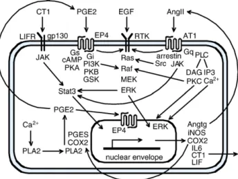
Fig. 1. Simplified scheme of the major routes connected with PGE2
signaling. PGE2is synthesized by enzymes on the nuclear envelope surface.
The PGE2receptor subtype EP4 locates to the cell membrane and to the
nuclear envelope. The EP4 receptor is able to couple with either Gs or Gi proteins; when coupling to Gi, the heterodimeric Gβ/γ component is thought to activate the PI3K-PKB path[13]. The PI3K-dependent activation of the MEK-ERK pathway is not yet defined in detail. Angtg is intracellularly transformed to AngII before secretion. The AngII receptor AT1 may signal via Gq as well as by direct interaction with several adapter proteins includingβ-arrestin, Src, and JAK. Both EP4 and AT1 receptors can transactivate the EGF receptor (a receptor tyrosine kinase = RTK). ERK1/2 occurs in the cytosolic and nuclear fractions. The IL6-related cytokines may be secreted and act in a paracrine and autocrine manner while PGE2may also act in an intracrine manner on the nuclear EP4 receptors. For
full transcriptional activation Stat3 requires phosphorylation at Tyr705 by JAK and at Ser727 by ERK. Abbreviations are in the text except for: DAG, diacylglycerol; GSK, glycogen synthase kinase; iNOS, inducible NO synthase; PI3K, phosphoinositide 3-kinase.
which emerges as the major communication route. Multi-farious direct and indirect (involving de novo protein syn-thesis) signaling loops connecting the PGE2-EP4, JAK/Stat, and MEK-ERK systems may account for fast as well as delayed activation by phosphorylation cascades. Despite many advances in our understanding of the intracellular signaling network, cardiac hypertrophy and heart failure remain a formidable challenge since an effective causative treatment strategy is still lacking.
References
[1] Schaub MC, Hefti MA, Zaugg M. Integration of calcium with the signaling network in cardiac myocytes. J Mol Cell Cardiol 2006;41:183214. [2] Chung J, Uchida E, Grammer TC, Blenis J. STAT3 serine
phosphorylation by ERK-dependent and -independent pathways negatively modulates its tyrosine phosphorylation. Mol Cell Biol 1997;17:650816.
[3] Park JY, Pillnger MH, Abramson SB. Prostaglandin E2 synthesis and secretion: the role of PGE2synthase. Clin Immunol 2006;119:22940.
[4] Mendez M, LaPointe MC. PGE2-induced hypertrophy of cardiac
myocytes involves EP4 receptor-dependent activation of p42/44 MAPK and EGFR transactivation. Am J Physiol Heart Circ Physiol 2005;288: H21117.
[5] Zhu T, Gobeil F, Vazquez-Tello A, Leduc M, Rihakova L, Bossolasco M, et al. Intracrine signaling through lipid mediators and their cognate nuclear G-protein-coupled receptors: a paradigm based on PGE2, PAF,
and LPA1 receptors. Can J Physiol Pharmacol 2006;84:37791.
[6] Frias MA, Rebsamen MC, Gerber-Wicht C, Lang U. Prostaglandin E2 activates Stat3 in neonatal rat ventricular cardiomyocytes: a role in cardiac hypertrophy. Cardiovasc Res 2007;73:5765, doi:10.1016/j. cardiores.2006.09.016[this issue].
[7] Steiner AA, Ivanov AI, Serrats J, Hosokawa H, Phayre AN, Robbins JR, et al. Cellular and molecular bases of the initial fever. PLoS Biol 2006;4:e284 [open access].
[8] Booz GW, Day JNE, Baker KM. Interplay between the cardiac renin angiotensin system and JAK-STAT signaling: role in cardiac hypertrophy, ischemia/reperfusion dysfunction, and heart Failure. J Mol Cell Cardiol 2002;34:144353.
[9] Hilfiker-Kleiner D, Hilfiker A, Drexler H. Many good reasons to have STAT3 in the heart. Pharmacol Ther 2004;107:1317.
[10] Xuan YT, Guo Y, Zhu Y, Wang OL, Rokosh G, Messing RO, et al. Role of the protein kinase C-ε-Raf-1-MEK-1/2-p44/42 MAPK signaling cascade in the activation of signal transducers and activators of transcription 1 and 3 and induction of cyclooxygenase-2 after ischemic preconditioning. Circulation 2005;112:19718.
[11] Sano M, Fukuda K, Kodama H, Takahashi T, Kato T, Hakuno D, et al. Autocrine/paracrine secretion of IL-6 family cytokines causes angio-tensin II-induced delayed STAT3 activation. Biochem Biophys Res Commun 2000;269:798802.
[12] Zhai P, Galeotti J, Liu J, Holle E, Yu X, Wagner T, et al. An angiotensin II type 1 receptor mutant lacking epidermal growth factor receptor transactivation does not induce angiotensin II-mediated cardiac hyper-trophy. Circ Res 2006;99:52836.
[13] Fujino H, Regan JW. EP4 prostanoid receptor coupling to a pertussis toxin-sensitive inhibitory G protein. Mol Pharmacol 2006;69:510.
5 M.C. Schaub, M.A. Hefti / Cardiovascular Research 73 (2007) 3–5