Université de Montréal
Angiotensin II Type 2 Receptor (AT2R) in Glomerulogenesis
Par Min-Chun Liao
Programme de sciences biomédicales Faculté de médecine
Thèse présentée en vue de l’obtention du grade de docteur en philosophie (Ph.D) en sciences biomédicales, option générale
Septembre 2019
Université de Montréal
Unité académique : Programme de sciences biomédicales, Faculté de médecine
Cette thèse intitulée
Angiotensin II Type 2 Receptor (AT2R) in Glomerulogenesis
Présenté par
Min-Chun Liao
A été évalué(e) par un jury composé des personnes suivantes
Dre. Jolanta Gutkowska
Président-rapporteur
Dre. Shao-Ling Zhang
Directeur de recherche
Dr. John S.D. Chan
Codirecteur
Dr. Casimiro Gararduzzi
Membre du jury
Dre. Elena Torban
Examinateur externe (pour une thèse)
Dre. Hélène Girouard
Résumé
Les données épidémiologiques indiquent que le diabète maternel est associé de manière significative aux anomalies congénitales des reins et des voies urinaires (CAKUT), ce qui implique un risque accru de CAKUT chez la progéniture des mères diabétiques par rapport à la population globale. Les causes de CAKUT sont multifactorielles, impliquant des facteurs génétiques et environnementaux. Le récepteur de l’angiotensine II de type 2 (AT2R) est l’un des gènes candidats impliqués dans le CAKUT humain et murin. Bien que de nombreuses études soutiennent l’influence des facteurs génétiques et environnementaux sur le développement rénal et la pathogenèse de CAKUT, les effets du gène AT2R et du milieu
hyperglycémique in utero sur le développement rénal et les effets à long terme chez les enfants de mères diabétiques ne sont pas clairs. Cette thèse a pour objectif d'étudier l'influence de chaque facteur individuellement, ainsi que l'interaction entre ces deux facteurs.
Premièrement, nous avons examiné si le déficit en AT2R (AT2RKO) altère la glomérulogenèse via la formation, la maturation et l'intégrité des podocytes. Nous avons observé que la glomérulogenèse était diminuée chez les embryons E15 AT2RKO, mais le nombre de néphrons ne présentaient aucune différence entre les nouveaux-nés AT2RKO et les souris de type sauvage. Les souris AT2RKO présentaient une dysplasie rénale avec un volume de touffes glomérulaires et un nombre de podocytes inférieurs à l’âge de trois semaines. Nos études ont démontré que la perte d’AT2R via l’augmentation de la génération des dérivés réactifs de l’oxygène (ROS) induite par la NADPH oxydase 4 (Nox4) stimulait l’interaction avec la protéine Hhip (‘Hedgehog interacting protein’), ce qui déclenchait en outre soit l’apoptose des podocytes par l’activation des voies de la caspase- 3 et de la p53, soit la transition épithéliale-mésenchymateuse des podocytes (EMT) par l’activation de la signalisation TGFβ1–Smad2/3. L'ARNm de Hhip glomérulaire était régulé positivement dans les biopsies rénales chez les patients atteints de glomérulosclérose segmentaire focale (FSGS). Les résultats suggèrent que le déficit en AT2R est associé à une perte ou un dysfonctionnement des podocytes et est dû, au moins en partie, à une expression accrue de Hhip ectopique dans les podocytes.
Deuxièmement, nous avons cherché à établir les mécanismes sous-jacents par lesquels un milieu hyperglycémique in utero et un régime riche en graisses (HFD) après le sevrage accélèrent la programmation périnatale des lésions rénales. Nous avons observé que la progéniture des mères atteintes de diabète sévère avait un phénotype de restriction de croissance intra-utérine (IUGR) et avait développé une hypertension légère et des signes d'atteinte rénale à l'âge adulte. De plus, la progéniture nourrie avec une HFD post-sevrage présentait un rattrapage rapide de la croissance puis des lésions rénales associées à une augmentation de l’expression rénale de TGFβ1 et du collagène de type IV, à la production de ROS et à une accumulation de lipides rénaux, mais sans hypertension systémique. Des études in vitro ont démontré que le HFD ou les acides gras libres accéléraient le processus de programmation périnatale des lésions rénales, via une expression accrue de CD36 et de la protéine de liaison aux acides gras (Fabp4) qui cible les ROS, le facteur nucléaire-kappa B et le TGFβ1. Ces résultats indiquent que l'exposition précoce à l'HFD chez les enfants de mères diabétiques ayant subi une IUGR augmente le risque d'apparition de lésions rénales à l’âge adulte, mais pas d'hypertension.
En résumé, AT2R joue un rôle essentiel dans la glomérulogenèse et influence l'intégrité et la fonction du podocyte via des altérations de l'expression de Hhip. En outre, les enfants de mères diabétiques ont un risque accru d'hypertension et de lésions rénales; la surnutrition postnatale accélère les lésions rénales chez ces enfants. Bien que le gène AT2R et le milieu
hyperglycémique in utero aient tous les deux un impact sur le développement du rein et sur les maladies rénales ultérieures, l'interaction entre ces deux facteurs doit encore faire l'objet d'études supplémentaires.
Mots-clés : CAKUT, diabète maternel, déficit en AT2R, glomérulogenèse, podocytes, Hhip, surnutrition postnatale, programmation périnatale, dérivés réactifs de l’oxygène
Abstract
Epidemiologic data indicate that maternal diabetes significantly associates with congenital anomalies of the kidney and urinary tract (CAKUT), which implies an increased chance of CAKUT in the offspring of mothers with diabetes compared to the general population. The causes of CAKUT are multifactorial, involving genetic and environmental factors. The angiotensin II receptor type 2 (AT2R) is one of the candidate genes to be implicated in both human and murine CAKUT. Although numerous studies support the influence of genetic and environmental factors on kidney development and the pathogenesis of CAKUT, the impacts of the AT2R gene and hyperglycemic milieu in utero on kidney
development and long-term outcomes in the offspring of diabetic mothers remain unclear. This thesis aims to investigate the influence of each factor individually, as well as their interaction.
Firstly, we investigated whether AT2R deficiency (AT2R knock-out (KO)) impairs glomerulogenesis via podocytes formation, maturation and integrity. We observed that glomerulogenesis is decreased in AT2RKO embryos at embryonic day 15 (E15), but actual nephron numbers are no different between AT2RKO and wild-type newborn mice. AT2RKO mice exhibited renal dysplasia with lower glomerular tuft volume and reduced podocyte numbers at the age of three weeks. Our studies demonstrated that loss of AT2R via NADPH oxidase 4 (Nox4)-derived reactive oxygen species (ROS) generation stimulates ectopic hedgehog interacting protein (Hhip) expression, which further triggers either podocyte apoptosis by the activation of the caspase-3 and p53 pathways or podocyte epithelial–to– mesenchymal transition (EMT) by the activation of TGFβ1–Smad2/3 signaling. Glomerular Hhip mRNA is upregulated in kidney biopsies of patients with focal segmental glomerulosclerosis (FSGS). The results suggest that AT2R deficiency is associated with podocyte loss/dysfunction and is mediated, at least in part, via increased ectopic Hhip expression in podocytes.
Secondly, we aimed to establish the underlying mechanisms by which a hyperglycemic milieu in utero and a post-weaning high-fat diet (HFD) accelerate the perinatal programming of kidney injury. We observed that the offspring of dams with severe maternal diabetes have
an intrauterine growth restriction (IUGR) phenotype and develop mild hypertension and evidence of kidney injury in adulthood. Moreover, those offspring fed with a post-weaning HFD result in rapid catch-up growth and subsequent profound kidney injury associated with the augmentation of renal TGFβ1 and collagen type IV expression, increased production of ROS, and accumulation of renal lipids, but not systemic hypertension. In vitro studies demonstrated that HFD or free fatty acids accelerate the process of perinatal programming of kidney injury, via increased CD36 and fatty acid-binding protein 4 (Fabp4) expression, which targets ROS, nuclear factor-kappa B and TGFβ1 signaling. These results indicate that early postnatal exposure to HFD in IUGR offspring of diabetic dams increases the risk of later developing kidney injury, but not hypertension.
In summary, AT2R plays an essential role in glomerulogenesis and influences the podocyte integrity and function via alterations of Hhip expression. In addition, the offspring of diabetic mothers have an increased risk of hypertension and kidney injury; postnatal overnutrition further accelerates kidney injury in those offspring. Although both AT2R and hyperglycemic milieu in utero have an impact on kidney development and later kidney diseases, the interaction between these two factors still needs further studies.
Keywords: CAKUT, maternal diabetes, AT2R deficiency, glomerulogenesis, podocytes, Hhip, postnatal overnutrition, perinatal programming, reactive oxygen species
Table of contents
Résumé ... 3 Abstract ... 5 Table of contents ... 7 List of Tables ... 9 List of Figures ... 10 List of Abbreviations ... 13 Acknowledgement ... 16 Thesis Outlines ... 17 CHAPTER 1 ... 18 1.1 Introduction ... 181.1.1 Angiotensin II Receptor Type 2 in Glomerulogenesis ... 18
1.1.1.1 Renin–Angiotensin System (RAS) ... 18
1.1.1.2 Systemic RAS ... 18
1.1.1.3 Local RAS ... 21
1.1.1.4 The Intrarenal RAS ... 22
1.1.2 AT2R in the Kidney ... 23
1.1.2.1 AT2R Expression in the Developing Kidney ... 24
1.1.2.2 AT2R Expression and Functions in the Adult Kidney ... 25
1.1.2.3 Mouse Models ... 27
1.1.2.4 Congenital anomalies of the kidney and urinary tract (CAKUT) ... 27
1.1.2.5. Hypertension and Chronic Kidney Disease ... 31
1.1.3. Glomerulogenesis and Podocyte ... 35
1.1.3.1 Kidney and Glomerulus ... 35
1.1.3.2 Glomerulogenesis ... 37
1.1.3.3 Podocyte ... 40
1.1.4 Hedgehog interacting protein (Hhip) ... 44
1.1.5 Reactive oxygen species (ROS) ... 47
1.2 Article 1 ... 52 1.3 Discussion ... 94 1.4 Summary ... 98 1.5 Before Chapter 2 ... 99 CHAPTER 2 ... 100 2.1 Introduction ... 100
2.1.1 Maternal Diabetes-induced Perinatal Programming ... 100
2.1.2 AT2R and Perinatal Programming ... 104
2.1.3 Sex and Gender Effects ... 105
2.1.4 Research Questions and Hypothesis ... 109
2.2 Article 2 ... 110
2.3 Discussion ... 133
2.4 Summary ... 136
CHAPTER 3 ... 137
3.1 Introduction ... 137
3.1.1 Interactions of AT2R with estrogen and its receptors ... 138
3.1.2 Objectives/ Hypothesis ... 139
3.2 Materials and Methods ... 140
3.3 Unpublished Results ... 144
3.4 Discussions ... 147
3.5 Future Works ... 149
List of Tables
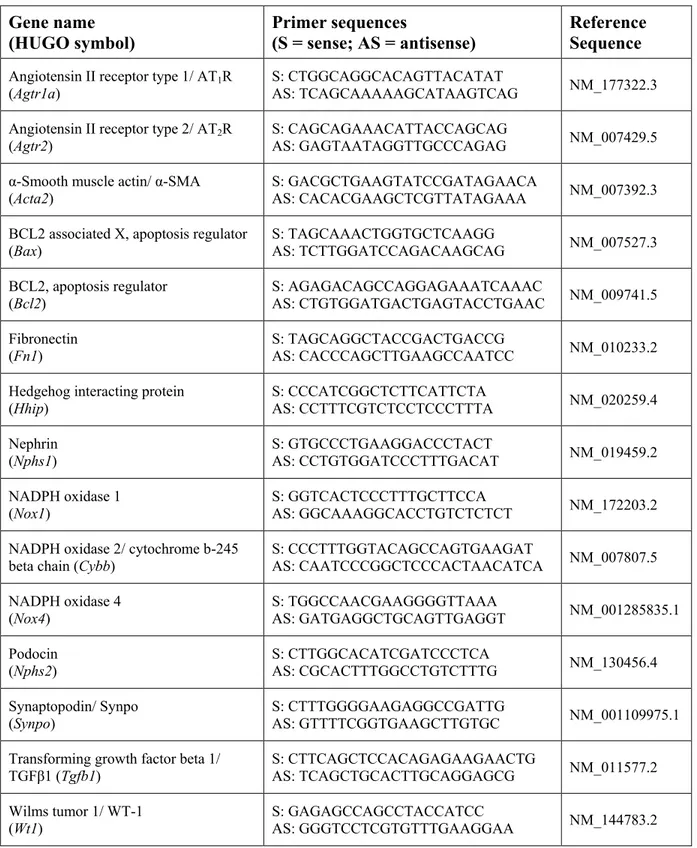
Table 1. Primer sequences ... 72
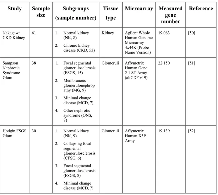
Table 2. Nephroseq analysis ... 73
Table 3. Patient information ... 74
Table 4. Biological measurements from WT and AT2RKO mice ... 74
List of Figures
Figure 1. An enzymatic cascade of angiotensin peptide formation ... 19
Figure 2. Schematic depiction of the RAS components and selected actions ... 21
Figure 3. A schematic illustration of the critical signaling pathways of AT2R ... 26
Figure 4. Illustrative 3D models of congenital abnormalities of the kidney and urinary tract (CAKUT) ... 28
Figure 5. A schematic representation of kidney development ... 30
Figure 6. Pathophysiologic mechanisms of hypertension in chronic kidney disease ... 32
Figure 7. Schematic depiction of a renal proximal tubule cell showing the principal Na+ transporters and angiotensin receptors that regulate them ... 33
Figure 8. Structure of the Bowman’s capsule and glomerular capillary tuft ... 36
Figure 9. Migration of endothelial cells into the developing glomerular tuft ... 39
Figure 10. Podocyte subcellular compartments showed by conventional SEM and TEM ... 41
Figure 11. The glomerular filtration barrier and its key molecular components ... 43
Figure 12. Sources of ROS and the intracellular anti-oxidative defense ... 48
Figure 13. Physiological measurements of glomerulogenesis and neonatal nephron number. 76 Figure 14. Podocyte marker analysis in vivo ... 78
Figure 15. Hhip expression in vivo and in vitro ... 80
Figure 16. Hhip expression and ROS generation ... 82
Figure 17. Hhip and TGFβ1-induced EMT ... 84
Figure 18. The interaction of Hhip and TGFβRI ... 86
Figure 19. Images of freshly isolated neonatal kidneys (whole-mount and frontal versus transverse sections) from Nephrin-CFP-Tg versus Nephrin/AT2RKO mice. ... 87
Figure 20. Proliferation study on silencing AT2R (siRNA) in mPODs under permissive (33°C with 10 U/ml INF-γ) and non-permissive conditions (37°C without INF-γ)... 87
Figure 21. Synpo IF staining in mPODs treated with or without PD123319 in a dose-dependent manner. ... 88
Figure 22. Renal AT1R protein expression in E18 and neonatal kidneys from wild-type versus AT2RKO mice analyzed by western blotting. ... 88
Figure 23. RT-qPCR assessment of AT2R mRNA expression in mPODs treated with AT2R
siRNA (50 nM). ... 88
Figure 24. RT-qPCR assessment of Nox1, Nox2, and Nox4 mRNA expression in mPODs treated with AT2R siRNA (50 nM). ... 89
Figure 25. RT-qPCR assessment of Hhip and Nox4 mRNA expression in mPODs treated with GKT (10−6 M), PD (10−8 M), and PD + GKT. ... 89
Figure 26. Semi-quantification of Figure 15G in vitro ... 89
Figure 27. Semi-quantification of Figure 15H in vitro ... 90
Figure 28. RT-qPCR assessment of Nephrin and Synpo mRNA expression in mPODs treated with rHhip in a dose-dependent manner. ... 90
Figure 29. Synpo IF expression in mPODs treated with rHhip in a dose-dependent manner. . 90
Figure 30. RT-qPCR assessment of Synpo, α-SMA, and Fn1 mRNA expression in mPODs treated with rTGFβ1 in a dose-dependent manner. ... 91
Figure 31. Nephroseq analysis of three publicly available microarray studies performed on human kidney biopsy samples: (1) Nakagawa Chronic Kidney Disease Study; (2) Hodgin Focal Segmental Glomerulosclerosis Glomerular Study; (3) Sampson Nephrotic Syndrome Glomerular Study. ... 93
Figure 32. A mechanistic proposal on the early developmental origins of ‘diabesity’ disposition, acquired by pre- and/or neonatal overfeeding, materno-fetal hyperglycemia, and perinatal hyperinsulinism ... 103
Figure 33. Metabolic parameters in offspring ... 127
Figure 34. Blood pressure, renal function, and adipocyte morphology ... 128
Figure 35. Renal morphology ... 129
Figure 36. Renal CD36 & Fabp 4 gene expression ... 130
Figure 37. Renal CD36 & Fabp 4 localization ... 131
Figure 38. BSA-PA effects in IRPTCs ... 132
Figure 39. Maternal diabetic murine model. ... 141
Figure 40. Systolic blood pressure in the offspring of non-diabetic (Con) and diabetic (Dia) dams of wild-type (WT) and AT2RKO (KO) mice ... 144
Figure 41. Renal morphology and function ... 145
Figure 43. Estrogen receptors expression in isolated glomeruli and effect of estradiol on estrogen receptors and AT2R expression in cultured podocytes ... 147 Figure 44. Proposed working model ... 150
List of Abbreviations
ACE : Angiotensin-converting enzyme ACE2 : Angiotensin-converting enzyme 2 ACR : Albumin/creatinine ratioAgt : Angiotensinogen Ang : Angiotensin Ang I : Angiotensin I Ang II : Angiotensin II Ang III : Angiotensin III APA : Aminopeptidase A APN : Aminopeptidase N
ARB : Angiotensin receptor blocker AT1R : Angiotensin II receptor type 1 AT2R : Angiotensin II receptor type 2 AT2RKO : AT2R deficiency
ATIP : AT2R interacting protein BK : Bradykinin
BP : Blood pressure
BSA : Bovine serum albumin
BSA-PA : BSA-bound sodium palmitate BW : Body weight
C21 : Compound 21
CAKUT : Congenital anomalies of the kidney and urinary tract CD2AP : CD2-associated protein
CFP : Cyan fluorescent protein CKD : Chronic kidney disease DHE : Dihydroethidium Dhh : Desert hedgehog
DOHaD : Developmental Origins of Health and Disease E : Embryonic day
EMT : Epithelial–to–mesenchymal transition ESRD : End-stage renal disease
Fabp4 : Fatty acid-binding protein 4 FAT1 : Fat cadherin 1
FSGS : Focal segmental glomerulosclerosis GBM : Glomerular basement membrane GDM : Gestational diabetes
GFR : Glomerular filtration rate GPCR : G-protein coupled receptor GWAS : Genome-wide association study H&E : Hematoxylin and eosin
H2O2 : Hydrogen peroxide
HAPO : Hyperglycemia and adverse pregnancy outcomes HFD : High-fat diet
Hh : Hedgehog
Hhip : Hedgehog interacting protein IF : immunofluorescence
IHC : Immunohistochemistry Ihh : Indian hedgehog
IRAP : Insulin-regulated aminopeptidase
IRPTC : Immortalized rat renal proximal tubular cell line IUGR : Intrauterine growth restriction
KO : Knockout KW : Kidney weight
MAPK : Mitogen-activated protein kinase MCD : Minimal change disease
MM : Metanephric mesenchyme
mPODs : Immortalized mouse podocytes cell line MrgD : Mas-related G protein-coupled D
ND : Normal diet
NEPH1 : Nephrin-like protein 1 NF-κB : Nuclear factor-kappa B
NHE-3 : Sodium-hydrogen exchanger-3
NKA : Sodium-potassium adenosine triphosphatase NO : Nitric oxide
Nox4 : NADPH oxidase 4 PAS : Periodic acid–Schiff PGDM : Pre-gestational diabetes
PLZF : Promyelocytic zinc finger protein RAS : Renin-angiotensin system
rHhip : Recombinant Hhip ROS : Reactive oxygen species SBP : systolic blood pressure Shh : Sonic hedgehog STZ : Streptozotocin Synpo : Synaptopodin Tg : Transgenic TGFβRI : TGFβ receptor I TL : Tibia length
TRPC6 : Transient receptor potential channel 6 UB : Ureteric bud
VEGF : Vascular endothelial growth factor
VEGFR2 : Vascular endothelial growth factor receptor 2 WB : Western blotting
Acknowledgement
It would not have been possible to complete this dissertation in a period of my study without the great support and help of a number of people whom I would like to acknowledge and thank.
First of all, I would like to express my most profound gratitude to my supervisor Dr. Shao-Ling Zhang, who offered me this opportunity to join her lab and guided me without preconditions throughout my entire journey of pursuing a Ph.D. Without her full support, inspiration and guidance, all the achievements are back to square one. Thank you for your patience, motivation, enthusiasm and perseverance. I am deeply indebted to my co-supervisor Dr. John Chan for his encouragement, helpful comments and academic direction.
I would like to express my deepest appreciation to my dissertation committee: Dr. Jolanta Gutkowska, Dr. Casimiro Gararduzzi, Dr. Elena Torban and Dr. Helene Girouard. I am grateful for their thoughtful insights, expert guidance and enthusiastic commitment.
I also want to offer my sincere thanks to my present colleagues and past lab members: Xing-Ping Zhao, Henry Nchienzia, Chao-Sheng Lo, Shuiling Zhao, Yessoufou Aliou, Shaaban Abdo, Yixuan Shi, Chin Han Wu and Anindya Ghosh for their valuable assistance in collecting data, discussing questions and cooperating experiments at the research site. I would like to extend my special thanks to Mrs. Isabelle Chénier and Dr. Kana Miyata for their insightful comments and suggestions on an earlier version of this dissertation and all the generous help in my studies and life. I owe a debt gratitude to all the people who have helped me throughout the journey of my Ph.D.
Finally, I wish to express my heartfelt gratitude to my parents, sister, brother and in-laws for their constant encouragement and support. The most important is to thank my lovely wife Shiao-Ying Chang for her enduring patience, unconditional support and understanding. Her love is my source of energy to complete this dissertation.
Thesis Outlines
My Ph.D. thesis contains three Chapters bellow:– Chapter 1: To study the impact of AT2R deficiency on glomerulogenesis. We aim to investigate whether AT2R deficiency impairs glomerulogenesis via altered podocytes formation, maturation, and integrity (Liao MC*, Zhao XP, Chang SY, Lo CS, Chenier I, Takano T, Ingelfinger JR, Zhang SL: AT2R deficiency mediated podocyte loss via activation of ectopic hedgehog interacting protein (Hhip) gene expression. The Journal of Pathology, 2017; 243 (3): 279-293, Impact Fact (IF): 6.253).
My paper was chosen as one of 21 original papers highlighted by The Journal
Pathology as “Recent Advances in Renal Pathology” to celebrate its 125th anniversary (Virtual Issue, September 2018).
– Chapter 2: To study the outcomes of perinatal programming occurring in the offspring of diabetic mothers postnatally fed with a high-fat diet (HFD). We aim to investigate the underlying mechanisms by which a post-weaning HFD accelerates the perinatal programming of kidney injury occurring (Aliou Y*, Liao MC*, Zhao XP, Chang SY, Chenier I, Ingelfinger JR, Zhang SL: Post-weaning high-fat diet accelerates kidney injury, but not hypertension programmed by maternal diabetes. Pediatric Research, 2016; 79(3): 416-424, Impact Fact (IF): 2.882. *The first two authors contributed equally to this work).
– Chapter 3: To study the impact of AT2R deficiency on maternal diabetes-induced perinatal programming. We aim to investigate whether AT2R deficiency accelerates perinatal programming induced by maternal diabetes. (Ongoing studies)
CHAPTER 1
Liao MC*, Zhao XP, Chang SY, Lo CS, Chenier I, Takano T, Ingelfinger JR, Zhang SL:
AT2R deficiency mediated podocyte loss via activation of ectopic hedgehog interacting protein (Hhip) gene expression. The Journal of Pathology, 2017; 243 (3): 279-293
1.1 Introduction
1.1.1 Angiotensin II Receptor Type 2 in Glomerulogenesis
1.1.1.1 Renin–Angiotensin System (RAS)
The renin–angiotensin system plays a pivotal role in controlling renal, vascular and cardiac physiology, and its activation is related to multiple diseases such as chronic kidney disease, hypertension, cardiovascular disease, and diabetes mellitus [1]. Historically, the RAS has been extensively discussed for more than 120 years since the first discovery of the enzyme renin by Tigerstedt and Bergmann in 1898 [2]. The original view of the RAS was thought to be a peptidergic system with endocrine features, which is the systemic RAS or also called the endocrine RAS. However, it is now generally recognized that this original view of the systemic RAS represents an insufficient description of the system. In addition to the systemic RAS, a local RAS exists in different organs and plays a crucial role in physiology and pathophysiology. There are several local RASs that function independently of each other and of the systemic RAS. For instance, angiotensin II (Ang II) generated in the kidney appears to have physiologic effects that are as important as circulating Ang II and, under some circumstances, even more important than circulating Ang II [3]. Thus, the RAS includes both a systemic system with endocrine effects and multiple local systems with autocrine, paracrine, and intracrine effects.
1.1.1.2 Systemic RAS
The systemic RAS begins with renin cleaving its substrate, angiotensinogen (Agt), to form the inactive peptide Angiotensin I (Ang I). Then, Ang I is converted to the biologically active octapeptide Ang II by a dipeptidyl carboxypeptidase named angiotensin-converting
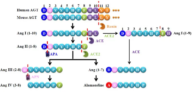
enzyme (ACE), a process that occurs most predominantly on the surface of endothelial cells [1]. Ang II binds to its two receptors, Ang II receptor type 1 (AT1R) and type 2 (AT2R), to regulate tissue and cell functions. For example, Ang II acts through AT1R to initiate and maintain actions that could be harmful to the body, including vasoconstriction, anti-natriuresis, aldosterone secretion, sympathetic nervous system activation, cellular dedifferentiation and growth, inflammation, reactive oxygen species production, and target organ damage [4]. In contrast, Ang II also binds to AT2R to counterbalance AT1R-mediated actions, including vasodilation, natriuresis, proliferation, inflammation, oxidative, anti-hypertrophic, and anti-fibrotic actions [5]. Although the expression of this receptor is low in healthy adults, it is activated in certain disease conditions. The RAS also includes several other enzymes, peptides (Ang II metabolites), and receptors, which collectively comprise a “protective arm” to counterbalance the harmful actions of the ACE/Ang II/AT1R pathway. The components of the protective arm are described below, and Figure 1 provides a schematic illustration of the formation of Ang II and its metabolites.
Figure 1. An enzymatic cascade of angiotensin peptide formation. AGT, angiotensinogen; APA, aminopeptidase A; APN, aminopeptidase N.
The RAS plays a critical role in regulating physiological and pathophysiological processes in the cardiovascular system and kidney through autocrine and paracrine as well as endocrine effects [1]. The system is involved in a large range of homeostatic and modulatory
processes that regulate vasoconstriction, salt and water balance, cell growth, tissue remodeling and dysfunction, hormone secretion and sympathetic activity. AT1R activation is considered to mediate the majority of the biological functions of Ang II [1]. However, the AT1R is also involved in a variety of pathophysiological conditions in which the RAS is abnormally activated, such as hypertension, cardiac hypertrophy, heart failure, diabetic nephropathy, fibrosis, and inflammation [6]. The AT1R is widely present in various tissues, including vascular smooth muscle, endothelium, heart, brain, kidney, adrenal gland, and adipose tissue. The classic ACE/Ang II/AT1R pathway promotes intracellular signaling pathways through the activation of protein kinases, growth factor receptor transactivation, subunits of nicotinamide adenine dinucleotide phosphate (NADPH) oxidase [1]. Those signaling transductions are mostly protein-dependent, including Gq/11α, G12/13α, and Giα [1]. The AT1R also elicits G-protein-independent signal transduction cascades, forms a heterodimer with other G-protein coupled receptors (GPCRs), and directly interacts with AT1R interacting proteins [1]. On the contrary, the biological functions of Ang II mediated through AT2R are considered as regulating vasodilation to counterbalance the vasoconstrictor effects mediated through AT1R.
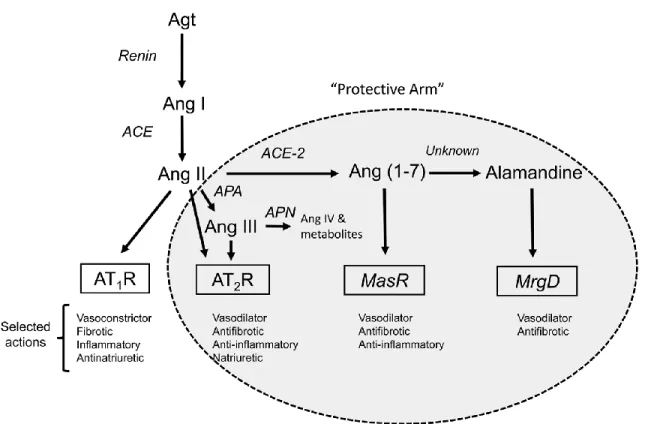
A protective arm of the RAS has more recently emerged, which includes the angiotensin-converting enzyme 2 (ACE2) /Ang (1-7)/Mas receptor pathway, the alamandine/Mas-related G protein-coupled D (MrgD) receptor pathway, and the Ang II/Ang III/AT2R pathway (Figure 2) [7]. In contrast to ACE, a novel mono-carboxypeptidase was identified and called ACE2, which is the pivotal enzyme of the protective arm by converting Ang I and Ang II into Ang (1-9) and Ang (1-7), respectively [1]. Ang (1-9) is then converted to Ang (1-7) by ACE. Ang (1-7) is a ligand of the Mas receptor, which triggers opposite actions to the classic ACE/Ang II/AT1R pathway, such as vasodilation and anti-fibrosis [1]. Alamandine can be formed from Ang (1-7) in the heart and also has vasodilative and anti-fibrotic effects through binding to MrgD [8]. Ang II is further converted to Ang III and Ang IV by different aminopeptidases (Figure 1). It has been demonstrated that Ang III is a ligand of both AT1R and AT2R, as well as the endogenous AT2R agonist in the kidney [5]. Ang IV binds to its receptor, AT4R (also known as insulin-regulated aminopeptidase (IRAP)), which causes vasodilation effects by increased endothelial nitric oxide (NO) synthase and also increases renal cortical blood flow and urinary sodium excretion [9]. The updated RAS,
including Ang peptides, their receptors, and actions are summarized in Figure 2. Together, these counter-regulatory pathways work to reduce the harmful impact of the classic ACE/Ang II/AT1R pathway on blood pressure and kidney function.
Figure 2. Schematic depiction of the RAS components and selected actions. Receptors are shown in the boxes. Components of the protective arm of the system are shown in the shaded ellipse. AT2R can be activated by Ang II or the Ang II metabolite, Ang III. Within the kidney, Ang III is thought to be the preferred agonist for AT2R-induced renal tubular inhibition of Na+ reabsorption. The “protective arm” of the RAS acts in a counter-regulatory manner to oppose the detrimental actions of Ang II via AT1R. Adapted from “AT2 Receptors: Potential Therapeutic Targets for Hypertension,” by R.M. Carey, 2017, American Journal of Hypertension, 30(4), p. 340, Copyright 2016, with permission from Oxford University Press. [7]
1.1.1.3 Local RAS
The concept of a local RAS is based on RAS components being expressed in “unlikely” places, and the functions of this system are mostly based on local Ang II synthesis.
Besides, the local RAS exerts diverse physiological and pathophysiological roles, and the RAS components are formed locally by various organs and cells (e.g., kidney, adrenal gland, heart, brain, reproductive tract, skin, digestive organs, white blood cells, and adipocytes) [2]. For example, although renin is secreted from the juxtaglomerular apparatus of the kidney, it is found in the brain after nephrectomy. Circulating renin and its substrate do not pass the blood-cerebrospinal fluid barrier, suggesting that in the brain, the local RAS appears to be regulated independently of the systemic RAS. However, there is the crosstalk between the systemic and local RAS. For instance, heart uptakes renin and Agt from the circulation and stores them locally. Thus, they are used to produce Ang II locally in the heart. Hence, in addition to the systemic RAS, local Ang II synthesis is an essential modulator of tissue function and structure. Also, locally produced Ang II might contribute to the tissue injury via the stimulation of inflammation, proliferation, and apoptosis, the regulation of some gene expressions, and the activation of several intracellular signaling pathways [3].
1.1.1.4 The Intrarenal RAS
Although every organ and tissue in the body has RAS components, the kidney is unique as it has all the RAS components to generate intrarenal Ang II. The level of intrarenal Ang II, expressed as content per mass of tissue, is much higher than the level of that delivered by the circulatory system. In rats, the level of plasma Ang II is in the picomolar range (50-100 pM), whereas the level of luminal Ang II in the renal proximal tubule is in the nanomolar range (30-40 nM) [10, 11]. Intrarenal Ang II plays a pivotal role in regulating renal hemodynamics and functions, which are associated with sodium balance and blood pressure homeostasis [12]. It has further been demonstrated that abnormally increased intrarenal Ang II causes hypertension and renal injury via AT1R [13].
The main fraction of Ang II in the kidney is produced locally from both plasma Agt and intrarenal Agt generated by the renal proximal tubule [3]. Plasma Ang I in the kidney can also be converted to Ang II by intrarenal ACE, which is expressed in the kidneys predominantly in renal proximal and distal tubules, the collecting ducts, and renal endothelial cells [14]. Renin synthesized by the juxtaglomerular apparatus cells is the primary origin of both plasma and intrarenal renin levels, which also can provide another pathway for the local
Ang I production [15]. Thus, all of the RAS components required to produce intrarenal Ang II are expressed along the nephron, indicating that there is specific compartmentalization of intrarenal RAS with distinct regulatory mechanisms predominating in the separate compartments.
In the view of physiology, the local RAS has abilities to maintain a balance or homeostasis at the tissue level to counterbalance the effects mediated by the systemic RAS [3]. The dual actions of Ang II on its receptors are thought to be the basis of the balance between the systemic and local RAS. Also, the possibility of alternative pathways including different tissue-expressed substrates for ACE, different receptors such as AT4R, and different Ang II metabolite products such as Ang (1-7) can be responsible for mediating the regulatory and counter-regulatory effects. If this balance is disarranged, the RAS turns into a mediator of the pathophysiological stimulus. Thus, inappropriate activation of the intrarenal RAS leads to alterations in the hemodynamic function that contributes to the development of hypertension. Persistence of this activation results in long-term consequences, including cellular proliferation and renal injury.
1.1.2 AT
2R in the Kidney
AT2R is generally considered as part of the “protective arm” of the RAS to counterbalance the actions of Ang II via AT1R [7]. The AGTR2, the gene symbol of AT2R, gene is localized on the X chromosome in human, mouse and rat [5]. The genomic DNA of all three species consists of three exons, two introns, and the whole protein-coding region is contained in the third exon [16]. Because of this uninterrupted protein-coding region, no AT2R subtypes or splice variants have been identified [17]. The homologous location and structure of the AGTR2 gene throughout all three species may have important implications in the genetics of the AGTR2 gene. AT2R cDNA encodes 363 amino acid residues correlated to a molecular weight of 41 kDa [5]. The AT2R protein belongs to the family of seven-transmembrane G-protein coupled receptor (GPCR) and functions as a receptor for Ang II [5]. Although AT1R and AT2R belong to the same family of GPCRs, both receptors share only approximately 34% amino acid sequence identity [18]. The homologous amino acid sequence of AT1R and AT2R is mainly localized in the transmembrane hydrophobic domain, which
forms seven-transmembrane helical columns. Amino acid residues located in these helical domains and considered to be necessary for Ang II binding to AT1R are also conserved in AT2R. The divergence regions between AT1R and AT2R are in the third intracellular loop and the carboxyl-terminal tail. These structural characteristics of AT2R establish a potential foundation for its weak coupling to G-proteins and lack of phosphorylation by GPCR kinases, as well as lack of desensitization after Ang II binding [19]. Therefore, due to the differences in amino acid sequence and structural features, it seems that the activation of AT1R and AT2R is likely to stimulate distinct signaling mechanisms and induce different biological actions.
1.1.2.1 AT2R Expression in the Developing Kidney
AT2R is highly expressed in fetal mesenchymal tissues, suggesting that the actions of Ang II via AT2R might play a pivotal role in regulating cellular differentiation and organ development [20]. Mouse metanephric development begins on embryonic day (E) 10.5, and kidney development continues until postnatal ten days after birth. In the rodent kidney, AT2R expression peaks during fetal metanephrogenesis and rapidly declines after birth. Also, AT2R is expressed earlier than AT1R. In mice, AT2R mRNA and protein can be detected in the
metanephric kidney as early as on E11.5 which is a rapidly developing period of the urogenital system, and its signals are located in the ureteric bud (UB) and the mesenchymal cells adjacent to the UB tip [20]. On E14, AT2R is expressed in UB branches, nephron progenitors and medullary stroma. AT2R is detected in inner cortical tubules (i.e., morphologically resembles proximal tubule) on E16 and reaches a maximal level on E19.
On the other hand, AT1R expression peaks at the end of gestation and subsequently declines gradually. On E12, AT1R protein is detected at low levels in the UB and the surrounding mesenchyme [20]. On E14, AT1R immunostaining is highly expressed in both the luminal and basolateral sides of UB branches. On E16, AT1R is present in proximal tubules and is weakly expressed in the UB branches, stromal mesenchyme and glomeruli.
Nearly simultaneously at E14, both AT1R and AT2R are expressed in the metanephros when UB is vigorous branching [21]. However, both receptors show a distinct cellular expression pattern during kidney development. In the fetal kidney, AT1R mRNA is expressed
mRNA is expressed primarily in the mesenchymal cells close to the UB tip, and later extended to the nephrogenic area of the superficial cortex and the cells between collecting ducts [21]. Furthermore, it has been demonstrated that in murine, the level of Ang II in the developing fetal kidney is higher than that of adult kidney [20]. In addition to AT1R and AT2R, the other RAS components are also expressed in the developing kidney, such as Agt, renin, and ACE, suggesting that the RAS locally plays a role in kidney development.
1.1.2.2 AT2R Expression and Functions in the Adult Kidney
AT2R decreases markedly after birth, but it is expressed at low levels in healthy adult tissues including the cardiovascular system, adrenal gland, kidney, brain, reproductive tissues, and skin [5]. In mature kidney, AT2R protein is expressed predominately in glomeruli and present in small quantities in cortical tubules and interstitial cells. AT2R is upregulated in adult tissues under certain pathological conditions, including fibrosis, wound healing, tissue remodeling, and inflammation [5]. It has been demonstrated that AT2R protein is upregulated in adult glomeruli, tubules and interstitium in response to dietary sodium depletion [22].
As mentioned before, the activation of AT2R is essential to counterbalance the detrimental effects mediated by AT1R and protect against the progression of organ damage and failure as a result of excessive action of Ang II. The possible signaling pathways involved in AT2R-mediated biological effects are schematically summarized in Figure 3 [5]. In a sodium-depletion animal model, AT2R is stimulated to protect the kidney from injury via the bradykinin–nitric oxide (NO)–cGMP pathway. In both vascular smooth muscle cells and cardiomyocytes, AT2R activation inhibits the phosphorylation of ERK1/2 via stimulation of a variety of phosphatases. It has been reported that inhibition of NADPH oxidase activity stimulates AT2R-mediated natriuresis [5]. Other potential pathways have been involved in AT2R-mediated effects, including interaction with AT2R interacting protein (ATIP) and transcription factor promyelocytic zinc finger protein (PLZF), and activation of several protein phosphatases. Zhang et al. [23] first reported that AT2R is able to couple the G-proteins Gi2α and Gi3α in the rat fetus and AT2R coupling to Giα is linked with activation of cGMP signaling. Another study has shown that AT2R-mediated activation of SHP-1 (i.e. phosphatases) is associated with Gsα [24]. Although AT2R has all of the classic motifs and signature residues of
a GPCR, it lacks the typical mechanisms of rapid desensitization, internalization and degradation. These atypical GPCR features cause a prolonged signaling response. Also, AT2R may have constitutive activity and exert ligand-independent cellular effects by dimerization with other GPCRs or interaction with different scaffold proteins to mediate its action [5].
Figure 3. A schematic illustration of the critical signaling pathways of AT2R. The AT2R has been shown to activate several pathways which are dependent on its specific action. AT2R-mediated natriuresis, vasodilation and anti-fibrotic effects involve the activation of a NO–cGMP–dependent pathway, which is likely mediated by the release of bradykinin and an increased NOS activity, whereas its mediated antioxidant, anti-inflammatory and growth inhibition involves the interaction with its interacting protein, ATIP1, and activation of several phosphatases (MKP-1, PP2A, SHP-1) to interfere with ERK1/2 and other kinases’ phosphorylation. On the other hand, the interaction with PLZF is involved in its mediated hypertrophic effect, primarily in cardiac tissues. Adapted from “Angiotensin II type 2 receptor (AT2R) in renal and cardiovascular disease,” by B.S. Chow, 2016, Clinical science (London, England: 1979), 130(15), p. 1312, Copyright 2016, with permission from Portland Press. [5]
1.1.2.3 Mouse Models
Gene knockout and overexpression mouse models have been useful tools to study the role of AT2R in cardiovascular and renal diseases. For example, the AT2R knockout (AT2RKO) mouse line was reported by two groups in 1995 [25, 26]. There are no dramatically abnormal phenotypes at baseline in AT2RKO mice, but blood pressure was reported to be either increased [25] or unchanged [26] in these AT2RKO mice. It has been reported that AT2RKO mice have increased vascular and anti-natriuretic sensitivity in response to exogenous Ang II, attenuation of exploratory behavior, delay differentiation in the vascular smooth muscle cells, and increased susceptibility to renal-tubular developmental disease [18]. AT2R deficiency leads to normal or slight blood pressure elevation and augmented vascular sensitivity to exogenous Ang II, suggesting that AT2R may exert a protective effect in blood pressure regulation by counteracting AT1R function via downregulation of AT1R expression [27] and upregulation of bradykinin–NO–cGMP signaling [28]. Although AT2RKO mice did not produce dramatic morphological abnormalities, those mice display congenital anomalies of the kidneys and urinary tract (CAKUT) with a >23% penetrance [18]. In contrast, transgenic mice that overexpress cardiac-specific AT2R were found to have no visible morphological or functional changes but a decreased response to AT1R-mediated vasoconstriction and chronotropic effects [29]. Also, transgenic mice that overexpress AT2R in vascular smooth muscle cells were found to have activated bradykinin–NO–cGMP pathway and diminished Ang II-induced vascular constriction as well [30].
1.1.2.4 Congenital anomalies of the kidney and urinary tract (CAKUT)
Congenital anomalies of the kidney and urinary tract (CAKUT) are frequent causes of chronic kidney disease (CKD) and end-stage renal disease (ESRD) in children [31]. CAKUT form approximately 20–30% of all congenital malformations, and their frequency has been predicted from three to six per 1,000 births [32, 33]. Because CAKUT play a causative role in 48–59% of cases of CKD and in 34–43% of cases of ESRD in children in developed countries [31, 34], it is imperative to diagnose these anomalies and establish a therapy to preserve renal function and prevent or delay the onset of kidney failure. CAKUT refer to a spectrum of structural malformations that are characterized by defects in morphogenesis of the kidney
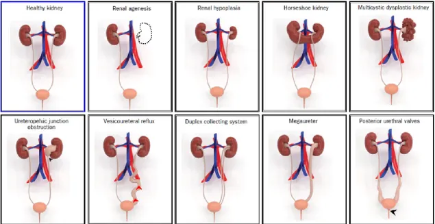
and/or urinary tract. The defects of CAKUT involve renal agenesis, renal hypoplasia, renal dysplasia, horseshoe kidney, multicystic dysplastic kidney, ureteropelvic junction obstruction, hydronephrosis, megaureter, duplex collecting system, ectopic ureter, vesicoureteral reflux, and posterior urethral valves (Figure 4) [35]. In Europe, CAKUT account for 41.3% of all children receiving renal replacement therapy [36]. In contrast, CAKUT are a much less frequent cause of ESRD in adult patients who received renal replacement therapy. It has been reported that the estimated median age at the start of renal replacement therapy is 31 and 61 years in patients with and without CAKUT, respectively [37]. However, recent evidence suggests that some patients with asymptomatic or milder forms of CAKUT might develop hypertension, proteinuria, and ESRD in adulthood [37]. Thus, CAKUT progress to ESRD more often at adulthood than in childhood.
Figure 4. Illustrative 3D models of congenital abnormalities of the kidney and urinary tract (CAKUT) that were created using the modeling tools ZBrush (Pixologic, Inc.) and 3ds Max (Autodesk, Inc.), to illustrate the phenotypic spectrum of CAKUT. The black arrows indicate the site of obstruction in ureteropelvic junction obstruction and posterior urethral valves. The red arrows indicate the abnormal flow of the urine from the bladder to the ureter or kidney occurring in vesicoureteral reflux. Adapted from “Genetic, environmental, and epigenetic factors involved in CAKUT,” by N. Nicolaou, 2015,
Nature reviews. Nephrology, 11(12), p. 722, Copyright 2015, with permission from Springer Nature. [35]
The etiology of CAKUT is multifactorial, including monogenetic causes, copy number variants, single nucleotide variants, epigenetic influences, and environmental risk factors [35]. Despite the diverse renal structural malformations, all forms of CAKUT stem from defective kidney development. In order to realize the basis of CAKUT, it is indispensable to consider how the kidney develops.
Kidney development (also called nephrogenesis) occurs in three stages, including the pronephros, mesonephros and metanephros, arising sequentially from the intermediate mesoderm [38]. The pronephros is a primitive and transient structure with a non-functional system. Degeneration of the pronephros is required for kidney development. The mesonephros develops caudally to the pronephros and is the first excretory organ, excreting urine via the mesonephric duct (also called Wolffian duct). In mice, the mesonephros becomes inactive and atrophies at E14.5, and, within 24 hours, nearly all of the tubules undergo apoptosis and disappear in a caudal to the cranial direction [39]. At this time point, AT2R mRNA is highly
expressed in the mesonephros, suggesting that AT2R expression might represent a group of specific cells that are programmed to undergo apoptosis [21]. While in the female the mesonephros disappears completely, in the male a part of it develops into the testicular efferent ducts. In contrast to the pronephros and mesonephros, the metanephric kidney persists and is characterized by reciprocal inductive interactions between UB and metanephric mesenchyme. Thus, kidney development occurs with UB outgrowth, followed by UB branching morphogenesis and mesenchyme–to–epithelial transition, and completes with nephron patterning and elongation [39]. The overview of kidney development is schematically summarized in Figure 5.
Figure 5. A schematic representation of kidney development. (A) In mammals, the kidney develops from the metanephric mesenchyme upon an invasion of the ureteric bud out of the nephric duct. (B and C) The ureteric bud starts branching within the growing metanephric mesenchyme. (D) The mesenchyme condenses around the ureteric bud tips forming the cap mesenchyme. (E) Renal vesicles form from the condensed cap mesenchyme. (F) A cleft develops in the comma-shaped bodies. (G) Podocyte progenitors start to attract angioblasts in the S-shaped body. (H) The developing nephron connects with the collecting duct. Adapted from “Glomerular development--shaping the multi-cellular filtration unit,” by C. Schell, 2014, Seminars in Cell & Developmental Biology, 36, p. 41, Copyright 2014, with permission from Elsevier. [40]
The underlying molecular control of nephrogenesis is governed by a large number of genes and signaling pathways. Perturbation in each step of nephrogenesis, due to the dysfunction of genes or exposure to environmental risk factors, can lead to the clinical phenotype of CAKUT. The current understanding of the genes involved in nephrogenesis and the molecular mechanisms involved in the pathogenesis of CAKUT has mainly been obtained from mouse models. As mentioned in the previous section, one of the phenotypes of AT2RKO
mice is CAKUT, which include a wide range of anatomical anomalies (Figure 4). The observed diversity of anatomical patterns in AT2RKO mice supports the notion that single-gene mutations might cause CAKUT. These murine and human CAKUT are similar to each other in terms of anatomical anomalies, male preponderance, frequent unilaterality, lack of other structural organ anomalies, and time of onset [41]. However, it has been reported that only 3.1% of AT2RKO mice have been observed with anatomical defects [41]. Besides, the penetrance of the CAKUT could increase to 23% by interbreeding [41]. Numerous studies demonstrated that epigenetic and gestational environmental risk factors can affect kidney development and might also contribute to the natural history of CAKUT. Thus, it is important to fully understand how disease progression is influenced by genetic and environmental interaction in order to prevent or delay the progression of kidney failure.
1.1.2.5. Hypertension and Chronic Kidney Disease
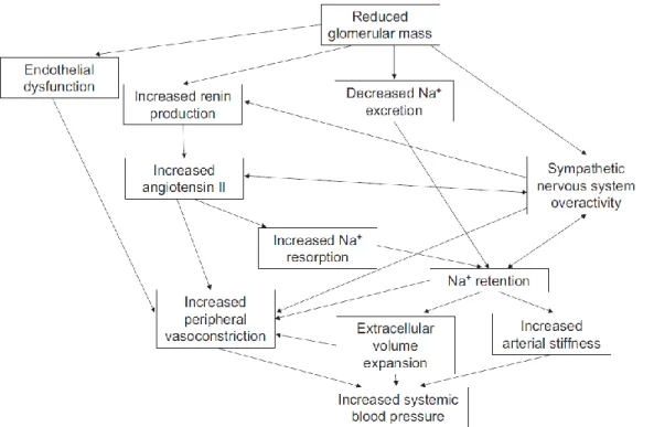
Hypertension and chronic kidney disease (CKD) are in a closely interlinked cause and effect relationship. Sustained elevations in blood pressure (BP) can lead to worsening kidney function, and the progressive decline in kidney function can conversely lead to worsening BP control. The leading causes of CKD are hypertension and diabetes. CKD affects as many as 10–15% of the adult population worldwide [42]. Patients with CKD are more likely to die from cardiovascular diseases than those without CKD. The prevalence of hypertension ranges from 60% to 90% in CKD patients, depending on the cause of CKD and its stages [43]. Also, it is gaining attention that hypertension and CKD in adulthood have childhood antecedents from as early as in utero and the perinatal period [31]. The pathophysiological mechanisms of hypertension in CKD involve multiple factors, including reduced nephron mass, increased sodium retention, activation of the RAS, volume overload, endothelial dysfunction, and sympathetic nervous system overactivity (Figure 6).
Figure 6. Pathophysiologic mechanisms of hypertension in chronic kidney disease. Adapted from “Hypertension in CKD: Core Curriculum 2019,” by E. Ku, 2014, American Journal of Kidney Diseases, 74(1), p. 121, Copyright 2019, with permission from Elsevier. [43]
Sodium homeostasis (i.e., the equivalent of sodium intake by sodium output) plays a pivotal role in extracellular fluid volume homeostasis, which is highly associated with long-term regulation of mean arterial blood pressure. The kidney is a principal organ to excrete sodium from the body, and it plays a central role in maintaining the long-term stability of mean arterial blood pressure. Numerous studies demonstrate that the inability to excrete sodium leads to increased BP in both humans and experimental animals [7]. AT2R is expressed continuously at a low level in physiological conditions but is upregulated in patients with certain cardiovascular and renal diseases. Accumulating evidences have demonstrated that renal AT2R plays an essential role in mediating natriuresis. AT2RKO mice were found to have a hypersensitive anti-natriuretic effect to exogenous Ang II and an abnormal pressure-natriuretic response, suggesting that the pressure-natriuretic action of AT2R is to maintain sodium homeostasis and further to regulate BP. It has been demonstrated that a highly selective
nonpeptide AT2R agonist, Compound 21 (C21), can markedly promote natriuresis by approximately 10-fold in Sprague-Dawley rats [44]. Besides, renal interstitial infusion of Ang III, an endogenous AT2R agonist, significantly increases AT2R-mediated natriuresis, which is abolished in the presence of an AT2R antagonist, PD123319 [45]. AT2R-mediated natriuresis is dependent on the bradykinin–NO–cGMP signaling pathway, which can internalize and inactivate major proximal tubule sodium transporters: sodium-hydrogen exchanger-3 (NHE-3) and sodium-potassium adenosine triphosphatase (NKA), therefore decreases sodium reabsorption (Figure 7) [7]. Thus, activation of AT2R prevents sodium retention and further lowers BP.
Figure 7. Schematic depiction of a renal proximal tubule cell showing the principal Na+ transporters and angiotensin receptors that regulate them. AT1Rs are depicted in light blue and AT2Rs in dark blue. Green arrows depict stimulation; red arrows depict internalization and inactivation; red lines depict inhibition. AT2R activation by endogenous agonist Ang III or exogenous nonpeptide agonist C-21 stimulates AT2R recruitment from intracellular sites to the apical plasma membranes of renal proximal tubule cells, reinforcing, and sustaining the natriuretic response. AT2R activation via
bradykinin (BK)–NO–cGMP signaling pathway internalizes and inactivates major Na+ transporter molecules NHE-3 and NKA, counterbalancing AT1R actions to increase Na+ reabsorption by stimulating these transporters. Adapted from “AT2 Receptors: Potential Therapeutic Targets for Hypertension,” by R.M. Carey, 2017, American Journal of Hypertension, 30(4), p. 342, Copyright 2016, with permission from Oxford University Press. [7]
AT2R-mediated vasodilatory responses oppose the vasoconstrictive effects of Ang II via the AT1R. AT2RKO mice exhibit higher basal BP than wild-type mice and have increased sensitivity to exogenous Ang II. However, these hemodynamic responses to Ang II are significantly blunted in mice over-expressing AT2R selectively in vascular smooth muscle cells [30]. Similar to the signaling of the kidney, AT2R-mediated vasodilation in the vasculature is also dependent on the bradykinin–NO–cGMP signaling pathway. Numerous studies have demonstrated that AT1R blockade (ARB) is essential to AT2R-mediated vasodilatory responses in both animal models and human arterioles [5]. For example, Bosnyak et al. [46] reported that a selective AT2R agonist (C21) alone did not lower BP in conscious experimental animals. However, when given in combination with ARB, C21 lowered BP, and this effect was abolished with the AT2R antagonist, PD123319. Thus, AT2R-mediated antihypertensive responses can be revealed by ARB. Nonetheless, the antihypertensive responses mediated by AT2R appear to be dependent on its expression level in the vasculature, the condition of activation of the RAS, and the presence or absence of ARB.
Fibrosis occurs with all forms of kidney diseases and it accelerates kidney failure. It is a persistent repair response of the normal wound healing process characterized by injury, inflammation, myofibroblast activation, as well as extracellular matrix deposition and remodeling. AT2R has been shown to have a renal protective ability by preventing fibrosis. It appears that AT2R deficiency aggravates kidney injury and decreases survival in mice with CKD [47]. Over-expression of the AT2R in a mouse remnant kidney model ((5/6) nephrectomy) ameliorates glomerular injury via decreased pro-fibrotic cytokines [48]. Thus, AT2R might be utilized as a therapeutic target to maintain cardiovascular and renal homeostasis in pathological conditions. The potential anti-fibrotic mechanisms of AT2R are likely to be associated with the reduction of inflammation and pro-fibrotic factors such as
transforming growth factor-β (TGFβ). A selective non-peptide AT2R agonist, C21, provides the opportunity for the understanding of AT2R function. It has been shown that C21 could exert an anti-inflammatory effect via inhibited NF-κB activation leading to reduced inflammatory cytokines and chemokines in a variety of animal models [49]. In addition, C21-mediated AT2R activation has been shown to increase NO and cGMP levels in the kidney and further to inhibit TGFβ signaling which in turn ameliorates TGFβ-mediated myofibroblast activation and extracellular matrix deposition, thereby providing another mechanism to modify fibrosis production [49]. It is reported that the AT2R may form heterodimers with other GPCR such as relaxin family peptide receptor 1 (RXFP1) to regulate fibrosis progression via the pERK1/2–NOS–NO–cGMP pathway [50].
1.1.3. Glomerulogenesis and Podocyte
1.1.3.1 Kidney and Glomerulus
The kidney is an essential excretory and homeostatic organ. It possesses two primary functions: it excretes various metabolic end products, removing them from the blood and excreting them in urine; and it concentrates certain constituents of the body’s fluids to regulate acid-base balance, extracellular fluid volume, and electrolyte concentrations by controlling their excretion and reabsorption. A nephron is the smallest functional unit of the kidney and consists of a renal corpuscle and a tubule unit including proximal tubule, loop of Henle, distal tubule, and collecting duct. The renal capsule, as the beginning of nephron, consists of glomerulus and Bowman’s capsule. Each component of the kidney plays distinct and pivotal roles in urine formation as well as in the homeostasis of extracellular fluid.
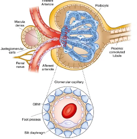
The glomerulus is an essential functional unit for kidney filtration and a specialized network of capillaries known as a tuft that is practically located between two resistance vessels (i.e., the afferent and efferent arterioles). These capillaries are surrounded by Bowman’s capsule and structurally supported by the glomerular mesangial cells. The filter itself has a unique and complex structure and is composed of a multi-layered and multi-cellular barrier, including the fenestrated endothelium of glomerular capillaries, the glomerular basement membrane (GBM), and the filtration slit diaphragm which is connecting adjacent podocyte foot processes (Figure 8). Thus, the primary glomerular filtrate goes through this unique
structure and enters a space defined by the visceral and parietal epithelial cells before flowing into the proximal tubule. Failure in any components of this unique structure causes the loss of size- and charge-selective filtration, clinically characterized by proteinuria [40]. The unique and complex structure of the glomerulus is derived from the metanephric mesenchyme and is based on a tightly regulated developmental program by the processes of glomerulogenesis.
Figure 8. Structure of the Bowman’s capsule and glomerular capillary tuft. The capsule is lined with parietal epithelium, which is connected to the cells of the proximal tubule at the urinary pole, which is the right part of the upper picture. Left part of the upper picture is called vascular pole and this includes both the afferent and efferent arterioles. The relationship between these arterioles and the specialized portion of the distal nephron called the macula densa is illustrated. (Inset) The layers that comprise the filtration barrier are displayed below. The outermost layer is composed of the visceral epithelial cells, the podocytes, next to the glomerular basement membrane (GBM) and finally, the fenestrated endothelial cells. Adapted from “The glomerulus: the sphere of influence,”
by M.R. Pollak, 2014, Clinical journal of the American Society of Nephrology: CJASN, 9(8), p. 1462, Copyright 2014, with permission from American Society of Nephrology. [51]
1.1.3.2 Glomerulogenesis
The permanent mammalian kidney (metanephros) stems from metanephric mesenchyme and starts to develop by UB outgrowth (Figure 5) [38, 40]. Kidney development is governed by a cascade of morphogenetic interactions among three cell lineages of different origins: the epithelial cells of the UB, the mesenchymal cells of the metanephric mesenchyme and the endothelial cells of the angioblasts. The development of the embryonic metanephros consists of two different processes: glomerulogenesis and tubulogenesis. Glomerulogenesis proceeds through five morphological stages in embryonic development: renal vesicle, comma-shaped body, S-comma-shaped body, capillary loop, and maturing glomerulus [40]. Glomerulogenesis is divided into two steps, before and after endothelial cell invasion (Figure 9) [52]. In the beginning, mesenchymal cells adjacent and inferior to the tips of UB start to condense, and this group of cells forms a pretubular aggregate. In response to signaling from the UB and surrounding stroma, the pretubular aggregate conducts a mesenchymal–to–epithelial transition forming a polarized renal vesicle [53], which then becomes a comma-shaped body composed of parietal cells and presumptive podocytes. At the same time, the UB continues to branch and induce new aggregates at the bud tips. The polarized renal vesicles remain attached and fuse to the ureteric bud epithelium at their distal part. The renal vesicles elongate along the proximal-distal axis and differentiate into nephron segments in which podocytes are located at the proximal end of the nephron epithelia. This proximal-distal polarity is related to the different gene expression levels [54]. For example, the proximal end of renal vesicle begins to express podocyte-related genes, such as Wt1and MafB, indicating the possibility that the cells within the proximal part have already been directed toward the podocyte lineage [54].
The functional glomerulus formation initiates at endothelial cells invasion into the vascular cleft observed in the S-shaped body. The S-shaped body is characterized by the presence of a layer of presumptive podocytes and a vascular cleft. At this stage, the S-shaped body is already patterned along the proximal-distal axis [38]. The distal end that had remained
in connect to the ureteric bud epithelium has now fused to form a continuous epithelial tubule. The proximal end assembles the glomerular tuft when endothelial cells migrate to the more proximal cleft. At this stage, the gradient of Wnt/β-catenin activity is generated along the proximal-distal nephron axis, and the proximal end of the S-shaped body, which contains presumptive podocytes, expresses the lowest β-catenin level, suggesting that Wnt signaling might regulate the development of podocytes [55]. The migration of endothelial cells into the vascular cleft depends on the signaling of angiogenic factors such as vascular endothelial growth factor (VEGF), which is produced by presumptive podocytes [52]. Then, endothelial cells proliferate and aggregate to form the first capillary loop without a lumen. It has been demonstrated that TGFβ signaling is associated with the process of lumenation [52]. After the initial migration of endothelial cells, mesangial cells migrate into the glomerulus around the first capillary loop and play a pivotal role in the formation of multiple capillary loops. In the mature glomerulus, the first capillary loop divides into six to eight capillary loops, and endothelial cells acquire a fenestrated morphology. Finally, the fully matured glomerulus includes four highly specified cell populations: the fenestrated endothelial cells, mesangial cells, podocytes, and the parietal epithelial cells of the Bowman’s capsule.
In the stages of comma-shaped body and S-shaped body, presumptive podocytes display a layer of columnar-shaped epithelial cells connected by apical junctions, having no foot processes and slit diaphragm, but produce VEGF to attract vascular endothelial growth factor receptor 2 (VEGFR2)-expressing angioblasts [52]. In the subsequent capillary loop stage, podocytes lose their lateral cell attachments to each other, but they remain attached at their basal membrane. At this stage, podocytes begin to form foot processes and slit diaphragm along the basal aspect of the lateral membrane, migrate around the capillary loops, and stop dividing by increasing the cyclin-dependent kinase inhibitors, p27 and p57 [56]. During podocyte development, podocytes gradually increase in size and extend their foot processes to a significant distance from the main cell body as well as exert normal physiological functions.
The mammalian kidney develops by the cells derived from three fundamental lineages: the UB lineage, the nephron lineage, and the stromal lineage. The metanephric mesenchyme consists of nephron progenitor cells (NPCs) and stromal progenitors. NPCs are characterized by expression of Six2 and differentiate into the entire nephron epithelium, including podocytes,
glomerular parietal epithelial cells, proximal tubule epithelial cells, loop of Henle, and distal tubule epithelial cells [57]. The stromal progenitors express Foxd1 and differentiate into interstitial fibroblasts, pericytes, mesangial cells and vascular smooth muscle [58]. The kidney vasculature derives from c-kit+/SCL+ and CD146+ endothelial progenitors [58-60]. The cell-cell interactions among those cell-cells are essential to the growth and continued patterning of the kidney. The Wilms’ tumor 1 (WT1) transcription factor is a potent regulator of NPCs in the development of kidney and involved in many genes with essential roles in kidney development [40]. The other transcription factors, including PAX2, SALL1, EYA1, and OSR1also play an important role in kidney development [40, 58].
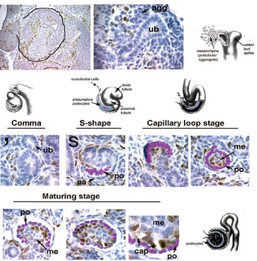
Figure 9. Migration of endothelial cells into the developing glomerular tuft. (Top) An E12.5 mouse metanephros is outlined in black. Endothelial cells express a VEGFR2-GFP transgene and stain brown. The glomerulus develops from a pretubular aggregate (agg) that forms immediately adjacent and below the tips of the ureteric buds (ub). (Middle and
Bottom) VEGFR2-positive cells are seen to “hug” the developing comma-shaped-stage nephron. The S-shaped stage is defined by the presence of a layer of presumptive podocytes (po) and a vascular cleft. Endothelial cells seem to be streaming into this cleft from the metanephric mesenchyme (MM). At the capillary loop stage, pockets of endothelial cells sit right next to the podocytes, and mesangial cells are soon found inside a single capillary loop. By the maturing stage, capillary lumens are beginning to form, and a large population of mesangial cells is present. Schematic diagrams of each developmental stage are shown above the photomicrographs. Podocytes have been digitally colorized for identification (purple). Abbreviation: pa, parietal epithelial cell; po, podocyte; me, mesangial cell; cap, capillary loop. Adapted from “How do mesangial and endothelial cells form the glomerular tuft?” by M.R. Vaughan, 2008, Journal of the American Society of Nephrology: JASN, 19(1), p. 25, Copyright 2008, with permission from American Society of Nephrology. [52]
1.1.3.3 Podocyte
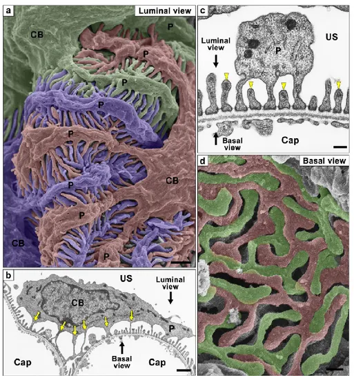
Podocyte is particularly specialized epithelial cell that resides on the visceral side of the Bowman’s capsule, wrap around glomerular capillaries, and serves as an essential component of the glomerular filtration barrier. These cells are thought to be the target cell in most forms of inherited and acquired glomerular disease including minimal change disease (MCD), focal segmental glomerulosclerosis (FSGS), and diffuse mesangial sclerosis. Podocytes are structurally divided into three kinds of subcellular compartment: cell body, primary process, and foot process (Figure 10). The cell body of podocyte has several thick primary processes, and many fine foot processes extending from each of the primary processes. The foot processes interdigitate with adjacent foot processes from neighboring podocytes. They are separated from each other by filtration slits and bridged with a specialized intercellular junction known as the slit diaphragm.
Figure 10. Podocyte subcellular compartments showed by conventional SEM and TEM. (a) Conventional SEM image. Three neighboring podocytes are individually colored with blue, green, and red. (b, c) Conventional TEM images. (d) Alkaline-maceration SEM image. CB, cell body; Cap, capillary lumen; P, primary process; US, urinary space of the Bowman’s capsule. Adapted from “Three-dimensional architecture of podocytes revealed by block-face scanning electron microscopy,” by K. Ichimura, 2015, Scientific reports, 5:8993, p. 2. [61]
The glomerular filtration barrier comprises three layers: glomerular endothelial cell, GBM, and epithelial podocyte. It is widely accepted that damage to any layer might result in leakage of macromolecules passing through the glomerular filtration barrier and ending up in the urine. The slit diaphragm plays a pivotal role as a size barrier for filtration. It is composed of nephrin, podocin, CD2AP (CD2-associated protein), TRPC6 (transient receptor potential