Original article
Limb salvage with isolated perfusion for soft tissue sarcoma:
could less TNF-a be better?
S. Bonvalot
1*, A. Laplanche
2, F. Lejeune
3, E. Stoeckle
4, C. Le Pe´choux
5, D. Vanel
6, P. Terrier
7,
J. Lumbroso
8, M. Ricard
8, G. Antoni
2, A. Cavalcanti
1, C. Robert
9, N. Lassau
6, J. Y. Blay
10& A. Le Cesne
9Departments of1Surgery,2Public Health,5Radiotherapy,6Radiology,7Pathology,8Nuclear Medicine and9Medical Oncology, Institut Gustave Roussy, Villejuif, France;3Multidisciplinary Oncology Center, Centre Hospitalier Universitaire Vaudois, Lausanne, Switzerland;4Department of Surgery, Institut Bergonie´, Bordeaux, France;10Department of Medical Oncology, Centre Le´on Be´rard, Lyon, France
Received 6 February 2005; revised 31 March 2005; accepted 7 April 2005
Background:
The optimal dose of TNF-a delivered by isolated limb perfusion (ILP) in patients
with locally advanced soft tissue sarcoma is still unknown.
Patients and methods:
Randomised phase II trial comparing hyperthermic ILP (38 – 408) with
melphalan and one of the four assigned doses of TNF-a: 0.5 mg, 1 mg, 2 mg, and 3/4 mg upper/lower
limb. The main end point was objective tumour response on MRI. Secondary end points were
histo-logical response, rate of amputation and toxicity. Resection of the remnant tumour was performed
2 – 3 months after ILP. The sample size was calculated assuming a linear increase of 10% in the
objective response rates between each dose level group.
Results:
One hundred patients (25 per arm) were included. Thirteen per cent of patients had a
sys-temic leakage with a cardiac toxicity in six patients correlated with high doses of TNF-a. Objective
tumour responses were: 68%, 56%, 72% and 64% in the 0.5 mg, 1 mg, 2 mg and 3 or 4 mg arms,
respectively (NS). Sixteen per cent of patients were not operated, 71% had a conservative surgery
and 13% were amputated with no difference between the groups. With a median follow-up of 24
months, the 2 year overall and disease-free survival rates (95% CI) were 82% (73% to 89%) and
49% (39% to 59%), respectively.
Conclusion:
At the range of TNF-a doses tested, there was no dose effect detected for the objective
tumour response, but systemic toxicity was significantly correlated with higher TNF-a doses.
Efficacy and safety of low-dose TNF-a could greatly facilitate ILP procedures in the near future.
Key words:
isolated limb perfusion, soft tissue sarcoma, TNF-a
Introduction
Isolated limb perfusion (ILP) was introduced by Creech in
1957 [1] for the treatment of patients with locoregionally
recurrent melanoma, with approximately 50% – 65% complete
responses with melphalan alone [2]. In contrast, ILP with
mel-phalan alone or other drugs has been used in the treatment of
patients with extremity soft tissue sarcoma with limited
suc-cess [3]. The addition of hyperthermia enhances cytotoxic
effects of alkylating agents such as melphalan [4]. Tumour
necrosis factor-alpha (TNF-a) was first described as an
antitu-mour factor present in the serum of animals that had been
primed with immunomodulators such as BCG, and then
treated with endotoxin [5]. Response rates seen with TNF-a
as a single systemic agent in different trials were very low,
with dose-limiting toxicity being constitutional symptoms and
hypotension [6]. Lie´nard et al. pioneered the administration
via ILP of high-dose TNFa and g – interferon with melphalan
[7]. The initial doses of 4 mg rHuTNF-a in the lower
extre-mity and 3 mg in the upper extreextre-mity were tested arbitrarily as
10 times the maximum tolerated dose in humans [8], and
equivalent to the effective dose in rats, i.e. around 50 mg/kg
[9]. Subsequently, this choice of dose has been continued
unaltered because of the high response rate. In a multicenter
European trial, ILP with high-dose TNF-a and melphalan
resulted in a 76% response rate and 71% limb salvage in
patients with limb-threatening soft-tissue sarcomas, leading to
the approval of TNF-a in Europe [10].
Until now, no other available treatment seems to give
com-parable results when applied to limb-threatening soft tissue
sarcomas. Mechanisms involved are an increase of melphalan
*Correspondence to: Dr S. Bonvalot, Department of Surgery, Institut Gustave Roussy, 39 rue Camille, Desmoulins, 94805 Villejuif Cedex, France. Tel: + 33-1-42-11-43-83; Fax: + 33-1-42-11-52-56; E-mail: [email protected]
Published online 1 June 2005
uptake in tumour tissue [11] and a selective destruction of
tumour vessels with the addition of TNF-a [7, 12, 13].
How-ever, the optimal dose of TNF-a delivered by isolated limb
perfusion in patients with advanced soft tissue sarcoma
(ASTS) is still unknown. In patients with melanoma,
increas-ing the TNF-a dose to 6 mg did not increase the complete
response rate but increased regional toxicity [14].
Pharmaco-kinetics data [15] showed plateau levels of micrograms of
TNF-a in perfusates during the whole 90 min ILP, suggesting
a saturation of the TNF-a receptors.
In a de-escalation study in rats, de Wilt demonstrated that
the synergy between rHuTNF-a and melphalan was lost at a
dose of TNF-a below 40 mg/kg [16]. Since rHuTNF-a in mice
binds only to the p55 receptor and not to the p75, its activity
is five to 10 times less than murine TNF-a (MuTNF) [16, 17].
This suggests that the dose of TNF-a currently used in clinical
setting (approximately 50 mg/kg) might be reduced five to
10-fold while retaining the synergistic effects. Preclinical studies
have showed that targeted delivery to tumour vessels of very
low doses (picograms) of TNF-a enhances the penetration of
doxorubicin in murine models [18] and that systemic
adminis-tration of low-dose TNF-a increases the antitumour activity of
a liposomal formulation of doxorubicin [19]. Clinical
obser-vations also favour a TNF-a dose decrease. Kinetics revealed
that with 3 or 4 mg of rHuTNF-a, the perfusion system is
supersaturated [20]. There is no significant difference in
tumour response between patients with leakage over 2%,
where the exposure of the perfused limb to TNF-a was 18.7%
lower, and those without leakage [21].
Severe systemic toxicity and haemodynamic changes after
ILP with TNF-a and melphalan, with or without interferon-g,
have been reported in several series, although reduced when
leakage is adequately controlled [20]. The potential advantage
of a lower dose of TNF-a includes a lower incidence of
sys-temic adverse events leading to a more simple and safe
pro-cedure with a significantly lower cost. Subsequently, this
approach could be expanded to a wider spectrum of
malignan-cies and patients.
In order to challenge the use of the usual high dose of
TNF-a (3 mg for the upper limb and 4 mg for the lower limb),
we conducted a randomised phase II comparing four doses of
TNF-a: 0.5 mg, 1 mg, 2 mg or the registered dose for ILP in
humans. Our primary end point was radiological response,
which is the main criterion to make the correct decision about
limb salvage or amputation.
Patients and methods
Eligibility criteria
It was required that the patient had a locally limb advanced soft tissue sar-coma considered non-resectable by the referring surgeon of one of the three participating centres, i.e. that could only be treated by amputation or functionally mutilating surgery. The tumours were considered unresectable because of either multifocal disease or fixation/invasion to the neuro-vascular bundle and/or bone. The diagnosis was confirmed by histological re-examination of the primary tumour specimen by an expert pathologist. Sarcomas were classified as low (I), intermediate (II) or high grade (III),
based upon histological examination of the primary according to the French cancer center’s histological grading [22]. Primary and recurrent sarcomas were included. Patients with synchronous metastasis were not excluded.
Patients had to be older than 16 years, with an Eastern Cooperative Oncology Group (ECOG) performance status of 0 or 1 and normal cardiac function. All patients provided written consent before randomisation. The protocol was approved by the Ethics Committees in the three participating institutions.
Randomisation
Eligible patients were randomly assigned to receive one of the four TNF-a doses: 0.5 mg, 1 mg, 2 mg or 3/4 mg for upper/lower limb. Ran-domisation was done by telephone or fax upon eligibility checklist, using a block allocation scheme.
ILP
Recombinant human TNF-a (TNF-a-1a, BeromunTM) was provided by Boehringer Ingelheim GmbH, Ingelheim/Rhein, Germany. Melphalan (Alkeran) was provided by Glaxo-Wellcome (London, UK). Under general anaesthesia, the main artery and vein of the affected limb were clamped and canulated after heparinisation. Cannulae were then connected to a heart – lung machine and a pneumatic tourniquet was applied proximally to prevent leakage into the general circulation. ILP consisted of extra-corporeal circulation with mild hyperthermia (tissue temperature 38 – 408C) obtained with a heat-exchanger. When sarcoma did not affect feet or hands, they were wrapped tightly with an Esmarch rubber bandage immediately before injection of the drugs into the perfusion circuit to pre-vent sequelae [23]. The allocated dose of TNF-a according to randomis-ation was injected into the arterial line when the limb tissue temperature was greater than 388C. Thirty minutes later, 10 mg/l (leg) or 13 mg/l (arm) of melphalan per limb volume was then added for the following 60 min, as performed in the European trial [10]. Limb volume was calculated using specifically dedicated software (Lie´nard D., unpublished). A 4 – 6 l washout of the limbs, using a mixture of Hartmann’s solution and Macro-dex, was performed at the end of the procedure. During the washout, the limb was extensively massaged.
Leakage from the isolated perfusion circuit to the systemic compart-ment was assessed with technetium 99m radiolabelled human serum albu-min (Vasculocis, Cis Biointernational, Schering, Gif-sur-Yvette, France). After injection of a small activity (4 MBq) in the systemic circulation (calibration of the blood volume and efficiency of a precordial NaI(Tl) scintillation probe), 100 MBq of radiolabelled human serum albumin were injected into the isolated circuit. Any increase in the precordial counting rate (continuous curve recording) was interpreted as a leakage from the isolated circuit to the systemic circulation, and was quantified according to calibration parameters.
Post ILP surgery and adjuvant treatments
A delayed resection of the remnant tumour was usually planned 2 – 3 months after ILP. The aim was to perform en bloc resection, with free margins whenever possible. Major vessels or nerve trunk included in the tumour were resected en bloc to avoid tumour rupture. Vascular graft, muscular flaps and skin grafts were used when necessary. Transpositions of tendons were used to palliate nerve trunk resection.
Adjuvant radiotherapy was considered in patients who had no previous radiotherapy and when the margins were close to histologically viable tumour. Adjuvant chemotherapy was optional for non-metastatic patients, its indication relying mainly upon high histological grade and young age of the patient.
End points and clinical follow-up
The primary end point was objective tumour response [complete response (CR) or partial response (PR)], assessed on MRI performed 1 and 2 months after ILP, immediately prior to surgery. The MRI examinations included T1-weighted SE and fast SE T2-weighted fat-saturated sequences, as well as dynamic sequences (T1-weighted SE repeated six times every 40 s), displaying the maximum intensity slope in each pixel [2]). CR was defined by complete necrosis of the tumour or disappearance of all measurable disease. A partial response (PR) was defined as a regression of the tumour size greater than 50% in the product of the bi-dimensional measurements. Progressive disease (PD) was defined as a greater than 25% disease progression or the appearance of any new lesion [24].
Secondary end points were the ability to perform a conservative sur-gery, histological response, toxicity and survival. Histopathological response was defined as complete response (pCR) if no residual identifi-able tumour cells were present, very good response between 1% and 10% of identifiable tumour cells, partial response between 11% and 50% of identifiable tumour cells, and no change if more than 50% identifiable tumour cells were present in the resection specimen. The R classification of UICC was used to classify the quality of resection [25]. Systemic tox-icity and peripheral neuro-toxtox-icity were graded according to the World Health Organisation (WHO) criteria scale [26]. Local toxicity was graded according to Wieberdink’s scale [27]. Late toxicity was evaluated 6 months after ILP.
Statistical design
The sample size was calculated assuming a linear increase of 10% in the objective response (OR) rate between each dose level group, i.e. between 45% (0.5 mg group) and 75% (3 mg/4 mg group). With a type I error of 5% and a type II error of 25%, 25 patients per group were required assum-ing this hypothesis [28]. Results are expressed as percentages with 95% confidence intervals (CIs) or as medians and range. The Wilcoxon test for trend was used to compare the percentages between the four dose groups.
Survival was calculated using the Kaplan and Meier method [29] with Rothman’s 95% CIs [30]. Median time of follow-up was calculated with the Schemper method [31]. In the calculation of the local recurrence rate, deaths without recurrence were censored. All tests were two-sided.
Results
Patient’s characteristics
One hundred consecutive patients were enrolled in three
centres between June 2000 and July 2003 (83 patients in
Gustave Roussy Institute, nine patients in Centre Hospitalier
Universitaire Vaudois and eight patients in Bergonie´ Institute).
Twenty-five patients were randomly assigned to each TNF-a
dose group. Initial characteristics per group are presented in
Table 1. Size, recurrences, grade and multifocality are well
balanced in the four groups.
Histopathological subtypes are: liposarcoma (n = 23),
undif-ferentiated sarcoma (n = 20), synovial sarcoma (n = 16),
epithe-lioid
sarcoma
(n = 8),
angiosarcoma
(n = 7),
malignant
peripheral nerve sheath tumours (n = 7), fibroblastic sarcoma
(n = 6), muscular sarcoma (n = 5), and miscellaneous (n = 8).
The median tumour size was 100 mm (range 20 – 208) for
lower limbs and 70 mm (range 10 – 125) for upper limbs. ILP
was performed in a previously irradiated volume for 27
patients.
ILP and leakage
All but four patients received the allocated TNF-a dose. Three
patients received a different dose in error: one patient of the
1 mg group received 2 mg, one patient of the 2 mg group
received 1 mg, and one patient of the 3 mg/4 mg group with
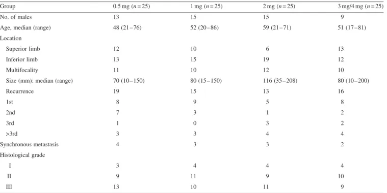
Table 1.Patient characteristics
Group 0.5 mg (n = 25) 1 mg (n = 25) 2 mg (n = 25) 3 mg/4 mg (n = 25)
No. of males 13 15 15 9
Age, median (range) 48 (21 – 76) 52 (20 – 86) 59 (21 – 71) 51 (17 – 81) Location
Superior limb 12 10 6 13
Inferior limb 13 15 19 12
Multifocality 11 10 12 10
Size (mm): median (range) 70 (10 – 150) 80 (15 – 150) 116 (35 – 208) 80 (10 – 200)
Recurrence 19 15 13 16 1st 8 9 5 8 2nd 7 3 1 2 3rd 1 0 3 2 >3rd 3 3 4 4 Synchronous metastasis 4 3 3 2 Histological grade I 3 4 4 4 II 9 11 9 10 III 13 10 11 9
a lower limb tumour received 3 mg instead of 4 mg. One
patient of the 3 mg/4 mg group did not have the perfusion
because of massive nodal involvement discovered during the
cannulation.
Median duration to reach the adequate tissue temperature
was 20 min (range 5 – 109). Median superficial/deep tissue
temperatures were 38.6/39.18C and 39.6/39.78C at the girdle
of the limb and distally, respectively. Thirteen per cent of
patients had a drug leakage after drug injection (median 3%,
range 1% – 12%). Reducing the perfusion flow rate corrected
the leakage most of the time. High leakages were due to a bad
venous return: for eight patients, reposition of the cannula of
the femoral vein under the level of the tourniquet corrected
this technical problem. Despite extensive washout, 17%
patients had leakage (median 3%, range 1% – 14%) after
reconnection of the limb circulation.
Toxicity
There was no toxic or surgery related deaths. Grade II and III
cardio-vascular toxicity (Table 2) was correlated with the
TNF-a dose (P <0.01). Only one patient (4 mg) needed
circu-latory support with dopamine infusion because of a ‘sepsis
like’ syndrome. Five patients (2, 3 or 4 mg) developed low
systolic blood pressure requiring fluid management. No
haematological, hepatic, pulmonary toxicity or allergic
reac-tion over grade I was observed. No compartment syndrome
was observed. Local toxicity did not differ according to
treat-ment arm (Table 2), gender (42% for women versus 37% for
men), pre-irradiation (37% for pre-irradiated patients versus
40%) or tumour location (34% upper limb versus 43% lower
limb).
Surgical morbidity after ILP consisted of two arterial
thromboses, one treated surgically and another percutaneously.
All patients were discharged from hospital, able to walk, after
a median stay of 7 days after ILP (range 4 – 15).
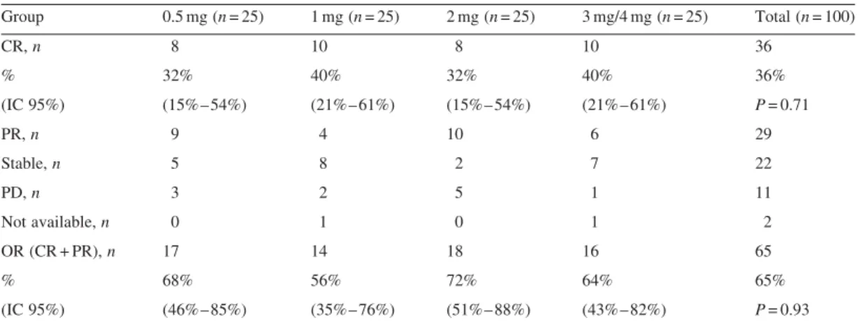
Tumour response
All but two patients were evaluated for response (one patient
did not have ILP, another was amputated outside the referring
centre before evaluation). MRI and histological responses
observed in the four groups are shown in Tables 3 and 4. The
percentages of OR are 68%, 56%, 72% and 64% in the
0.5 mg, 1 mg, 2 mg and 3 mg/4 mg groups, respectively
(P = 0.93). OR did not differ significantly according to tumour
grade (53%, 56% and 77% in grades I, II and III),
pre-irradiation (52% for pre-irradiated patients versus 70%) or
Table 2.ILP toxicity
Group 0.5 mg (n = 25) 1 mg (n = 25) 2 mg (n = 25) 3 mg/4 mg (n = 24) Acute regional tissue reactions 12 (48%) 10 (40%) 8 (32%) 9 (38%)
Grade II 9 8 6 8
Grade III 3 2 1 1
Grade IV 0 0 1 0
Paraesthesiae (grade I) 6 (24%) 3 (12%) 1 (4%) 3 (13%)
Nerve palsy (grade III) 3 (12%) 4 (16%) 0 (0%) 3 (13%)
Stiffness 5 (20%) 4 (16%) 3 (12%) 6 (25%)
Hemodynamic grade II toxicity 0 (0%) 0 (0%) 1 (4%) 4 (16.6%)
Hemodynamic grade III toxicity 0 (0%) 0 (0%) 0 (0%) 1 (4%)
Acute regional tissue reactions according to Wieberdink et al. [26]. Hemodynamic and peripheral neurotoxicity according to WHO grading [25].
Table 3. MRI response
Group 0.5 mg (n = 25) 1 mg (n = 25) 2 mg (n = 25) 3 mg/4 mg (n = 25) Total (n = 100) CR, n 8 10 8 10 36 % 32% 40% 32% 40% 36% (IC 95%) (15% – 54%) (21% – 61%) (15%– 54%) (21% – 61%) P = 0.71 PR, n 9 4 10 6 29 Stable, n 5 8 2 7 22 PD, n 3 2 5 1 11 Not available, n 0 1 0 1 2 OR (CR + PR), n 17 14 18 16 65 % 68% 56% 72% 64% 65% (IC 95%) (46% – 85%) (35% – 76%) (51%– 88%) (43% – 82%) P = 0.93
topography (61% upper limb versus 68% lower limb).
Correlation between radiological (CR or not on MRI) and
pathological responses (less than 11 viable cells or not) was
good (82%) (Table 5).
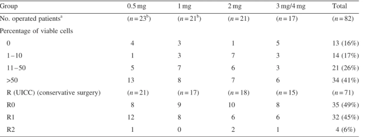
Post ILP surgery
Sixteen patients were not operated (nine metastasis
pro-gression, four patient refusals, one second cancer, two other
reasons), 71 underwent conservative surgery and 13 were
amputated. The percentages of conservative surgery (Table 6)
are 84%, 68%, 72% and 60% in the 0.5 mg, 1 mg, 2 mg and
3 mg/4 mg
groups,
respectively
(P = 0.10).
Conservative
surgery required a myocutaneous flap in 24 patients (two
MacGregors, two pedicled latissimus dorsi, 20 latissimus dorsi
free flaps) and four vascular grafts. Quality of surgery in the
71 patients with conservative surgery was: R0 in 35 patients,
R1 in 32 patients and R2 in four patients. Two patients were
amputated after vascular complications (one occlusion of a
vascular graft associated with a muscular free flap 1 month
after surgery, and one vascular rupture). Rates of limb salvage
were 88%, 80%, 88%, 92% in the 0.5 mg, 1 mg, 2 mg and
3 mg/4 mg groups, respectively (NS).
Further treatments
Thirty-seven patients received post-operative radiotherapy at a
median dose of 50 Gy (range 15 – 65) concerning 10, 10, nine
and eight patients in the 0.5 mg, 1 mg, 2 mg and 3 mg/4 mg
groups, respectively. Adjuvant chemotherapy was delivered in
18 patients: three, six, three and six patients in the 0.5 mg,
1 mg, 2 mg and 3 mg/4 mg groups, respectively.
Late toxicity
Late toxicity consisted of a muscular atrophy (5%), oedema
(11%), grade 1 sensitive disorders (6%), grade 3 nerve palsy
(3%) and stiffness (16%). Late toxicity was not significantly
different whether a patient received post-ILP radiotherapy
(21%) or not (33%). However, three post-ILP irradiated
patients experienced a spontaneous fracture between 6 and 12
months after the end of treatment.
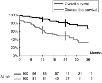
Survival and local recurrence
No patient was lost to follow-up. With a median follow-up of
24 months, the 2-year overall and disease-free survival rates
(95% CI) were 82% (73% – 89%) and 49% (39% – 59%),
respectively (Figure 1), with no significant difference between
the four groups. Median disease-free survival was 24 months.
Twenty-four local recurrences were recorded with a median
time to progression of 13 months: 6, 5, 8 and 5 months in the
0.5 mg, 1 mg, 2 mg and 3 mg/4 mg groups, respectively, with
no statistical difference between the four groups (Figure 2).
The 2-year local recurrence rate (95% CI) was 27%
(18% – 38%; Figure 3). Recurrences were correlated with
qual-ity (R) of post-ILP surgical resection (log rank P <0.00 001).
Table 4. Pathological response and quality of surgery (R)
Group 0.5 mg 1 mg 2 mg 3 mg/4 mg Total
No. operated patientsa (n = 23b) (n = 21b) (n = 21) (n = 17) (n = 82)
Percentage of viable cells
0 4 3 1 5 13 (16%)
1 – 10 1 3 7 3 14 (17%)
11 – 50 5 7 6 3 21 (26%)
>50 13 8 7 6 34 (41%)
R (UICC) (conservative surgery) (n = 21) (n = 17) (n = 18) (n = 15) (n = 71)
R0 8 9 10 8 35 (49%)
R1 12 8 6 6 32 (45%)
R2 1 0 2 1 4 (6%)
a
Operated patients: amputations + conservative surgery.
b
Two pathological responses missing.
Table 5.Correlation between MRI and pathological responses
MRI complete responses
% viable cells Yes No Total
0 – 10 21 (26%) 6 27
>11 9 46 (56%) 55
Total 30 52 82%
Table 6.Post-ILP surgery
Group 0.5 mg (n = 25) 1 mg (n = 25) 2 mg (n = 25) 3 mg/4 mg (n = 25) No surgery 1 3 4 8 Amputation 3 5 3 2 Conservative surgery 21 (84%) 17 (68%) 18 (72%) 15 (60%) Limb salvagea 22 (88%) 20 (80%) 22 (88%) 23 (92%)
A total of 13 amputations were initially performed (Table 6)
and eight additional amputations were performed during the
follow-up due to recurrence (six patients) or complication of
post-ILP surgery (two patients). The amputation rate at 2
years (95% CI) was 22% (14% – 32%; Figure 3). A total of 21
amputations were thus performed: five, five, six and five
amputations in the 0.5 mg, 1 mg, 2 mg and 3 mg/4 mg groups,
respectively.
Discussion
This trial was conducted over a short period of time (3 years
inclusion), with a high homogeneity of the ILP procedures, as
well as in the indications and evaluation of results. In Gustave
Roussy Institute, the 83 patients included represented 12% of
the operated sarcomas by the same surgeon during this period,
reflecting the strict criteria of inclusion.
The present study is the first to randomise different doses of
TNF-a with the registered dose of melphalan in sarcomas.
Animal models and kinetic findings suggested that very high
doses of TNF-a used traditionally in the clinical setting may
well be reduced. In a pilot clinical study [32], nine sarcoma
patients experienced a CR with a reduced dose of TNF-a
(0.5 – 0.925 mg) albeit with significant local toxicity. In 20
patients with melanoma [33], low-dose TNF-a ILP achieved
tumour responses comparable with those historically reported
with a higher dose.
Our primary end point was radiological response, which is
the main criterion to make the correct decision about limb
sal-vage or amputation. Our study failed to show a 10%
differ-ence between the different dose groups, which was the
statistical prerequisite to find a TNF-a dose effect (see
Patients and methods) at least at this range of TNF-a doses.
The rates of conservative surgery, which is the final issue,
were equivalent in the four groups. These results suggest that
synergism between low-dose TNF-a and melphalan was
con-served. Moreover, overall response rates and limb salvage
rates are similar to those of previous studies with high doses
of TNF-a [10, 34, 35].
There was no significant difference in terms of CR
accord-ing to tumour grade, although there was a trend for better
responses in patients with high-grade sarcoma. This is
sup-ported by the observation of better clinical responses
corre-lated with high mitotic activity and a low amount of apoptosis
in tumour samples taken prior to ILP with TNF-a and
melpha-lan [36]. It should be emphasised that the rate of CR in
low-grade sarcomas is far higher than that obtained with systemic
chemotherapy.
Correlation between radiological and pathological responses
was good (82%). Nevertheless, assessment of the percentage
of viable tumour cells is difficult because its evaluation in soft
tissue sarcoma after pre-operative treatment is based on the
residual volume of the tumour. If the tumour shrinks but still
contains viable tumour cells, the percentage of residual
tumour is often overestimated. This is why our main objective
was to compare the clinical OR of each therapeutic arm on
MRI, which is more reliable for deciding further surgery.
Systemic toxicity, mostly cardiovascular, is significantly
correlated with the administrated dose of TNF-a. Systemic
toxicity with melphalan is rarely severe, with nausea and
vomiting being the most frequently encountered side-effects
Figure 2. Local recurrences by group. Figure 1. Overall and disease-free survival.
[37]. It is remarkable that no patient with low-dose TNF-a
had systemic toxicity. This is in contrast with previous studies
with high-dose TNF-a [10, 34] where all patients developed a
hyperdynamic state and went through a phase of lowered
blood pressure that may require fluid loading and vasopressor
drugs [38]. These cardiovascular side-effects [39] increase
with the exposure of the patient to systemic circulation of
TNF-a [21]. In a review of studies using TNF-a [40], 4.2% of
patients had a shock, 2.7% acute pulmonary oedema and 1.7%
anuria. The possible occurrence of such serious adverse
events until now has demanded the use of isotopic leakage
monitoring in order that corrective measures can be taken if
a systemic leak should occur. Insertion of a Swan Ganz
cath-eter was recommended with 24 h observation in an intensive
care unit [38, 41]. With the current use of 1 mg TNF-a,
patients are now directly admitted to the surgery ward in two
of the centres.
The increase of the TNF-a dose does not add any regional
toxicity. The local toxicity was low compared with other
series; probably because mild hyperthermia (38 – 408C) was
used [16]. In our study, grade 4 toxicity was only observed in
one of 99 patients compared with 15/186 grade 4 and 5 in the
multicentric European study [10]. This toxicity was not
differ-ent for males and females and was not increased when ILP
was performed in a pre-irradiated field. The main disturbing
local toxicity was neurological (paraesthesia and nerve palsy)
which has been reported in a wide range of perfused patients
[42]. Nerve palsy was observed mainly in the upper limb at
the beginning of this study. These neurological side-effects
are observed with melphalan alone; TNF-a clearly does not
add any regional toxicity. Therefore, after inclusion of the first
five upper limb sarcomas, we decided to modify the protocol
and use the same concentration of melphalan (10 mg/l) for
upper and lower limbs. From that moment on, no more
paraly-sis was observed and there was no statistical difference in
terms of local toxicity or response rates between upper and
lower limbs in the whole study.
Patients who received post-ILP radiotherapy did not
experi-ence more significant morbidity. Olieman [43] demonstrated
that adjuvant radiotherapy after ILP with TNF-a and
melpha-lan and delayed tumour resection was feasible and could
increase local tumour control without increasing treatment
morbidity.
The complete surgery rate with free margins is high
regard-ing criteria of inclusion, but many of them (24/71) required
reconstructive surgery to achieve these results. The rate of
local recurrences is obviously higher than those observed in
primary non-selected patients [44, 45], but all the patients of
our study had recurrences or locally advanced high-grade
pri-mary. Median disease-free survival (24 months) was similar to
that reported in the Stojadinovic study evaluating the outcome
of recurrent soft tissue sarcoma of the extremity where no
sig-nificant difference in OS was found between patients
under-going amputation and limb sparing surgery [46]. In our study,
all local failures or progressions after ILP occurred after
incomplete surgery or no surgery at all. This underlines the
aim that post-ILP surgery should be a wide resection
when-ever possible.
Conclusion
ILP with TNF-a and melphalan is an effective neo-adjuvant
treatment with high response rates that can achieve limb
sal-vage for most patients with locally advanced soft tissue
sar-coma. At the range of TNF-a doses tested, there was no dose
effect detected for OR and the rate of conservative surgery
was similar, but systemic toxicity was significantly related to
high doses of TNF-a. Thus, 1 mg TNF-a might be an
effec-tive dose in ILP for advanced soft tissue sarcoma. Such a
decrease of the recommended dose of TNF-a might greatly
facilitate ILP procedures in the near future.
Acknowledgements
The authors thank Dr Arriagada for reviewing the script and
Mrs M. Luboinski and Mrs G. Goma for their help with data
management.
References
1. Creech O, Krementz ET, Ryan RF, Winblad JN. Chemotherapy of cancer: regional perfusion utilizing an extracorporeal circuit. Ann Surg 1958; 148: 616 – 632.
2. Lienard D, Eggermont AM, Kroon BB et al. Isolated limb perfusion in primary and recurrent melanoma: indications and results. Semin Surg Oncol 1998; 14: 202 – 209.
3. Krementz ET, Carter RD, Sutherlan CM, Hutton I. Chemotherapy of sarcomas of the limbs by regional perfusion. Ann Surg 1977; 185: 555 – 564.
4. Abdel-Wahab OI, Grubbs E, Viglianti BL et al. The role of hyperther-mia in regional alkylating agent chemotherapy. Clin Cancer Res 2004; 10: 5919 – 5929.
5. Carswell EA, Old LJ, Kassel RL et al. An endotoxin-induced serum factor that causes necrosis of tumors. Proc Natl Acad Sci USA 1975; 72: 3666 – 3670.
6. Hersh EM, Metch BS, Muggia FM et al. Phase II studies of recombi-nant human tumor necrosis factor alpha in patients with maligrecombi-nant disease: a summary of the Southwest oncology Group experience. J Immunother 1991; 10: 426 – 431.
7. Lienard D, Ewalenko P, Delmotte JJ, Renard N, Lejeune FJ. High-dose recombinant tumor necrosis factor alpha in combination with interferon gamma and melphalan in isolation perfusion of the limbs for melanoma and sarcoma. J Clin Oncol 1992; 10: 52 – 60.
8. Creagan ET, Kovach JS, Moertel CG et al. A phase I clinical trial of recombinant human tumor necrosis factor. Cancer 1988; 62: 2467 – 2471.
9. Old LJ. Tumor necrosis factor. Science 1985; 230: 630 – 632. 10. Eggermont AM, Schraffordt Koops H et al. Klausner JM. Isolated
limb perfusion with tumor necrosis factor and melphalan for limb sal-vage in 186 patients with locally advanced soft tissue extremity sarco-mas. The cumulative multicenter European experience. Ann Surg 1996; 224: 756 – 764.
11. de Wilt JH, ten Hagen TL, de Boeck G et al. Tumour necrosis factor alpha increases melphalan concentration in tumour tissue after isolated limb perfusion. Br J Cancer 2000; 82: 1000 – 1003.
12. Renard N, Nooijen PT, Schalkwijk L et al. Early endothelium acti-vation and polymorphonuclear cell invasion precede specific necrosis of human melanoma and sarcoma treated by intravascular high-dose tumour necrosis factor alpha (rTNF alpha). Int J Cancer 1994; 57: 656 – 663.
13. Eggermont A, Schraffordt Koops, Lie´nard J, Ouderk M. Destruction of tumor associated vessels by isolated limb perfusion with TNF: angiographic observations in sarcoma patients. Eur J Surg Oncol 1994; 20: 403 – 404.
14. Fraker DL, Alexander HR, Andrich M, Rosenberg SA. Treatment of patients with melanoma of the extremity using hyperthermic isolated limb perfusion with melphalan, tumor necrosis factor, and interferon gamma: results of a tumor necrosis factor dose-escalation study. J Clin Oncol 1996; 14: 479 – 489.
15. Lienard D, Lejeune FJ, Ewalenko P. In transit metastases of malig-nant melanoma treated by high dose rTNF alpha in combination with interferon-gamma and melphalan in isolation perfusion. World J Surg 1992; 16: 234 – 240.
16. De Wilt JH, Manusama ER, van Tiel ST et al. Prerequisites for effec-tive isolated limb perfusion using tumour necrosis factor alpha and melphalan in rats. Br J Cancer 1999; 80: 161 – 166.
17. Broukaert P, Libert C, Everaerdt B, Fiers W. Selective species speci-ficity of tumor necrosis factor for toxicity in the mouse. Lymphokine Cytokine Res 1992; 11: 193 – 196.
18. Curnis F, Sacchi A, Corti A. Improving chemotherapeutic drug pen-etration in tumors by vascular targeting and barrier alteration. J Clin Invest 2002; 110: 475 – 482.
19. Ten Hagen TL, Van Der Veen AH, Nooijen PT et al. Low-dose tumor necrosis factor-alpha augments antitumor activity of stealth liposomal doxorubicin (DOXIL) in soft tissue sarcoma-bearing rats. Int J Cancer 2000; 87: 829 – 837.
20. Vrouenraets BC, Kroon BB, Ogilvie AC et al. Absence of severe sys-temic toxicity after leakage-controlled isolated limb perfusion with tumor necrosis factor-alpha and melphalan. Ann Surg Oncol 1999; 6: 405 – 412.
21. van Ginkel RJ, Limburg PC, Piers DA et al. Value of continuous leakage monitoring with radioactive iodine-131-labeled human serum albumin during hyperthermic isolated limb perfusion with tumor necrosis factor-alpha and melphalan. Ann Surg Oncol 2002; 9: 355 – 363.
22. Trojani M, Contesso G, Coindre JM et al. Soft tissue sarcoma of adults, study of pathological variables and definition of histopatholo-gical grading system. Int J Cancer 1984; 33: 37 – 42.
23. Thompson JF, Lai DT, Ingvar C, Kam PC. Maximizing efficacy and minimizing toxicity in isolated limb perfusion for melanoma. Melanoma Res 1994; 4 (Suppl 1) 45 – 50.
24. Vanel D, Bonvalot S, Guinebretiere JM et al. MR imaging in the evaluation of isolated limb perfusion: a prospective study of 18 cases. Skeletal Radiol 2004; 33: 150 – 156.
25. Tumor of bone and soft tissues. R classification. TNM Classification of Malignant Tumours UICC. In Sobin LH, Wittekind Ch (eds): 6th edition. New York: Wiley Liss 2002; 110.
26. World Health Organisation (WHO). WHO handbook for reporting results of cancer treatment. Geneva: WHO offset publication No. 48, 1979.
27. Wieberdink J, Benckhuysen C, Braat RP et al. Dosimetry in isolation perfusion of the limbs by assessment of perfused tissue volume and grading of toxic tissue reactions. Eur J Cancer Clin Oncol 1982; 18: 905 – 910.
28. Nam J. A simple approximation for calculating sample sizes for detecting linear trend in proportions. Biometrics 1987; 43: 701 – 705.
29. Kaplan EL, Meier P. Non parametric estimation from incomplete observations. J Am Statist Assoc 1958; 53: 457 – 481.
30. Rothman KJ. Estimation of confidence limits for the cumulative prob-ability of survival in life table analysis. J Chron Dis 1978; 31: 557 – 560.
31. Schemper M, Smith T. A note on quantifying follow-up in studies of failure time. Controlled clinical trials 1996; 17: 343 – 346.
32. Hill S, Thomas JM. Low-dose tumour necrosis factor-alpha (TNF-alpha) and melphalan in hyperthermic isolated limb perfusion. Results from a pilot study performed in the United Kingdom. Melanoma Res 1994; 4 (Suppl 1) 31 – 34.
33. Rossi CR, Foletto M, Mocellin S, Pilati P, Lise M. Hyperthermic iso-lated limb perfusion with low-dose tumor necrosis factor-alpha and melphalan for bulky in-transit melanoma metastases. Ann Surg Oncol 2004; 11: 173 – 177.
34. Lejeune FJ, Pujol N, Lienard D et al. Limb salvage by neoadjuvant isolated perfusion with TNFalpha and melphalan for non-resectable soft tissue sarcoma of the extremities. Eur J Surg Oncol 2000; 26: 669 – 678.
35. Gutman M, Inbar M, Lev-Shlush D et al. High dose tumor necrosis factor-alpha and melphalan administred via isolated limb perfusion for advanced limb soft tissue sarcoma results in a >90% response rate and limb preservation. Cancer 1997; 79: 1129 – 1137.
36. Plaat BE, Molenaar WM, Mastik MF et al. Hyperthermic isolated limb perfusion with tumor necrosis factor-alpha and melphalan in patients with locally advanced soft tissue sarcomas: treatment response and clinical outcome related to changes in proliferation and apoptosis. Clin Cancer Res 1999; 5: 1650 – 1657.
37. Sonneveld EJ, Vrouenraets BC, van Geel BN et al. Systemic toxicity after isolated limb perfusion with melphalan for melanoma. Eur J Surg Oncol 1996; 22: 521 – 527.
38. Sigurdsson GH, Nachbur B, Lejeune F. Anesthesiologist’s manage-ment of isolated limb perfusion with ‘high’ doses TNFa. Anesthesiol-ogy 1993; 79: 1433 – 1437.
39. Zwaveling JH, Maring JK, Clarke FL et al. High plasma TNF concen-trations and a sepsis like syndrome in patients undergoing hyperther-mic isolated limb perfusion with recombinant TNF, interferon-gamma and melphalan. Crit Care Med 1996; 24: 765 – 770.
40. Eggermont A, Lejeune F, Mann B. Isolated limb perfusion with Beromun: a neoadjuvant induction bio-therapy for the treatment of irresectable soft tissue sarcoma of the extremities. Boehringer Ingel-heim Pharma GmbH 2003.
41. Christoforidis D, Chassot P-G, Mosimann F et al. Isolated limb perfusion: distinct tourniquet and tumor necrosis factor effects on the early hemodynamic response. Arch Surg 2003; 138: 17 – 25.
42. Vrouenraets BC, Eggermont AL, Klaase JM. Long term neuropathy after regional isolated perfusion with melphalan for melanoma of limbs. Eur J Surg Oncol 1994; 20: 681 – 685.
43. Olieman AF, Pras E, van Ginkel RJ et al. Feasibility and efficacy of external beam radiotherapy after hyperthermic isolated limb perfusion with TNF-alpha and melphalan for limb-saving treatment in locally advanced extremity soft-tissue sarcoma. Int J Radiat Oncol Biol Phys 1998; 40: 807 – 814.
44. Pitcher ME, Ramanathan RC, Fish S, A’Hern R, Thomas JM. Out-come of treatment for limb and limb girdle sarcomas at the Royal Marsden Hospital. Eur J Surg Oncol 2000; 26: 548 – 551.
45. Pisters PW, Leung DH, Woodruff J, Shi W, Brennan MF. Analysis of prognostic factors in 1,041 patients with localized soft tissue sarcomas of the extremities. J Clin Oncol 1996; 14: 1679 – 1689.
46. Stojadinovic A, Jaques DP, Leung DH, Healey JH, Brennan MF. Amputation for recurrent soft tissue sarcoma of the extremity: indi-cations and outcome. Ann Surg Oncol 2001; 8: 509 – 518.