Volatile anaesthetics and positive pressure ventilation reduce
left atrial performance: a transthoracic echocardiographic
study in young healthy adults
D. Freiermuth
1*, K. Skarvan
1, M. Filipovic
2, M. D. Seeberger
1and D. Bolliger
11Department of Anaesthesia and Intensive Care Medicine, University Hospital Basel, CH-4031 Basel, Switzerland 2
Institute for Anaesthesia, Cantonal Hospital St Gallen, CH-9007 St Gallen, Switzerland * Corresponding author. E-mail: [email protected]
Editor’s key points
† The authors usedtransthoracic echocardiography to assess the influence of volatile anaesthetics on left atrial (LA)
performance. † Volatile anaesthetic
agents significantly impaired LA function. † This impairment was
worse during mechanical ventilation than during spontaneous breathing. † There were no significant
differences between sevoflurane, desflurane, or isoflurane.
Background.Animal and in vitro studies suggest that volatile anaesthetics affect left atrial (LA) performance. We hypothesized that human LA pump function and dimensions are altered by volatile anaesthetics in vivo.
Methods.We performed transthoracic echocardiographic (TTE) measurements in 59 healthy subjects (aged 18 –48 yr) undergoing minor surgery under general anaesthesia. The unpremedicated patients were randomly assigned to anaesthesia with sevoflurane, desflurane, or isoflurane. TTE examinations were performed at baseline and after induction of anaesthesia and upon placement of a laryngeal mask during spontaneous breathing. After changing to intermittent positive pressure ventilation (IPPV), an additional TTE was performed. The study focused on the velocity –time integral of late peak transmitral inflow velocity (AVTI) and maximum LA volume.
Results.We found no evidence for relevant differences in the effects of the three volatile anaesthetics. AVTI decreased significantly from 4.1 (1.2) cm at baseline to 3.2 (1.1) cm
during spontaneous breathing of 1 minimum alveolar concentration of volatile anaesthetics. AVTIdecreased further to 2.8 (1.0) cm after changing to IPPV. The maximum
LA volume was 45.4 (18.6) cm3at baseline and remained unchanged during spontaneous breathing but decreased to 34.5 (16.7) cm3 during IPPV. Other parameters of LA pump function and dimensions decreased similarly.
Conclusions. Volatile anaesthetics reduced active LA pump function in humans in vivo. Addition of IPPV decreased LA dimensions and further reduced LA pump function. Effects in vivo were less pronounced than previously found in in vitro and animal studies. Further studies are warranted to evaluate the clinical implications of these findings.
Clinical trial registration.NCT0024451.
Keywords:anaesthetic effects; echocardiography, transthoracic; inhalation anaesthetics; left atrial function
Accepted for publication: 21 November 2013
The left atrium (LA) plays a key role in cardiac performance. It serves as a reservoir and passive conduit for blood entering the LA from the pulmonary veins and filling the left ventricle (LV) during diastole. In addition, it acts as an active booster pump, which completes the LV filling during late diastole.1
The critical role of LA function in maintaining normal LV filling and stroke volume becomes obvious when atrio-ventricular coupling is disturbed or when atrial fibrillation develops. The latter may decrease cardiac output by up to 20–25% even in healthy subjects.2 3
The cardiovascular effects of the volatile anaesthetics com-monly used as a component of general anaesthesia are still not
fully elucidated, particularly with regard to the balance of their beneficial and adverse cardiovascular effects. Volatile anaes-thetics were reported to impair LV systolic function4 5due to
interaction with intracellular Ca2+homeostasis, whereas two recent echocardiographic studies in healthy adults showed that they did not relevantly affect LV systolic function at age-adjusted minimum alveolar concentration (MAC) of 1.0.6 7The latter studies also showed no clinically relevant negative
effect on early diastolic relaxation, whereas late diastolic func-tion seemed to be impaired.6 7Late diastolic filling of the LV
is mainly due to active atrial contraction. In the early stage of LV diastolic dysfunction, LV filling becomes increasingly
dependent on the LA booster pump, which compensates for the impaired passive filling.1Our knowledge about the in vivo effects of volatile anaesthetics and/or positive pressure venti-lation on atrial size and different atrial functions is limited. If volatile anaesthetics, positive pressure ventilation, or both po-tentially change atrial size and function, the interpretation of echocardiographic studies focused on LV function must be questioned. A former in vitro study showed an impairment of atrial myocardium by volatile anaesthetics.8 Canine in vivo studies showed a reduction in LA contractility by about 50% in the presence of different volatile anaesthetics and mechan-ical ventilation.9–11 However, animal and in vitro studies cannot be directly transferred to the human patient during an-aesthesia, and human in vivo data are needed.
In this study, we aimed to evaluate the effect of volatile anaesthetics on LA dimensions and functions as evaluated by complementary analyses of transthoracic echocardio-graphic (TTE) examinations in a human in vivo model. We hypothesized that—in contrast to LV systolic function7— active LA pump function and LA dimensions will be relevantly reduced by volatile anaesthetics during spontaneous breath-ing, positive pressure ventilation, or both.
Methods
Patients
This study is based on electronically stored echocardiographic data that were collected during a previously published study.7 The actual and the former study were approved by the local ethics committee (Ethikkommission beider Basel, Basel, Switz-erland), and written informed consent was received from a total of 61 patients undergoing minor surgical procedures under general anaesthesia. Exclusion criteria were any history or signs of cardiac, pulmonary, or systemic disease; any medica-tion with cardiovascular effects or side-effects; age ,18 or .50 yr, and BMI .30 kg m22. Of those enrolled, two patients were excluded from this analysis because of diastolic dysfunction at baseline in one patient and because the echocardiographic data were of insufficient quality to be analysed in the other patient, leaving 59 patients in the study (Table1).
The study design has been described in detail before.7In short, patients were randomly assigned to anaesthesia with sevoflurane, desflurane, or isoflurane. Fifty per cent of the cal-culated fluid deficit after overnight fasting was replaced by Ringer’s lactate before the start of the study, and a total of 66% was replaced until the end of the study. Patients were con-tinuously monitored with two-lead electrocardiogram, pulse oximetry, and non-invasive arterial pressure measurements, and for end-tidal CO2tension, concentrations of the volatile
an-aesthetic, and body temperature. Body temperature was kept above 368C. Hypotension, defined as a .30% decrease in sys-tolic arterial pressure from the baseline, was treated with i.v. boluses of 25–50 mg phenylephrine.
Three echocardiographic measurements were performed. The first TTE (baseline) was performed in the awake and unpre-medicated patient in a partial left lateral position. The same position was used during all further measurements. After
completion of the baseline TTE, anaesthesia was induced by i.v. infusion of remifentanil (Ultivaw, GlaxoSmithKline, London,
UK) delivered by a target-controlled infusion system (Orchestra& Base Primera, Fresenius Vial, Brezins, France) at a calculated end-organ concentration of 2.0 ng ml21. Oxygen was delivered
at 100% by a facemask. Induction was completed by inhalation of sevoflurane (Sevoranew, Abbott International Ltd, Abbott
Park, IL, USA), desflurane (Supranew, Baxter, Deerfield, IL,
USA), or isoflurane (Foranew, Abbott International Ltd). No
other narcotics, opioids, or neuromuscular blocking agents were used. After placement of a laryngeal mask, the inspiratory fraction of oxygen was adjusted to 0.4, the remifentanil infusion stopped, and the administration of volatile anaesthetics reduced to an end-tidal concentration corresponding to 1 MAC. The MAC value was age-adjusted according to the formula published by Eger.12As soon as remifentanil end-organ
concentration in the patient reached ,0.1 ng ml21(as
calcu-lated by the target-controlled infusion system) and anaesthetic and haemodynamic steady-state conditions were reached, a second TTE was performed under spontaneous breathing (step I). After completion of data acquisition, intermittent posi-tive pressure ventilation (IPPV) was started to achieve normo-ventilation (end-tidal CO2 4.5–5 kPa). Tidal volumes were
adjusted to 7–8 ml kg21. The concentration of the volatile
an-aesthetic was kept at 1.0 MAC. During haemodynamic steady-state conditions, a third TTE (step II) was performed, which completed the study. Subsequently, each patient under-went scheduled surgery and anaesthesia, which was not influ-enced by the study protocol.
Echocardiography
All echocardiograms were obtained with a SonosTM5500
ultra-sonographic system and a 1.8–2.1/3.6 –4.1 MHz S4 probe (Philips Medical Systems, Best, The Netherlands). All echocar-diographic data were digitally stored for subsequent off-line analysis. Standard parasternal short-axis and apical four-and two-chamber views (4-CV four-and 2-CV) were obtained according to the current guidelines for TTE.13For recordings of pulsed-wave tissue Doppler imaging (TDI), the sample volume was placed at the septal and lateral sides of the
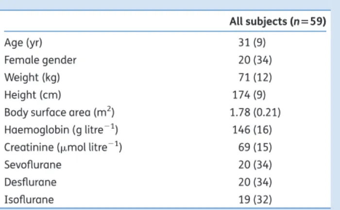
Table 1 Patient characteristics. Values are mean (SD) or numbers
(%) All subjects (n559) Age (yr) 31 (9) Female gender 20 (34) Weight (kg) 71 (12) Height (cm) 174 (9)
Body surface area (m2) 1.78 (0.21)
Haemoglobin (g litre21) 146 (16)
Creatinine (mmol litre21) 69 (15)
Sevoflurane 20 (34)
Desflurane 20 (34)
mitral annulus and the presettings of the SonosTM5000 for TDI
were used (Software Version D.1). For the pulsed-wave Doppler recordings of the mitral inflow, the sample volume was posi-tioned between the tips of the open mitral leaflets using optimal alignment with transmitral blood flow. For the pulsed-wave Doppler recordings of the pulmonary venous flow, the sample volume was positioned about 1 cm within the upper right or left pulmonary vein.
The apical 4-CV and 2-CV served for assessment of LA area, LA length, and LA volume (Fig.1). Measurements were per-formed at maximum and minimum volume on the frame pre-ceding the opening and closure of the mitral valve, respectively. The mitral annulus plane represented the inferior border of the LA. The confluences of the pulmonary veins and the LA ap-pendage were excluded from the LA cavity. LA length was mea-sured as the distance between the middle of the mitral annulus plane and the roof of the LA. The biplane LA volume was calcu-lated according to the area – length model (ALM) of the LA using the formula: LA volume (ml)¼8/3 q (A1×A2/L), where
A1and A2are the LA areas derived from the 4-CV and 2-CV,
re-spectively, and L is the length.14In addition, biplane Simpson’s method of discs (MOD) was used to calculate LA volume in the apical 4-CV and 2-CV.14Further Doppler variables measured include: peak early (E) and late (A) transmitral filling velocities, and early (e′) and late (a′) peak diastolic and peak systolic (s′) velocities of the mitral annulus predefined as the average of septal and lateral mitral annulus measurements obtained by TDI (Fig.1). Further, the velocity –time integrals of A (AVTI), E
(EVTI), and a′wave (a′VTI) were measured from mitral inflow
and tissue Doppler measurements of the mitral annulus,
respectively. Finally, peak velocities of systolic (SPVF), diastolic
(DPVF), and atrial reverse (APVF) waves of pulmonary venous
flow were measured.15
The following parameters were calculated: LA fractional area change (LA-FAC) as maximum LA area minus the minimum LA area in the 4-CV divided by maximum LA area×100; LA empty-ing fraction (LA-EF) as maximum LAvolume minus minimum LA volume divided by maximum LAvolume×100; LA fraction of the ventricular diastolic filling as [AVTI/(EVTI+AVTI)]×100; LA ejection
force as 0.5×r×MAA×A2, where r is the blood density (1.06 g cm23) and MAA the mitral annulus area (calculated as p×mitral annulus diameter in 4-CV/2×mitralannulusdiameterin2-CV/2) and A2the square of the peak late transmitral filling velocity.16 LA reservoir volume was calculated as maximum minus minimum LA volume; conduit volume was calculated as total LV stroke volume minus the reservoir volume, whereby stroke volume was estimated by the formula (EVTI+AVTI)×MAA.17LA
active emptying volume was estimated as AVTI×MAA.
For comparison of LA and LV performance during anaesthe-sia with spontaneous breathing and IPPV, we additionally mea-sured the LVend-diastolic area (LV-EDA) and the LVend-systolic area (LV-ESA) in the parasternal short-axis view. Using these values, we calculated the LV-FAC as [(EDA2ESA)/EDA]×100.
All variables were measured at end-expiration over three preferably consecutive cardiac cycles and were averaged by an experienced physician-echocardiographer blinded to all other study data. Due care was taken to avoid a foreshortening of the LA cavity in all measurements.
To determine intra- and inter-rater variabilities, a random sample of 25% of LA volumes, transmitral filling velocities,
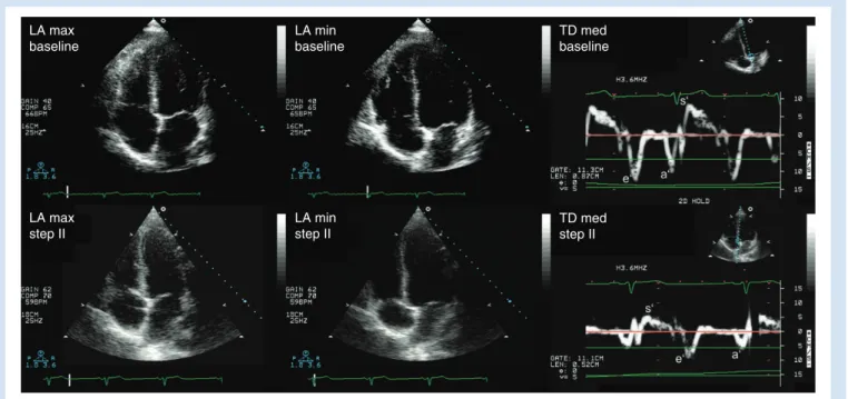
LA max baseline LA min baseline LA max step II LA min step II TD med baseline TD med step II
Fig 1 LA area and TDI parameters at baseline and during positive pressure ventilation. The upper row shows maximum and minimum LA area in the apical four-chamber view as tissue Doppler parameters in the awake patient (baseline). The lower row shows the corresponding LA areas and tissue Doppler parameters during anaesthesia with volatile anaesthetics and positive pressure ventilation (step II). LA, left atrium; TD, tissue Doppler; a′, late peak diastolic velocity of the mitral annulus; e′, early peak diastolic velocity of the mitral annulus; s′, peak systolic velocity of the mitral annulus.
and tissue Doppler recordings was submitted twice to the first investigator and once to a second investigator. The variabilities were then calculated as the mean absolute difference between both readings divided by their mean and expressed as percen-tages and their 95% confidence intervals (CIs).
Statistical analysis
The study focused on AVTIand LA maximum volume as the
primary parameter of active LA pump function and dimension, respectively.18No formal sample size calculation was performed. However, a post hoc power analysis showed that a sample size of 59 patients would allow detecting a decrease of 15% and 20% in AVTIand maximum LA volume, respectively (a,0.05 and
b≥0.8), by one-way analysis of variance (ANOVA) for repeated
measurements followed by Bonferroni’s post hoc test.
Continuous variables are presented as mean (SD), and
di-chotomous variables as number (%). Based on the former study,7normal distribution of echocardiographic parameters was assumed. To evaluate potential differences in the effects of the volatile anaesthetics, we performed a general linear model for repeated measurements to test differences between the three different anaesthetics (P¼0.025 was con-sidered significant). Influence of anaesthesia with volatile agents and positive pressure ventilation on LA size and func-tion was subsequently analysed byANOVAfor repeated
measure-ments followed by Bonferroni’s post hoc test for differences at step I and step II compared with baseline (P¼0.025 was con-sidered significant). Finally, LA and LV maximum area and sys-tolic function (a′vs s′and LA-FACvs LV-FAC)19 20were compared after normalization for baseline (set as 100%). Again, a general linear model for repeated measurements was used to test the differences between the LA vs the LV and at the different time points. Influence of heart rate on echocardiographic LA para-meters was evaluated using the Pearson’s correlation coeffi-cient. All statistical analyses were performed using IBM SPSS Statistics 21 (IBM Corp., Armonk, NY, USA).
Results
Haemodynamics
Mean arterial pressure decreased after administration of the volatile anaesthetics, and heart rate increased. However, the increase in heart rate became significant only after changing to IPPV (Table2). Phenylephrine but no other vasoactive medi-cation was administered during the study to five patients (25 – 500 mg): to four during both study steps I and II and to one during step II only. Heart rate was negatively correlated with LA volumes; however, correlation was weak (R≤0.4). Heart rate was not correlated with functional parameters including a′, a′VTI, APVF, and LA active emptying volume, but there was
a weak positive correlation with A and AVTI(R≤0.3).
Influence of different volatile anaesthetics
We found no evidence for significant differences between the three volatile anaesthetics (sevoflurane, desflurane, and iso-flurane) regarding their effect on LA function and dimensions. Also, systolic LV function as evaluated by s′and LV-FAC was
similar between all three. All P-values in the general linear model were above the significance level of 0.025 with the ex-ception of a′, where P¼0.007 (Supplementary Appendix S1). However, there were no significant differences in A, AVTI, a′VTI,
and APVF. Furthermore, a′ decreased consequently in each
study step with each volatile anaesthetic (Supplementary Ap-pendix S2). Therefore, the data from all patients anaesthetized with these three anaesthetics were pooled for the following analyses.
Influence of volatile anaesthetics and IPPV
Measured and calculated echocardiographic LA parameters at baseline and with volatile anaesthetics during spontaneous breathing and IPPV are shown in Table2. Active LA pump func-tion, as evaluated by AVTI, decreased significantly from 4.1 (1.2)
cm at baseline to 3.2 (1.1) and 2.8 (1.0) cm after the addition of volatile anaesthetics during spontaneous breathing and IPPV, respectively. Similar changes were found with other echocar-diographic parameters of active LA pump function, including A, a′, a′VTI, LA ejection forces, and filling fraction. They were
all significantly affected by volatile anaesthetics during spon-taneous breathing compared with baseline, and were further impaired by IPPV, but impairment did not regularly reach stat-istical significance compared with spontaneous breathing. AVTI, A, a′, a′VTI, and ejection forces were similarly reduced by
30–40% from baseline to IPPV.
LA volume and area were differently affected. Minimum LA area and volume were similar during all study steps. LA maximum area and volume were similar at baseline and with volatile anaesthetics during spontaneous breathing but decreased significantly after changing to IPPV. Maximum area and volume and also FAC and EF were reduced by 16–25% from baseline to IPPV.
LA reservoir volume decreased only after changing to IPPV, whereas LA active emptying volume significantly decreased with volatile anaesthetics during spontaneous breathing but showed no further significant decrease with IPPV. LA conduit volume was well preserved during administration of volatile anaesthetics with spontaneous breathing and slightly decreased after changing of IPPV (Fig.2). Pulmonary venous flow velocity during LV systole (SPVF) decreased already during
spontaneous breathing and decreased further after changing to IPPV. In contrast, the flow during LV diastole (DPVF) did not
change throughout the study.
Comparison between LA and LV dimensions and
function
For comparison of changes of LA and LV size and pump func-tion, all values were normalized to their respective baseline values. Pump function of the LA as evaluated by a′ and LA-FAC decreased consequently with volatile anaesthetics during both spontaneous breathing and IPPV, whereas systolic function of the LV as evaluated by s′and LV-FAC did not change significantly at any step during administration of volatile anaesthetics (P,0.001 and 0.001 for the general linear model) (Fig. 3). LV-EDA was similar during all study steps,
whereas maximum LA area in the apical 4-CV decreased with IPPV. However, these differences did not become significant (P¼0.143).
Inter- and intra-rater variability
Intra- and inter-rater variabilities for different echocardio-graphic parameters are shown in Table3. Variabilities within the three study steps were comparable.
Discussion
Our study in healthy adults found that volatile anaesthetics reduced active LA pump function as primarily evaluated by AVTIbut not LA minimum and maximum volume during
spon-taneous breathing. After changing from sponspon-taneous breath-ing to IPPV, there was a marked decrease in maximum LA volume and a further reduction in LA pump function. Other parameters of active LA pump function including A, a′, a′VTI,
calculated ejection forces, and active emptying volume went in parallel with AVTI, whereas LA reservoir volume decreased
after changing to IPPV in parallel with maximum volume. In contrast, LA conduit volume remained preserved during all study steps.
The mechanical function of the LA plays a critical role in cardiac performance as a reservoir, passive conduit, and active booster pump, thereby draining the pulmonary veins and facili-tating LV filling.1The active contraction and passive relaxation of
the LA myocardium are dependent on calcium inflow into myo-cardial muscle cells and the dissociation of calcium from troponin C and active re-uptake into the sarcoplasmic reticulum. Volatile anaesthetics are known to alter calcium homeostasis at several subcellular targets of the myocardial cell.21These
inter-actions are thought to be the molecular basis for impairment of both systolic and diastolic cardiac function by volatile anaes-thetics.4 5However, own previous studies have shown that
dif-ferent volatile anaesthetics including halothane, sevoflurane,
Table 2 Effects of volatile anaesthetics with spontaneous breathing and positive pressure ventilation on echocardiographic parameters of the LA. Values are mean (SD). P-values were calculated byANOVAfor repeated measurements followed by Bonferroni’s post hoc test (*P,0.025 baseline vs step I;†P,0.025 baseline vs step II;‡P,0.025 step I vs step II.). 2-CV, two-chamber view; 4-CV, four-chamber view; A, late transmitral filling velocity; a′, late peak diastolic velocity of the mitral annulus; ALM, area –length model; APVF, peak velocity of atrial reverse wave in pulmonary venous flow;
AVTI, velocity –time integral of A; a′VTI, velocity – time integral of a′; DPVF, diastolic peak velocity of pulmonary venous flow; E, early transmitral filling
velocity; IPPV, intermittent positive pressure ventilation; LA-EF, left atrial emptying fraction; LA-FAC, left atrial fractional area change; MAP, mean arterial pressure; MOD, method of discs; NA, not applicable; PE′
CO2, end-tidal concentration of carbon dioxide; SPVF, systolic peak velocity of pulmonary venous flow
Baseline Spontaneous breathing (step I) IPPV (step II) P-value
Mean arterial pressure (mm Hg) 81 (8)*,† 67 (7)* 68 (7)† ,0.001
Heart rate (beats min21) 62 (10)† 67 (10)‡ 73 (12)†,‡ ,0.001
PE′ CO2(kPa) NA 6.8 (0.8) 4.7 (0.1) ,0.001 Maximum area, 4-CV (cm2) 14.9 (3.7)† 14.4 (3.8)‡ 12.3 (3.6)†,‡ 0.001 Minimum area, 4-CV (cm2) 7.5 (2.5) 7.7 (2.7) 7.5 (2.5) 0.810 Maximum area, 2-CV (cm2) 16.1 (4.7)† 15.4 (4.5)‡ 12.8 (4.1)†,‡ 0.001 Minimum area, 2-CV (cm2) 9.2 (3.4) 9.8 (3.1) 8.7 (3.1) 0.200
Maximum volume (cm3), MOD 45.4 (18.6)† 44.0 (16.7)‡ 34.5 (16.7)†,‡ 0.003
Minimum volume (cm3), MOD 18.4 (9.3) 19.6 (8.8) 17.7 (9.4) 0.595
Maximum volume (cm3), ALM 45.4 (16.5)† 43.1 (16.4) 36.3 (16.5)† 0.013
Minimum volume (cm3), ALM 18.4 (8.9) 19.1 (8.5) 18.5 (8.9) 0.895
LA-FAC, 4-CV (%) 51 (9)† 47 (10)‡ 40 (9)†,‡ ,0.001 LA-FAC, 2-CV (%) 45(8)† 37 (10)‡ 33 (10)†,‡ ,0.001 LA-EF (%), MOD 61 (9)† 56 (10)‡ 50 (11)†,‡ ,0.001 LA-EF (%), ALM 61 (10)† 56 (11)‡ 50 (11)†,‡ ,0.001 LA reservoir volume (ml) 27.5 (10.6)† 24.4 (10.2)‡ 17.1 (8.4)†,‡ ,0.001 LA conduit volume (ml) 69.5 (20.6) 68.7 (15.9) 62.0 (14.8) 0.085
LA active emptying volume (ml) 33.5 (12.6)*,† 25.8 (9.2)* 21.6 (7.8)† ,0.001
A (cm s21) 47 (10)*,† 40 (8)* 37 (10)† ,0.001 AVTI(cm) 4.1 (1.2)*,† 3.2 (1.1)* 2.8 (1.0)† ,0.001 a′(cm s21) 7.6 (1.5)*,† 6.1 (1.7)*,‡ 4.6 (1.6)†,‡ ,0.001 a′VTI(cm) 0.59 (0.13)*,† 0.47 (0.16)*,‡ 0.36 (0.14)†,‡ ,0.001 LA filling fraction (%) 24 (6)*,† 20 (7)* 21 (8)† 0.003 SPVF(cm s21) 54.5 (12.7)*,† 46.1 (12.4)*,‡ 17.8 (10.8)†,‡ ,0.001 DPVF(cm s21) 55.6 (12.3) 57.2 (10.9) 53.5 (9.1) 0.891 APVF(cm s21) 23.0 (3.8)† 21.3 (4.0)‡ 14.0 (10.8)†,‡ ,0.001
desflurane, and isoflurane in concentrations of 1 MAC do not relevantly affect LV systolic and diastolic function in healthy adults free from cardiovascular diseases6 7or in patients with diastolic dysfunction.22In contrast, the present study showed a relevant impairment of active LA pump function, which is in agreement with preliminary data in a recent own study.7
We found no relevant differences between sevoflurane, des-flurane, and isoflurane regarding their effect on LA dimension and function in the actual analyses. We, therefore, decided to pool the data from all subjects, which are supported by human and canine in vivo studies.6 7 10
Maximum LA area and volume were similar to baseline when volatile anaesthetics were administered during spontan-eous breathing. Changing to IPPV significantly decreased maximum LA area and volume. In contrast, active LA pump function as evaluated by AVTI, A, a′, a′VTI, LA ejection force,
and filling fraction were significantly decreased by administra-tion of volatile anaesthetics during spontaneous breathing. Changing to IPPV differently affected the echocardiographic parameters of LA pump function. AVTIand A did not
significant-ly decrease during IPPV, whereas a′VTIand a′, which can be
used as a marker for global LA pump function,23 24 gave evidence for a further significant impairment of LA function. We might suggest that the preservation of the transmitral forward flow (as assessed by A and AVTI) during IPPV
compared with spontaneous breathing occurred at the expense of the markedly reduced retrograde flow into the pulmonary veins as evidenced by the marked reduction in APFVduring IPPV. P e rcenta g es of baseline v alues 40 60 80 100 120 40 60 80 100 120 40 60 80 100 LA max area EDA (P=0.143) 120 LA-FAC LV-FAC (P=0.001) a′ s′ (P<0.001)
Fig 3Comparative changes of pump function and dimensions between LA and LV. LA pump function as evaluated by late peak diastolic velocity of the mitral annulus (a′) and LA fractional area change (FAC) was significantly more impaired than LV pump func-tion as evaluated by peak systolic velocity of the mitral annulus (s′) and LV FAC. Maximum LA area and end-diastolic area (EDA) of the LV were similarly affected by volatile anaesthetics during spontan-eous breathing and IPPV.
Reservoir volume Mean per centa g e c hang e 10 0 –10 –20 –30 –40 –50 *† * *
Active emptying volume Conduit volume Step I Step II
Fig 2 Percentage changes of reservoir, contractile, and conduit volumes compared with baseline. Whereas reservoir volume decreased significant-ly onsignificant-ly in step II (volatile anaesthetic and positive pressure ventilation), LA active emptying volume decreased significantsignificant-ly in both step I (volatile anaesthetic and spontaneous breathing) and step II. Conduit volume did not change significantly during steps I and II. *P,0.025 vs baseline;
In agreement with the above-described findings, the LA phasic volumes (reservoir, conduit, and active emptying volume) were affected differently by volatile anaesthetics. LA active emptying volume was significantly decreased by volatile anaesthetics during spontaneous breathing, whereas chan-ging to IPPV resulted in a marked decrease in reservoir volume. The passive LA conduit volume was not affected by volatile anaesthetic during spontaneous breathing and IPPV. These changes in LA phasic volumes are reflected by pulmon-ary venous flow. SPFV, representing an index of LA reservoir
function,25 decreased in parallel with LA reservoir volume, whereas DPVF, reflecting the conduit function,25remained
un-changed during all study steps. Thus, LV filling became less active and more passive, which is in agreement with former findings in an animal in vivo study.10
Altered loading conditions and relative hypovolaemia due to vasodilation induced by volatile anaesthetics are unlikely to be causative for our findings, as LA area and volume did not change during spontaneous breathing, but enforce the rele-vance of negative inotropic effect of the volatile anaesthetics on active LA pump function. Two former studies in patients undergoing haemodialysis showed that induced hypovolaemia did not change A and a′velocities.26 27The different changes of parameters of active LA pump function in non-anaesthetized patients with hypovolaemia compared with the changes found in our anaesthetized study population imply that our findings cannot be simply explained by decreased LA preload associated with blood volume distribution. Further, volatile anaesthetics in spontaneously breathing patients will always induce hypercapnia, which potentially influences LA function. However, hypercapnia is an unlikely confounder, as previous studies have found that arterial CO2levels do not influence
myo-cardial contractility and LV end-diastolic pressure in animals28 and in young healthy subjects.7 29 30Hypercapnia has even been found to increase late peak velocity of transmitral inflow (A)30due to sympathicoadrenergic stimulation. Therefore, the depressive effects of volatile anaesthetics on this parameter might have been even more pronounced if normocapnia had been present during study step I.
The marked decrease in maximum and the unchanged minimum LA volume during IPPV resulted in a significant de-crease in LA reservoir volume. This reduction in LA volumes
can be caused by impaired LA relaxation and compliance, and by external compression of the LA due to mechanical ventila-tion. However, the most likely explanations are the redistribution of circulating blood volume from the central compartment to the periphery31 and the expiratory pooling of blood in the pulmonary vascular bed, resulting in reduced LA filling after changing to IPPV. The reduced preload might have altered LA muscle mechanics due to reduced myocardial fibre length, and therefore, further reduced LA pump function.
While our findings are in principle agreement with in vitro findings8 and with animal studies,10 there are marked differences in the magnitude of observed effects. We found a reduction of about 10–20% in the different functional para-meters under volatile anaesthetics at 1 MAC during spontan-eous breathing and a reduction of about 20% to maximally 40% during IPPV (Fig. 3). This is substantially less than described in a canine model showing a reduction in LA con-tractility by approximately 50% at 1.2 MAC.11Differences in MAC, the use of additional cardiodepressive drugs as thiopen-tal, open chest conditions, differences in ventilation, and in-strumentation of the LA might have contributed to the more pronounced effect of volatile anaesthetics in those animal studies compared with our study. In fact, the enlargement of the LA in another canine study10suggests relevant myocardial depression, which was absent in our patients.
Further, our results are in agreement with two clinical studies in patients undergoing surgery under general anaesthesia,32 33 both suggesting impaired LA systolic function by volatile anaes-thetics. However, these studies are limited due to the fact that the authors compared preoperative TTE measurements with intraoperative transoesophageal echocardiographic measure-ments in ventilated patients anaesthetized with isoflurane in combination with opioids, sedatives, and neuromuscular block-ing agents. Moreover, the evaluation of the LA function was solely based on transmitral A, mitral annular a′, or both velocities.
In our previous human studies that used the same or similar study models,6 7 22we found that LV function was not relevant-ly impaired by volatile anaesthetics, which is in contrast to earlier animal studies. In the present study, we found signifi-cantly different effects of volatile anaesthetics on LA vs LV pump function. This is in disagreement with a former canine
Table 3Inter- and intra-rater variabilities for echocardiographic parameters. Values are expressed as percentages (95% CI). P-values were calculated withANOVAfor repeated measurements. 4-CV, four-chamber view; A, late transmitral filling velocity; a′, late peak diastolic velocity of the mitral annulus; AVTI, velocity –time integral of A; EVTI, velocity – time integral of early transmitral filling velocity; EF, emptying fraction; MOD, method
of discs
Intra-rater variability P-value (within study steps) Inter-rater variability P-value (within study steps)
Maximum volume, 4-CV, MOD 7.8 (6.1– 9.9) 0.871 7.6 (5.8– 9.5) 0.559
Minimum volume, 4-CV, MOD 15.3 (11.6 –19.0) 0.846 15.3 (11.1 –19.6) 0.852
EF, MOD 11.2 (9.2– 13.2) 0.147 10.1 (6.8– 13.3) 0.199
A 8.8 (6.0– 11.7) 0.419 3.7 (2.4– 5.1) 0.941
AVTI 11.3 (9.0– 13.7) 0.386 7.2 (4.7– 9.6) 0.224
EVTI 7.5 (5.3– 9.6) 0.182 6.2 (4.4– 8.0) 0.224
study9 10suggesting similar effects of volatile anaesthetics on the LA and LV. However, volatile anaesthetics decrease after-load of the LV but not necessarily of the LA. Whereas LV-FAC is well preserved, LA-FAC might be relevantly decreased by impaired myocardial contractility due to volatile anaesthetics or impaired LV diastolic function. However, we could not show impaired diastolic function in the former study,7and there were also significant differences in the systolic TDI para-meters of the LA (i.e. a′) and LV (i.e. s′).19 20Further, the differ-ence between LA-FAC and LV-FAC might underestimate the real difference as they do not reflect the same function. While LV-FAC results from systolic area change only, LA-FAC includes both the passive and active LA function. Finally, myo-cardial cells in the atria and ventricles are morphologically, mo-lecularly and functionally distinct.34Differences in contractility between the atria and ventricles have been ascribed to the dif-ferential regulation and expression of sarcoplasmic reticulum calcium ATPase.35It seems plausible that volatile anaesthetics could, therefore, differently affect the inotropic action of atrial and ventricular myocytes.21 To summarize, our findings suggest different impairment of systolic pump function of the LA and LV by volatile anaesthetics.
Strengths of this study include the human origin of the data, the adequate power of the study, the standardized TTE assess-ment based on current guidelines,13and the established re-search model both in awake and anaesthetized subjects under mono-anaesthesia with volatile anaesthetics. Limita-tions of the study include that this study is an additional ana-lysis of echocardiographic data from a study, which aimed to evaluate LV diastolic function. The study may have been con-ducted slightly differently if LA function and volumes had been the primary endpoints. Further, our TTE examinations were limited to 2D echocardiography, pulsed-wave Doppler, and TDI. The application of novel techniques such as LA strain measurement by 2D or 3D speckle tracking might have provided additional insight into changes in LA function and geometry. Nevertheless, our results were consistent over a broad range of echocardiographic parameters and according to different assessments (e.g. MOD vs ALM, 4-CV vs 2-CV, con-ventional Doppler vs TDI), which reinforces the relevance of our findings. The inter-rater and intra-rater variabilities were slightly higher compared with former own studies.6 7 22 36 However, the variabilities were comparable with reported values in LA assessment in healthy subjects,37 38 and the lower variabilities found in a recent study39have been favoured by the markedly dilated LA present in those patients. In add-ition, the variabilities were consistent within the three study steps and over almost every measured echocardiographic par-ameter; this supports the credibility of our findings. Further, volume measurements did not include the LA appendage (LAA). In the absence of published evidence, one might specu-late that the LAA responded similarly to volatile anaesthetics as the LA. However, even if that was not the case, LAA volumes of reportedly only 3 ml in healthy adults40make differ-ent behaviour of LAA an unlikely confounder of our results. Another potential limitation is that an a-adrenergic agonist (phenylephrine) was given to five participants due to
hypotension during echocardiographic evaluation. The admin-istration of vasoactive drugs might potentially influence LA function by positive inotropic effects on myocardium and increased venous return.41 42However, the low number of patients treated with small doses of phenylephrine is unlikely to be a confounder of our results. Further, heart rate slightly increased during the study after changing to IPPV. However, the observation that heart rate was not correlated with most functional echocardiographic parameters, and that its correl-ation with LA volumes and AVTIwas weak questions the
in-crease in heart rate as a relevant confounder of our findings. Finally, we included only young healthy patients without car-diopulmonary diseases. Airway pressures were, therefore, low in our patients, but increased ventilation pressures in patients with pulmonary diseases might relevantly influence LA function and volumes. The study was also not designed to assess the clinical importance of the effects of volatile anaes-thetics, and it remains unclear whether the effects of volatile anaesthetics on LA dimension and function might be clinically important, for example, in patients with impaired LV function who are more dependent on the contribution of atrial function. Potentially, the use of volatile anaesthetics might increase the risk of intraoperative haemodynamic instability in such patients.43
In conclusion, our study found that volatile anaesthetics impair active LA pump function in healthy adults. LA maximum dimensions decreased only after changing to IPPV, and LA pump function was further impaired. The clinical im-portance of these findings needs to be assessed in further studies in patients with pre-existing cardiac dysfunction.
Supplementary material
Supplementary material is available at British Journal of Anaesthesia online.
Authors’ contribution
D.F.: study design and conduction, data analysis, and prepar-ation of the manuscript. K.S.: study design and conduction, data collection and analysis, and preparation of the manu-script. M.F.: study design and conduction, patient recruitment, data collection, and preparation of the manuscript. M.D.S.: study design and conduction, patient recruitment, data collec-tion, and preparation of the manuscript. D.B.: study design and conduction, patient recruitment, data collection and analysis, and preparation of the manuscript.
Acknowledgements
The authors thank Regina M. Schumann, MD, Jorge Kasper, MD, Claudia Werner, RN, and Esther Seeberger, DAS, Department of Anaesthesia and Intensive Care Medicine, University Hospital Basel, Basel, Switzerland, for help with data acquisition and Allison Dwileski, BS, Department of Anaesthesia and Intensive Care Medicine, University Hospital Basel, Basel, Switzerland, for editorial assistance.
Declaration of interest
None declared.
Funding
This work was supported by grants from the Swiss National Science Foundation, Bern, Switzerland (grant no. 3200BO-116229) and from the ‘Verein zur Fo¨rderung von Ausbildung und Forschung’, Department of Anaesthesia and Intensive Care Medicine, University Hospital Basel, Basel, Switzerland.
References
1 Stefanadis C, Dernellis J, Toutouzas P. A clinical appraisal of left atrial function. Eur Heart J 2001; 22: 22 –36
2 Kagawa K, Arakawa M, Miwa H, et al. Left atrial function during left ventricular diastole evaluated by left atrial angiography and left ventriculography. J Cardiol 1994; 24: 317 –25
3 Mitchell JH, Shapiro W. Atrial function and the hemodynamic con-sequences of atrial fibrillation in man. Am J Cardiol 1969; 23: 556– 67
4 Weiskopf RB. Cardiovascular effects of desflurane in experimental animals and volunteers. Anaesthesia 1995; 50(Suppl.): 14 –7 5 De Hert SG, Van der Linden PJ, ten Broecke PW, Vermeylen KT,
Rodrigus IE, Stockman BA. Effects of desflurane and sevoflurane on length-dependent regulation of myocardial function in coronary surgery patients. Anesthesiology 2001; 95: 357 –63
6 Filipovic M, Wang J, Michaux I, Hunziker P, Skarvan K, Seeberger MD. Effects of halothane, sevoflurane and propofol on left ventricular diastolic function in humans during spontaneous and mechanical ventilation. Br J Anaesth 2005; 94: 186 –92
7 Bolliger D, Seeberger MD, Kasper J, et al. Different effects of sevoflur-ane, desflursevoflur-ane, and isoflurane on early and late left ventricular dia-stolic function in young healthy adults. Br J Anaesth 2010; 104: 547– 54
8 Hanouz JL, Massetti M, Guesne G, et al. In vitro effects of desflurane, sevoflurane, isoflurane, and halothane in isolated human right atria. Anesthesiology 2000; 92: 116– 24
9 Pagel PS, Kehl F, Gare M, Hettrick DA, Kersten JR, Warltier DC. Mech-anical function of the left atrium: new insights based on analysis of pressure –volume relations and Doppler echocardiography. Anes-thesiology 2003; 98: 975 –94
10 Gare M, Schwabe DA, Hettrick DA, Kersten JR, Warltier DC, Pagel PS. Desflurane, sevoflurane, and isoflurane affect left atrial active and passive mechanical properties and impair left atrial– left ventricu-lar coupling in vivo: analysis using pressure– volume relations. An-esthesiology 2001; 95: 689 –98
11 Hettrick DA, Pagel PS, Warltier DC. Desflurane, sevoflurane, and iso-flurane impair canine left ventricular – arterial coupling and mech-anical efficiency. Anesthesiology 1996; 85: 403 –13
12 Eger EI II. Age, minimum alveolar anesthetic concentration, and minimum alveolar anesthetic concentration-awake. Anesth Analg 2001; 93: 947–53
13 Gottdiener JS, Bednarz J, Devereux R, et al. American Society of Echocardiography recommendations for use of echocardiography in clinical trials. J Am Soc Echocardiogr 2004; 17: 1086–119 14 Lang RM, Bierig M, Devereux RB, et al. Recommendations for
chamber quantification: a report from the American Society of Echocardiography’s Guidelines and Standards Committee and the Chamber Quantification Writing Group, developed in conjunction with the European Association of Echocardiography, a branch of
the European Society of Cardiology. J Am Soc Echocardiogr 2005; 18: 1440– 63
15 Tabata T, Thomas JD, Klein AL. Pulmonary venous flow by Doppler echocardiography: revisited 12 years later. J Am Coll Cardiol 2003; 41: 1243– 50
16 Manning WJ, Silverman DI, Katz SE, Douglas PS. Atrial ejection force: a noninvasive assessment of atrial systolic function. J Am Coll Cardiol 1993; 22: 221 –5
17 Blume GG, McLeod CJ, Barnes ME, et al. Left atrial function: physi-ology, assessment, and clinical implications. Eur J Echocardiogr 2011; 12: 421–30
18 Leung DY, Boyd A, Ng AA, Chi C, Thomas L. Echocardiographic evalu-ation of left atrial size and function: current understanding, patho-physiologic correlates, and prognostic implications. Am Heart J 2008; 156: 1056– 64
19 Seo JS, Kim DH, Kim WJ, Song JM, Kang DH, Song JK. Peak systolic velocity of mitral annular longitudinal movement mea-sured by pulsed tissue Doppler imaging as an index of global left ventricular contractility. Am J Physiol Heart Circ Physiol 2010; 298: H1608–15
20 Ho CY, Solomon SD. A clinician’s guide to tissue Doppler imaging. Circulation 2006; 113: e396– 8
21 Hanley PJ, ter Keurs HE, Cannell MB. Excitation –contraction coup-ling in the heart and the negative inotropic action of volatile anes-thetics. Anesthesiology 2004; 101: 999– 1014
22 Filipovic M, Michaux I, Wang J, Hunziker P, Skarvan K, Seeberger M. Effects of sevoflurane and propofol on left ventricular diastolic function in patients with pre-existing diastolic dysfunction. Br J Anaesth 2007; 98: 12 –8
23 Masuda M, Iwakura K, Inoue K, et al. Estimation of left atrial pump function by mitral annular velocity. Circ J 2012; 76: 1430–5 24 Hesse B, Schuele SU, Thamilasaran M, Thomas J, Rodriguez L. A
rapid method to quantify left atrial contractile function: Doppler tissue imaging of the mitral annulus during atrial systole. Eur J Echocardiogr 2004; 5: 86 –92
25 Appleton CP. Hemodynamic determinants of Doppler pulmonary venous flow velocity components: new insights from studies in lightly sedated normal dogs. J Am Coll Cardiol 1997; 30: 1562– 74
26 Park CS, Kim YK, Song HC, et al. Effect of preload on left atrial func-tion: evaluated by tissue Doppler and strain imaging. Eur Heart J Cardiovasc Imaging 2012; 13: 938 –47
27 Oguzhan A, Arinc H, Abaci A, et al. Preload dependence of Doppler tissue imaging derived indexes of left ventricular diastolic function. Echocardiography 2005; 22: 320 –5
28 Foex P, Prys-Roberts C. Effects of changes in PaCO2on pulmonary
input impedance. J Appl Physiol 1975; 38: 52– 7
29 Kiely DG, Cargill RI, Lipworth BJ. Effects of hypercapnia on hemo-dynamic, inotropic, lusitropic, and electrophysiologic indices in humans. Chest 1996; 109: 1215–21
30 Tzou WS, Korcarz CE, Aeschlimann SE, Morgan BJ, Skatrud JB, Stein JH. Coronary flow velocity changes in response to hypercap-nia: assessment by transthoracic Doppler echocardiography. J Am Soc Echocardiogr 2007; 20: 421– 6
31 Hedenstierna G, Strandberg A, Brismar B, Lundquist H, Svensson L, Tokics L. Functional residual capacity, thoracoabdominal dimen-sions, and central blood volume during general anesthesia with muscle paralysis and mechanical ventilation. Anesthesiology 1985; 62: 247–54
32 Oxorn D, Edelist G, Harrington E, Tsang S. Echocardiographic assess-ment of left ventricular filling during isoflurane anaesthesia. Can J Anaesth 1996; 43: 569 –74
33 Couture P, Denault AY, Shi Y, et al. Effects of anesthetic induction in patients with diastolic dysfunction. Can J Anaesth 2009; 56: 357– 65 34 Ng SY, Wong CK, Tsang SY. Differential gene expressions in atrial and ventricular myocytes: insights into the road of applying embryonic stem cell-derived cardiomyocytes for future therapies. Am J Physiol Cell Physiol 2010; 299: C1234–49
35 Zhao XS, Gallardo TD, Lin L, Schageman JJ, Shohet RV. Transcriptional mapping and genomic analysis of the cardiac atria and ventricles. Physiol Genomics 2002; 12: 53– 60
36 Bolliger D, Seeberger MD, Kasper J, et al. Remifentanil does not impair left ventricular systolic and diastolic function in young healthy patients. Br J Anaesth 2011; 106: 573–9
37 Aune E, Baekkevar M, Roislien J, Rodevand O, Otterstad JE. Normal reference ranges for left and right atrial volume indexes and ejection fractions obtained with real-time three-dimensional echocardiography. Eur J Echocardiogr 2009; 10: 738– 44
38 Pela G, Regolisti G, Coghi P, et al. Effects of the reduction of preload on left and right ventricular myocardial velocities analyzed by Doppler tissue echocardiography in healthy subjects. Eur J Echocar-diogr 2004; 5: 262– 71
39 Buechel RR, Stephan FP, Sommer G, Bremerich J, Zellweger MJ, Kaufmann BA. Head-to-head comparison of two-dimensional and three-dimensional echocardiographic methods for left atrial chamber quantification with magnetic resonance imaging. J Am Soc Echocardiogr 2013; 26: 428– 35
40 Yakar Tuluce S, Kayikcioglu M, Tuluce K, et al. Assessment of left atrial appendage function during sinus rhythm in patients with hypertrophic cardiomyopathy: transesophageal echocardiog-raphy and tissue Doppler study. J Am Soc Echocardiogr 2010; 23: 1207– 16
41 Thiele RH, Nemergut EC, Lynch C III. The clinical implications of isolated alpha(1) adrenergic stimulation. Anesth Analg 2011; 113: 297– 304
42 Landzberg JS, Parker JD, Gauthier DF, Colucci WS. Effects of myocardial alpha 1-adrenergic receptor stimulation and blockade on contractility in humans. Circulation 1991; 84: 1608–14 43 Calleja AM, Dommaraju S, Gaddam R, Cha S, Khandheria BK,
Chaliki HP. Cardiac risk in patients aged .75 years with asymptomatic, severe aortic stenosis undergoing noncardiac surgery. Am J Cardiol 2010; 105: 1159–63