THE INNERVATION OF THE ORGAN
OF CORTI*f
By H. SPOENDLIN (Zurich)
ACCORDING to our present knowledge three different types of nerve fibres participate in the innervation of the cochlea:
1. The sensory neurons of the cochlear nerve. 2. The efferent neurons of the olivo-cochlear bundle. 3. Autonomic nerve fibres.
The major portion of the sensory and of the efferent neurons consists of myelinated nerve fibres. Their peripheral ramifications within the organ of Corti, however, are unmyelinated (Fig. 1).
The myelinated portions of the cochlear sensory neurons are well known from light microscopic studies. They extend from the habenula perforata to the cochlear nuclei and have their bipolar ganglion cells in the spiral ganglion. They are unbranched without connections between the individual fibres. From the habenula to the spiral ganglion they take a regular radial course. In the main cochlear nerve the fibres of the different cochlear turns wind spirally around each other (Sando). At the level of the cochlear nuclei each fibre divides into two or more branches making synaptic contacts with second order neurons of all three cochlear nuclei (Sando, Rose et al.).
The myelin sheaths begin immediately below the habenula perforata with the same characteristic arrangement of the myelin lamellae as it is found at the nodes of Ranvier where the myelin sheath is interrupted for a short distance (Fig. 2). Also the bipolar spiral ganglion cells are entirely covered by a layer of myelin with somewhat coarser lamellae preventing any synaptic contact of the ganglion cell body with other nerve fibres (Fig. 3). Their main role seems to be the metabolic centres of the neurons. The abundance of ribosomes in the cytoplasm indicates a very intense protein metabolism. In contrast to the ganglion
* A more detailed report is published in "The Organization of the Cochlear Receptor" by H. Spoendlin, Advances in Oto-Rhino-Laryngology. Vol. 13, S. Karger, Basel—New York.
f Read at Symposium on Electron microscopy at the Institute af Laryngology and Otology, London, October 1966.
cell body, ribosomes are completely missing within axons and dendrites where no protein synthesis is taking place.
The ganglion cells of the second order neurons in the cochlear nuclei are different. Their unmyelinated surface is very irregular with many villosities in numerous synaptic contacts with "bouttons terminaux" of the cochlear and probably other neurons (Fig. 4).
(A)
(B)
FIG. I
Transverse section through two myelinated nerve fibres in the osseous spiral lamina Typical regularly arranged myelin layers (M) surround the axon (A) which contains predominantly neurofilaments. Phenoments (small points in cross section).
Transverse section through a group of outer spiral fibres (D) which are the terminal dendnte ramifications of the cochlear neurons. They are unmyelinated and only surrounded by extensions of the supporting cells (S) of the organ of Corti These dendrites contain exclusively neurocanaliculi in their axoplasm, which are well visible in this glutaraldehyde-fixed material.
The terminal portion of a myelin sheath (M) around the axon (A) of a cochlear neuron below the habenula perforata. The myelin lamellae are spread out in a typical manner around the axon, forming saclike enlargements adjacent to the axon membrane. This area of the neuron corresponds probably to the so called "initial segment" where the action
potentials of the nerve take their origin.
FIG. 3
Portion of a bipolar ganglion cell (G) of the spiral ganglion in the modiolus, with the origin of one axon (A). The entire ganglion cell is surrounded by a myelin sheath (M)
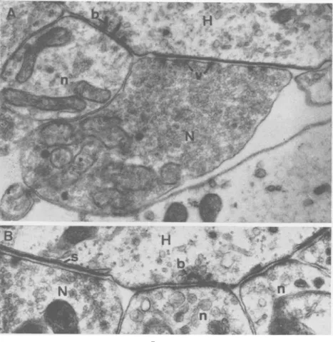
(A) A portion of a ganglion cell (G) of the cochlear nuclei with a very irregular unmyelinated surface which is in contact with a great number of nerve endings (N) or so called "boutton terminaux".
(B) Two synaptic zones (S) at higher magnification between a nerve ending (X) and a ganglion cell (G). The thickening of the pre- and post-synaptic membranes and the condensation of synaptic vesicles at the presynaptic side is clearly visible.
As in all centrifugal, efferent neurons the ganglion cells of the olivo-cochlear fibres are situated within the central nervous system. Rasmussen counted about 500 olivo-cochlear fibres of which about 2/3 are coming from the contralateral accessory olive and 1/3 from the homolateral superior olive. According to Rossi some fibres originate also in the recticular formation. The contralateral fibres cross the midline at the floor of the fourth ventricle, join the homolateral bundle and run with the vestibular root and nerve as far as the internal acoustic meatus. Only at this level they cross over to the cochlear nerve through the anastomosis of Oort (Fig. 5). Within the modiolus they form the intraganglionic spiral bundle, from where they branch out to send regular projections to the habenula perforata. In contrast to the sensory neurons the olivo-cochlear fibres show considerable branching within the osseous spiral lamina.
The unmyelinated portions of afferent and efferent neurons begin below the habenula perforata and enter the organ of Corti. They can hardly be followed by light microscopy. Their complicated distribution pattern and inter-relations offer, however, a special interest as a very important part of the cochlear receptor. With phase-contrast microscopy or special silver stains (Fernandez) different groups of fibres can be
vestibular nerve
B,
cochlear nerve
F I G . 5
Schematic representation of the vestibular and cochlear nerve including the olivo-cochlear bundle (interrupted line) which originates partly from ganglion cells of the contralateral accessory olive and partly from ganglion cells of the homolateral superior olive. It reaches the periphery with the vestibular root and nerve and crosses over to the cochlear nerve only in the periphery. The places where the olivo-cochlear fibres can be selectively sectioned
without damaging the cochlear neurons are indicated by arrows:
The floor of the fourth ventricle where only the contralateral olivo-cochlear fibres are (A)
(B) (C)
destroyed.
At the level of the vestibular root.
Within the vestibular nerve where the contra and homolateral olivo-cochlear fibres are destroyed.
FIG. 6
(A) Phase contrast picture of the organ of Corti. The upper tunnel radial fibres (R) and the outer spiral fibres (OS) are clearly outlined. The inner spiral fibres (IS) and the basilar fibres at the bottom of the tunnel (B) are more difficult to be distinguished. The nerve fibres penetrate the organ of Corti at the habenula perforta (HB). Basilar membrane (BM), inner hair cells (IH), outer hair cells (OH) and Hensen cells (H). Space of the tunnel (T), tectorial membrane (TE). The vertical black line through the tunnel indicates the tangential plane of section as represented in Figs. B and C. (B) Phase contrast picture of a tangential section through the tunnel where the tunnel
radial fibres are transversely cut and appear as a row of black points (R).
(C) Phase contrast picture of a tangential section through the tunnel of a cat where the olivo-cochlear bundle has been sectioned two weeks previously. The tunnel space is completely empty and no tunnel radial fibres are visible (X).
distinguished (Fig. 6). A much better differentiation is, however, possible with electron-microscopy. In general terms the traditional topographic classification of nerve fibres in the following groups (Lorente de No) is still valid:
The short radial fibres to the internal hair cells with an average diameter of 0-5 micron (Fig. 8).
The tunnel radial fibres which cross the tunnel at a middle level in small fascicles of three to four fibres with usually large diameters up to 1-5 micron (Fig. 6).
B
FIG. 7
Electronmicrograph of the area as indicated in the schematic representation of the organ of Corti and the lower right corner. The outer spiral fibres (OS) are arranged in regular rows between the Deiter's cells (D). Each nerve fibre is individually surrounded by
The outer spiral fibres which run in a spiral direction in regular rows of approximately twenty independent fibres between the Deiters cells. Their diameter is, in the cat, around 0*4 micron (Fig. 7).
The inner spiral fibres which are closely packed together in two or three bundles of predominantly very small fibres (frequently less than o-1 micron diameter) below the internal hair cells (Fig. 8).
The tunnel spiral fibres which are situated in the tunnel adjacent to the inner pillars. They are very similar and probably closely associated to the inner spiral fibres (Fig. 6).
The nerve endings at the inner hair cells are simple enlargements of the terminal nerve fibres irregularly distributed over the greater part of the hair cell (Fig. 8). Synaptic regions with thickening of pre- and post-synaptic membranes and with accessory post-synaptic structures such as synaptic bars (Smith and Sjostrand) are frequently seen. In the cat there is only this one simple type of nerve ending connected with the inner hair cell. In the guinea pig another type of nerve ending, filled with small vesicles, is occasionally found in contact with the hair cell (Fig. 9).
The distinction of two types of nerve endings is much clearer at the basis of the outer hair cells. In all species so far studied, including man (Kimura, et al.), small so called type I nerve endings containing only very few irregular vesicles and large so called type II endings, filled with syn-aptic vesicles, are regularly observed adjacent to the outer hair cells. The synaptic contacts between type I nerve ending and sensory cell are characterized by simple membrane thickenings and, in the guinea pig, synaptic bars. Accessory membranes or subsynaptic cisternae are always found along the sensory cells plasma membrane adjacent to type II nerve endings (Fig. 10).
The great number of synaptic vesicles and the association with subsynaptic cisternae suggests an efferent nature of the type II nerve endings in analogy to the endings of other known centrifugal nerve fibres such as motor end-plates. However, only by means of degeneration studies, which were carried out in different animals, was it possible to prove that the type II endings belong to the efferent olivo-cochlear fibres. Because the olivo-cochlear bundle runs separated from the afferent cochlear neurons it can be sectioned selectively at the floor of the 4th ventricle for the contralateral fibres only (Kimura et al.) or in the vesti-bular root (Iurato, Smith, Spoendlin and Gacek) and in the vestivesti-bular nerve (Spoendlin) for the homo- and contralateral fibres (Fig. 5). Within a few days after selective transection of the olivo-cochlear bundle the type II nerve endings degenerate and disappear completely after a few weeks. According to our findings the totality of type II endings is affected when all olivo-cochlear fibres are interrupted.
Once it is known that the vesiculated endings are efferent in nature it is necessary to trace backwards the efferent nerve fibres down to the
FIG. 8
Electronmicrograph of the area below the internal hair cell of a guinea pig as indicated at the lower right corner. The basal extension of an inner hair cell (H) is in synaptic contact with several fairly large nerve endings (N). Vesicle filled enlargements of efferent nerve fibres (E) are predominantly in synaptic contact with afferent nerve endings (N) or afferent dendrites (D) but not directly with the hair cell. The great majority of the inner spiral fibres (S) are very small in diameter. They are usually closely intermingled with
FIG. 9
(A) Nerve endings at the base of an inner hair cell of a guinea pig from the area indicated at the lower left corner. A large afferent nerve ending (N) shows typical synaptic differentiations in form of a marked thickening of the postsynaptic membrane (S) and a presynaptic bar (b.) surrounded from small vesicles inside the cytoplasm of the hair cell (H). Next to this afferent ending there is an enlargement of efferent nerve fibre (E) filled with synaptic vesicles. As a rule those efferent elements do not have synaptic contact with the inner hair cell. Such contacts can only occasionally be seen in the guinea pig, characterized by an agglomeration of synaptic vesicles in the nerve ending and a subsynaptic cisterna (X) inside the hair cell.
(B) High magnification of a synapse between inner hair cell and afferent nerve ending. The unit-membrane pattern (a double dark line) is clearly visible in the pre- and postsynaptic membrane (S). The fairly homogenous synaptic bar (b) is surrounded by a great number of small synaptic vesicles (v) which show again the unit-membrane pattern.
habenula perforata. In our light microscopic preparations of the organ of Corti of cats where the olivo-cochlear fibres have been cut we always noted missing tunnel radial fibres, whereas they are almost always visible in normal preparations. Using tangential sections by cutting the tunnel in a longitudinal vertical plane normally a great number of transversely cut tunnel radial fibres are visible as a row of small spots in the middle of the tunnel. In this way a quantitative evaluation of those fibres is possible.
FIG. IO
(A) Xerve endings at the base of an outer hair cell (H). The type I nerve ending (n) is relatively small and contains only a few irregular vesicles. The synaptic contact with the hair cell is characterized by a membrane thickening and a presynaptic bar (b). The type II nerve ending which is efferent in nature (X) is filled with fairly regularly sized vesicles which are agglomerated and condensed (v) towards the hair cell indi-cating synaptic activities in this area.
(B) The contact zone between efferent nerve ending (X") and the outer hair cell (H) is not only characterized by an agglomeration of synaptic vesicles in the nerve ending but also by a subsynaptic cysterna inside the hair cell (s). The afferent nerve endings (n) contain very irregular and fewer vesicles. A synaptic bar is partially seen at (b). In the synaptic cleft between these nerve endings and the hair cell frequently dense material can be seen.
After transection of the olivo-cochlear bundle in the cat we find indeed a complete disappearance of all tunnel radial fibres (Fig. 6 B and C). This demonstrates very clearly that in the cat all the tunnel radial fibres belong to the efferent olivo-cochlear innervation.
In addition to the tunnel radial fibres the greatest part of the inner and tunnel spiral fibres also degenerates after a lesion of the olivo-cochlear fibres. Iurato has even found in a recent study that the internal spiral fibres seem to originate mainly from the homolateral olivo-cochlear fibres whereas the efferent fibres destined for the outer hair cells originate mainly from the contralateral olivo-cochlear bundle.
This very extensive efferent innervation of the cochlear receptor is certainly surprising. In the basal turn in cats there is about an equal number of efferent and afferent nerve endings at the outer hair cells. More apical-wards in the cochlea the relation between the two types of nerve endings becomes altered in favour of the afferents. Already at the end of the first turn the efferent endings disappear from the third
FIG. I I
Base of an outer hair cell (H) of the basal turn of a guinea pig cochlea. There is a majority of very large efferent nerve endings (X) filled with small synaptic vesicles surrounding the base of the hair cell. Only two small afferent nerve endings (n) are seen in this section.
row outer hair cells and in the apical turn they are only present in the first row. The total number of efferent endings in the cochlea is certainly inferior to the afferent ones. However, the efferent nerve endings usually have a much larger contact area with the hair cell than the afferent endings. Their total anatomical representation in the organ of Corti is just as extensive as the afferent innervation (Fig. n ) .
The total number of efferent nerve endings can roughly be estimated to be around 40,000 in the cochlea of the cat. All those nerve endings depend from the olivo-cochlear bundle with its 500 fibres. Considerable branching must therefore occur in the course of the olivo-cochlear fibres. Such ramifications are found in the osseous spiral lamina (Schuknecht and Nomura), below the inner hair cells and below the outer hair cells. The afferent dendrites in the organ of Corti are differently organized. After complete degeneration of the efferent nerve fibres and endings we can assume that all remaining fibres belong to the afferent sensory neurons. They loose their myelin sheaths proximal to the habenula perforata and enter as densely packed unmyelinated fibres the organ of Corti through the openings of the habenula perforata. In this area they are surrounded by a special type of satellite cell, where one single cell surrounds a great number of nerve fibres. As seen in horizontal sections an average of 10-20 fibres enter the organ of Corti through one opening of the habenula. The greater part of them goes straight up to the inner hair cells where they usually end with a single nerve ending.
Only few fibres destined for the outer hair cells take a short spiral course and turn outwards between the inner pillar cells. They cross the tunnel as basilar fibres at the bottom frequently embedded in the cyto-plasm of the pillar cells (Fig. 12). After passing between the bottom parts of the outer pillars these fibres turn basalwards as outer spiral fibres and climb gradually up towards the base of the outer hair cells. Their spiral extension can be estimated by comparing the average number of basilar fibres penetrating between two outer pillars with the average total number of outer spiral fibres at the same place. The ratio is about 1 to 90 suggesting that one outer spiral fibre extends at least for a distance which corresponds to about 90 pillar cells, or approximately o-6 mm. (cp. Engstrom et al.).
After a long spiral course the nerve fibres give off small collaterals to the outer hair cells. One fibre sends collaterals to many hair cells and one hair cell is innervated by several different nerve fibres, thus providing a multiple innervation of the sensory cells.
The actual nerve ending is a small button-like enlargement of the nerve fibre in close contact to the plasma membrane of the sensory cell with distinct synaptic membrane differentiations (Figs. 10, 11).
Because of the clear topographical separation of efferent and afferent fibres in the organ of Corti of cats it was possible to find also distinctive
(A) Area of the basal portion of the outer pillars as indicated in the right upper corner showing the pillar (P) and some nerve fibres (F) crossing the pillar at this level. (B) Horizontal section through the bottom of the tunnel as indicated in the right upper
corner where we see some basilar nerve fibres partially embedded in the cytoplasm of the supporting cells over- and undercrossing each other frequently.
(C) Tangential section through the area of outer pillar feet (P) as indicated in the right upper corner showing always one nerve fibre (F) passing between two pillars.
structural features. After gluteraldhyde-fixation the typical afferent dendrite such as an outer spiral or a basilar fibre contains a great number of small neurotubules, which extend over a considerable distance (Fig. 7). The efferent fibres such as the tunnel radial fibres on the other hand contain almost no neurotubules but exclusively neurofilaments (Spoendlin). Such ultrastructural differentiations might indicate a different functional behaviour of efferent and afferent terminal fibres.
The relationships of the efferent fibres and endings to the hair cells and to the afferent dendrites is of eminent importance. Synaptic contacts of efferent fibres are characterized by an agglomeration and condensation of synaptic vesicles at the presynaptic membrane (Fig. 10). At the outer hair cells the great majority of such synaptic contacts are located between nerve endings and sensory cells and only very few are found between efferent fibres and afferent dendrites (Fig. 11). Just the opposite is the case at the level of the internal hair cells where efferent elements are almost exclusively in synaptic contact with the afferent dendrites and only occasionally with the sensory cell. This difference of innervation pattern between outer and inner hair cells is much more pronounced in cats than in guinea pigs.
The area below the internal hair cells, where afferent dendrites from inner and outer hair cells intimately intermingle with the efferent elements of the inner spiral bundle seems to be a very important centre of inter-action between afferent and efferent fibres. The efferent fibres of the inner spiral bundle form numerous vesicle-filled enlargements which extend between the afferent dendrites making frequently synaptic contacts. How far the internal spiral fibre extend in a spiral direction is not known (Fig. 13).
The afferent neurons exhibit a peculiarity in their way of degeneration after transection of the cochlear nerve. In contrast to the efferent fibres, which, after transection, degenerate promptly down to the nerve endings, the afferent dendrites degenerate not further than the habenula perforata. The unmyelinated dendrites within the organ of Corti remain unchanged even after complete degeneration of the rest of the neurons including the spiral ganglion cells. As long as 16 months after transection of the cochlear nerve we found the dendrites in the organ of Corti structurally intact in spite of an almost complete degeneration of the spiral ganglion cells (Spoendlin and Gacek). The reason of this peculiar behaviour of the afferent dendrites is not known. It might be that the supporting cells of the organ of Corti play an important role in the independent survival of these peripheral dendrites.
The third type of innervation consists of an adrenergic nerve plexus at the level of the habenula perforata. The adrenergic nerve fibres in the inner ear can be demonstrated by the histo-chemical method of Falk and Hillarp (Spoendlin and Lichtensteiger, Terayama et al.). According
to our findings the relatively rich peripheral adrenergic nerve plexus is independent from blood vessels and one wonders whether it might have a direct influence on the initial segments of the cochlear neurons below the habenula perforata.
FIG. 13
Horizontal section through the area of the inner spiral fibres as indicated in the right upper corner. The inner spiral fibres (S) frequently have enlargements in their course, filled with synaptic vesicles (E). These enlargements extend between afferent dendrites (D)
and are in synaptic contact with them.
Summarizing all our findings a number of basic features of the peri-pheral innervation pattern of the organ of Corti are recognized (Fig. 14). (1) The afferent and efferent terminal fibres appear to have a reciprocal innervation modus. The afferent dendrites have an almost exclusively spiral distribution on the outer hair cells and a clearly radial distribution on the inner hair cells. The efferent terminal branches, however, present a predominantly radial distribution with a relatively limited spiral extension as compared with the afferent dendrites on the outer hair cells and a spiral expansion in the inner spiral plexus. These opposite reciprocal principles of fibre distributions certainly are meaningful.
(2) A great majority of afferent neurons contribute to the innervation of the inner hair cells whereas only a minority is destinated for the outer hair cells.
(3) The efferent neurons are characterized by an extensive rami-fication at different levels. The afferent bipolar neurons on the other hand show very limited branching if any, with the exception of the outer spiral fibres which give off their terminal collaterals to the outer hair cells.
(4) Each outer hair cell is innervated by several different neurons and one neuron sends its terminal branches to a great number of different hair cells, according to the principle of multiple innervation. The inner hair cells are provided with an even greater number of nerve endings of different neurons. These neurons, however, are unbranched and correlated
oH
] and endings1 efferent nervefibres \ afferent nerve fibres } and endingsiH
HA
FIG. 14
Schematic drawing of the organ of Corti showing the principles of the innervation pattern of the sensory cells with efferent and afferent nerve fibres in the cat. Afferent fibres are represented by full lines, efferent fibres by interrupted lines. Afferent and efferent fibres for the outer hair cells are drawn with thick lines, afferent and efferent fibres for the inner hair cells with thin lines. Outer hair cells (OH), inner hair cells (IH) and openings of the habenula perforata (HA). Only representative examples of nerve fibres arriving to the organ of Corti through two habenular openings are shown in their approximate relative numbers. The full spiral basalward extension of the afferent fibres from the outer hair cells (outer spiral fibres) cannot be shown because of the limited space. The indicated nerve
with only one inner hair cell. At the inner hair cell the principle of multiple innervation is therefore only half way accomplished.
(5) There are synaptic contacts between all hair cells and afferent dendrites, between the efferent endings and the outer hair cells and between the efferent terminal fibres and afferent dendrites predominantly in the inner spiral plexus.
(6) Afferent and efferent unmyelinated fibres are in very close re-lationships in the area below the inner hair cells.
(7) The afferent and efferent nerve fibres in the organ of Corti can not only topographically be distinguished but they show also a different characteristic ultrastructure.
(8) There is a rich adrenergic nerve plexus independent from the blood vessels at the region of the habenula perforta.
This rather complicated peripheral innervation system controls the acoustic input to the cochlear nerve. The interaction of afferent and efferent elements as well as receptor cells renders a first coding of the acoustic message possible.
The differentiation of the cochlear neurons in a major myelinated portion and a smaller unmyelinated portion within the peripheral receptor is probably of great importance.
Myelinated axons and unmyelinated dendrites show a basically different behaviour (Davis).
In the myelinated axon the impulse transmission is saltatory from one node of Ranvier to the other. The impulse is an all or none response which is triggered off with a certain threshold at the initial segment of the axon. There arises either a full blown action potential or none at all. The conduction is very fast without a decrement. Such a system is highly qualified for the transmission of impulses but it allows no modification of the nerve impulses.
The typical response of an unmyelinated dendrite on the other hand is not all or none but occurs in the form of a graded potential which is directly proportional to the stimulus without a threshold. It spreads much slower by electrotonus with a certain decrement. In contrast to the situation in myelinated fibres there is a certain waste of energy but the electric activity can easily be modified in the sense of spatial or temporal sum-mation (Davis).
The system of the myelinated fibres works on the same principles as a digital computer with its "yes" or "no" responses whereas the system of the unmyelinated dendrites corresponds more to an analogue com-puter where basic mathematical operations such as addition or sub-traction are performed.
According to their qualities the myelinated portions of the cochlear neurons appear to be mainly concerned with impulse transmission rather than with integration.
If we consider the unmyelinated peripheral ramifications of the afferent cochlear neurons as typical dendrites in the above mentioned sense and there is no reason not to do so, the electric activities (post-synaptic potentials) of the dendrite-ramifications will be collected and summed up in the main stem of the dendrite. After such a spatial sum-mation of the activities of different dendrite ramifications the potential will be conducted by electronic spread to the initial segment of the axon (the beginning of the myelin sheaths below the habenual perforata) where an action potential is triggered off if the threshold is reached (Fig. 15). Such a mechanism corresponds to a simple analogue computer
nner hair cells
initial segments
3 * - .«is- —A-— action potentials
FIG. 15
Schematic representation of the afferent innervation pattern of outer and inner hair cells in the cat. One neuron innervates a great number of outer hair cells whereas several neurons innervate one inner hair cell. According to the principles of spatial summation a relatively weak stimulus on the outer hair cells (small arrows) creates a limited activation of hair cell and nerve ending ( + ) which are then summated to trigger off an action potential at the initial segment. According to the anatomical distribution of nerve fibres and endings no spatial summation does occur at the level of the inner hair cell. A greater activation by a stronger stimulus (large arrow) is therefore needed at this level in order to activate each independent nerve ending and fibre enough (}}) that the threshold for firing an action potential at the initial segment is reached. Such a difference in innervation pattern would
with additive capacities. The situation is, however, certainly not as simple as outlined here for one single dendrite. The great number of synaptic connections of efferent neurons with the afferent dendrites and the hair cells suggests that the activities in the afferent dendrites are influenced in a high degree by the efferent innervation. It has been clearly demon-strated that the olivo-cochlear fibres have an inhibitory effect on the afferent activity of the cochlear neurons (Galambos, Katsuki, Fex). We must therefore assume that the efferent synapses in the organ of Corti are of the inhibitory hyperpolarizing type (Eccles). Thus the sum-mated potential (depolarization) of the afferent dendrite is probably constantly modified by inhibitory (hyperpolarizing) impulses of the efferent fibres. According to the localization of the synapses such in-hibitory influences are, at the outer hair cells, mainly effected on the sensory cell itself, at the level of the inner spiral plexus, however, directly on the afferent dendrites. Addition and subtraction of nervous activities might just be two examples of the more elaborate integrative capacities of the peripheral innervation system of the cochlear receptor.
This integrated nervous message from the peripheral unmyelinated nerve fibres is presumably digitalized (transferred in all or none responses) at the initial segments and transported to the central nervous system as a digital message.
Some characteristics of the receptor such as a difference in sensitivity of the inner and outer hair cells as it was postulated a long time ago by von Bekesy could be explained by the innervation pattern. Since the excitations of a great number of outer hair cells are summed up in the ramifications of the dendrites it needs only relatively weak stimuli until the depolarization threshold for firing off an action potential at the initial segment is reached. For the inner hair cells on the other hand, where a greater number of individual unbranched dendrites contact each sensory cell it needs a stronger stimulus until the threshold of the action potential is reached (Fig. 15). Once, however, the stimulation of the inner hair cell is strong enough to induce sufficient activity in one dendrite for the initiation of an action potential all other dendrites associated with this inner hair cell will also be activated which results in a very strong nervous response. In other words the outer hair cells would have a much greater sensitivity than the inner hair cells whereas the inner hair cells present a more localized radial innervation, favourable for spatial analysis. This is in agreement with the observation that pitch discrimination improves with increasing loudness.
The tonotopic localization of the stimulation of the organ of Corti is probably for the greater part of the cochlea a basic principle. It has been shown by von Bekesy that the outer hair cells respond mainly to radial and the inner hair cells mainly to longitudinal shearing motions. Along the travelling wave in the cochlea the radial shearing motion lies for one
given frequency more basally than the longitudinal shearing motion. This means that one travelling wave excites a group of internal hair cells and a group of outer hair cells a certain distance apart from each other. The fact that the dendrites for the outer hair cells extend in a spiral basalward direction over a considerable distance before they innervate the sensory cells might be important for the spatial co-ordination of the two distant fields of excitation for outer and inner hair cells in order to maintain or improve the tonotopic localization.
The role of the efferent fibres for the coding of the acoustic message at the level of the organ of Corti is not yet entirely understood. The only directly demonstrated action is an inhibitory effect on the afferent nerve impulses (Desmedt, Katsuki, Fex). This inhibition is, however, not very strong and it is hard to believe that such an extensive efferent innervation system in the cochlear receptor would have only such a limited function. It is more likely that the efferent fibres have a much more complex function than this relafively restricted inhibition which can be measured. They might influence the afferent impulses in a more qualitative than only quantitative fashion.
One of the astonishing capacities of the cochlear receptor is the frequency discrimination. Mechanically the cochlea could discriminate approximately forty frequencies whereas we actually can distinguish about 2,000 frequencies. This discrepancy between the mechanical frequency analysis in the cochlea and the actual discrimination power of the auditory system must be matched by contrast increasing mechanisms which occur stepwise along the entire acoustic pathway (Bekesy, Katsuki). However this process of sharpening up the sensation takes place partially already in the first order neuron, as illustrated by the relatively high frequency— specificity of the cochlear neurons which is much better than one would expect from the travelling wave pattern in the cochlea. The only place where this first sharpening of the acoustic message can be achieved is the peripheral nerve fibre plexus with its numerous interconnections between afferent and efferent elements in the organ of Corti.
Many different phenomena of the auditory physiology might depend on the efferent innervation of the cochlea. However, only a few have been directly demonstrated hitherto as for instance the adaptation phenomenon (Leibbrandt).
_In general conclusion it should be emphasized how important the peripheral unmyelinated portions of the nerve fibres, their complicated but well defined distribution pattern and their interactions must be for the integration and coding of the acoustic message. The organ of Corti is not only a mechano-electrical transducer but it seems to have also important integrative functions. As long as these coding mechanisms can not be simulated an artificial cochlea will only provide a very rudimentary function (Simmons).
R E F E R E N C E S BEKESY, G. v. (i960) Experiments in hearing. New York. DAVIS, H. (1961) Physiol. Rev., 41, 391.
DESMEDT, J. E. Auditory-evoked potentials from cochlea to cortex as influenced by
activation of the efferent olivo-cochlear bundle.
ECCLES, J. C. (1964) The physiology of synapses. Berlin.
ENGSTROM, H. ADES, H. W., and ANDERSSON, A. (1966) Structural pattern of the
organ of corli. Stockholm.
FERNANDEZ, C. (1951) Laryngoscope (St. Louis), 61, 1152. F E X , J. (1962) Ada physiol. scand., 55, Suppl. 189. GALAMBOS, R. (1956) / . Neurophysiol., 19, 424. IURATO, S. (1962) Exp. Cell. Res., 27, 162.
KATSUKI, Y., SUMI, T., UCHIYAMA, H., and WATANABE, T. (1958) / . Neurophysiol., 21, 569.
KIMURA, R. S., SCHUKNECHT, H., and SANDO, I. (1964) Ada oto-laryng. (Stockh.), 58, 390.
, and WERSALL, J. (1962) Ada oto-laryng. (Stockh.), 55, 11. LEIBBRANDT, C. C. (1965) Ada oto-laryng. (Stockh.), 59, 124. LORENTE DE No R. (1937) Laryngoscope (St. Louis), 47, 373.
NOMURA, Y., and SCHUKNECHT, H. H. (1965) Ann. Otol. (St. Louis), 74, 289. ROSE, J. E., GALAMBOS, R., and HUGHES, J. R. (1957) Anat. Rec, 127, 358. Rossi, G., and CORTESINA, G. (1962) Panminerva med., 4, 478.
SANDO, I. (1965) Ada oto-laryng. (Stockh.), 59, 417. SIMMONS, F. B. (1966) Arch. Otolaryng., 84, 2.
SMITH, C. A., and RASMUSSEN, G. L. (1963) Ann. Otol. (St. Louis), 72, 489. , and SJOSTRAND, F. (1961) / . Ultrastrud. Res., 5, 184.
SPOENDLIN, H. (1966) Fortschr. Hals-Kas-Ohrenheilk., 13. , and GACEK, R. R. (1963) Ann. Otol. 72, 660
, , (1965) Proceedings of the Yth Internat. Congress of Neurophatho-logy, In Excerpta med. (Amst.) Int. Congr. Ser., No. 100, 926.
and LICHTENSTEIGER, \V. (1965) Ada oto-laryng. (Stockh.), 61, 423. TERAYAMA, Y., HOLZ, E., and BECK, C. (1966) Ann. Otol. (St. Louis), 75, 69. Otorhinolaryngologische Klinik,
Kautonspital, Zurich, Switzerland.
![Amplitude analysis of B[superscript +] →J/ψϕK[superscript +] decays](data:image/gif;base64,R0lGODlhAQABAIAAAP///wAAACH5BAEAAAAALAAAAAABAAEAAAICRAEAOw==)