1094
Detection of Capsular Polysaccharide in Serum for the Diagnosis of
Pneumococcal Pneumonia: Clinical and Experimental Evaluation
Andreas Schaffner, Claudia Michel-Harder, and Sibel Yeginsoy
Department of Medicine, University of Zurich Medical School, Switzerland
Toimprove diagnostic options for pneumococcal pneumonia, an ELISA systemwas developed that can detect~6ng/mI capsular polysaccharideinserum. The test was limited to39serotypes causing>95%of pneumococcal infections. In clinical evaluation the test identified 14of 15 cases (missing one serotype not included). No false-positivereaction occurred. However, the duration and level of antigenemia were variable(~500-2.5ng/mI) and seemed nottodepend solely on the severity of infection. Therefore, the question of whether the extent of antigenemia was deter-mined by a serotype-dependent variation in the elimination rates of polysaccharides was inves-tigated. Clearance rates for 12 serotypes varied in rabbits and rats by a factor of>250.This remarkable variability appearedtoaffect the extent of clinical antigenemia. Thus, only very sen-sitive systems can detect circulating antigen from rapidly cleared polysaccharide serotypes. Fur-thermore, the question arises whether slowpolysaccharide clearance contributes to the virulence of some pneumococcal serotypes.
Streptococcus pneumoniae
still is one of the most impor-tant respiratory pathogens. Despite extensive research [1-8], diagnosis of nonbacteremic pneumococcal pneumonia remains an enigma [9, 10]. It is estimated that nonbacteremic cases account for 70%-80% ofpneumonias caused by S.pneumo-niae.
Results of sputum cultures or of sputum inoculation into mice are hampered mainly by a lack of specificity [2, 9-11], particularly in populations with a high incidence of chronic obstructive lung disease in which bronchial colonization with S.pneumoniae
is common [12]. Furthermore, an appreciable number of patients with pneumonia do not produce sputum at the time they need antibiotic therapy [7, 9]. Percutaneous lung aspiration has an impressive diagnostic yield with an ac-ceptable complication rate when done by an experienced prac-titioner but has not gained wide popularity for fear of serious complications [3, 10, 13].Detection of pneumococcal antigens in body fluids for the diagnosis of pneumococcal pneumonia has a tradition of>70 years [14]. The method for detection of specific capsular poly-saccharide (SCP) in blood and urine by counterimmunoe1ec-trophoresis has been well characterized [1, 4] and is used in many centers despite its limited and serotype-dependent sen-sitivity [1, 7, 9]. The more convenient agglutination tests have provided a somewhat higher sensitivity in serum but not urine [5, 7, 8]. By using ELISA techniques and commercial sera,
Received 9 August 1990; revised 26 November 1990.
Presented in part: 28th Interscience Conference on Antimicrobial Agents and Chemotherapy, Los Angeles, October 1988 (abstract 463).
Grant support: Swiss National Science Foundation (3.875.0.85). Reprints or correspondence: Dr. A. Schaffner, Laboratory for Clinical Mycology, Room AA23/28, Department of Medicine, University Hospital, CH-8091 Zurich, Switzerland.
The Journal of Infectious Diseases 1991;163:1094-1102
©1991 byThe University of Chicago. All rights reserved. 0022-1899/91/6305-0024$01.00
the test sensitivity can be significantly improved for many but not all important SCP types [7, 15, 16]. In a small study, 12 of 17 cases ofbacteremic pneumococcal pneumonia were de-tected by an ELISA technique based on IgG prepared from serum containing antibodies to all 84 known capsular poly-saccharide types (Omniserum; Statens Serum Institute, Co-penhagen), but for some SCP types this test system was relatively blind [7, 16].
Commercial sera are not specifically raised to react uni-formly in ELISA systems but to produce uniform quellung reactions [17]. Avidity of an antibody is essential in ELISA systems [18] but might not be particularly important in the quellung reaction. Therefore, it seems possible that IgG raised specifically for use in an ELISA would make detection thresholds for different SCP types more similar. A test sys-tem reacting uniformly with all included SCP types would also permit estimation of blood levels of SCPs and provide insight into clearance rates of SCP types from blood, which might differ among the 84 pneumococcal serotypes.
Here we report an ELISA system developed with polyclonal rabbit IgG raised in our laboratory to ELISA specifications. The characterization and clinical evaluation of this ELISA sys-tem confirmed that by this tactic, uniformly low detection thresholds can be obtained for all included SCP types. For practical reasons we limited our test system to 39 serotypes that are responsible for >95 % of invasive pneumococcal in-fections [17, 19]. We were, however, intrigued that serum levels of SCPs varied by a factor of >150 and that serum levels did not reflect the severity of pneumococcal infection. Thus, we systematically studied elimination of 12 different SCP types from blood in rats and rabbits.
Methods
Animals. Male New Zealand white rabbits(1.5-2.2kg; (Mador-ing, Fullingsdorf, Switzerland) were kept singly under standard
con-ditions. Female outbred, specific pathogen-free Wistar rats (Institute for Laboratory Animal Breeding, University of Zurich) were housed in groups of two to three and offered pelleted food and acidified wa-ter ad libitum.
Rabbitantibody. Rabbit antibody was raised in five pools, tak-ing advantage of known cross-reactivities among SCP types [17], against the following pneumococcal strains (Danish nomenclature; Statens Serum Institute): pool A: 1, 2, 4, 5, 18F, 18A, 18B, 18C; pool B: 3, 6A, 6B, 8, 19A, 19F, 22F, 31; pool C: 7F, 7C, 13, 20, 24, 34, 35B; pool D: 9N, 9V, lOF, lOA, llA, I1B, 17F, 23F; and pool E: 12F, 12A, 15A, 15B, 14, 16,21, 33F. S.pneumoniaegrown
inserum-free Todd-Hewitt broth (BBL Microbiology Systems, Cock-eysville, MD) to late-log phase was formol-fixed, washed, and stored as described by Lund and Henrichsen [17]. Male New Zealand white rabbits (1.5-2 kg) were immunized intravenously by three weekly injections of 1mlof the appropriate pool of formol-fixed bacteria adjusted to a density equivalent to 75%light transmission at 600 nm. Initially the bacterial pools were madebyadding the same amount of each serotype (light transmission) to vaccines. Four weeks into immunization the pools were empirically adjusted for each rabbit by adjusting the proportion of a given serotype according to the an-tibody activity determined by ELISA every 2-3 weeks. Immuniza-tion was continued over 4-6 months before exsanguinaImmuniza-tion. Antisera to single serotypes and serogroups, Omniserum, and anti-pool A serum for comparison with our sera were purchased from Statens Serum Institute. IgG was prepared by two consecutive NazS04 precipitations, followed by elution of IgG from a DEAE-Sephacel column [20]. IgG was coupled to horseradish peroxidase (RZ 3.6; Sigma, S1. Louis) according to the periodate method of Wilson and Nakane [21].
Polysaccharide antigen was either obtained as mixture of23 poly-saccharides (Pneumovax-23; gift of Merck Sharp& Dohme, West Point, PA) or as individual purified polysaccharides (ATCC, Rock-ville, MD) or prepared by threefold cold ethanol precipitation from deoxycholate-lysed pneumococci as described [17].
EliSA systems. Antibody titers in rabbits were measured in a whole-cell sandwich ELISA with washed, formol-fixed suspensions of pneumococci in V-bottomed micro titer plates. For the washing steps, plates with pneumococci were centrifuged at 1500g,and the supernatant was aspirated through a 21-gauge needle. Rabbit IgG was detected with a commercial anti-rabbit IgG-peroxidase conjugate (DAKO, Glostrup, Denmark). After production of the first anti-SCP IgG-peroxidase conjugates, titers were also followed by measuring SCP binding of serially diluted sera bound to microtiter plates. An-tigen was detected and quantified by an anAn-tigen capture ELISA in which the solid phase was formed by ELISA microtiter plates (Im-munlon type I; Dynatech, Alexandria, VA)coated overnight at 4°C with serotype- or serogroup-specific IgG in 0.05 M NazC03, pH 9.5. For capture of antigen, specimens were serially diluted in PBS and incubated at room temperature for 90 min, followed by three washes and addition of the appropriate peroxidase-anti-pool antibody con-jugate for 90 min more. Peroxidase was quantified by measuring peroxydation of o-phenylenediamine dihydrochloride (Sigma) in the presence of HzOzbymeasuring the absorbance at 492 nm in an au-tomated ELISA reader (Dynatech) after addition ofHzS04to stop the reaction. Antibody dilutions for coating microtiter plates and of peroxidase conjugates were optimized by checkerboard titration. Antigen concentrations were computed from linear regression curves obtained from serial dilutions of pure antigen in the appropriate diluent.
Studypopulation. The ward teams of our medical department, a tertiary referral center, were invited during a 12-month period to submit blood, urine, sputum, and cerebrospinal and pleural fluid from patients in whom pneumococcal infection was considered at admission. Only patients from whom a blood specimen was submit-ted within 36 h of admission were evaluasubmit-ted. A total of 144 epi-sodes was studied in 143 patients. In 98 epiepi-sodes, pneumococcal infection was considered possible by the ward teams; those later ex-cluded on clinical grounds served as controls. Blood samples from 46 additional patients hospitalized for elective cardiovascular sur-gery without signs of infection or chronic bronchitis were included as additional controls. Pneumonia was defined as a disease present-ing with an unequivocal radiologic lung infiltrate and systemic signs of inflammation without evidence for an alternate diagnosis (e.g. , pulmonary infarction).
Preparation ofclinicalspecimens for antigendetection. Serum or plasma was diluted 1:5 and 1:10 with PBS before heating for 10 min in boiling water to denature proteins and centrifugation at 3000
gfor 10 min. Cerebrospinal and pleural fluid was equally heated. Urine was concentrated 20-fold by cold ethanol precipitation and resuspension of the precipitate in PBS [17].
Kineticstudies in animals. For kinetic studies in rabbits, poly-saccharide in a dose of 250 or 500 f1.g suspended in 5 or 10mlof isotonic saline was injected into an ear vein, after bleeding to deter-mine background activity. At indicated times, 5mlof blood was obtained from the ear arteries. In rats, blood antigen levels were studied after intraperitoneal injection of antigen or log-phase pneu-mococcal cultures resuspended in PBS to a light transmission of 75% (600 nm). Colony-forming units of the challenge dose were enumer-ated after culture of serial dilutions on blood agar plates in candle jars. Blood from rats was obtained from the retroorbital venous plexus of anesthetized animals. Within 20 min after challenge with live bac-teria, rats were administered 106units of procaine penicillin
in-tramuscularly to halt infection.
Statistical analysis. Simple linear regression curves were con-structed by using the Inplot Graphpad program (Graphpad, San Diego) on an mM AT computer. Mean values were compared by unpairedttest. For intertest comparison of absorbance readings, ELISA readings were adjusted for each microtiter plate by subtract-ing the mean background and multiplysubtract-ing the readsubtract-ings from sam-ples with a factor derived from interior positive standards included in all microtiter plates (corrected optical density [OD]). The for-mula was (Ol) of the test sample - Ol) of the test background) . [(mean Ol) of all standards - mean Ol) of the background)/(OD of the test standard - OD of the test background)].Inkinetic studies, serum half-lives of antigens were computed from linear regression curves (Graphpad). Volumes of distribution were computed by divid-ing the injected dose of polysaccharide by the serum concentration at the time of injection computed from the elimination curve. Clear-ance rates were computed by the formula: clearClear-ance
=
(0.7 . vol-ume of distribution)/T'h.Results
Production ofantisera and characterization ofEUSA. By strictly adhering to the procedures outlined by Lund and Hen-richsen [17] for the production of vaccines and immunization of rabbits, anti-SCP pool antisera were obtained without difficulty. Starting 4 weeks after the beginning of
immuniza-1096 Schaffner et al. JID 1991;163 (May)
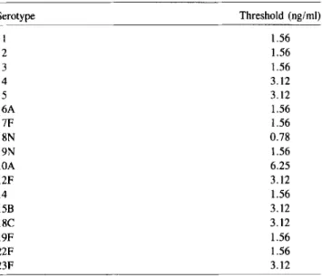
Table 2. Detection threshold of ELISA for specific capsular poly-saccharides in normal human serum.
NOTE. Normal human serum was spiked with 100 ng/ml of individual purified polysaccharides (ATCC) and serially diluted 1:2 with normal human serum. Thresh-old concentration is original polysaccharide concentration before 1:5 dilution and heat inactivation detectable in spiked normal human serum. Cutoff was mean of all nega-tive samples +2 SD.
*Pneumonia, 8; meningitis, 4; bacteremia without definite pulmonary infiltrates, 3; required isolation ofStreptococcus pneumoniae from blood.
tFourteen noninfectious conditions (malignancies, pulmonary infarction, cardiac failure, chronic obstructive lung disease), 14 pneumonias caused by alternate patho-gens diagnosed by culture or serology(Qfever, legionellosis, subphrenic empyema with pleural effusion, tuberculosis [2],Streptococcus pyogenes pneumonia [2], Staph-ylococcusaureus pneumonia [2], chronic obstructive lung disease with viral infection
[5]).
tPulmonary infiltrate on chest radiograph, fever, laboratory findings of inflamma-tion but no etiologic diagnosis (three sets of blood cultures, negative serologic workup).
ture-proven pneumococcal infection. One blood isolate, which reacted with Omniserum but with none of our pool sera or the full set of 39 commercial anti-serotype or -serogroup sera, was not included in our five seropools, explaining the one nega-tive test. This episode of pneumococcal bacteremia occurred in an individual who had been vaccinated several months pre-viously with the 23-valent pneumococcal vaccine after post-traumatic splenectomy, a setting possibly favoring infection with a less common serotype. No serum was positive in the 28 control patients in which pneumococcal infection was con-sidered possible at entry but excluded on clinical grounds. False-positive results also were not a problem in 46 control patients with cardiovascular disease without signs of infec-tion or inflammainfec-tion (table 3).
The ELISA also was evaluated in other body fluids submit-Table 1. Antibody titer of a commercial anti-pool A serum raised
to give uniform quellung reactions and an anti-pool A serum raised to give uniform ELISA titers: relation of antibody titer by ELISA to the sensitivity of the antigen-detection ELISA.
Commercial serum Elisa serum
Detection Detection
Antibody threshold Antibody threshold
Serotype titer (ng/ml) titer (ng/ml)
1 I :64,000 0.63 1:256,000 0.16
2 1:32,000 1.33 1:256,000 0.16
4 1:32,000 2.5 1:256,000 0.63
5 1:32,000 0.63 I :64,000 0.63
18C 1:8000 2.5 I:128,000 0.63
NOTE. Commercial anti-pool A rabbit serum was compared with a serum spe-cifically raised to obtain uniformly high titers by whole-cell ELISA. The detection threshold for purified commercial antigen (ATCC) was measured in buffer by capture sandwich ELISA optimized for each antigen by checkerboard titration.
tion, the composition of the seven or eight serotypes making up a pool vaccine was adjusted individually for each rabbit by augmenting the fraction of formol-fixed bacteria from the serotype to which the antibody response was inferior com-pared with the average for the other serotypes in the same pool or reducing the fraction of a serotype if antibody to one serotype prevailed in the pool. Immunotolerance was never a problem after increasing the vaccine dose. After 4-6 months of continuous immunization, sera deemed adequate were ob-tained for all five antigen pools.
Antibody titers and detection threshold for five purified an-tigens are shown for anti-pool A serum in table 1. So that we could compare our sera raised to ELISA specification with a commercial serum raised to produce a uniform quellung reaction (Statens Serum Institute), we included the same num-ber and types of pneumococci in our anti-pool A serum as in theirs. While both sera showed comparable variability in reactivity with individual serotypes, the serum raised to ELISA specification had a >5 times higher mean antibody titer by ELISA and detected on average a 3.4 times lower an-tigen concentration, regardless of concentrations of capture antibodies and conjugates established for each individual an-tigen, serum, and conjugate. These observations indicated that it was advantageous to follow the antibody response during serum production by ELISA, which in contrast to the quel-lung reaction also permitted an objective quantitation of an-tibody.
When we tested the quality of our ELISA system with a representative panel of the most commonly isolated pneumo-coccal serotypes, the ELISA detected relatively uniformly low concentrations of polysaccharides in human serum (mean
±
SD, 2.3±
1.24 ng/ml; table 2), even though the samples were diluted 1:5 in PBS for heat inactivation of nonspecific reac-tions between rabbit serum and clinical specimens.Clinical evaluation. Sera from 143 patients (144 episodes) obtained within 36 h of admission were studied. Antigen was detected in sera from 14 of 15 patients with blood
cul-Serotype I 2 3 4 5 6A 7F 8N 9N lOA 12F 14 15B 18C 19F 22F 23F
Table 3. Study population.
Group
I, definite* pneumococcal infection II, pneumococcal disease excluded'[ III, cardiovascular control group IV, pneumonia of unknown cause+
Threshold (ng/ml) 1.56 1.56 1.56 3.12 3.12 1.56 1.56 0.78 1.56 6.25 3.12 1.56 3.12 3.12 1.56 1.56 3.12 No. positive No. patients by ELISA
15 14
28 0
46 0
Table 5. Serotyping of antigen in body fluids by ELISA. Table 4. Detection of polysaccharide in body fluids other than serum.
NOTE. A set of anti-serotype or -serogroup sera corresponding to the reactive pool was used as capture antibody to bind type- or group-specific antigen followed by the detection step with anti-pool conjugate. S, serum; PLF, pleural fluid; D, 20-times-concentrated urine; CSF, cerebrospinal fluid.
28
n
cut off=mean+2SD
i~l~
~
tro
.Q u 't:J ClJ - J - J - J - J - Ja
a a a a
a
a a a a
Q Q Q Q QTRUE NEGATIVE SAMPLES
14
-Ja
a Q~
~ f--'-1 C/)a
Q1.1
"
I 46 I,
n
,
0.9
=,
,
I,
,
0.7
II I ~ I II I II I II J0.5 -
I II I,
II I I I0.3
II I I I I0.1
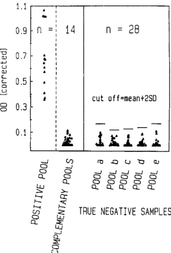
I I I I o o u OJ -+-' U OJ c:.... c:.... a ufection was excluded. Furthermore, antigen did not cross-react with complementary pools in positive sera (figure 1). How-ever, when we looked at the nonbacteremic individuals with pneumococcal pneumonia, diagnosed on the basis of a posi-tive ELISA test, we observed a discrete distribution of ELISA readings, ranging from clear-cut positive values to readings barely above the cutoff point, fixed 2 SD above the mean value of the negative readings from bacteremic patients (complemen-tary pools) and those in which pneumococcal infection was excluded (figures 1 and 2). Because our ELISA system reacted uniformly with all included antigens, this observation indi-cated that the level of antigenemia was disturbingly inconsis-tent. This was confirmed by computing the antigen level from linear regression curves plotted for each seropool. This anal-ysis showed, not unexpectedly, that bacteremic pneumococ-cal infections were usually associated with higher levels of Figure 1. ELISA readings from 14patients with blood culture-positive pneumococcal infection (culture-positive pool and complementary negative pools). There was no cross-reaction between positive seropool and four complementary pools. A cutoff 2 SD above mean value of negative controls, in whom pneumococcal infection was ex-cluded (table 3, group II), and negative complementary pools of proven cases frankly discriminates positive and negative test results. Optical density (OD) read at 492 nm was corrected for intertest vari-ations by internal standards OD.
7/7 0/9 NA 0/2 4/5 8/16 5/5 5/8 5/8 6/31 Cerebrospinal or pleural Urine Sputum fluid
10/10 1/13
ted together with the sera (table 4). These studies confirmed [11, 12] that the airways in patients with chronic obstructive lung disease are frequently colonized by one or multiple pneu-mococcal serotypes, so that little gain can be expected from an increase in sensitivity of a pneumococcal detection system. Furthermore, in some patients with mucopurulent chronic bronchitis, antigen of the same pool type as in sputum was detected in 20-fold-concentrated urine, indicating that by lowering the detection threshold for antigen in urine, anti-genuria associated with bronchial colonization could produce false-positive results.
By using commercial serotype- and serogroup-specific an-tisera as capture antibody, antigen could be typed correctly in body fluids by ELISA from the seven patients from whom an isolate was available for typing by the quellung reaction (table 5).
Extent ofantigenemia. The specifications required for a test system to be adequately sensitive to diagnose pneumo-coccal infection by detecting SCP in serum is governed by the level of clinical antigenemia. When we looked at the ELISA readings from 14 positive serum samples from bacteremic patients, it was not a problem to discriminate their positive readings from that of 28 patients in which pneumococcal
in-Group
I, definite pneumococcal infection II, pneumococcal disease excluded IV, pneumonia of unknown cause
A: positive serum ELISA B: negative serum ELISA
Reactive Reactive Serotype or
pool type or Source serogroup of
(ELISA) group (body fluid) isolate
A 2 S, U 2 A 4 S, PLF, U 4 C 7 S, PLF 7 C 35 S, CSF 35 D 9 S, CSF 9 D 9 S, U 9 D 23 S 23
NOTE. Data are number positive/number tested. Positive urine and cerebrospi-nal and pleural fluid samples always reacted in the same seropool as the serum sam-ple. In contrast, sputum samples reacted frequently in additional seropools in groups I and IVA. All group II and most group IVB patients with positive sputum reactions and all patients with positive urine reactions had a history of chronic productive bron-chitis. The urine reaction was always concordant with one of the frequently multiple reactions of the five seropools with sputum. NA: no specimen available.
1098 Schaffner etal. JID 1991;163 (May)
1.1 , . . . - - - ,
Table 6. Antigen concentrations detected in serum by ELISA. GroupI: Bacteremic pneumococcal pneumonia GroupIVA: Nonbacteremic pneumococcal pneumonia*
0.9
Pool Type or group Level (ng/ml) Type or Pool group Level (ng/ml)0.7
fo-
~ QJ ~ U QJ c, c,0.5
0 ~ Cl 00.3
•
•
•
•
•
•
••
•
A A A B B B B C C D D D D D Mean±SD 2 360.0 A It 39.0 B 4 10.0 B 22t 500.0 B 81.0 C 6.0 C 4.5 C 7 242.0 C 35 51.0 C 9 18.0 D 16.0 D 9 10.0 23 7.5 3.5 96.3±
156*8t
9t
10.0 8.0 3.5 2.5 15.5 11.5 5.0 3.5 3.0 15.5 13.5 8.3 ±5*•
---~---_.0.1 .
.~
..
~ ~
111111l
.
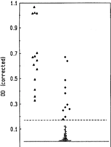
Figure 2. ELISA readings from 55 patients with blood culture-and serologically negative pneumonia, 11 with antigen detected in serum by ELISA ( • ) and 44 with no antigen detected (.). Note that these ELISA readings, in contrast to those from bacteremic pneu-mococcal infections(Ll), show discrete distribution without clear-cut bimodal distribution of positives and negatives. Dashed horizontal line corresponds to highest cutoff for negative readings, which was for pool A (see figure 1).
antigenemia than were nonbacteremic cases (table 6). But the variability of antigen levels by a factor of >150 at the time of bacteremia was unexpected. Furthermore, in many patients antigen levels (table 6) were below the detection threshold reported for antigen detection systems based on commercial sera [5, 7, 8, 15, 16].
These observations on the variability of antigenemia pointed to unknown factors that affected the level of antigenemia. Further insight was gained into such possible factors by com-paring the evolution of antigenemia in two patients with bac-teremic pneumococcal pneumonia, both adequately treated with penicillin G, to which the isolates were highly sensitive. One patient with pneumonia and sepsis caused by S. pneu-moniaeserotype 7F was initially in septic shock but cleared antigenemia within the first 3 days. The second patient with comparable pulmonary infiltrates caused by S. pneumoniae serotype 2 was unable to clear antigenemia over the full length of his hospital stay (figure 3). These observations indicated that the elimination rate of SCP could differ dramatically and
*Diagnosis dependent on antigen detection by ELISA.
tSerotyped by ELISA only.
:I:p<.04. E
400
0/\
::J c, Q..l350
(J) 4-300
aserotype 2
E250
<,200
0", _ _ _ _ _ _ _ 0 - 0 c 0 - 0 Q..l150
D1 .r-< +.l100
c rn ~serotype
7F D150
\\ C0
..
0
4
8
12
days after start of therapy
Figure 3. Course of antigenemia in a patient with serotype 2 and a patient with serotype 7F pneumonia of similar severity.presumably affected blood antigen levels. We therefore turned to experimental studies on the variability of SCP clearance from blood.
Kineticstudiesofantigenemia in animals. In accordance with our clinical observations, rats rapidly eliminated im-munoreactive SCP from serotype 7F but not from serotype 2 from their blood (figure 4).
Next we compared systematically the elimination rates of 12 SCP types in rabbits. These studies showed that the serum half-life of immunologically detected SCPs could varybya factor of >70, ranging for the 12 examined serotypes from 24 min to >29 h (figure 5). In addition to serum half-life of
4
SCPs, their volume of distributionand therefore their clear-ance varied appreciably (table 7). The mechanisms behind the variability in SCP serum half-lives and their distribution volumes appeared not to be uniformly the same because no correlationwas seenbetweenthe two kineticparameters (ta-ble 7). According to thevariationsinserumhalf-life, theperiod during which antigencould be detected in serum varied sig-nificantly (table 7) and correlated well with the serum half-life but not with the volume of distribution.
Nextweexplored the possibility thatpolysaccharides would preferentially associate withformed bloodelements. Wecom-pared the antigen concentration measured in blood plasma with that of sedimented blood cells for 11 of the 12 selected serotypes(all but lOA). The ratio of the polysaccharide con-centration in the plasma to that in the cell sediment was >1 for all antigens testedexcept for lIA and 20, whichwerepref-erentially associated with formed blood elements. When we attempted to correlatethe volume of distribution or the elimi-nation rate from bloodto the relativedistributionof polysac-charide betweenplasma and the formed blood elements we found no significant correlation between theseparameters (not shown). Nevertheless, this study indicated that for a few selected serotypes sedimented bloodcells werea more prom-ising source for diagnostic antigen detection than serum or plasma.
24
18
12
HOURS
7F
6
a
3
1
Figure 4. Elimination of specific capsular polysaccharides 2 and 7F from blood. Groups of five rats were injected with 1 mg (sero-type 2) or 2 mg (sero(sero-type 7F) of a crude preparation of capsular polysaccharide into the peritoneal cavity, and antigen levels were followed by ELISA for 24 h. Data are mean
±
SD. LN = loge.o 24 48 72 96 120 144 168 hours TYPE 14 8 • TYPE 158
~
7 ~ 7\
~ '" go c c 5 c ...J ...J o!8~
TYPE 2 '"c C ...J o 24 48 72 96 120 144 168 hours o 24 48 72hours96 120 144 168 o 24 48 72 96 120 144 168 hours TYPE 7F 72 96 120 144 168 hours TYPE 9N&N o 24 48 72 96 120 144 168 hours TYPE lac o 24 48 72 96 120 144 168 hours TYPE lOA o 24 48 72 96 120 144 168 hours ~ 7 ~ '" c TYPE llA o 24 48 72 96 120 144 168 hours '" c S 5 TYPE 20 o 24 48 72 96 120 144 168 hours TYPE 22F~:~
S 5 ~ o 24 48 72 96 120 144 168 hoursFigure 5. Kinetic of the disappearance of pneumococcal polysaccharides from serum of a rabbit injected intravenously with 500J.Lg (cir-cles) or 250J.Lg(diamonds) of polysaccharide. In some instances the phase of initial distribution was not completed 15 min after injection of the antigens (open symbols). Linear regression was computed from antigen levels after completion of initial distribution (solid symbols). Type lOA did not fit a model of a first-order elimination kinetic but showed two distinct phases, with a first half-time of 2.7 h followed bya slower terminal phase with a half-time of 39 h. LN
=
loge.1100 Schaffner et al. JID 1991;163 (May)
Discussion
type 2 and 7F did not result from an artifact stemming from in vitro antigen preparation because the difference between the two eliminationrates was comparable in the experiments with purified antigen and live bacteria (Figures 4 and 6). Figure 6. Kinetics of antigenemia ofratschallenged withlivepneu-mococciserotypes 2 and7F into theperitonealcavity20 min before administration of 106units of procaine penicillinintramuscularly.
Data are mean
±
SD from four or five rats per time point. Chal-lenge dose was 4.8 x 10⁸cfu. LN=
loge.24
48
72
HOURS AFTER CHALLENGE
8
~6
--
c: <l> C1'> ..-4
c: <t: C1'> c:2
z ~a
a
These studies showthat by raising antisera to pneumococ-cal polysaccharides specifipneumococ-cally for use in ELISA, it is possi-ble to obtain a clinically useful and highly and uniformly sensitivetool for the diagnosisof invasive pneumococcal in-fection. Our ELISAtest correctly identifiedat admission all 14bacteremic cases causedby serotypesincludedin the test. The only infection missed wascausedby a pneumococcal sero-type not contained in our pools of 39 serosero-types. The test also detected antigenemia in 11 of 55 patients with blood cul-ture-negative pneumoniathat remained without etiologicdi-agnosis after a complete workup, increasing the number of diagnosed pneumococcal pneumonias from 8 to 19 among 63 prospectively studied pneumonia patients (table 3).
The diagnostic yieldof our ELISA comparesfavorably with previousstudiesevaluating the increasedsensitivity of ELISA over that of counterimmunoelectrophoresis or agglutination techniques for diagnosing pneumococcal pneumonia [6-9, 15, 16]. In the best-documented study [7], in which an ELISA basedon Omniserumwasevaluated, 9 of 17bacteremicpneu-mococcalinfections were diagnosedon the initial sampleand 12 of 17with subsequent blood samples. In viewof the limited sensitivity of this Omniserum-based ELISA, which ranged from 200 to 3000 ng/ml of serum for single SCP types, it is not surprisingthat this ELISAmissedfive bacteremiccases and was not useful for the diagnosis of nonbacteremic pneu-Duration of
Volume of Serum
anti-distribution half-life Clearance genemia
Serotype (ml/kg) (h) (ml/min/kg) (h) 2 47.3 ± 8 20.0 ± 8.0 1.6 83 3 88.8 ± 14 27.5 ± 10.0 2.3 144 7F 90.3 ± 14 3.1 ± 1.0 20.4 13 9N&V 245.2 ± 15 24.0 ± 9.0 7.2 83 lOA 1026.0 ± 103 19.3 ± 1.0 37.2 40 11A. 1231.0 ± 38 7.1 ±5.0 123.1 8 14 214.5 ± 22 0.4 ± 0.1 375.4 2 15B 62.4 ± 15 2.6 ± 1.0 16.8 9 17F 129.5 ± 30 23.7 ± 12.0 3.8 75 18C 479.0 ± 82 29.1 ± 8.0 11.5 99 20 192.0 ± 3 13.7 ± 1.0 9.8 67 22F 265.9 ± 54 26.1 ± 8.0 7.1 83
We next ascertained that the examined antigens were sta-ble in heparinized rabbit blood and serum by demonstrating that the concentration of all 12 antigens was not altered by incubation in blood or serum over 48 h at 37°C.
Becauseantibody to SCP might affectits eliminationfrom the blood, we studied whether antibody was responsible for the observed variations between serum half-lives of SCPs. We examined whether natural antibody was responsible for the disparities, because adaptive humoral immunity developing in response to injected SCP could not be of importance in viewof the stableelimination rates attainedwithina few hours after challenge with antigen. When we looked at the poten-tial of serum from rats or rabbits obtained before challenge to capturepolysaccharide in our sandwichELISA system, we could not detect any SCP bindingto the solid phase mediated by the sera. Similarly, formol-fixed pneumococci did notbind detectable amounts of rabbit or rat serum immunoglobulin from the animals under study.
We anticipatedthat SCP types with a short serum half-life would not easily build up high blood SCP levels during the course of an infection. Toconfirmthis we turnedagainto rats; we used a model of infection with our initially studied pair of SCP-types with variable elimination rates to study the ki-netics of antigenemiaafter infectionwith livebacteria (figure 6). These studiesconfirmedthat the rapidlycleared SCP type 7F does not build up high antigen levels in the course of in-fection, whileinfection withS.pneumoniaeserotype2 results in consistentlyhigh SCP levels,which evenincrease after the beginning of penicillin treatment. This experiment also confirmed that the discrepantelimination ratesof SCP of
sero-NOTE. Serum half-life was computed from linear regression after completion of the initial distribution of injected antigen. Clearance was computed from the apparent volume of distribution of the injected antigen and the serum half-life. Data are mean or mean±SO from three animals. Antigen (Pneumovax-23) was injected intrave-nously in a dose of 250 ug in two animals and 500 ug in one animal for 7 days for each serotype. Data were pooled because elimination rates appeared not to depend on antigen load at these doses.
monias, in which we found antigen levels 10-100 times be-low the detection limit of the Omniserum-based test (table 6). ELISAsystems developed with commercial anti-poolsera have been found to be more sensitive than Omniserum sys-tems [16], but evensuchan ELISA system wasrelatively blind for certain common serotypes, resulting in false-negative re-sults eveninculturally positive cerebrospinal fluid samples [16]. In any event, these studies indicate that detection of SCP in blood by an ELISA able reliably to detect antigen levels as low as 1-6 ng/ml of serum can significantly contribute to the diagnosis of pneumococcal infection, evenin settings such as nonbacteremic pneumococcal pneumonia. Furthermore, the ability to serotype antigen in clinical specimens would allowfurther insight into the epidemiology of nonbacteremic pneumococcal pneumoniaor evenrapid predictionof the risk for penicillin resistance that is associated with certain sero-types[22].In contrastto detection bybloodtests, the increased sensitivityfor detection of SCP in concentrated urine speci-mens or sputum was offsetby the many positive reactions in sputum and urine of patients with chronic obstructive lung disease accompaniedby chronic bronchitis and pneumococ-cal colonization (table 4).
Our studydocuments an impressivevariability in the blood levels of SCP antigen, by a factor of >150; this variability could not be relatedto the severity of the infection alone. Bac-teremic patients had higher antigen levels, as shown previ-ouslyby others [23], but evenduringbacteremia the variability remained impressive(table 6). On one hand the inconsistent antigen levels, with concentrations as low as 3-10 ng/ml in bacteremiccases and levelsat the detectionlimit of our assay in nonbacteremic cases, documented that to be clinically re-liable, a test for detection of antigenemiamust have a detec-tion threshold of as little as 1-2 ng/ml or even less. On the other hand, the variability of antigenemia indicatedthat apart from the severity of infectionand the presenceof bacteremia, additional, hitherto-unknown factors determine the extent of antigenemia. Because we observed an impressivedifference in the elimination rate of SCP in two patients with compara-bly severe infections caused by disparate SCP serotypes, the question occurred whether the eliminationof SCP was sero-type dependent.
Animalstudiesof experimentalantigenemia confirmedthat disposal of immunoreactivecapsular pneumococcal polysac-charides varies significantly among different serotypes. Se-rum half-life, the volume of distribution, and the clearance rates of the 12 examined SCP types differed by a factor of >70, 25, and 250, respectively, explaining why some SCP types were detectable for >7 days in experimental antigene-mia while others were eliminated from blood within hours (table 7).
The observation that the elimination of SCP types 2 and 7F from the blood of rabbits and rats wascomparablein both species and, aboveall, corresponded to the clinical observa-tions with these two SCP types that originallypromptedthese
studies(figures 3-6) indicates thatthe variability of SCPclear-anceratesis, at leastinprinciple, not species dependent. These findings haveobviousimplications for the clinicaluse of SCP detection in body fluids for diagnosisof pneumococcal infec-tion.Itis conceivable that SCP types rapidly eliminated from blood are more difficult to detect in circulation, building up lower serum levelsthat last for shorter time periods. Accord-ingly, the experimental observations are in agreement with our clinical findings that the extent of antigenemia did not correlate with the severity of pneumococcal infections and that the antigen blood levelsmeasured in a population of pa-tients with serious pneumococcal infectionshowed a disturb-ing variability, with values from as high as several hundred ng/ml to barely measurable levels no longer clearly distinct from antigen-free samples in a sensitive ELISA system (figure 2). Furthermore, the significantvariations in the clearance of polysaccharide from blood and the important differences between the volume of distribution for the studied polysac-charides makeit plausible thatexcretion ofantigen in the urine, another body fluid used for antigen detection [7, 24], must also be quite variable.
Our observations also raisethe questionwhetherprolonged persistence of some SCP types contributes to virulence. The injectionof homologous pneumococcal capsular polysaccha-rides impressively enhances the susceptibility ofmiceto pneu-mococci [25, 26]. Circulating polysaccharide antagonizes homologous humoral anti-pneumococcal defenses by neu-tralizing antibody [25-28], and it appears logical that SCP types reaching higher levels and circulating for longer time periods havemore impact on humoral defenses by neutraliz-ing antibodythan do polysaccharides that fail to circulate for extendedperiods. In this context, it is interestingthat Coon-rod and Drennan [23] noted that pneumococci with low serotype numbers, which might be more virulent than high-numbered serotypes, cause higher SCP blood levelsthan do high-numbered serotypesand that high levels of antigenemia are associated with delayed appearance of measurable anti-body. Finally, failure to eliminate polysaccharide from cir-culationmightcontributeto the phenomenon of immunologic paralysis [29, 30], reported clinically withpolysaccharide 18C [31], which had the longest circulation time among the 12 SCP types studies in our experiments.
References
1.CoonrodJD, RytelMW. Detectionof type specificpneumococcal anti-gensbycounterimmunoelectrophoresis. Etiologic diagnosis of pneu-mococcal pneumonia. J Lab Clin Moo 1973;81:778-86.
2. LeachRP,CoonrodJD. Detectionofpneumococcal antigensin the spu-tum in pneumococcal pneumonia. AmRev Respir Dis 1977;116: 847-51.
3. Davidson M, Tempest B, Palmer DL. Bacteriologic diagnosisof acute pneumonia: comparisonof sputum,transtrachealaspirates,and lung aspirates. JAMA 1976;235:158-63.
4. Perlino A. Laboratory diagnosis of pneumonia due to Streptococcus
1102 Schaffner et al. liD1991;163 (May)
5. MartinSI,Hoganson DA, Thomass ET. Detection ofStreptococcus pneu-moniaeandHaemophilus irifluenzaetype b antigens in acute nonbac-teremic pneumonia.I Clin Microbiol 1987;25:248-50.
6. Holmberg H, Krook A. Comparison of enzyme-linked immunosorbent assay with coagglutination and latex agglutination for rapid diagno-sis of pneumococcal pneumonia by detecting antigen in sputa. Eur
I Clin Microbiol Infect Dis 1986;5:282-6.
7. Lenthe-Eboa S, Brighouse G, Auckenthaler R, et al. Comparison of im-munological methods for diagnosis of pneumococcal pneumonia in biological fluids. EurI Clin Microbiol Infect Dis 1987;6:28-34. 8. Ortquist A, Ionsson I, Kalin M, Krook A. Comparison of three methods
for detection of pneumococcal antigen in sputum of patients with community-acquired pneumonia. Eur I Clin Microbiol Infect Dis 1989;8:956-61.
9. Rytel MW, Preheim LC. Antigen detection in the diagnosis and in the prognostic assessment of bacterial pneumonias. Diagn Microbiol In-fect Dis 1986;4:S35-46.
10. Palmer DL, Iones CC. Diagnosis of pneumococcal pneumonia. Semin Respir Infect 1988;3:131-9.
11. Barrett-Connor E. The nonvalue of sputum culture in the diagnosis of pneumococcal pneumonia. Am Rev Respir Dis 1971;103:845-8. 12. Haas, H, MorrisIF,Samson S, KilbournIP,KimIP.Bacterial flora
of the respiratory tract in chronic' bronchitis: comparison of trans-tracheal, fiber-bronchoscopic and oropharyngeal sampling methods. Am Rev Respir Dis 1977;116:41-7.
13. BarnesDI,Naraqi S, IgoID.The role of percutaneous lung aspiration in the bacteriological diagnosis of pneumonia in adults. Aust N Z
I Med 1988;18:754-7.
14. Dochez AR, Averyor.The elaboration of specific soluble substance by pneumococcus during growth. I Exp Med 1917;26:477-92. 15. Harding SA, Sheld WM, McGowan MD, Sande MA. Enzyme-linked
immunosorbent assay for detection ofStreptococcus pneumoniae an-tigen.I Clin Microbiol 1979;10:339-42.
16. Da Costa Castro1M,Deschamps F, Benbachir M, HenrichsenI,Volle
PI,Guinet RMF. Highly sensitive biotin-avidin sandwich ELISA for the rapid detection of pneumococcal capsular polysaccharide anti-gens.I Immunol Methods 1987;104:265-70.
17. Lund E, HenrichsenI.Laboratory diagnosis, serology and epidemiol-ogy of streptococcus pneumoniae. Methods Microbioll978;12:241-62.
18. Roitt I. Essential immunology. Oxford: Blackwell Scientific Publica-tions, 1980.
19. AustrianR.Some observations on the pneumococcus and on the cur-rent status of pneumococcal disease and its prevention. Rev Infect Dis 1981;3:S1-17.
20. Iohnstone A, Thorpe R. Immunochemistry in practice. Oxford: Black-well Scientific Publications, 1982.
21. Wilson MB, Nakane PK. Recent development in the periodate method of conjugating horseradish peroxidase (HRPO) to antibodies.In:Knapp W, Holulas K, Wick G, eds. Immunofluorescence and related stain-ing techniques. New York: Elsevier-North Holland Biomedical Press, 1978:215-24.
22. Austrian R. Pneumococcal polysaccharide vaccines. Rev Infect Dis 1989;11:S598-612.
23. CoonrodID,Drennan DP. Pneumococcal pneumonia: capsular poly-saccharide antigenemia and antibody responses. Ann Intern Med 1976;84:254-60.
24. CoonrodID.Urine as an antigen reservoir for diagnosis of infectious diseases. AmI Med 1983;75:85-92.
25. Felton LD, Bailey GH. Biological significance of the soluble specific substances of pneumococci.I Infect Dis 1926;38:131-44. 26. Felton LD, Kaufmann G, Prescott B, Ottinger B. Studies on the
mecha-nism of the immunologic paralysis induced in mice by pneumococ-cal polysaccharides.I Immunol 1955;74:17-26.
27. Rosenow EC. Human pneumococcal opsonin and the anti-opsonic sub-stance in virulent pneumococci.I Infect Dis 1907;4:285-96. 28. Cole R. The neutralization of antipneumococcus immune bodies by
in-fected exudates and sera. I Exp Med 1918;27:453-575.
29. Stark OK. Studies on pneumococcal polysaccharide. II. Mechanism in-volved in production of "immunologic paralysis" by type I pneumo-coccal polysaccharide.I Immunol 1955;74:130-3.
30. HowardIG,Christie GH, IakobMI,Elson1. Studies on immunological paralysis. III. Recirculation and antibody-neutralizing activity of14C_ labeled type III pneumococcal polysaccharide in paralyzed mice. Clin Exp Immunol 1970;7:583-96.
31. Pichiero ME. Immunological paralysis to pneumococcal polysaccha-ride in man. Lancet 1985;2:468-71.