International Immunology, Vb/. 7, No. 11, pp. 1741-1752
Presentation of peptides by cultured
monocytes or activated T cells allows
specific priming of human cytotoxic T
lymphocytes in vitro
Maria Cristina Gagliardi, Guido De Petrillo, Simonetta Salemi, Laura Boffa,
Maria Grazia Longobardi, Paolo Dellabona
1, Giulia Casorati
1, Nabuyuki Tanigaki
2,
Reuben Harris
3, Antonio Lanzavecchia
3and Vincenzo Barnaba
Istituto I Clinica Medica, Universita di Roma 'La Sapienza', Policlinico Umberto I, Viale del Policlinico 155, 00161 Roma, Italy
1DIBIT, H. S. Raffaele, Milan, Italy
2lnstitute of Cellular Biology, CNR, Rome, Italy 3Basel Institute for Immunology, Basel, Switzerland
Keywords: CTL priming, monocytes, peptides
Abstract
The conditions favouring effective specific cytotoxic T lymphocyte (CTL) priming have been exploited to set up a simple and reproducible method to Induce a primary CTL response In vitro. We report that cultured monocytes, as well as activated T cells, pulsed with exogenous HLA-A2 binding immunogenic peptides, can induce primary peptlde-specific CTL responses In vitro in a Th-lndependent manner. Primary viral peptide-induced CTL were HLA-A2 restricted, and recognized
both peptlde-pulsed target cells and targets infected with recomblnant vaccinia virus expressing viral endogenous antigens. In addition, both cultured monocytes and activated T cells primed peptide-speciflc CD8+ T cells depleted from the CD45RO+ memory cell fraction. The efficiency of
CTL priming by monocytes was dependent upon the strong up-regulation of class I, adhesion and co-stimulatory molecules occurring spontaneously upon In vitro culture. The Inability of
unseparated peripheral blood mononuclear cells to mount a peptlde-speclflc CTL response could be reverted by direct co-stimulation of responding CD8+ T cells by soluble B7.1 or a stimulatory
anti-CD28 antibody, that allowed a specific response to take place. Although co-stimulation via the B7-CD28 interaction appeared sufficient to trigger CTL responses, it was not essential for CTL priming, since neither anti-B7.1 mAb nor soluble CTLA-4 Inhibited Induction of primary CTL response. This new method for Induction of specific CD8+ T cell response In vitro may be
exploited in adoptive Immunotherapy in cancer or In HIV-lnfected patients.
Introduction
Recent advances in understanding the interaction between naturally processed peptides and MHC molecules allow the identification of the likely immunogenic peptides in a protein sequence on the basis of the presence of specific anchors (1). Peptides binding to MHC class I molecules, which are in general 9-10 residues long, usually contain within their sequence two anchor residues interacting with corresponding binding pockets in the MHC molecule (2-4). The binding can be measured in vitro by demonstrating the capacity of the synthetic peptide to assist the folding of a particular class I
molecule (1,5-7). Definition of specific MHC motifs allows us to predict those peptides derived from viral or tumor antigens, potentially immunogenic for CTL.
Synthetic peptides have been used not only to identify the epitopes of a protein recognized by specific cytotoxic T lymphocytes (CTL), but also as vaccines to induce protective CTL in mouse (8-20). Moreover, peptides conjugated to either a helper epitope or lipid have been used to induce protective CTL responses (21,22). Alternatively, with regard to adoptive immunotherapy, i.e. in cancer patients or in severe
immuno-Correspondence to. V. Barnaba
compromised HIV carriers, one of the two following approaches may be considered: either the in vivo transfer of
in wrro-primed, well characterized peptide-specific CTL, or
peptide vaccination using well characterized peptides, that could be delivered on professional APC.
However, it would be desirable to set up a simple and reproducible method to induce primary antigen-specific CTL response in vitro. Indeed, such an approach may be essential to identify not only immunogenic peptides, but also the most appropriate professional APC capable of triggering CTL precursors. The prerequisite for such APC is to present a high density of peptide-MHC molecule complexes (signal 1) and to simultaneously deliver the co-stimulatory signals (signal 2) required for T cell activation, which has been defined as B7.1 or B7.2 interacting with CD28/CTLA-4 on T cells (23-26). The strategies to induce primary CTL responses in vitro and the conditions that determine effective CTL priming as well as unfavourable conditions are reported herein.
Methods
Identification of HLA-A2.1 binding peptides
Peptides of HIVgp120 and hepatitis B envelope antigen (HBenvAg; subtype ADW2) carrying HLA-A2.1 motif (1) were synthesized by the solid-phase method on an automated multiple peptide synthesizer (AMS 422; Abimed, Langenfeld, Germany) using Fmoc chemistry. The purity of peptides was determined by reverse-phase HPLC. Peptides were diluted to a concentration of 2 mg/ml and stored at -20°C. The peptides were screened for their ability to stabilize HLA-A2.1 molecules on the surface of transporter defective mutant T2 cells (27,28). T2 cells were cultured in RPMI 1640 (HyClone, Logan, UT) supplemented with 10% FCS (HyClone), 2 mM L-glutamine, 1% sodium pyruvate, 100 U/ml penicillin, 100 jig/ ml streptomycin and 2 jig/ml fungizone (Flow, Irvine, UK) (complete medium). T2 cells were washed twice and resus-pended in either serum-free medium in the presence of 10 \iglm\ human f^microglobulin (fern) (Sigma, St Louis, MO) or complete medium and incubated in 96-well flat-bottom plates in the presence or absence of different concentrations of peptide overnight at 37°C, 5% CO2. Cells were washed and stained with an anti-HLA-A2.1 mAb (lgG2a, BB7.2; ATCC, Rockville, MD) for 30 min at 4°C followed by FITC-F(ab)'2 goat anti-mouse Ig (GAM). Cells were washed twice and analysed using a FACScan (Becton Dickinson, Mountain View, CA).
Peptides showing high levels of stabilization of class I on T2 cells were studied for their ability to bind HLA-A2.1 by a direct binding assay, that is based on serologic detection of the conformational change of HLA class I a-chains induced by binding to specific peptides in the presence of fem, as described (29). Briefly, Epstein-Barr virus transformed-B (EBV-B) cells were lysed in Tris-HCI, pH 7.5, containing 0.5% NP-40 and protease inhibitors, and the lysates were denaturated by alkaline dissociation. The unfolded a-chain was separated from fern and peptides by gel filtration. The fractions containing the first major protein peak were pooled and incubated with test peptides and excess p2m for 16 h at 25CC. The increase in folded a-chain activity induced by
peptide binding was quantitated by a specific radioimmuno-assay involving a rabbit anti-HLA class I serum and 125l-labelled purified HLA-A2.1 molecules.
Purification of CD8* T cells
Human peripheral blood mononuclear cells (PBMC) were isolated from HLA-A2+ healthy donors on Lymphoprep cush-ions (LSM; Organon Teknika, Durham, NC). Donors were negative for HBV and HIV serological markers. PBMC were then washed in serum-free medium and allowed to adhere to 24-well plates (Falcon) in RPMI-1% human AB serum. After 90 min at 37°C the non-adherent cells were removed and used for cell purification as described (30). Briefly, CD8+ T cells were isolated by immunomagnetic separation with anti-CD8 mAb attached to Dynabeads (Dynal, Oslo, Norway). Positively selected cells were detached from magnetic beads by incubation with Detachabead (Dynal) according to the manufacturer's instructions. After the treatment, purified CD8+ and CD8"T cells were >98% CD8+ and < 1 % CD8~ respect-ively. In some experiments, a CD45RA+CD45RO~ population was isolated from purified CD8+ T cells by depletion of CD45R0"1" cells with anti-CD45RO+ mAb attached to mag-netic beads.
In vitro priming with peptide
PBMC (4-5X10⁶) were incubated in RPMI-1% human AB serum in culture 24-well culture plates (Falcon) for 90 min at 37°C, 5% CO2; the non-adherent cells were removed, and the adherent fraction was pulsed with different concentrations of peptide in serum-free medium for 4 h and used as APC. Adherent cells were then incubated with 1.5X10⁶ responding purified CD8+ T cells in the presence or absence of different concentrations of an anti-B7.1 mAb (31) or a fusion protein between human CTLA-4 and human lgG1 (huCTLA-4-hulgGI) (32). In some experiments we used as APC either an irradiated (3000 rad) autologous T cell clone, or irradiated (3000 rad) autologous PBMC or irradiated (13,000 rad) autol-ogous EBV-B cells. In some experiments, either a fusion protein between human B7.1 and IgM (huB7.1-lgM) or an anti-CD28 mAb were added to cultures in which irradiated PBMC were used as APC.
In all cases after 2 days of culture, 50 U/ml rlL-2 (Proleukin; Eurocetus, Emeryville, CA) was added and after a further 5 days, CD8+ T cells were re-stimulated with irradiated autolog-ous phytohaemagluttinin (PHA)-T cell blasts pulsed with 10 ng/ml peptide. After a second administration of IL-2, viable CD8+ T cells from each culture were tested for specific cytotoxicity on day 7 from the secondary stimulation.
Generation of CTL clones
T cell clones were isolated and maintained as previously described (33). Briefly, primary peptide-specific CTL were cloned by limiting dilution at 0.3 cells/well onto 96-well U-bottom plates in the presence of 0.5 ng/ml PHA-P (Wellcome Beckenham, UK), 50 U/ml rlL-2 and irradiated allogeneic feeder cells. After 2-3 weeks, cell growth was detected using an inverted microscope and growing cultures were tested for their capacity to mount a specific cytotoxic response to peptide-pulsed 51Cr-labelled target cells. Peptide-specific CTL clones were then expanded in rll-2-containing medium
and maintained in culture with 2 week cycles of re-stimulation with PHA plus allogeneic APC.
CTL assay
Cytotoxicity of CD8+ T cells, primed with peptide-pulsed APC, was tested in a 6 h 51Cr-release assay. T cells were used as effector cells at an E:T ratio ranging from 50:1 to 20:1. Effector cells were incubated in triplicate in U-bottom microtitre wells (Falcon), containing 5X10³ 51Cr-labelled homozygous HLA-A2.1+ EBV-B cells. Target cells were labelled with 100 nCi of Na51Cr (Amersham, Buckinghamshire, UK) for 2 h and then pulsed with 10 ng/ml peptide for 1 h at 4°C or left unpulsed. In some experiments anti-HLA-A2 mAb or an anti-HLA-B27 mAb were added at the initiation of the assay.
In some experiments, EBV-B cells infected with recombinant vaccinia virus (rW) expressing gp160 (VPE16) or with VSC8 as control were used as target cells. The r W were kindly donated by Andrea De Maria (University of Genova, Italy). EBV-B cells (1.5X10⁶) were incubated with 1 p.f.u./ixiO6 cells of the different preparations of r W at 4°C for 10 min, washed and resuspended in 5 ml complete medium and incubated at 37°C, 5% CO2, for 12 h before being used as target cells.
F7\CS analysis
The following purified specific mAb were used: anti-CD3 (lgG1, TR66), anti-CD4 (IgGI, 6D10), anti-HLA-A2.1 (lgG2a, BB7.2), anti-HLA-DR (lgG2a, L243), anti-HLA-DQ (lgG2a, SPVL3), anti-HLA-DP (lgG1, B7.21) and anti-B7.1 (lgG2a, B7.24); anti-CD28 (IgM, CK248) was kindly donated by Sandro Poggi (1ST National Institute for Cancer Research, Genova, Italy) (34); anti-HLA-B27 (lgG1, MEI) was kindly donated by Rossella Sorrentino (Department of Experimental Medicine, University of L'Aquila, Italy); CD19 (IgGI, J4.119), CD13 (IgGI, SJ.1D1), CD14 (lgG2a, RMO52), CD45RA (IgGI, ALB11), CD45RO (lgG2A, UCHL1), anti-CD1a (IgGI, BL6), anti-CD1b (lgG2a, 4.A7.6) and anti-CD1c (IgGI, L161) were purchased from Immunotech (Marseille, France); anti-CD11a (LFA-1, IgG, TEC-NK2) was purchased from TechnoGenetics (Milan, Italy); anti-HLA-ABC (lgG2a, W6.32) and anti-CD54 (ICAM-1, IgGI, 15.2) were purchased from Sera-Lab (Crawley Down, UK); and FITC-F(ab)'2 GAM was purchased from TechnoGenetics.
Cells were labelled with mAb on ice for 30 min, washed four times, incubated for 30 min on ice with FITC-F(ab)'2 GAM Ig, washed again and immediately analysed on FACScan flow cytometer (Becton Dickinson) equipped with a 15 mW air-cooled 488 nm argon-ion laser. The cytometer was calibrated using three different types of CaliBRITE (Becton Dickinson) beads of -6.6 |im in diameter. Gating was performed using a combination of forward and orthogonal light scatter (linear amplification). Propidium iodide was used to exclude dead cells. Data of 10,000 events was acquired and stored in list mode using the FACScan research software. The Lysys 111.1 program was used for analysis of data.
Soluble human B7.1-lgM molecule
The generation and characterization of the soluble huB7.1-IgM will be described in detail elsewhere (P. Dellabona and G. Casorati, manuscript in preparation). Briefly, total
cytoplasmic RNA extracted from an EBV-transformed cell line was reverse transcribed into cDNA, and the sequence encoding for the extracellular domains of the human B7.1 molecule was amplified by PCR using the following oligonucle-otide: 5'-CCTGAGCTCCTGAAGCCATGGGCCACACACGG-3' and 5'-TCTGACTTACCATCAGGAAAATGCTCTTGCTT-5'-CCTGAGCTCCTGAAGCCATGGGCCACACACGG-3' (underlined is a consensus splicing donor site). The PCR was run with the following conditions: 20 s at 94°C, 30 s at 60°C and 30 s at 72°C for 20 cycles. The amplified product was cloned blunt ended into the pBluescript vector, sequenced to rule out PCR errors and subcloned into the Ssfl-SaA digested pCD4-hCu expression vector. The huB7.1-lgM was transfected by protoplast fusion into the mouse plasmocytoma cell line J558L as described (35) and the clones secreting the highest amount of proteins (3-5 |ig/ml) were selected for expansion.
The approximate size of the B7.1-lgM was determined on an SDS-PAGE gel. Briefly, the B7.1-lgM protein was precipitated from culture supernatant using the anti-B7.1 mAb B7.24 (31) and Protein G (Pharmacia, Uppsala, Sweden), run on an SDS-PAGE both in reducing and non-reducing conditions, and blotted on a nitrocellulose filter. The filter was decorated with alkaline phosphatase-labelled GAM Ig (Southern Biotechnology, Birmingham, AL) followed by ECL (Amersham). Under reducing conditions, the B7.1-lgM migrated as a single molecular species of -130 kDa, while under non-reducing condition it migrated with an apparent Mr of 700, consistent with the pentameric structure determined by the IgM Fc portion. To prepare the B7.1-lgM containing supernatant, the J558L clone secreting it was grown in standard RPMI medium (Hyclone) containing 7% FCS in a Miniperm fermenter (Haereus, Germany). We determined as 1:10 the optimal dilution of B7.1-lgM containing super-natant giving the highest proliferative response on purified human CD4+ CD45RO T cells, stimulated with a sub-optimal dose of anti-CD3 mAb. The proliferative response was measured by [3H]thymidine incorporation in a stand-ard 72 h assay, and for each B7.1-lgM dilution was 1:10 = 47,000 C.p.m., 1:20 = 40,000 c.p.m., 1:40 = 38,000 c.p.m., 1:80 = 32,000 c.p.m. and 1:100 = 25,000 c.p.m. A spare culture supernatant used as a control gave the same back-ground incorporation of 2000 c.p.m. at all dilutions tested.
Results
Identification of HLA-A2 binding peptides
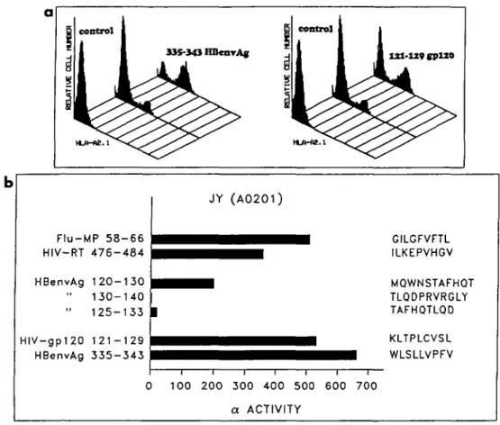
T cell determinants to be used for the induction of CTL responses in vitro were identified using the known HLA-A2 binding motif (5,6). Forty-eight nonamers carrying the A2 motif were screened for their capacity to stabilize HLA-A2 molecules on the surface of the transporter defective mutant T2 cells essentially as described (36). A peptide of HIVgp120 (121-129; KLTPLCVSL) and a peptide of HBenvAg (335-343; WLSLLVPFV), that showed high levels of binding, were selected for functional experiments, according to our prelimin-ary experiments demonstrating that CTL specific for those peptides efficiently cross-reacted on endogenous antigen presenting target cells. As shown in Rg. 1, these peptides can stabilize cell surface A2 molecules on T2 cells and can
335-343 HBenrAg XXI-tt9gpX» HLA-AE. 1
Flu-MP 58-66
HIV-RT 476-484
HBenvAg 120-130
130-140
" 125-133
HIV-gp120 121-129
HBenvAg 335-343
CJY (A0201)
^ ^ ^ ^ ^ • • • ^ • ^ • 1 GILGFVFTL
• • • • ^ ^ ^ ^ ILKEPVHGV
l ^ ^ ^ H M0WNSTAFH0T TLQDPRVRGLY | TAFHOTLQD i i i i i i i 100 2 0 0 300 400 500 600 7 0 0 a ACTIVITYFig. 1. Identification of HLA-A2 binding peptides. (a) Stabilization of surface HLA-A2 molecules in T2 cells by HIVgp12O(121-129) and by HBenvAg(335-343) peptides as detected by surface staining with anti-A2 antibody, (b) Effect of peptide and fem on refolding of HLA-A2 a-chains isolated from the HLA homozygous cell line, JY (A0201, B7, X), as detected by a specific radioimmunoassay. One unit of activity detected by the radioimmunoassay is defined as the amount of test sample that induces a 50% inhibition of the specific binding involved in the assay system. The results are presented as a activity/ml of test sample. Bars indicate a activity above the control (a-chain - Bom only). Flu-MP58-66 and HIV-RT476-484 represent positive controls, while HBV120-130,130-140 and 125-133 represent negative controls.
assist the refolding of isolated A2 a chains in the presence
of fern.
Characterization of APC for in vitro priming
Initial attempts to induce primary CTL responses by directly adding the HLA-A2 binding peptides to PBMC of HLA A2+ donors were unsuccessful (data not shown). We reasoned that the failure to induce a specific response might be due either to insufficient number of sites available on APC or to inappropriate presentation, i.e. presentation on non-profes-sional APC (26) or on the responding T cells themselves (37). We therefore asked whether selective display of the peptide on professional APC may favour stimulatory interactions and lead to CTL priming.
We tested different sources of APC: PBMC, EBV-B cells, the transporter mutant T2 that can be efficiently loaded with peptide, autologous T cell clones and adherent cells. Consistent stimulation of a peptide-specific response was obtained only when either adherent cells or T cell clones were used as APC. EBV-transformed autologous B cells and to a higher extent, T2 cells induced a very strong non-peptide-specific response that obscured a non-peptide-specific response.
Table 1. Phenotypic analysis of adherent cells at different
times of in vitro culture
CD13 CD14 CD4 HLA-ABC HLA-A2 HLA-DR HLA-DP HLA-DQ CD1a B7.1 CD54 CD11a CD3 CD19 Time of 0 20" 126 9 113 52 56 20 9 7 0 19 165 < 1 %c < 1 % in vitro culture 2 67 193 14 508 181 545 50 20 11 7 42 217 (h)a 6 143 322 24 550 194 663 156 45 46 27 96 344 24 346 282 9 392 154 151 142 33 38 38 236 264
"Adherent cells from HLA-A2+ donor were analysed for surface markers at different times of in vitro culture.
bMean fluorescence intensity. The background was subtracted, cvalues are expressed as percentage because they represent
The population of adherent cells was characterized by surface staining (Table 1). This population consisted mainly of CD13+CD14+ monocytes. It should be noted that upon
in vitro culture, monocytes underwent dramatic changes in
surface expression of various markers within the first few hours of culture (Table 1). The surface expression of HLA-A2 molecules increased at least three times. A similar increase
was evident for total class I as well as class II molecules. B7.1 was undetectable on freshly isolated monocytes, but was rapidly up-regulated after in vitro culture. The expression of ICAM-1 increased five times over 24 h while LFA-1 was unchanged. These results show that the simple in vitro culture of fresh monocytes is sufficient to up-regulate MHC, adhesion and co-stimulatory molecules, thus explaining their efficient
Primary stimulus of purified C T L a s s aY CD8+ T colls Specific CTL lines/ total cultures IPBMC/ -iPBMC/ 121-129 p«p. Mo./ Mo./ 121-129 pep. (10/ig/ml) Mo./ 121-129 pep. Mo./ 121-129 pep. (100/ig/ml) tTCC/
-rrcc/
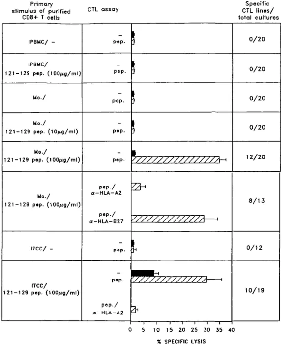
1 2 1 - 1 2 9 pep. ( 1 0 0 / i g / m l ) POP-pep. pep. pep. pep. pep./ a-HLA-A2 pep./ a-HLA-B27 pep. pep. pep./ O-HLA-A2 0/20 0/20 0/20 0/20 12/20 8/13 0/12 10/19 5 10 15 20 25 30 35 40 X SPECIFIC LYSISFig. 2. Induction of primary in vitro peptide-specific CTL responses by peptide-pulsed activated monocytes. Representative experiments are shown, in which purified CD8+ T cells from an HLA-A2+ donor were stimulated in replicate cultures with either irradiated (i) PBMC or adherent monocytes or autologous activated (i) T cell clones pulsed or not with different concentrations of HIV gp120 (121-129 peptide). After 7 days, cultures were re-stimulated with peptide-pulsed (i) autologous T cell blasts and after a further 7 days the cytotoxic activity of the individual cultures was measured against 51Cr-labelled target cells pulsed (hatched bars) or not (solid bars) with 10 jig/ml peptide, in the presence or absence of anti-HLA-A2 or anti-HLA-B27 mAb at an E:T ratio 25:1. On the far right of the figure the number of specific CTL lines above the total cultures is reported. Similar results were obtained using HBenvAg(335-343). Results represent the percentage mean of specific lysis expressed by the specific CTL lines.
presentation of peptide and T cell priming. As previously reported, human activated T cell clones express high levels of the same molecules and display 'professional' antigen presenting capacity (38,39).
Conditions for the generation of primary CTL responses using peptide-pulsed APC
A general protocol for the generation of specific CTL responses in vitro was developed. PBMC (4-5X10⁶) were incubated in 24-well culture plates for 90 min; the non-adherent cells were removed and the non-adherent fraction was extensively washed to deplete contaminating T and B cells and pulsed with different concentrations of peptide. Due to the low frequency of peptide-specific T cells in an unprimed donor, we set up several replicate 2 ml cultures containing adherent cells and 1.5X10⁶ responding purified (>98%) CD8+ T cells. Alternatively, responding CD8+ T cells were cultured with the aforementioned APC (1 x10⁶), that had been previously pulsed with different concentrations of peptide. Anyway, after 2 days, IL-2 (50 U/ml) was added and the cultures were incubated for an additional 5 days before secondary stimulation with 106 irradiated autologous T cell blasts that had been pulsed with 10 u,g/ml peptide. After a further 7 days, the cultures were individually tested for specific cytotoxicity against homozygous HLA-A2+ EBV-B cells pulsed or not with peptide.
As evident from a typical experiment reported in Fig. 2, monocytes pulsed with 100 ng/ml peptide induce a peptide-specific cytotoxic response detectable in 12 out of 20 replicate cultures. When autologous activated T cell clones were used as a source of peptide-pulsed APC a similar cytotoxic response was obtained (Fig. 2). In contrast, no specific response was obtained when either peptide-pulsed PBMC (Fig. 2), T2 cells or EBV-B cells (not shown) were used as APC. In all cases the responding T cells were peptide specific and HLA-A2 restricted, as shown by the inhibition of killing by an anti-HLA-A2 antibody. Similar results were obtained in 10 out of 14 healthy donors tested. Figure 3 clearly shows that CTL activity was specific for the peptide used in the priming, since target cells incubated with an HLA-A2.1 binding control peptide were not lysed.
Further evidence for selective priming of CTL by activated monocytes was obtained from cytofluorimetric analysis. When unfractionated HLA-A2+ PBMC were cultured with peptide-pulsed monocytes a selective expansion of CD8+ T cells was detected on day 7 (not shown).
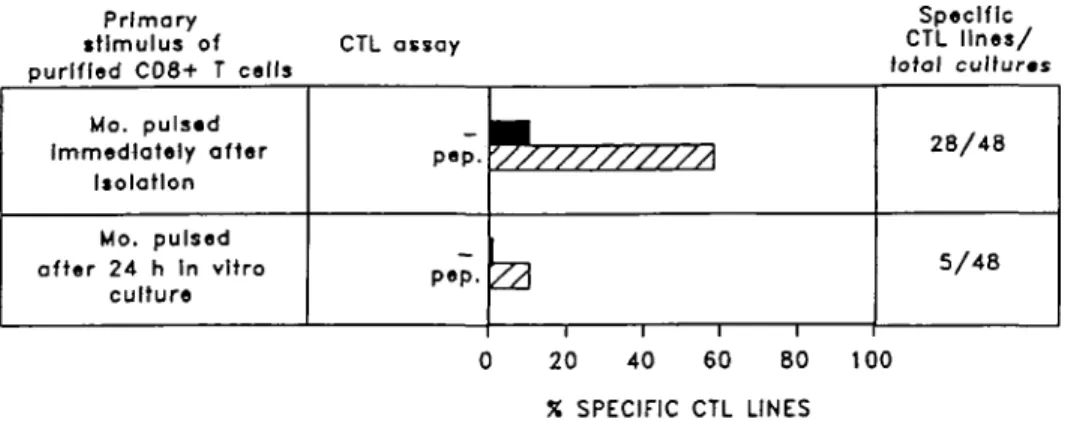
Interestingly, we noticed that the optimal timing for peptide loading on monocytes coincides with the first 4 h of in vitro culture when there is a maximum increase of surface class I molecules (Fig. 4 and Table 1).
We conclude that (i) when the only interactions allowed are those between specific CTL precursor and monocytes or T cell clones, a CTL response can be readily induced; (ii) in this case, induction of a primary CTL response triggered by professional APC does not require CD4+ T cells; and (iii) failure of total PBMC to mount a CTL response to soluble peptide cannot be explained by insufficient peptide loading, but rather by the presence of inappropriate interactions.
M 20
100
24 36 48
E:T r a t i o
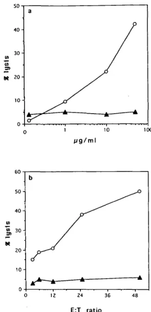
Fig. 3. Dose-response curves of in wf/o-primed CTL. (a) HIVgp120(121-129J-primed CTL showed cytotoxic activity, at an E:T ratio 25:1, against '1Cr-labelled target cells, previously pulsed with increasing concentrations of HIVgp12O(121-129) peptide (O), but not when pulsed with a non-relevant HLA-A2-binding peptide, as the HBenvAg(335-343) (A); (b) HIVgp120(121-129)-primed CTL were compared in their lytic responses against either HIVgp120(121-129)-sensitized (O) or HBenvAg(335-343)-HIVgp120(121-129)-sensitized (A) target cells at different E:T ratios.
Co-stimulatory requirements for the induction of a primary CTL response
The fact that peptide presentation by monocytes or T cell clones is effective, while presentation by total PBMC (which include monocytes as well) is not, suggests that total PBMC contain cells capable of presenting the peptide in a 'sup-pressing' fashion. To test whether the inability of PBMC to mount a peptide-specific CTL response was determined by presentation of peptide on cells that lack co-stimulatory signals, we asked whether soluble molecules that directly
Primary stimulus of purified CD8+ T cells CTL assay Specific CTL lines/ total cultures Mo. pulsed Immediately after Isolation Mo. pulsed after 24 h In vitro culture P • P • \//////////A
1
P<>P- p ^ l
1 1 1 1 28/48 5/48 0 20 40 60 80 100 X SPECIFIC CTL LINESFig. 4. Peptide pulsing of monocytes is most effective in the first hours of in vitro culture. Highly purified CD8+ T cells were primed with monocytes that were pulsed with gp 120(121-129) peptide for 4 h either immediately after 90 min adherence or after 24 h in vitro culture. Thereafter, they were re-stimulated and assayed for cytotoxicity against peptide-pulsed (hatched bars) or unpulsed (solid bars) target cells, as described, at an E:T ratio 25:1. The percentage of the specific CTL lines generated with the two conditions was reported. A CTL line was defined specific when it expressed a specific lysisof >15%.
Primary stimulus of purified CD8+ T cells CTL assay CTL llnss/Specific total cultures IPBMC/ 121-129 pep. (lOO/ig/ml) IPBMC/ 121-129 p«p. (100^g/ml) HuB7.1-lgM (1:10) IPBMC/ 121-129 p»p.(100/ig/ml) ontl-CD28 (1:100) p e p . W<
_ [
P.P. V///////////M
p«p- y///////////A—i
0/16 6/16 8/16 5 10 15 20 X SPECIFIC LYSIS 25Fig. 5. Co-stimulation by soluble B7.1 and anti-CD28 allows the response of unseparated peptide-pulsed PBMC. Peptide-pulsed unseparated PBMC, used as a source of APC, were cultured with purified CD8+ T cells in the absence or presence of either chimeric huB7.1-lgM molecules Of mouse anti-CD28 IgM antibodies. After 7 days, the cells were re-stimulated, as previously described, and after a further 7 days, were tested in a cytotoxicity assay against peptide-pulsed (hatched bars) or unpulsed (solid bars) target cells, as described, at an E:T ratio 25:1. Results represent the percentage mean of specific lysis expressed by all the specific CTL lines.
deliver the co-stimulatory signal to T cells might reverse this effect. When a huB7.1-lgM chimeric protein or an IgM anti-CD28 mAb was added to unseparated PBMC and peptide, a clear CTL response was detected (Fig. 5). These results demonstrate that co-stimulation via CD28 is sufficient for the induction of the CTL response and suggest that peptide presentation on frequent non-professional APC present among PBMC (resting T and B cells expressing class I but not co-stimulatory molecules) may induce peptide-specific anergy in CTL precursors.
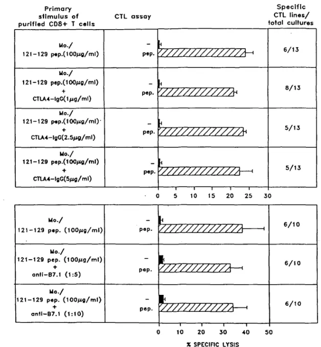
To further study the co-stimulatory requirements for the
induction of a primary anti-peptide response, we tested the blocking effect of anti-B7.1 mAb or soluble CTLA-4, that can bind with high affinity to both B7.1 and B7.2. Interestingly, neither anti-B7.1 nor soluble CTLA-4 was able to significantly inhibit the induction of a primary peptide-specific CTL response by activated monocytes (Fig. 6). Similar results were obtained using autologous activated T cells as APC (not shown). Control experiments showed that both anti-B7.1 mAb and soluble CTLA-4, both added at 5 u.g/ml at the initiation of culture, significantly blocked the proliferative response by resting T cells in a primary mixed lymphocyte reaction
Primary stimulus of purified CD8+ T cells CTL assay Specific CTL lines/ total cultures Mo./ 121-129 p«p.(100Atg/ml) Mo./ 121-129 pep.(100/xo/ml) + CTLA4-lgG(1/xg/ml) Mo./ 121-129 p«p.(100M0/ml)-+ CTLA4-lgG(2.5/*g/ml) Mo./ 121-129 pep.(100/xg/ml) + CTLA4-lgG(5/ifl/ml) pep. pep. pep. pep. <
'//////////////A-*
iV///////////M
i//////////////A*
(
'/////////////A—>
6 / 1 3 8/13 5/13 5/13 0 5 10 15 20 25 3 0 Mo./ 121-129 pep. (100/xg/ml) Mo./ 121-129 pep. (lOO^g/ml) ont!-B7.1 (1:5) Mo./ 121-129 pep. (100/xg/ml) ant1-B7.1 (1:10)_ I
pep. V/////////////A 1_ L
pap. Y///////////A '1
_ L
Deo. Y////////////A '
1
6/10 6/10 6/10 10 20 3 0 40 X SPECIFIC LYSIS 50Fig. 6. The induction of a specific CTL response by activated monocytes is not blocked by anti-B7.1 mAb nor by soluble huCTLA-4-hulgG. Peptide-pulsed monocytes were used as APC to prime a specific CTL response in the presence or absence of different concentrations of either anti-B7.1 mAb or soluble huCTLA-4-lgG. Results represent the percentage mean of specific lysis expressed by the specific CTL lines against peptide-pulsed (hatched bars) or unpulsed (solid bars) target cells.
stimulated by either T cell clones or monocytes or dendritic cells (not shown).
These results suggest that, in addition to B7.1/B7.2 molec-ules, professional APC may possess additional co-stimulatory ligands that are sufficient for the induction of a CTL response. Thus, it appears that each of the two pathways is in itself sufficient, although not strictly necessary for CTL activation.
Characterization of the responding T cells
To investigate whether the CTL response was due to in vitro priming of virgin CTL precursors or to the reactivation of memory cells that had been primed in vivo by the same or cross-reacting peptides, we compared the response of total
CD8+ T cells with that of CD8+ T cells depleted of CD45RO+ putative memory T cells. As shown in Table 2, the CTL response was comparable in both populations, indicating that the contribution to the response of CD45RO+ cells (usually only 10-20% of total CD8+) is indeed negligible. We cannot exclude, however, that the responding cells may belong to a population of resting CD45RA"1" cells that had reverted from CD45RO+ cells.
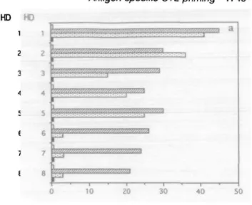
CTL obtained by in vitro priming with synthetic peptides recognize naturally processed peptides on infected cells
We tested whether the primary peptide-specific CD8+ T cells were able to recognize naturally processed peptides derived
Table 2. Primary peptide-induced CD8+CD45RCTT cells lyse peptide-pulsed target cells
Responding population8 Unseparated CD8+ T cells CD8+CD45RCTT cells Specific lysis (%) Peptide-pulsed Unpulsed targets targets 27 29 0 0
"Unseparated CD8+ T cells or CD45RO depleted-CD8+ T cells (CD8+CD45RO" T cells), sorted from peripheral blood as described in Methods, were cultured with peptide-pulsed adherent monocytes for 7 days followed by re-stimulation with peptide-pulsed autologous T cell blasts. Afterwards they were tested for their capacity to kill 51Cr-labelled EBV-B cells, pulsed or not with peptide. Percentage of specific lysis is expressed as mean of triplicate determinations.
from endogenously synthesized proteins. Out of eight HIVgp120(121-129) peptide-specific CTL lines tested, five efficiently cross-reacted on HLA-A2+ EBV-B cells infected with r W expressing HIVgp160 (Fig. 7). Figure 7 also shows that two representative CD8+ T cell clones, generated from HIVgp120(121-129) peptide-primed T cells, killed both the peptide-sensitized A2+ EBV-B cells and the same targets endogenously expressing HIVgp160. Similarly, CD8+ T cell clones specific for HBenvAg(335-343) peptide recognized EBV-B cells infected with r W expressing HBenvAg (not shown).
Discussion
In the perspective of an adoptive immunotherapy, it is import-ant to generate in vitro primary CTL responses, i.e. responses to antigens to which the individual has not been primed
in vivo. We used two nonamer HLA-A2 binding peptides from
HIVgp120 and HBenvAg to define the conditions for the generation of such CTL responses by CD8+ cells from seronegative donors. A critical factor appeared to be the mode of antigen presentation. While peptide pulsing of unseparated PBMC was invariably ineffective, pulsing of adherent mono-cytes or activated T cells with peptide induced a peptide-specific, HLA-A2-restricted CTL response. The responding CD8+ T cells recognize naturally processed peptides on target cells infected with r W expressing HIVgp160. This last is a fundamental requirement in the perspective of adoptive immunotherapy, in which specific CTL have to recognize peptides expressed in association with class I molecules on the surface of host tumor or infected cells, as a product of endogenous processing. Indeed, the majority of our peptide-specific CTL lines tested, as well as the CTL clones derived from them, efficiently killed infected target cells, confirming previous reports indicating that the immunodominant peptides always belong to the highest HLA-binding peptides (10,14,17).
Previous attempts to generate primary in vitro CTL responses in the mouse system involved the use of either dendritic cells, which possess high stimulatory capacity (40), or of the transporter defective mutant RMA-S, which expresses high levels of empty MHC molecules (12). Recently, human
HD 1 2 3 c i ; f I 2 3 4 5 e 7 e ^//%OTK«J^//'/^w/«^^^
r""*
10 20 30 % Specific Lysis 40 50 9A1 9A11 IF 1F4 % Specific LysisFig. 7. Primary viral peptide-induced CD8+ T cells recognize naturally processed peptide on infected cells, (a) Purified CD8+ T cells, derived from healthy donors (HD), were primed with HIVgp120(121-129)-sensitized adherent monocytes, followed by re-stimulation with peptide-sensitized autologous T cell blasts. After, they were tested for their capacity to kill (at an E:T ratio 25:1): 51 Cr-labelled HLA-A2+ EBV-B cells (JY line: HLA-A2.1, B7, C7) infected with wild-type W ( • ) , or same targets both infected with wild type W and pulsed with peptide (E3), or same targets infected with r W expressing HIVgp160 (EZI), or MCr-labelled HLA-A2" EBV-B cells (SA line: HLA-A24, B7, C7) infected with r W expressing HIVgp160 ( • ) . (b) Two representative CD8+ T cell clones (9A11 and 1F4), generated from gp120(121-129}-primed T cells, were tested for their ability to kill both HU\-A2+ EBV-B cells sensitized with HIVgp120(121-129)-peptide (0), and same targets infected with either wild-type W ( • ) or r W expressing HIV gp160 (ED, at an E.T ratio 10:1. Percentage of specific lysis is expressed as mean of triplicate determinations.
dendritic cells have been demonstrated to be very efficient APC in priming both naive CD4+ and CD8+ T cells (41,42). Moreover, the report by Houbiers et al. describes the genera-tion of primary peptide-specific CTL responses in humans
after in vitro stimulation with mutant T2 cells (43). This protocol was not successful in our hands. Indeed, in preliminary experiments we found that the human transporter defective mutant T2 cells, although able to bind high peptide levels (Fig. 1), was not suitable for in vitro priming, because of the very high level of background activation.
Out of all APC tested, only monocytes and T cell clones gave reproducible results. The fact that cultured monocytes are efficient for CTL priming implies that they can bind sufficient amounts of peptide and present it in the appropriate co-stimulatory context. This efficient presentation may be dependent upon the strong up-regulation of MHC class I, as well as adhesion and co-stimulatory molecules that appear spontaneously upon in vitro culture within the first 2-6 hours (Table 1) (44), as shown by the fact that adherent monocytes are much more effective if pulsed in the first few hours of culture. The peptide pulsing, done during the up-regulation of class I molecules by APC, may favour a more efficient peptide binding to that small percentage of newly synthesized empty class I molecules, available to bind exogenous peptides.
We also found that autologous activated T cell clones can function as APC for CTL priming with antigenic peptides. An advantage in the use of activated T cells is the lack of a very low non-specific background of stimulation in the absence of peptide. We and others have previously demonstrated that activated T cell clones are indeed professional APC and very effective in priming other T cells (38,39).
Recently, an alternative approach for inducing primary anti-tumor CTL in humans was successfully carried out using as APC non-transformed B cell blasts (45). This together with the finding that EBV-transformed B cells used as APC were not able, in our system, to prime antigen-specific CTL responses, suggests that the two B cell preparations have different APC capabilities.
It has been previously reported that the density of T cell epitopes required for the induction of primary CTL response is much higher than the concentration required for secondary responses (12). This difference is evident also in the induction of human CTL, since a 10-fold lower peptide concentration is required to sensitize target cells for killing.
There are three points that need further discussion. First, as reported in several experimental systems (12,32,46-49), our data demonstrate that, when antigen is presented by professional APC, the requirement for antigen-specific Th cells in the induction of CTL may not be evident. This result gives rise to speculation that CTL precursors primed by professional APC do not require Th cooperation, that is instead essential when antigen is presented by non-professional APC; in this last instance, activated T cells themselves, working as professional APC (39), could simultaneously present peptide and provide co-stimulation for an appropriate CTL priming.
The second point concerns the pathway of co-stimulation. Our results demonstrate that direct co-stimulation of responding CD8+ T cells by soluble ligands via CD28 can revert the inability of peptide presentation by non-professional APC. In apparent contrast, however, we found that anti-B7.1 antibody as well as soluble CTLA-4 that blocks both B7.1 and B7.2 (24,25) fail to inhibit induction of a specific CTL
response by activated monocytes. These results suggest that the B7^CD28 interaction, although by itself sufficient for priming, is not strictly necessary and that alternative co-stimulatory pathways may exist. Similar conclusions have been recently reached by Johnson and Jenkins in an anti-CD3-dependent T cell activation system (50).
Finally, our data favour the idea that a real CTL priming is carried out by virgin T cells in our system for two reasons: (i) the donors were healthy and seronegative and (ii) the depletion of CD45RO+ cells did not affect the CTL response, indicating that most of the responding cells belong to the RA+RO~ compartment, even though we cannot exclude that this last population may contain some CD45RA+ resting T cells that have reverted from memory CD45RO+ phenotype (51,52).
In conclusion, exploiting the strategies affected by the immune system for inducing a primary T cell response, we define a protocol of CTL priming, by using activated mono-cytes or T cell clones as professional APC, well defined HLA-binding peptides as immunogenic antigen, and the conditions favouring productive interactions between specific CTL pre-cursors and professional APC.
Acknowledgements
This work was supported in part by the Ministero della Sanita-lstituto Superiore di Sanita-Progetto AIDS-Roma-ltalia, by the Ministero della Universita e della Ricerca Scientifica e Tecnologica 40% grant 051503097 and by the Andrea Cesalpino Foundation.
Abbreviations
( V1 [)2-microglobulin
CTL cytotoxic T lymphocyte
EBV-B Epstein-Barr virus-transformed B cell GAM goat anti-mouse Ig
HBenvAg hepatitis B envelope antigen PBMC peripheral blood mononuclear cell PHA phytohaemagglutinin
W vaccinia virus
References
1 Rammensee, H. G., Falk, K. and Rotzschke, O. 1993. Peptides naturally presented by MHC class I molecules. Annu. Rev. Immunol. 11:213.
2 Madden, D. R., Gorga, J. C , Strominger, J. and Wiley, D. 1991. The structure of HLA-B27 reveals nonamer self-peptides bound in an extended conformation. Nature 353:321.
3 Matzumura, M., Fremont, D. H., Peterson, P. A. and Wilson, I. A. 1992. Emerging principles for the recognition of peptide antigens by MHC class I molecules. Science 257:927.
4 Germain, R. N. 1994. MHC-dependent antigen processing and peptide presentation providing ligands for T lymphocyte activation. Cell 76:287.
5 Falk, K., Rotzschkle, O., Stevanovic, S., Jung, G. and Rammensee, H.-G. 1994. Allele-specific motifs revealed by sequencing of self-peptides eluted from MHC molecules. 1991. Nature 351:290. 6 Hunt, D. R, Henderson, R. A., Shabanowitz, J., Sakaguchi, K.,
Michel, H., Sevilir, N., Cox, A. L., Appella, E. and Engelhard, V. H. 1994. Characterization of peptides bound to the class I MHC molecule HLA-A2.1 by mass spectrometry. 1992. Science 255:1261.
7 Ruppert, J., Sidney, J., Celis, E., Kubo, R. T, Grey, H. M. and Sette, A. 1993. Prominent role of secondary anchor residues in peptide binding to HLA-A2.1 molecules. Cell 74:929.
8 Carbone, F. Ft., Moore, M. W, Sheil, J. M. and Bevan, M. J. 1993. Induction of cytotoxic T lymphocytes by primary in vitro stimulation with peptides. J. Exp. Med. 167:1767.
9 Carbone, F. R. and Bevan, M. J. 1993. Induction of ovalbumin-specific cytotoxic T cells by in vivo peptide immunization. J. Exp. Med. 169:603.
10 Kast, W. M., Offringa, R., Peters, P. J., Vbordouw, A. C , Meloen, R. H., van der Eb, A. J. and Melief, C. J. 1989. Eradication of adenovirus E1-induced tumors by E1A-specific cytotoxic T lymphocytes. Cell 59:603.
11 Aichele, P., Hengartner, H., Zinkernagel, R. M. and Schulz, M. 1990. Antiviral cytotoxic T cell response induced by in vivo priming with a free synthetic peptide. J. Exp. Med. 171:1815. 12 De Brujin, M. L H., Schumacher, T. N., Nieland. J. D., Ploegh,
H. L, Kast, W. M. and Melief, C. J. 1991. Peptide loading of empty major histocompatibility complex molecules on RMA-S cells allows the induction of primary cytoxic T lymphocyte responses. Eur. J. Immunol. 21:2963.
13 De Brujin, M. L. H., Nieland, J. D., Schumacher, T. N., Ploegh, H. L, Kast, W. M. and Melief, C. J. 1991. Mechanisms of induction of primary virus-specific cytotoxic T lymphocyte responses. Eur. J. Immunol. 22:3013.
14 Kast, W. M., Roux, L, Curren, J., Blom, H. J., Vbordouw, A. C , Meloen, R. H., Kolakofsky, D. and Melief, C. J. 1991. Protection against lethal Sendai virus infection by in vitro priming of virus-specific cytotoxic T lymphocytes with a free synthetic peptide. Proc. Natl Acad. Sci. USA 88:2283.
15 Schulz, M., Zinkernagel, R. M. and Hengartner, H. 1991. Peptide-induced antiviral protection by cytotoxic T cells. Proc. Natl Acad. Sci. USA 88:991.
16 Riddell, S. R., Watanabe, K. S., Goodrich, J. M., Li, C. R., Agha, M. E. and Greenberg, P. D. 1992. Restoration of viral immunity in immunodeficient humans by the adoptive transfer of T cell clones. Science 257:238.
17 Feltcamp, M. C. W., Smits, H. L, Vlerboom, M. P. M, Minnaar, R. P., de Jongh, B. M., Drijfhout, J. W., Schegget, J. ter, Melief, C. J. M. and Kast, W. M. 1993. Vaccination with cytotoxic T lymphocyte epitope-containing peptide protects against a tumor induced by human papillomavirus type 16-transformed cells. Eur. J. Immunol. 23:2242.
18 Lanzavecchia, A. 1993. Identifying strategies for immune intervention. Science 260:937.
19 van der Bruggen, P., Traversari, C , Chomez, P., Lurquin, C , De Plaen, E., Van den Eynde, B., Knuth, A. and Boon, T. 1991. A gene encoding an antigen recognized by cytolytic T lymphocytes on human melanoma. Science 254:1643.
20 Traversari, C , van der Bruggen, P., Leuscher, I. F., Lurquin, C , Chomez, P., Van Pel, A., De Plaen, E., Amar-Costesec, A. and Boon, T. 1992. A nonapeptide encoded by human gene MAGE-1 is recognized on HLA-AMAGE-1 by cytolytic T lymphocytes directed against tumor antigen MZ2-E. J. Exp. Med. 176:1453.
21 Deres, K., Schild, H., Wiesmuller, K. H., Jung, G. and Rammensee, H. G. 1989. In vivo priming of virus-specific cytotoxic T lymphocytes with synthetic lipopeptide vaccine. Nature 342:561. 22 Widmann, C ., Romero, P., Marjanski, J. L., Corradin, G. and Valmori, D. J. 1992. T helper epitopes enhance the cytotoxic response of mice immunized with MHC class l-restricted malaria peptides. Immunol. Methods 155:95.
23 Linsley, P. S., Brady, W., Grosmaire, L S., Aruffo, A., Damle, N. K. and Ledbetter, J. A. 1991. Binding of the B cell activation antigen B7 to CD28 co-stimulates T cell proliferation and interleukin 2 mRNA accumulation. J. Exp. Med. 173:721. 24 Linsley, P. S., Brady, W., Urnes, M., Grosmaire, L. S., Damle, N. K.
and Ledbetter, J. A. J1991. CTLA-4 is a second receptor for the B cell activation antigen B7. J. Exp. Med. 174:561.
25 Freeman, G. J., Greiben, J. G., Boussiotis, V. A., Ng, J. W., Restivo, V. A., Jr, Lombard, L. A., Gray, G. S. and Nadler, L. M. 1993. Cloning of B7-2: a CTLA-4 counter-receptor that costimulates human T cell proliferation. Science 262:909. 26 Janeway, C. A., Jr and Bottomly, K. 1994. Signals and signs for
lymphocyte responses. Cell 76:275.
27 Salter, R. D. and Cresswell, P. 1986. Impaired assembly and
transport HLA-A and -B antigens in a mutant T x B cell hybrid. EMBO J. 5:943.
28 DeMars, R. and Spies, T. 1992. New genes in the MHC that encode proteins for antigen processing. Trends Cell Biol. 2:81. 29 Fruci, D., Rovero, P., Falasca, G., Chersi, A., Sofrentino, R., Butler, R., Tanigaki, N. and Tosi, R. 1993. Anchor residue motifs of HLA class-l-binding peptides analyzed by the direct binding of synthetic peptides to HLA class-l alpha chains. Human Immunol. 38:187.
30 Barnaba, V., Franco, A., Paroli, M., Benvenuto, R., De Petrillo, G., Burgio, V. L., Santilio, I., Balsano, C , Bonavita, M. S., Cappelli, G., Colizzi, V., Cutrona, G. and Ferrarini, M. 1994. Selective expansion of cytotoxic T lymphocytes with a CD4+ CD56+ surface phenotype and a T helper type 1 profile of cytokine secretion in the liver of patients chronically infected with hepatitis B virus. J. Immunol. 152. 3047.
31 De Boer, M., Parren, P., Dove., J., Ossendorp, F., van der Horst, G. and Reeder, J. 1992. Functional characterisation of a novel anti-B7 monoclonal antibody. Eur. J. Immunol. 22:3071. 32 Dohring, C , Angman, L., Spagnoli, G. and Lanzavecchia, A. 1994.
T-helper- and accessory-cell-independent cytotoxic responses to human tumor cells transfected with a B7 retroviral vector. Int. J. Cancer57A.
33 Barnaba, V., Franco, A., Alberti, A., Benvenuto, R. and Balsano, F. 1990. Selective killing of hepatitis B envelope antigen-specific B cells by class l-restricted, exogenous antigen-antigen-specific T lymphocytes. Nature 345:258.
34 Poggi, A., Bottino, C , Zocchi, M. R., Pantaleo, G., Ciccone, E., Mingari, C , Moretta, L. and Moretta, A. 1987. CD3+ WT31~ peripheral T lymphocytes lack T44 (CD28), a surface molecule involved in activation of T cells bearing the alpha/beta heterodimer. Eur. J. Immunol. 17:1065.
35 Traunecker, A., Olivieri, F. and Karjalainen, K. 1991. Myeloma based extrusion system for production of large mammalian proteins. Trends Bhtechnol. 9:109.
36 Cerundolo, B., Alexander, J., Anderson, K., Lamb, C , Cresswell, P., McMichael, A., Gotch, F. and Townsend, A. 1990. Presentation of viral antigen controlled by a gene in the major histocompatibility complex. Nature 345:449.
37 Celis, E. and Saibara, T. 1992. Binding of T cell receptor to major histocompatibility complex class ll-peptide complexes at the single-cell level results in the induction of antigen unresponsiveness (anergy). Eur. J. Immunol. 22:3127.
38 Azuma, M., Yssel, H., Phillips, J. H., Spits, H. and Lanier, L. L 1993. Functional expression of B7/BB1 on activated T lymphocytes. J. Exp. Med. 177:845.
39 Barnaba, V, Watts, C , De Boer, M., Lane, P. and Lanzavecchia, A. 1994. Professional presentation of antigen by activated human T cells. Eur. J. Immunol. 24:71.
40 Macatonia, S. E., Taylor, P. M., Knight, S. C. and Askonas, B. A. 1989. Primary stimulation by dendritic cells induces antiviral proliferate and cytotoxic T cell responses in vitro. J. Exp. Med. 169:1255.
41 Sallusto, F. and Lanzavecchia, A. 1994. Efficient presentation of soluble antigen by cultured human dendritic cells is maintained by granulocyte/macrophage colony stimulating factor plus interleukin 4 and downregulated by tumor necrosis factor a. J. Exp. Med. 178:1109.
42 Mehta-Damani, A., Markowicz, S. and Engleman E. G. 1994. Generation of antigen-specific CD8+ CTLs from naive precursors.
J. Immunol. 153:996.
43 Houbiers, J. G. A., Nijman, H. W., van der Burg, S. H., Drijfhourt, J. W., Kenemans, P., van de velde, C. J. H., Brand, A., Momburg, F., Kast, W. M. and Melief, C. J. M. 1993. In vitro induction of human cytotoxic T lymphocyte responses against peptides of mutant and wild-type p53. Eur. J. Immunol. 23:2072. 44 Thomas, R., Davis, L. S. and Lipsky, P. E. 1993. Comparative
accessory cell function of human peripheral blood dendritic cells and monocytes. J. Immunol. 151:6840.
45 Celis, E., Tsai, V, Crimi, C , DeMars, R., Wentworth, P. A., Chestnut, R. W., Grey, H. M., Sette, A. and Serra, H. M. 1994. Induction of anti-tumor cytotoxic T lymphocytes in normal humans using
primary cultures and synthetic peptide epitopes. Proc. NatlAcad. Sci. USA 91:2105.
46 Chen, L, Ashe, S., Brady, W. A., Hellstrom, I., Hellstrom, K. E., Ledbetter, J. A., McGowan, P. and LJnsley, P. S. 1992. Costimulation of antitumor immunity by the B7 counter-receptor for the T lymphocyte molecules CD28 and CTLA-4. Cell. 71:1093. 47 Azuma, M., Cayabyab, M., Buck, D., Phillips, J. H. and Lanier, L. L.
1992. CD28 interaction with B7 costimulates primary allogeneic proliferative responses and cytotoxicity mediated by small, resting T lymphocytes. J. Exp. Med. 175:353.
48 Townsend, S. E. and Allison, J. P. 1993. Tumor rejection after direct costimulation of CD8+ T cells by B7-transfected melanoma
cells. Science 259:368.
49 Harding, F. A. and Allison, J. P. 1993. CD28-B7 interactions allow the induction of CD8+ cytotoxic T lymphocytes in the absence of exogenous help. J. Exp. Med. 177:1791.
50 Johnson, J. G. and Jenkins, M. K. 1994. Monocytes provide a novel costimulatory signal to T cells that is not mediated by the CD28/B7 interaction. J. Immunol. 152:429.
51 Bell, E. B. and Sparshott, S. M. 1990. Interconversion of CD45R subsets of CD4 T cells in vivo. Nature 348:163.
52 Michie, C. A., McLean, A., Alcock, C. and Beverley, P. C. L. 1992. Lifespan of human lymphocyte subset defined by CD45 isoforms. Nature 360:264.