Talking in Fury: The Cortico-Subcortical Network Underlying Angry Vocalizations
Sascha Frühholz
1,2, Hannah S. Klaas
1, Sona Patel
2and Didier Grandjean
1,21
Neuroscience of Emotion and Affective Dynamics Laboratory (NEAD), Department of Psychology, University of Geneva, Geneva,
Switzerland and
2Swiss Center for Affective Sciences, University of Geneva, Geneva, Switzerland
Sascha Frühholz and Hannah S. Klaas contributed equally to this study.
Address correspondence to Sascha Frühholz, University of Geneva, Swiss Center for Affective Sciences, 9 Chemin des Mines, CH-1202 Geneva, Switzerland. Email: [email protected]
Although the neural basis for the
perception of vocal emotions has
been described extensively, the neural basis for the
expression of
vocal emotions is almost unknown. Here, we asked participants both
to repeat and to express high-arousing angry vocalizations to
command (i.e., evoked expressions). First, repeated expressions
eli-cited activity in the left middle superior temporal gyrus (STG),
point-ing to a short auditory memory trace for the repetition of vocal
expressions. Evoked expressions activated the left hippocampus,
suggesting the retrieval of long-term stored scripts. Secondly, angry
compared with neutral expressions elicited activity in the inferior
frontal cortex IFC and the dorsal basal ganglia (BG), speci
fically
during evoked expressions. Angry expressions also activated the
amygdala and anterior cingulate cortex (ACC), and the latter
corre-lated with pupil size as an indicator of bodily arousal during
emotion-al output behavior. Though uncorrelated, both ACC activity and pupil
diameter were also increased during repetition trials indicating
in-creased control demands during the more constraint production type
of precisely repeating prosodic intonations. Finally, different acoustic
measures of angry expressions were associated with activity in the
left STG, bilateral inferior frontal gyrus, and dorsal BG.
Keywords: ACC, amygdala, basal ganglia, fMRI, vocal emotions
Introduction
Emotional prosody is crucial for signaling affective states
during social interactions. An impairment in the expression of
emotional prosody can have severe consequences in social
contexts and for social and emotional development (
Bell et al.
1990
). Early evidence for the neural basis of emotional vocal
expressions comes from patient studies. Brain lesions leading
to affective aprosodia point to a dominant role of the right
frontal cortex in emotional prosody production (
Borod et al.
2002
;
Ross and Monnot 2008
). These studies also point to an
involvement of the basal ganglia (BG;
Cohen et al. 1994
).
The importance of the right frontal cortex and of the BG has
also been supported by recent neuroimaging studies on
emotional vocal productions. They reported activations in the
inferior frontal gyrus (IFG;
Aziz-Zadeh et al. 2010
;
Laukka
et al. 2011
;
Pichon and Kell 2013
) and several subregions of
the BG (
Laukka et al. 2011
;
Pichon and Kell 2013
). In addition,
activations were reported in the STG (
Dogil et al. 2002
;
Aziz-Zadeh et al. 2010
;
Laukka et al. 2011
;
Pichon and Kell 2013
).
These
findings of STG involvement have been interpreted as
arising from phonological feedback processing (
Dogil et al.
2002
), as well as from the provision of articulatory maps (
Aziz-Zadeh et al. 2010
) in relation to acoustic features of emotional
vocal output, especially in the right STG (
Pichon and Kell
2013
). The IFG is supposed to have a role in articulatory
moni-toring and modulation of vocal expressions (
Dogil et al. 2002
;
Aziz-Zadeh et al. 2010
;
Laukka et al. 2011
). The BG are assumed
to be responsible for volitional control of vocally expressed
affect (
Laukka et al. 2011
), especially in their dorsal part, while
the ventral part seems to add the emotional component during
the preparation of vocal expressions (
Pichon and Kell 2013
).
These results together provide evidence for an extended
cortico-subcortical network involved in the production of
emotional prosody that partly overlaps with a neural model of
mammalian vocalizations (
Jurgens 2009
;
Hage 2010
). Some
in-consistencies remain in the previous
findings, however, as well
as some open questions, especially concerning the BG,
amyg-dala, anterior cingulate cortex (ACC), STG, and IFG, and also
concerning different types of vocal productions.
In this study, we addressed many of these open questions
concerning these brain regions by investigating the neural
basis of angry vocalizations. First, though the BG are an
inte-gral part of the mammalian vocalizations network (
Jurgens
2009
;
Hage 2010
), the importance of the BG for the expression
of emotional prosody in human vocalizations is still under
dis-cussion (
Ross and Monnot 2008
).
Laukka et al. (2011)
claimed
that vocal affect is regulated by the BG by showing that
increas-ing BG activation was inversely related to levels of nervousness
in the voice. Nervousness, however, is only one aspect of vocal
affect among several other important vocal acoustic features.
Pichon and Kell (2013)
found an involvement of the BG,
especially of the ventral parts, during an emotional induction
phase prior to vocalizations, which showed a strong
connec-tivity to the dorsal BG during the production phase of
vocaliza-tions that followed. This is indicative of a functional
segregation in the BG related to emotional and sensorimotor
processing in the ventral and dorsal BG, respectively (
Yelnik
2008
;
Péron et al. 2013
). However,
Pichon and Kell (2013)
were not able to properly validate the accuracy and validity of
vocal production during the experimental phase. Thus, their
results only indirectly provide evidence for a role of different
BG subregions for the production of vocal emotions.
Hence, the results of previous studies provide only limited
evidence for direct involvement of the BG in producing vocal
affect. There are reasons, however, for a speci
fic and important
role of the BG during vocal output behavior and speci
fically
for prosody production (
Péron et al. 2013
). It has been recently
suggested that the BG have a speci
fic role during propositional
speech production, with particular involvement in the
sequen-cing of speech units and their decoding (
Riecker et al. 2002
;
Kotz and Schwartze 2010
;
Paulmann et al. 2011
). This role has
currently been proposed only for propositional, but not for
emotional, speech. For emotional prosody and especially for
© The Author 2014. Published by Oxford University Press. All rights reserved.Cerebral Cortex September 2015;25:2752–2762 doi:10.1093/cercor/bhu074
anger prosody (
Banse and Scherer 1996
;
Patel et al. 2011
), the
sequencing of speech units is especially relevant (
Péron et al.
2013
). Emotional compared with neutral prosody can be
de-scribed by a change in the timing of speech sequences
indi-cated by the dynamics of acoustical features. Hence, we
expected to
find especially activations in the dorsal BG in our
study according to the demands of dynamic speech patterning,
because the dorsal BG seem to be strongly linked to the
sen-sorimotor output components of vocal expressions (
Pichon
and Kell 2013
).
Besides the BG, the amygdala is another important brain
structure that is strongly involved in the processing of
emotion-al stimuli (
LeDoux 2012
). This structure is particularly involved
in the processing of high-arousal vocal emotions (
Grandjean
et al. 2005
;
Sander et al. 2005
;
Wiethoff et al. 2009
;
Frühholz
and Grandjean 2013a
). In addition, it is also important for
emotional output behavior by regulating the autonomous
nervous system (
Coccaro et al. 2011
;
LeDoux 2012
). The
amyg-dala also regulates autonomic reactions that support motor
execution, especially in emotional contexts (
LeDoux 2000
).
Thus, it should also be involved in emotional output behavior,
such as vocal emotions, but a strong link to the amygdala in a
recent model of mammalian vocalizations is largely missing yet
(
Jurgens 2009
;
Hage 2010
). One recent study in humans
re-ported amygdala activity during emotional vocalizations, but
only during an emotion induction phase and not during the
pro-duction of vocal expressions (
Pichon and Kell 2013
). The relative
lack of support for amygdala activations during the production
of vocal expressions may have been due to the use of
low-arousal emotions (such as neutral and sad) (
Dogil et al. 2002
;
Aziz-Zadeh et al. 2010
) or of less distinguished and vaguely
de
fined emotions (such as nervousness) (
Laukka et al. 2011
).
Here, we expected to
find amygdala activation during
wrath-ful vocal expressions of anger, a vocal expression of negative
valence and of high arousal. Being a phylogenetically old
emotion that is negative in valence and high in arousal and
power (
Banse and Scherer 1996
;
Patel et al. 2011
), vocal wrath
or
“hot anger” should be especially conducive to elicit
acti-vations in subcortical structures during its expression. In
addition,
“hot” specifically compared with “cold” anger and
generally compared with other emotional vocalizations can be
reliably analyzed for acoustic and voice quality features (
Patel
et al. 2011
). Vocal anger usually involves a strong activation in
the autonomous nervous system. A brain structure engaged in
arousal and the generation of autonomous reactions during
emotional states is the ACC. Together with the amygdala, this
region is implicated in a system of emotional control and
affec-tive autonomic response generation (
Critchley 2009
). The ACC
is also supposed to volitionally and motivationally control the
initialization of primate vocalizations in general (
Jurgens 2009
;
Hage 2010
). Thus, along with activation in the amygdala
during the expression of high-arousal and negative vocal
expressions of anger, we expected activation in the ACC.
The
final and crucial question we addressed here was
whether different types of emotional prosody production,
speci
fically repetition (i.e., imitation) and evoked production
(see Fig.
1
), activate different neural regions. This has not been
studied yet using functional neuroimaging, but patient studies
provide some evidence for a neuronal dissociation of these
production types (
Heilman et al. 2004
;
Ross and Monnot
2008
). All the reported patients had lesions in right frontal
areas, with medial frontal lobe lesions leading to stronger
impairments in evoked expressions of prosody (
Heilman et al.
2004
), while especially small focal lesions in the lateral frontal
operculum (fOP) can lead to stronger impairments in
repeat-ing prosody compared with larger posterior fOP lesions
leading to evoked production de
ficits (
Ross and Monnot 2008
).
However, lesion studies were not able to precisely locate the 2
different production types, because there was much variation
in the size and location of lesions in the right frontal areas
(
Ross and Monnot 2008
) and in the brain regions additionally
involved, such as the ACC (
Heilman et al. 2004
), the insula, or
the BG (
Ross and Monnot 2008
). For the latter we especially
might expect higher dorsal BG activity during the evoked
pro-duction of prosody, since this mode more strongly requires
self-generation of prosodic sequences and sensorimotor
control (
Pichon and Kell 2013
).
Materials and Methods
Participants
Fifteen healthy, native French-speaking and right-handed volunteers (8 females, mean age 23.67 years, SD 3.50 years) participated in the experiment. All participants had normal or corrected-to-normal vision and normal hearing, and no history of psychiatric or neurologic inci-dents. Participants gave their informed and written consent for their participation in the experiment. The study was approved by the local ethics committee in accordance with ethical and data security guide-lines of the University of Geneva. After a postevaluation of the stimulus recordings (see below), it was determined that <40% of the angry re-cordings of 2 participants were categorized as being angry and <40% of their neutral recordings were categorized as being neutral. There-fore, these 2 participants were excluded from analyses, resulting in 13 participants in thefinal sample (7 females, mean age 23.85 years, SD 3.69 years, age range 19–32 years).
Stimulus Material
During the main experiment, participants had to express neutral and angry prosody using 2-syllable, 5-letter pseudowords consisting of a C–V–C–V–C combination (C = consonant and V = vowel) as stimulus material. Pseudowords were chosen to avoid any semantic and emotional meaning that might influence the production of emotional intonations. Four different pseudowords (“belam,” “lagod,” “minad,” and“namil”) were selected, which were similar to pseudowords that are already incorporated in the Geneva Multimodal Emotion Portrayal corpus (Bänziger and Scherer 2010). The 4 pseudowords were chosen from a sample of different pseudowords (2-syllable pseudowords, voiced sounds, no fricatives) spoken by 2 male and 2 female actors in a neutral and angry tone before the experiment. Thirty-two pseudo-words (2 male actors/2 female actors × 4 pseudopseudo-words × 2 emotions) were selected after a behavioral evaluation of the database by 12 par-ticipants (9 females, mean age 27.17 years, SD 4.39 years). All selected words spoken in an angry tone were significantly evaluated as being angry (F4,44= 65.099, P < 0.001). All words spoken in a neutral tone
were significantly rated as being neutral (F4,44= 148.731, P < 0.001).
Angry words were judged as higher in arousal than in neutral words (F4,44= 159.415, P < 0.001). The selected stimuli were then normalized
for the mean energy across all stimuli.
Task Procedure
Prior to the experiment, each participant was trained with a short version of the experiment using pseudowords that were not included in the main experiment. Participants were especially trained not to move their heads while speaking. To further reduce head movements during scanning, participants’ heads were tightly fixed in the func-tional magnetic resonance imaging (fMRI) scanner.
The main experiment consisted of 4 experimental blocks (2 rep-etition blocks and 2 evoked production blocks), each consisting of 38
trials. Repetition and evoked production blocks alternated across the experiment and the block sequence was counterbalanced across par-ticipants. The 38 trials of each block consisted of 32 trials that included the production of prosody and 6 null events without auditory stimu-lation and vocal productions. During null event trials, no stimulus would appear and participants were told to rest. The order of the trials was randomized for each participant.
In repetition blocks (Fig.1A), participants were asked to repeat the prosodic intonations, which they had immediately heard spoken beforehand by the actors. In evoked production blocks (Fig.1B), par-ticipants had to produce the prosody freely. This evoked production task included a freely acted production of prosody with no constraint of imitating or repeating a certain prosodic style. We have to note, however, that the evoked task did not represent the production of vo-calizations resulting from really experiencing the underlying emotion or feeling of anger, but rather a relatively unconstraint production of
vocalizations on demand. In both the repetition and the evoked pro-duction blocks, the pseudoword wasfirst presented on a gray screen for 800 ms starting 250 ms after the last volume acquisition (Fig.1C). It was presented either in uppercase letters (indicating angry prosody production), or in lowercase letters (indicating neutral prosody production). For the repetition trials, the word was pre-sented together with the voice of the actors. Afterwards, a black cross appeared on the screen during one volume acquisition (TA = 1580 ms, see below). After the volume acquisition, the black cross turned into a white cross. The white cross indicated that participants should produce the prosody asked for. The white cross stayed on the screen for 1580 ms, after which the cross turned black again and the next volume was acquired. Participants’ prosody productions were re-corded in the silent gap during volume acquisitions using an fMRI-compatible Sennheiser optical microphone (16 bits and 44.1 kHz) and a digital voice recorder.
Figure 1. (A) During the repetition task, participants had to repeat and imitate the vocal production of neutral and angry prosody on 4 two-syllable pseudowords (“belam,” “lagod,” “minad,” and “namil”), which were presented visually on a screen and which they had heard immediately beforehand when listening to recordings of actors (“listening to actor”). The production of neutral prosody was indicated by words written in lowercase letters (e.g.,“belam”) and the production of angry prosody was indicated by words written in uppercase letters (e.g.,“BELAM”). The spectrograms show example recordings of the actors (left column in A and B) and of the participants (right column in A and B) for producing prosody on the pseudoword“belam.” (B) During the evoked task, participants were asked to freely produce prosodic intonations without hearing the recording of an actor beforehand (“no actor”). This task included evoked production of prosody to the degree that participants could freely produce acted angry prosody without the constraint to imitate a certain prosodic style. (C) Shown are 2 example trials. The first trial depicts a repetition trial for the word “belam,” which had to be spoken in neutral tone (indicated by visually presented lower case letters) as previously heard from listening to an actor recording (auditory stimulation). The second trial depicts an evoked trial for the word“namil” spoken in an angry tone (indicated by visually presented upper case letters). Both auditory stimulation and vocal recording occurred during the silent gap between image acquisitions. (D) Results of the acoustical feature analysis of prosody productions during the repetition task (red) and the evoked task (green), separately for pitch-related acoustic features (left panel), for intensity-related features (middle panel), and for other voice quality features (right panel). The bars represent the ratio between feature scores for angry trials divided by the same feature scores for neutral trials. The“e” indicates significant effects of the factor emotion (angry > neutral) for a 2 × 2 repeated-measures ANOVA, “-e” indicates the reverse effect (neutral > anger);“t” indicates a significant effect for the factor task, and “t*e” indicates a significant task * emotion interaction. Acoustical features: f0m, mean of
pitch;f0sd, standard deviation of pitch;f0min, minimum of pitch;f0max, maximum of pitch;f0range, range of pitch;Im, mean of intensity;Isd, standard deviation of intensity;Imin,
minimum of normalized intensity;Imax, maximum of normalized intensity;Irange, range of intensity; ENdur, relative energy normalized to total duration; REN, relative energy in 0–500
Functional Voice Localizer Scanning
We used 8 s sound clips taken from an existing database (seehttp://vnl. psy.gla.ac.uk/) (Belin and Zatorre 2000) to determine human voice-sensitive regions in the bilateral superior temporal cortex. The sound clips consisted of 20 sequences of nonemotional human voices and 20 sequences of animal or environmental sounds. Each sound clip was pre-sented once with afixation cross on the screen and a 4-s gap between each clip. The scanning sequence also contained twenty 8 s silent events. Participants listened passively to the stimuli.
Mouth Movement Localizer Scanning
We were interested only in the activations related primarily to the pro-duction of emotional prosody, not to the movement of the vocal appar-atus during speaking. Thus, to be able to exclude sensorimotor regions showing activations due to mouth movement only, we conducted a movement localizer scanning in the experiment. The movement locali-zer consisted of 8 movement blocks and 8 resting blocks. In each block, the same word appeared 10 times, alternating with a cross. The word and the cross each appeared for 1 s on the screen. In movement blocks, the color of the words and crosses was green, and participants were instructed to form the word with their lips as soon as it appeared on the screen. In resting blocks, words and crosses were red and par-ticipants were instructed not to move their lips and watch. Between each block, there was a 5-s gap indicated by a blank screen. The 4 stimulus words of the main experiment were used. Each word was used in 2 movement blocks and in 2 resting blocks.
Image Acquisition and Image Processing
All functional imaging data were recorded on a 3-T Siemens Trio System (Siemens, Erlangen, Germany) using a T2*-weighted gradient
echo-planar imaging sequence [time to repetition (TR) = 3290 ms, time of acquisition TA = 1580 ms, time to echo (TE) = 30 ms,flip angle FA = 90°, 28 slices, slice thickness 4 mm, distance factor = 20%, 64 matrix (3 × 3 mm)]. We used a sparse temporal acquisition protocol for the main experiment, which allowed presentation of auditory stimuli in the silent gap between volume acquisitions. It also allowed us to record the prosody productions of the participants (see below), which are unaffected by the background scanner noise. A high-resolution magnetization-prepared rapid acquisition gradient echo, T1-weighted
sequence [1 mm slices, TR = 1900 ms, TE = 2.27 ms, time to inversion (TI) = 900 ms, FoV 296 mm, in-plane 1 × 1 mm] was obtained in sagittal orientation to obtain structural brain images from each subject.
Images from the main experiment and from both localizer scans were preprocessed and analyzed using the Statistical Parametric Mapping software SPM8 (Welcome Department of Cognitive Neurol-ogy, London, UK). Functional images were realigned and coregistered to the anatomical image. During realignment we ensured that head motion in any spatial dimension of each participant was <1.5 mm, which is less than half of the voxel size used for image acquisition. Seg-mentation of the anatomical image revealed warping parameters that were used to normalize the functional images to the Montreal Neuro-logical Institute (MNI) stereotactic template brain. Functional images were resampled to a 2-mm3voxel size and spatially smoothed using an
isotropic Gaussian kernel of 8 mm3full-width at half-maximum.
Pupil Diameter Measurement and Analysis
We recorded the pupil diameter of each participant continuously throughout the main experiment by using an MRI-compatible long-range eye tracker system (EyeTrac 6, Applied Science Laboratories, USA) at a sampling rate of 60 Hz. Eye blinks in the pupil data were interpolated. The pupil diameter was supposed to be an indicator of the bodily arousal states (Partala and Surakka 2003) during the emotional vocalizations of the participants. For the cases in which blinks affected >20% of a trial, the entire trial was excluded from further statistical analyses. The average percentage of valid trials was 86.95% (SD = 7.24). For valid trials, the time course of the pupil diam-eter was extracted for a window of−1000– to 3000 ms, time locked to the appearance of the whitefixation cross (the signal to the partici-pants to produce prosody). The time courses were baseline corrected
according to the mean signal in the baseline period−1000 to 0 ms. The mean pupil diameter was scored in the time period 0–1580 ms. This was the silent gap interval during which participants were asked to produce prosody. The mean pupil size was determined separately for each experimental condition. Two participants had to be excluded from this analysis because of bad or missing pupil data due to acqui-sition problems during the experiment. The mean scores were sub-sequently subjected to a 2 × 2 repeated-measures analysis of variance (ANOVA) with the within-subject factors task (repeated and evoked) and emotion (neutral and anger). A statistical threshold of P < 0.05 was used for this analysis.
Statistical Analyses of Functional Data
Main Experiment
Each trial was modeled with a boxcar function defined by the onset of auditory stimulation and the onset of vocal productions, including the duration of each event. The boxcar function was convolved with a stan-dard hemodynamic response function (HRF) on a single-subject level taking into account the temporal sparse acquisition pattern (seeKumar et al. 2007;Frühholz et al. 2012). For the main experiment, separate re-gressors were created for each of the 4 experimental conditions (2 tasks × 2 emotions) for correct trials as defined by the perceptual evalu-ation of the prosody productions (see below). Only trials were in-cluded in thefirst 4 regressors, which were reliably classified as neutral or angry prosody in the perceptual evaluation of the recorded prosody productions. Afifth regressor modeled all trials with unreliable classifi-cation. Six motion correction parameters werefinally included as re-gressors of no interest to minimize false-positive activations that were due to task-correlated motion. Simple contrasts for each experimental condition for each participant were taken to a second-level random-effects ANOVA group analysis.
To obtain the differences in blood oxygen level-dependent responses, the following contrasts were computed for the group analysis using a single ANOVA (i.e., theflexible factorial design option in SPM) including all experimental conditions. To reveal the main effect of task, we com-pared repetition trials with evoked trials and vice versa. To reveal the main effect of emotion, we compared angry production trials with neutral production trials. The effect of angry compared with neutral trials was also computed separately for each the repetition and evoked task. Finally, we also computed interaction contrasts tofind specific activation for angry trials during the repetition and during the evoked task. All con-trasts were thresholded at P < 0.001 and a cluster extent of k = 33. This combined voxel and cluster threshold corresponds to P < 0.05 corrected at the cluster level and was determined by the 3DClustSim algorithm implemented in the AFNI software (http://afni.nimh.nih.gov/afni) using the estimated smoothness of the data across all contrasts computed. Across all contrasts, this procedure resulted in a maximum k = 33, and this was set as cluster threshold for all contrasts.
We extracted beta estimates in several ROIs, including the bilateral IFG (left IFG, right IFG, and right IFGor), the bilateral STG [left mSTG, left posterior STG ( pSTG), and right pSTG], the bilateral amygdala, left hippocampus (HC), and bilateral BG (left putamen and right caudate nucleus), and the ACC. Peak activations for these ROIs were taken from the analysis of experimental main effects to directly investigate experimental interaction effects in these ROIs additionally to the inter-action analyses described above. The ROIs were created in regions which were of primary interest in this study. Multiple ROIs were created in the IFG and the STG, because it was previously shown that subregions in these areas can have different functional roles during the processing and production of vocalizations (Frühholz and Grandjean 2013b,c). For these ROI analyses, we scored mean beta estimates in 3 mm radius spheres around peak activations. Since we did notfind peak activations for the amygdala in the main analysis, we extracted mean beta estimates in an anatomical amygdala mask as defined by the AAL brain atlas (Tzourio-Mazoyer et al. 2002), because the amygdala was one of our main ROIs. Beta estimates were subjected to a 2 × 2 repeated-measures ANOVA with the within-subject factors task (re-peated and evoked) and emotion (neutral and anger), especially to de-termine interaction effects in these ROIs between the experimental factors.
We additionally performed a correlation analysis on the extracted beta estimates. For each participant, we separately computed the mean values for each acoustical feature, the mean values of the beta estimates for each ROI, and the mean value for the pupil diameter. This was done separately for each of the 4 experimental conditions. Correlations were computed on difference scores between conditions for which we found functional activation according to the main effects of emotion and task. We computed correlations between each of the ROI differ-ence scores and the differdiffer-ence scores of the acoustical features. The ROI difference scores and the difference scores of the acoustical fea-tures were also correlated with the pupil diameter difference scores.
Voice Localizer
Trials with vocal and nonvocal stimuli were modeled with a boxcar function aligned to the onset of each stimulus, 8 s in duration. The boxcar function was convolved with a standard HRF. We compared vocal against nonvocal animal and nonvocal environmental stimuli for each subject. These single-subject contrasts were taken to a group level. The group results were again thresholded at P < 0.001 and a cluster extent of k = 33. Using this contrast, we determined voice-sensitive regions along the STG and the STS in both hemispheres (Sup-plementary Fig. 1).
Movement Localizer
Blocks with mouth movement and resting blocks were modeled with a boxcar function aligned to the onset of each block, 20 s in duration. The boxcar function was convolved with a standard HRF. We com-pared movement blocks against resting blocks for each subject. These single-subject contrasts were taken to a group level. The group results were thresholded at P < 0.001 and a cluster extent of k = 33. Using this contrast, we determined regions in the primary motor cortex involved in mouth movements during speaking (Supplementary Fig. 1).
Vocal Production Analysis
An acoustic analysis was performed on the recorded voice samples of the participants to validate the emotionality of the samples and to de-termine whether there were differences between the evoked and rep-etition conditions. Rather than computing a large number of acoustic features, a selected group of parameters were examined in PRAAT (Boersma 2001). These included the duration, the mean and standard deviation of fundamental frequency (f0) and intensity, and the harmonics-to-noise ratio (HNR; the proportion of the periodic and aperiodic components that are present in the signal). These features were specifically chosen as a substantial amount of evidence has shown that anger can be differentiated from neutral prosody using these features. Specifically, the mean f0 and intensity, as well as the SD of f0 and intensity, were predicted to be greater for anger than for neutral prosody, and HNR was predicted to be smaller for anger than for neutral prosody (Banse and Scherer 1996;Patel et al. 2011).
Perceptual Evaluation of Prosody Production
We conducted a post-experimental perceptual evaluation of all vocali-zations in order to include trials for functional analysis that were reliably perceived as neutral and angry expressions. The test included 48 participants (24 females, mean age 24.2 years, SD 4.2 years, age range 18–35 years). Each recording had to be judged on a continuous scale ranging from 100% neutral to“0” to 100% angry. Participants were asked to judge the emotion and its magnitude by using a visual-analog scale with 3 labeled anchors (“100% neutral,” “100%” angry, and“0” as the midpoint). Judgments were made by moving a bar to any point on the scale (shown on a computer screen), indicating participants’ impression of how angry or neutral the emotion was based on the speaker’s prosody. Participants were told to choose “100% neutral” for an unambiguous perception of a neutral voice and to choose“100% angry” for the unambiguous perception of an angry voice. The“0” represented the midpoint of the scale for the case that the prosody could not reliably be identified either as angry or as neutral (i.e., in case of an ambiguous perception of the vocal prosody). Participants were also told that they can rate the voices on any point on
the scale indicating the level of unambiguity or perceptual clearness of the vocal expression according to a dimensional approach to emotional perception (Fontaine et al. 2007). For example, a rating of 75% on the scale between“0” and “100% angry” would indicate a less ambiguous angry vocalization compared with a rating of 25%. The same applied to the scale between“0” and “100% neutral.”
From the perceptual evaluation results, we defined threshold values to classify angry and neutral prosody productions as angry or neutral. Therefore, for each production, the mean of the judgment values was computed. A bimodal distribution of mean values resulted in 2 peaks. The 2 peaks were computed by the median values for the neutral and the angry stimuli distributions. With reference to these 2 peak values (Mdneutral= 55.75%/SD = 18.10 on the scale between “0” and “100%
neutral”; Mdanger= 48.77%/SD = 23.61 on the scale between “0” and
“100% angry”), cutoffs were defined for the neutral and the angry stimuli productions separately by adding one standard deviation in the direction of the zero point (i.e.,“0” as the midpoint of the scale). The neutral productions were included in the analyses above a threshold of 37.65% for neutral trials (i.e., trials with ratings between 37.65 and 100% on the scale toward the“100% neutral” anchor point), and only angry productions that lay over a threshold of 25.16% were included for anger trials (i.e., trials with ratings between 25.16 and 100% on the scale toward the“100% anger” anchor point). According to these cutoff values, it revealed that 84.32% of the angry prosody productions were judged as angry and 80.91% of the neutral prosody productions as neutral. Follow-up Wilcoxon tests for paired samples revealed no sig-nificant (Z = 0.637, P = 0.524) median differences of evaluations between the repeated (Mdrep= 53.25, SD = 22.78) and the evoked
pro-ductions (Mdspo= 52.33, SD = 22.78).
Results of the perceptual evaluation indicated that 84.32% of the angry prosody productions were judged as angry and 80.91% of the neutral prosody productions as neutral, thus indicating a similar task-related effort to produce neutral and angry vocalizations. We identified “bad” or unreliable speakers as those for whom <40% of their angry recordings were judged as being angry or <40% of their neutral recordings were categorized as being neutral. Using these cri-teria, 2 participants were excluded from the functional analyses, result-ing in afinal sample of 13 participants (see the Materials and Methods section above). Furthermore, only trials that were identified as angry or neutral were included in the functional analysis. This resulted in the inclusion of 82.61% of the trials.
Results
Vocal Production Analysis
Separate 2 × 2 repeated-measures ANOVAs, including the
within-subject factors task (repeated and evoked) and emotion
(neutral and anger), were conducted on acoustical features
(Fig.
1
D and Supplementary Table 1) including a statistical
threshold of P < 0.05. The duration of vocal productions did
not differ across emotions (F
1,12= 1.613, P = 0.228) and across
tasks (F
1,12= 3.420, P = 0.089). There were signi
ficant emotion
effects for features related to fundamental frequency (f0) (all
F
1,12> 111.112, all P < 0.001), such as mean f0, standard
devi-ation of f0, minimum and maximum f0, and f0 range. These
effects indicated higher values for angry than for neutral
prosody productions.
Furthermore, signi
ficant emotion effects were revealed for
intensity-related features (i.e., loudness) (all F
1,12> 53.290, all
P < 0.001), such as mean intensity (I
m), minimum intensity
(I
min), and standard deviation of intensity (I
sd), indicating
higher values for angry than for neutral trials. The emotion
effect for the maximum intensity (I
max) and the intensity range
(I
range) did not reach signi
ficance (all F
1,12< 2.593, P > 0.133).
For some acoustical features, there was a reverse effect of
emotion pointing to higher values for neutral than for angry
trials. The acoustical measures of jitter (F
1,12= 15.236, P =
0.002) and shimmer (F
1,12= 20.672, P = 0.001) revealed such
an effect. Furthermore, also the HNR revealed higher values
during neutral than during angry trials (F
1,12= 37.036, P < 0.001).
For this feature, also the interaction between the factors emotion
and task reached signi
ficance (F
1,12= 6.972, P = 0.022),
indicat-ing a higher mean HNR durindicat-ing the repetition of neutral
prosody compared with that of angry prosody (t
12= 5.159,
P < 0.001). There were no other signi
ficant interactions (all
F
1,12< 3.360, all P > 0.090).
There was one task effect for I
sdindicating higher values for
repetition than for evoked trials (F
1,12= 4.893, P = 0.047). All
other task effects for the remaining acoustical features did not
reach signi
ficance (all F
1,12< 4.386, all P > 0.058).
Functional Brain Data
Because we were only interested in functional activity which
was speci
fically related to the production of angry prosody and
not to general activation due to mouth movements, we only
report functional activations here, which were located outside
the primary sensorimotor regions as determined by the mouth
movement localizer scan (Supplementary Fig. 1).
Repeated and Evoked Production of Prosody
For repeated compared with evoked prosody production, we
found signi
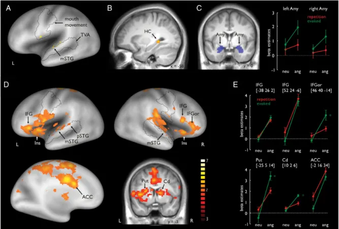
ficant activation in the left mSTG (Fig.
2
A; see
Sup-plementary Table 2A and B). The mSTG activation was located
within the temporal voice area (TVA), as de
fined by the voice
localizer scan (Supplementary Fig. 1B). When comparing
evoked with repetitive prosody productions, we found activity
in the left HC (see Fig.
2
B).
Functional Activations for Angry Compared with Neutral
Trials
For angry trials, we found increased activations in the left and
right STG, bilateral IFG, and left ACC, and in several
subre-gions of the BG, such as the putamen and the caudate nucleus
(Fig.
2
D; Supplementary Table 2C). Activations extended from
the left mSTG to the pSTG, and an activation peak was also
found in the right mSTG. STG activation was again located
within the TVA. A repeated-measures ANOVA on beta estimates
including the factors task (repeated and evoked) and emotion
(neutral and angry) indicated a signi
ficant main effect for
the factor task (F
1,12= 10.418, P = 0.007) for the ROI within
the right mSTG, indicating increased activity for repetition
Figure 2. (A) Repeated compared with evoked production of prosody revealed activity in the left mSTG located within the TVA. The black outline represents the frontal and temporal areas related to silent mouth movement according to the functional mouth movement localizer scan and the voice-sensitive temporal cortex (see Supplementary material). (B) Evoked compared with the repeated production of prosody revealed activity in the left HC. (C) Mean activity in the bilateral amygdala was extracted by using a brain mask defined by the AAL atlas (blue inserts in the right panel) (Tzourio-Mazoyer et al. 2002). Functional activity in the left amygdala revealed a significant signal increase for angry (ang) compared with neutral (neu) voices, as well as for the evoked compared with the repetition condition. (D) Functional activations for the production of angry compared with neutral prosody revealed activity in the left mSTG and pSTG, right STG, ACC, bilateral BG (left putamen and right caudate nucleus), and bilateral IFG. Additional activity was also found in the right pars orbitals of the IFG (IFGor). (E) Signal plots for the main regions of interest. Numbers in brackets represent peak MNI coordinates. Asterisks indicate a significant signal increase in the bilateral BG and IFGor for angry compared with neutral prosody, especially for the evoked condition. ACC, anterior cingulate cortex; Cd: caudate nucleus; HC: hippocampus; IFG: inferior frontal gyrus; IFGor: inferior frontal gyrus pars orbitalis; Ins: insula; MNI: Montreal Neurological Institute; mSTG: middle temporal gyrus; pSTG: posterior temporal gyrus; Put: putamen; TVA: temporal voice area.
compared with evoked trials. There was no signi
ficant
inter-action (F
1,12= 0.087, P = 0.774). The reverse contrasts of
neutral compared with angry trials did not reveal any signi
fi-cant functional activations.
Furthermore, activations were found in the bilateral IFG for
angry productions (Fig.
2
D). One peak was located in the left
IFG and 2 peaks were found in the right IFG. For the right IFG,
one peak of activity was located in the pars orbitalis of the
IFG (IFGor) and the other peak was located more posterior in
the pars opercularis of the IFG. Only in the IFGor peak did we
find a significant interaction between the factors emotion and
task (F
1,12= 12.437, P = 0.004) (Fig.
2
E). Here, the activation
difference was especially pronounced during evoked angry
compared with evoked neutral prosody production (t
12=
4.381, P < 0.001). No other post hoc tests revealed a signi
ficant
difference between experimental conditions (all t
’s < 1.941, all
P
’s > 0.076).
Regarding the left ACC (Fig.
2
D), there was a signi
ficant
main effect for emotion, indicating an increase of activity in
this region when prosody was produced angrily compared
with neutral productions. An ANOVA on ROI beta estimates in
the ACC also revealed a signi
ficant main effect for task (F
1,12=
9.938, P = 0.008), indicating greater involvement of the ACC in
the repetition compared with the evoked production. We also
found activity in the right insula in angry compared with
neutral production.
Furthermore, for angry productions, we found activity in the
bilateral dorsal BG. There were activation peaks in the left
putamen and the right caudate nucleus (Fig.
3
C). According to
the ROI analysis, this effect was especially pronounced for
evoked angry prosody production in both the left putamen
(F
1,12= 26.538, P < 0.001) and the right caudate nucleus (F
1,12-= 10.902, P 1,12-= 0.006), as indicated by signi
ficant interaction
effects. For the left putamen, paired post hoc t-tests revealed a
signi
ficant difference between angry and neutral productions
for evoked prosody (t
12= 8.664, P < 0.001). For the right
caudate nucleus, post hoc tests revealed a signi
ficant difference
between angry and neutral productions for evoked prosody
(t
12= 6.926, P < 0.001). No other post hoc test revealed a
sig-ni
ficant difference between experimental conditions (all t’s <
1.465, all P
’s > 0.169).
Functional Activations for Angry Compared with Neutral
Trials Within Each Task
For angry trials in the repetition task, we obtained activations
in the bilateral IFG, bilateral insula, and middle and left dorsal
ACC (Supplementary Table 3B). For angry trials in evoked
tasks, there were activation peaks in the right IFG, the pars
or-bitalis of the IFG (IFGor), the right insula, the left middle
cin-gulate gyrus, the right STG, the bilateral putamen, and the
right caudate nucleus (Supplementary Table 3A). We
further-more computed interaction contrasts to
find activity during the
production of angry prosody that was speci
fic to each task. No
signi
ficant activation was revealed in this analysis.
Functional Amygdala Activity
Our whole-brain analysis did not reveal signi
ficant activity in
the bilateral amygdala. However, since the amygdala was one
of our primary ROIs, we performed a ROI analysis on mean
beta estimates extracted from the bilateral amygdala masks
(Fig.
2
C). Repeated-measures ANOVAs on the beta estimates
revealed a signi
ficant effect for the factor emotion for the left
amygdala (F
1,12= 4.899, P = 0.047), indicating higher activation
in the amygdala during angry than during neutral prosody
pro-duction. There was also a signi
ficant effect for the factor task
(F
1,12= 2.957, P = 0.031), indicating a greater involvement of
the amygdala during evoked prosody production than during
repetition of prosody. An interaction did not reach signi
ficance
(F
1,12= 2.290, P = 0.278). No signi
ficant main effects or
inter-actions were found in the right amygdala (all F
1,12< 2.947, all
P > 0.112).
Correlations of Functional Activations with Features of Vocal
Productions
To
find out which functional activations in our primary ROIs
are related to speci
fic acoustic features of vocal productions,
we computed correlations between functional activations in
our ROIs and vocal features of the emotional prosody
pro-ductions. Signi
ficant correlations are reported in Table
1
, all
other correlations were not signi
ficant (all r’s < |0.533|, all
P
’s > 0.061). Correlations were computed on difference scores
that resulted from comparing the different experimental
con-ditions. During the repetition compared with the evoked
Figure 3. (A) Grand average time course (left panel) of the pupil size time locked to the onset of the silent gap (time “0”) during the sparse temporal acquisition sequence. Mean pupil (right panel) size was scored in a time window of 500–1500 ms after the onset of the silent scanning gap (gray underlay in the right panel). (B) Significant positive signal relationship between the pupil size difference, comparing angry with neutral prosody productions, and the respective signal in the anterior cingulate cortex (ACC).
production of prosody, an increase in the maximum f0, standard
deviation of f0, and f0 range was associated with an increase of
activity in the left mSTG. Similarly, activation in the left pSTG
was associated with an increase in the standard deviation of f0
for repetition trials. The bilateral IFG correlated positively with
mean intensity for repetition trials. During evoked prosody
production trials, the minimum intensity level was correlated
with activity in the left mSTG. The standard deviation of
inten-sity correlated positively with activity in the right insula. The
mean HNR was positively associated with activation in the left
putamen during evoked angry prosody production.
Pupil Diameter
Besides the functional brain activity, we also recorded the
pupil diameter of participants as an indicator of the bodily
arousal (Fig.
3
). The ANOVA for the mean pupil diameter data
revealed a main effect both for the factor task (F
1,10= 7.641, P
= 0.020) and for the factor emotion (F
1,10= 32.647, P < 0.001),
indicating increased diameter (i.e., arousal) for repetition (M =
0.88, SD = 0.26) compared with evoked trials (M = 0.414, SD =
0.414), as well as for angry (M = 1.33, SD = 0.36) compared
with neutral productions (M =
−0.03, SD = 0.19), respectively.
There was no interaction between the factors (F
1,10= 2.617, P
= 0.137). A positive correlation was found between pupil
diam-eter and activity in the left dorsal ACC (r = 0.706, P = 0.015) for
the comparison of angry versus neutral trials, but this effect
might have been driven by one participant showing
consider-ably higher values on both measures. Excluding this
partici-pant from the analysis resulted in a nonsigni
ficant correlation
(r = 0.385, P = 0.271), indicating that the original effect might
have to be taken with some caution. However, the
Kolmogor-ov
–Smirnoff test including all participants revealed that both
variables (all P
’s > 0.132) were normally distributed.
Further-more, the data of the speci
fic participant were close the
com-monly de
fined threshold of data outliers (i.e., 1.5 times the
interquartile range), thus indicating some validity of the
orig-inal data including all participants. No correlation was found
between the pupil diameter and the ACC activity for the
com-parison of repetition with evoked trials (r = 0.106, P = 0.756).
Discussion
This study aimed to determine the neural network and the
functional role of several cortical and subcortical brain regions
involved in the production of wrathful vocalizations of anger.
We revealed 3 main
findings. First, besides activity in the STG
and IFG, we found activity in several parts of the dorsal BG,
the dorsal ACC, and the amygdala in response to angry
com-pared with neutral prosody productions. Secondly, we found
that the repetition and evoked production of prosody relied on
different brain networks supporting previous notions from
patient studies about a neural separation for these different
types of vocalizations. Speci
fically, the evoked production of
hot anger revealed an extended and speci
fic cortical and
sub-cortical brain network. Finally, we found that many of the
vocal features of prosody production were directly associated
with activity in speci
fic brain regions, indicating that several
acoustic features of emotional vocalizations are directly
con-trolled by speci
fic brain regions.
Producing Angry Prosody
We expected to
find a widespread cortico-subcortical network,
which we supposed to be directly and functionally involved in
the production of angry prosody, consisting of subcortical
regions, such as the BG and the amygdala, but also of cortical
regions, such as the STG, the ACC, and the IFG.
We found bilateral IFG activity for the production of angry
prosody, including several IFG subregions in the right
hemi-sphere. Left hemispheric IFG activation could be attributed to
different articulatory plans for vocal tract coordination while
producing angry prosody (
Ackermann and Riecker 2010
). The
right IFG might be associated with monitoring and regulation
functions in emotional expressive behavior (
Phillips et al.
2008
) and aggressive behavior (
Potegal 2012
). In our study,
the right IFG, in close interaction with the phonological
feed-back processing in the STG (as discussed in the next section),
could
serve
higher-order
controlling
functions
in
the
expression of emotional prosody. Whereas a more posterior
activation in the right IFG during angry prosody production
was similarly found for both production tasks, activation in the
anterior IFGor was more speci
fically enhanced when angry
prosody was produced during the evoked condition. This
might resemble a general posterior-to-anterior organization
within the IFG, from
first-order monitoring in the posterior
IFG to complex monitoring functions in the anterior IFG (
Pet-rides 2005
). The evoked production of prosody might involve
2 levels: First creating a production script for the expression of
prosody and then monitoring the production process. Thus,
regulation and coordination demands were more complex
during evoked prosody production, pointing to a more
anterior activation in the IFGor. This result is also in
accord-ance with a distinction within the orbitofrontal cortex, in
which the processing of more complex stimuli is located more
anteriorly (
Kringelbach 2004
).
Along with greater activation in the IFGor, activation of the
dorsal BG (i.e., putamen and the caudate nucleus) was found
for angry prosody production, which was again more
pro-nounced in the evoked task. Besides a general role of the BG
in motor planning and (emotional) output behavior (
Yelnik
2008
;
Péron et al. 2013
), the dorsal BG might have a speci
fic
role in the dynamic and temporal sequencing of speech in
terms of sensorimotor control (
Kotz and Schwartze 2010
;
Pichon and Kell 2013
). Although this function can be seen as a
common feature of both linguistic and affective prosody,
tem-poral dynamics are essential features of angry prosody,
indi-cated by an increased mean and variation in f0 and intensity
(
Banse and Scherer 1996
;
Patel et al. 2011
). Both f0 and
Table 1
Correlation coefficients (r) and statistical values (P) for the relationship between functional brain activity and acoustic features of vocal productions
Condition Brain region Acoustical feature r P-values
(A) Repetition trials Left mSTG f0max 0.606 0.028
f0sd 0.712 0.006
f0range 0.594 0.032
Imin 0.567 0.006
Left IFG Im 0.586 0.035
Right IFG Im 0.783 0.002
(B) Evoked trials Left pSTG f0sd 0.663 0.013
Right Ins Isd 0.600 0.030
(C) Evoked angry trials Left Put HNRm 0.562 0.046
(A) Repetition compared with evoked trials. (B) Evoked compared with repetition trials. (C) Angry productions during the evoked trials. For abbreviations, see Figure1.
intensity dynamics determine the temporal and rhythmic
un-folding of emotional prosody, and this rhythmic aspect during
vocal productions can be impaired in individuals with BG
lesions (
Péron et al. 2010
). In our study, the repetition task
in-volved actor recordings as an external cue, thereby minimizing
the need to self-generate the temporal dynamics for prosody
production. In contrast, the dorsal BG showed stronger activity
in the evoked task for producing angry prosody, which may
re
flect stronger self-generated prosody dynamics.
While the BG might be associated with the temporal and
dynamic sequencing of emotional speech output, the ACC
might have 2 different functions during emotional
vocaliza-tions. First, the emotion effect that we observed in the ACC
might point to its function of regulating the arousal level and
thus in the control of the autonomous nervous system for
emotional output behavior (
Critchley et al. 2003
). Activity in
the ACC was positively correlated with the individual
’s pupil
diameter as a physiological measure of bodily arousal (
Partala
and Surakka 2003
) during the production of angry compared
with neutral prosody. This result is further supported by our
finding of activation in the right insula together with the ACC
during angry prosody production, since the insula is similarly
associated with the generation of autonomic responses (
Ull-sperger et al. 2010
). Thus, both the ACC and the insula seem to
regulate autonomic arousal when people speak in an
aggres-sive angry tone.
Though uncorrelated, the increased activity of the ACC and
the increased pupil size for repetition compared with evoked
trials together might point to the second function of ACC
serving increased performance monitoring during the more
demanding repetition/imitation of emotional vocalizations.
The pupil size has been shown to be an indicator of cognitive
load especially during language-related tasks (
Hyona et al.
1995
), and the ACC seems a central brain area serving
perform-ance and error monitoring during increased demands of
cogni-tive control (
Kerns et al. 2004
). The more constraint
production type of exactly repeating prosodic intonation
should have involved increased cognitive load and
perform-ance and error monitoring demands. Thus, beyond the
func-tions of the ACC for volitional initiation of vocalizafunc-tions
(
Jurgens 2009
;
Hage 2010
), our data seem to suggest that the
ACC is also involved regulating the arousal level as well as
monitoring the vocal performance depending on the
pro-duction type.
The present study also found activation in the amygdala
during the production of angry prosody and provides strong
evidence to extend the general vocalization network (
Jurgens
2009
;
Hage 2010
) by the limbic brain system, which plays an
important role during emotional vocalizations. Our results
em-phasize the importance of the amygdala underlying emotional
output behavior, but contradict the prevailing view of the
amygdala as a structure mainly involved in the detection of
stimuli and conditioning (
Cardinal et al. 2002
). One important
role of the amygdala is its involvement in the expression of
emotionally relevant behavior. The amygdala is known to be
involved in the expression and regulation of angry behavior,
probably in the experience of an aggressive impulse (
Coccaro
et al. 2011
). The amygdala was also more activated in our
study during evoked productions, indicating that evoked
prosody might be associated with stronger emotional
regulat-ory effects by the amygdala. This interpretation is supported
by recent
findings of amygdala involvement during emotional
prosody preparation (
Pichon and Kell 2013
). One alternative
interpretation might be that, instead of regulating emotional
output behavior, amygdala activity might also originate from
auditory feedback processing of own emotional vocalizations.
While this might partly explain higher amygdala activity
during angry compared with neutral vocalizations, evoked
compared with repetition trials also revealed stronger
amygda-la activity, and both are baamygda-lanced in terms of expressing
neutral and angry vocalizations. A considerable proportion of
the amygdala activity thus might be modulated by the vocal
production type. In the present study, evoked trials were less
constraint than repetition trials in terms of how to vocalize.
This provides some evidence for its differential regulatory role
underlying emotional and especially angry vocalizations
(
Coccaro et al. 2011
) under conditions of less constraint
pro-duction modes, which might also have been a more stressful
and thus more
“emotional” production mode. This is also
sup-ported by the important regulatory role of the amygdala in
pathological expressions of vocal emotions (
Lauterbach et al.
2013
).
The
final brain structure found for producing angry prosody
was the STG. During the production of prosody, speci
fic
pro-sodic speech elements have to be derived and translated into
autonomous motor output. Evidence suggests that the STG
supports sensory-motor integration (
Hickok 2009
;
Peschke
et al. 2009
), the gating of amygdala connections (
Pehrs et al.
2013
), and phonological feedback processing (
Zheng et al.
2010
), which are necessary for accurate production of angry
prosody. This is supported by our
finding that left STG activity
correlated with pitch features of vocal productions, supporting
the online adjustments of vocal output behavior by auditory
feedback processes (
Aziz-Zadeh et al. 2010
). The STG might
also serve as a phonological short-term store facilitating
prosody production, especially in cases when there is a delay
between the perception and production of prosody, such as
during the repetition task.
Repeated and Evoked Vocal Expressions
Besides our
first experimental questions about the brain
network underlying angry vocalizations, the second questions
concerned the differential modulation of brain activity
depend-ing on the type of vocal productions. We thus compared brain
activity during prosody production for the repetition and the
evoked production of prosody. The repetition task elicited
activity in the left mSTG and pSTG. The STG seems to function
as a phonological store during the repetition of prosody,
which is corroborated by recent
findings for a short-term
storage system in the STG (
Ravizza et al. 2010
;
Acheson et al.
2011
) and the closely located planum temporale (
Hickok et al.
2009
). The planum temporale was found in a recent study
during the nonaffective repetition of pseudowords (
McGetti-gan et al. 2011
), and the same region has been proposed for
sensory-motor integration that maps perceived speech directly
to motor speech output. During the repetition task in the
present study, the actors
’ intonations had to be perceived and
stored for subsequent repetition. Hence, it is likely that left
STG activation is due to phonological short-term storage of the
pseudoword before its production.
The evoked task elicited activity in the left HC. The HC is
generally involved in long-term memory functions, as well as
emotional regulation functions (
Fanselow and Dong 2010
).
Our
finding of HC activity might indicate the retrieval of
long-term stored production rules during evoked prosody
pro-duction for speech. During the evoked task, participants had
to produce prosody naturally without relying on a prosody
template that was immediately heard beforehand. For this
evoked production, the participants had to retrieve a prototype
script from long-term memory following the production rules
for prosody intonations. This might be also explained by the
fact that the evoked task did not involve vocal productions
trig-gered by underlying emotional states, but rather required
rela-tively unconstraint vocal productions on demand, which
makes the use of prototype scripts more likely. Another
expla-nation for the HC activation might be that the pseudowords
were learned by being processed and constantly repeated
(
Paulesu et al. 2009
). Yet, as the same words were used in both
the repetition and evoked production conditions, learning
should have occurred in both conditions and not only in the
evoked production of prosody. This, however, is not the case
since there was no HC activation during the repeated
pro-duction of prosody.
Linking Brain Activity in the STG, BG, and IFG with
Speci
fic Voice Features
Our
final experimental question concerned the involvement of
speci
fic brain areas, which underlie the production of specific
acoustic features during angry prosody. We accordingly
ob-served that some of the neural activations mentioned earlier
were associated with speci
fic acoustical features of the vocal
expressions. Activation within the STG was positively
associ-ated with pitch-relassoci-ated features, such as the maximum,
vari-ation, and range of the f0. As the STG is responsible for
sensory-motor integration (
Hickok 2009
;
Peschke et al. 2009
)
and phonological feedback processing (
Zheng et al. 2010
),
this association might indicate regulatory effort and online
ad-justments for accurate f0 production in angry prosody. Thus,
emotional vocalizations strongly depend on auditory feedback
and perceptual processing of own-vocalizations in the auditory
cortex. Our results strongly suggest to extend recent models of
mammalian vocalizations (
Jurgens 2009
;
Hage 2010
) by
additionally including auditory-motor loops as an integral part
of vocal expressions. Activation in the IFG was positively
associated with the intensity of vocal productions, potentially
due to the monitoring and regulation functions of the IFG.
In-tensity was higher in angry than in neutral trials. The
pro-duction of angry prosody probably required to a greater extent
the regulation and monitoring of intensity, resulting in an
association of intensity and IFG activation. Finally, dorsal BG
activations were positively correlated with the HNR of the
pro-duced speech, con
firming results from BG lesion studies (
Van
Lancker Sidtis et al. 2010
).
Conclusions
The production of high-arousing emotional prosody comprises
a cortical
–subcortical network encompassing not only the
bilateral IFG and STG, but also the bilateral dorsal BG, the left
amygdala, and the ACC. Several of these structures directly
regulate the bodily arousal or activation level as well as the
acoustic properties underlying emotional vocalization. The
results implicate both left and right hemispheric structures in
the production of emotional prosody, which contrasts with a
prominent model stating that affective prosody is processed
dominantly by the right hemisphere and that its organization is
analogous to propositional prosody in the left hemisphere
(
Ross and Monnot 2008
). The results about the central role of
the BG might inspire research on the neural basis of
impair-ments in the production of emotional prosody such as they
occur in individuals with Parkinson disease (
Péron et al. 2010
,
2013
). The results might also inspire future research on the
neural basis of angry vocalizations that are based on real
experiences of emotions or feelings instead of vocalizations
produced on command.
Supplementary Material
Supplementary material can be found at: http://www.cercor.oxford-journals.org/.
Funding
This study was supported by the Swiss National Science
Foun-dation (SNSF, 105314_124572/1
—D.G.) and by the NCCR in
Affective Sciences at the University of Geneva (51NF40-104897
—D.G.).
Notes
Conflict of Interest: None declared.
References
Acheson DJ, Hamidi M, Binder JR, Postle BR. 2011. A common neural substrate for language production and verbal working memory. J Cogn Neurosci. 23:1358–1367.
Ackermann H, Riecker A. 2010. The contribution(s) of the insula to speech production: a review of the clinical and functional imaging literature. Brain Struct Funct. 214:419–433.
Aziz-Zadeh L, Sheng T, Gheytanchi A. 2010. Common premotor regions for the perception and production of prosody and corre-lations with empathy and prosodic ability. PLoS ONE. 5. e8759.
Banse R, Scherer KR. 1996. Acoustic profiles in vocal emotion
expression. J Pers Soc Psychol. 70:614–636.
Bänziger T, Scherer KR. 2010. Introducing the Geneva Multimodal Emotion Portrayal (GEMEP) Corpus. In: Bänziger T, Scherer KR, Roesch EB, editors. Blueprint for affective computing: a source-book Oxford. UK: Oxford University Press. p. 271–294.
Belin P, Zatorre RJ. 2000. Voice-selective areas in human auditory cortex. Nature. 403:309.
Bell WL, Davis DL, Morgan-Fisher A, Ross ED. 1990. Acquired aproso-dia in children. J Child Neurol. 5:19.
Boersma P. 2001. Praat, a system for doing phonetics by computer. Glot Int. 5:341–345.
Borod JC, Bloom RL, Brickman AM, Nakhutina L, Curko EA. 2002. Emotional processing deficits in individuals with unilateral brain damage. Appl Neuropsychol. 9:23–36.
Cardinal RN, Parkinson JA, Hall J, Everitt BJ. 2002. Emotion and motiv-ation: the role of the amygdala, ventral striatum, and prefrontal cortex. Neurosci Biobehav Rev. 26:321–352.
Coccaro EF, Sripada CS, Yanowitch RN, Phan KL. 2011. Corticolimbic function in impulsive aggressive behavior. Biol Psychiatry. 69:1153–1159.
Cohen MJ, Riccio CA, Flannery AM. 1994. Expressive aprosodia follow-ing stroke to the right basal ganglia: a case report. Neuropsychol-ogy. 8:242–245.
Critchley HD. 2009. Psychophysiology of neural, cognitive and affec-tive integration: fMRI and autonomic indicants. Int J Psychophysiol. 73:88–94.
Critchley HD, Mathias CJ, Josephs O, O’Doherty J, Zanini S, Dewar BK, Cipolotti L, Shallice T, Dolan RJ. 2003. Human cingulate cortex and