Précis on The Cognitive-Emotional
Brain
Luiz Pessoa
Department of Psychology, University of Maryland, College Park, MD 20742
http://www.emotioncognition.org
Abstract: In The Cognitive-Emotional Brain (Pessoa 2013), I describe the many ways that emotion and cognition interact and are integrated in the brain. The book summarizesfive areas of research that support this integrative view and makes four arguments to organize each area. (1) Based on rodent and human data, I propose that the amygdala’s functions go beyond emotion as traditionally conceived. Furthermore, the processing of emotion-laden information is capacity limited, thus not independent of attention and awareness. (2) Cognitive-emotional interactions in the human prefrontal cortex (PFC) assume diverse forms and are not limited to mutual suppression. Particularly, the lateral PFC is a focal point for cognitive-emotional interactions. (3) Interactions between motivation and cognition can be seen across a range of perceptual and cognitive tasks. Motivation shapes behavior in specific ways – for example, by reducing response conflict or via selective effects on working memory. Traditional accounts, by contrast, typically describe motivation as a global activation independent of particular control demands. (4) Perception and cognition are directly influenced by information with affective or motivational content in powerful ways. A dual competition model outlines a framework for such interactions at the perceptual and executive levels. A specific neural architecture is proposed that embeds emotional and motivational signals into perception and cognition through multiple channels. (5) A network perspective should supplant the strategy of understanding the brain in terms of individual regions. More broadly, in a network view of brain architecture, “emotion” and “cognition” may be used as labels of certain behaviors, but will not map cleanly into compartmentalized pieces of the brain.
Keywords: brain; cognition; emotion; integration; prefrontal cortex; motivation
1. Introduction
In The Cognitive-Emotional Brain (Pessoa
2013
), I
describe how emotion and cognition interact and are
inte-grated in the brain. I believe that brain research has been
ill-served by the dichotomization of large concepts like
cog-nition and emotion. Further, strict a priori deficog-nitions of
these concepts fuel dichotomization. We need a vocabulary
that views concepts as complementary pairs that mutually
define each other and, critically, do not exclude each
other. As in the book, I will not define here terms such as
emotion, motivation, and cognition; they will be used
de-scriptively to refer to paradigms, task conditions, or
“pro-cesses” that are closer to the traditional intended
meanings of these terms. The book is aimed at students
and investigators interested in the brain basis of emotion,
especially those interested in understanding how
percep-tion and cognipercep-tion reflect and embed affective/motivapercep-tional
significance. The book integrates findings from nonhuman
animal research and human imaging research, the latter
being the area my research focuses on.
The structure of this précis is as follows. Section 2 briefly
reviews issues covered in chapters 2–4 of the book, which
propose how to conceptualize amygdala function in
broader terms than typically adopted in the
field, and
argue against the notion of the
“automaticity of emotion.”
Sections 3–6 focus on chapters 5–7 of the book, which
describe how cognitive-emotional/motivational interactions
and integration take place in the brain. Section 7 addresses
the general issue of structure-function mapping in the
brain. There, I argue for a
“network” view of brain function
but also describe several problems with this view that are
underappreciated in the literature. Potential tools to
char-acterize complex structure-function mappings are
de-scribed. Finally, in Section 8, I draw some conclusions of
the network perspective to the understanding of emotion
and the brain.
2. Amygdala and the automaticity of emotion
2.1. AmygdalaChapter 2 discusses the ever-important amygdala and its
role in brain function. Based on rodent and human data,
I describe how the amygdala’s functions go beyond
LUIZPESSOAis a Professor of Psychology and the direc-tor of the Maryland Neuroimaging Center, University of Maryland, College Park. He received BS and MS degrees in Computer Science from the Federal Univer-sity of Rio de Janeiro, Brazil. He then received a PhD in computational neuroscience at Boston University. After a few years as Computer Science faculty at the Federal University of Rio de Janeiro, he returned to the United States as a Visiting Fellow at the National Institute of Mental Health. Interested in the interactions between emotion/motivation and cognition in the brain, he has published more than 100 papers and chapters.
BEHAVIORAL AND BRAIN SCIENCES (2015), Page 1 of 66
emotion as traditionally conceived, reflecting a trend
toward viewing this structure not simply in terms of
“fear.”
A key function of the amygdala is to shape selective
infor-mation processing. Selection of inforinfor-mation for further
analysis is, of course, a central problem that needs to be
solved for effective behavior (Grossberg & Levine
1987
).
The amygdala is a core structure in a system involved in
“What is it?” processing and thus contributes to
highlight-ing what is of significance to the organism (Pribram &
McGuinness
1975
). However, the functions of the
amygda-la also involve
“What’s to be done?” A key reason for this is
that the amygdala participates in the representation of
value (including positive value) and in decision making.
For example, amygdala lesions impair behavior on the
Iowa Gambling Task in humans and alter delay-based
deci-sion making in rats (e.g., they become more impulsive).
The amygdala thus takes part in an impressive array of
processes that far exceed some of its proposed functions,
such as vigilance, arousal, salience detection, novelty
detec-tion, and relevance detection.
“Information gathering”
(Whalen
1998
) better captures several of its functions but
comes short, too. In the end, it is better simply to refrain
from overly summarizing its functional repertoire so as to
better appreciate the wide scope of the amygdala’s
contri-butions to brain mechanisms and behavior.
2.2. Subcortical“low road” pathway and emotional processing
A purported division of labor between cortical and
subcort-ical regions has been present from the time of the earliest
circuit models of emotion (e.g., Papez
1937
). Many versions
of this type of dual processing model exist, including some
variants that have captured the popular imagination, such
as the
“triune brain” (MacLean
1970
;
1990
).
In the case of vision, it has been suggested that a
subcort-ical pathway from the retina to superior colliculus to
pulvi-nar to amygdala that entirely bypasses cortex enables the
processing of emotion-laden visual stimuli to be fast,
auto-matic, and nonconscious. In chapter 3, I argue against this
notion on several general grounds: (1) Affective visual
in-formation is not handled qualitatively faster than other
visual information; (2) processing of affective visual
stimuli involves both low- and high-spatial frequency
infor-mation; and (3) the amygdala is not required for rapid,
non-conscious detection of affective information. For these and
many other reasons, Ralph Adolphs and I proposed the
“multiple waves” model as an alternative to the low-road
pathway scheme (Pessoa & Adolphs
2010
). The model
shifts the debate away from whether there is a unique
sub-cortical pathway to whether a processing architecture exists
that is capable of rapidly transmitting information via
mul-tiple pathways. The resulting mulmul-tiple waves model
empha-sizes the role of the pulvinar in coordinating and regulating
the
flow of multimodal information, which is accomplished
via a series of thalamo-cortical loops. In this role, the
pulvi-nar moves from being a passive relay station of the
“stan-dard hypothesis” to being an active element of
information processing.
2.3. What kind of unawareness matters?
The research literature is replete with paradigms such as
backward masking and the attentional blink that challenge
the visual system so that awareness can be studied. At
times, much is made about neuroimaging responses
ob-served in the amygdala for very brief stimuli (e.g., 15–30
ms). In such cases, subjects may report not seeing them
(“subjective unawareness”). In the book, I argue that this
type of
“subliminal” unawareness is not the most relevant
one to understand the impact of affective content on
behavior and on clinical conditions such as anxiety. A
more important sense is associated with the idea of
unin-tentional processing, which may prove to be more
impor-tant to the understanding of human behavior. Whether
the unintentional unconscious is sophisticated and
flexible,
as argued by social psychologists (see Bargh & Morsella
2008
), is a matter of debate. But there can be no doubt
that it is qualitatively different from the type of subliminal
unconscious sometimes emphasized in the emotion
litera-ture (for evidence that the
“subliminal unconscious” may
be quite
“dumb,” see Loftus & Klinger
1992
– if at all
present; Pessoa
2005
).
2.4. Why is the amygdala important?
In the broader neuroscience literature, the amygdala is
viewed as a central node in emotional processing in part
because of the
“low-level” properties ascribed to the
sub-cortical pathway. Defects in the amygdala system are said
to underlie phobias, mood disorders, and post-traumatic
stress syndrome, and variability in its functioning to
reflect individual differences at the genotypic and
personal-ity level.
Although in chapter 3 I challenge many of the properties
typically ascribed to the subcortical pathway, the amygdala
is indeed important for behavior and mental health.
First, together with the hypothalamus and medial PFC,
the amygdala has extensive projections to downstream
regions in the brainstem that are capable of mobilizing
the body; indeed, its central nucleus is at times described
as a
“controller of the brainstem.” The autonomic and
neu-roendocrine connections of these brain regions are part of
sympathetic and parasympathetic networks that coordinate
bodily responses in the face of challenges to the organism.
Second, the amygdala, hypothalamus, medial PFC, and
related regions, being among the most extensively
connect-ed parts of the brain, are optimally positionconnect-ed to influence
information processing. As hubs through which evaluative
signals are communicated, they are thought to have
wide-spread effects on mental function and to play a significant
role in affective and cognitive impairments observed in
mood disorders. Metaphorically speaking, as one of these
hubs, the amygdala is strategically positioned to
“ignite”
both body and brain.
2.5. Processing of emotion-laden information and automaticity
Shiffrin and Schneider (
1977
, pp. 155–156) defined an
“au-tomatic process
… as a sequence of nodes that nearly
always becomes active in response to a particular input
con-figuration.” Because automatic and controlled processes
appear to be qualitatively opposed, it is natural to
dichoto-mize mental phenomena into these two classes. But such a
dichotomy has simply not held up in the face of data.
Reports of automaticity have invariably been countered
by reports of capacity limitation; behavioral effects
Pessoa: Précis on The Cognitive-Emotional Brain
assumed to operate automatically are influenced in ways
that belie that assumption.
The argument that I make in chapter 3 is that a better
framework is one where performance is always considered
capacity limited and described as a performance-resource
function (Norman & Bobrow
1975
). Some behaviors will
exhibit shallower performance-resource relationships,
where performance only rises slowly based on the mental
effort exerted
– these behaviors are hence “controlled.”
Other behaviors exhibit steeper relationships, and ceiling
performance is reached even when conditions are degraded
(e.g., under short exposure)
– these behaviors are hence
“efficient.” Although the performance-resource function
may seem to be an abstract construct when little is known
about the task at hand, it forces researchers to consider a
spectrum of scenarios when studying how a behavior
depends on multiple factors that influence performance.
Why is a continuous framework better than a
dichoto-mous one? For one thing, it
fits the empirical data better:
Researchers have repeatedly found capacity limitations
for
“automatic” phenomena (e.g., Pashler
1998
). For
another, the dichotomous framework is plagued by
serious conceptual issues (Moors & De Houwer
2006
).
Another reason a continuous framework is better is that
we still have an incipient understanding of competition
–
and, hence, of whether interference will result when
mul-tiple items are involved. The notion of competition, as
accepted by most researchers, goes roughly as follows.
Because processing capacity is limited, competition is
pro-posed to
“select” the most relevant information at any given
time (Desimone & Duncan
1995
; Grossberg
1980
); when
resources are not fully consumed, spare capacity is used
to process task-irrelevant items (Lavie
1995
). The
problem is that we do not always know whether
interfer-ence will occur in any given situation. Generally, multiple
factors determine how information competes in visual
cortex and beyond, including task difficulty, set size,
spatial arrangement, cuing, and the like. Finally, a
continu-ous framework demystifies the processing of certain
complex features. For example, processes such as reading
and the perception of elaborate emotional images are at
times depicted as
“automatic” in a sense that is almost
magical (for a cogent in-depth discussion, see Pourtois
et al. 2012). Indeed, the underlying mechanisms of abilities
such as proficient reading and the perception of emotional
scenes are remarkably fast. That we do not understand why
they are so fast, however, simply means that we are still
quite some way from a better mechanistic description of
these processes.
2.6. Dual process models
The discussion of automatic versus controlled processes is
also pertinent to dual process models. Common to these
models is the strong assumption of the existence of two
qualitatively different mental systems, for example,
“intui-tion” and “reasoning” (see Keren and Schul
2009
). A
popular trend is to call the two components
“system 1”
and
“system 2,” where the first is
automatic/heuristic/reflex-ive and the second is controlled/analytic/reflectautomatic/heuristic/reflex-ive (Evans
2008
). But as others have expressed in the past, the idea
of a dual system model is both slippery and conceptually
unclear (see Keren & Schul
2009
). For one, nearly all
dual process models have as a central component the
automatic versus controlled dichotomy, which as discussed
above is not a viable distinction. In fact, as with the question
of automatic versus controlled processing of emotion-laden
stimuli, the question of whether there are two systems in
dual process models is not an entirely empirical one. This
is because no single critical experiment can provide a
final, definitive answer. In the end, however irresistible
di-chotomies are to the human mind (Kelso & Engstrøm
2006
; Newell
1973
), dichotomizing implies oversimplifying
(Keren & Schul
2009
; Kruglanski et al.
2006
). A continuous
framework is better, albeit more complex (Kruglanski et al.
2006
).
3. Diverse forms of cognitive-emotional
interactions are not limited to mutual suppression
Nauta (
1971
, p. 182) suggested that the PFC could be
con-sidered
“the major – though not the only – neocortical
rep-resentative of the limbic system.” Yet, most proposals in the
literature portray the PFC’s core function as cognitive, or
compartmentalize it into cognitive and affective regions
(see Bush et al.
2000
). In particular, the lateral PFC is
still viewed as a quintessential cognitive region, especially
the portion that is loosely referred to as the
“dorsal-lateral PFC.” This section reviews human studies that
have investigated cognitive and emotional processing in
the human PFC (see also Dolcos et al.
2011
) to explore
how emotion and cognition, domains traditionally thought
of as mutually antagonistic, interact there. The section
does not discuss the part of the PFC called the orbitofrontal
cortex, whose contributions to emotion are well accepted
(Zald & Rauch
2007
). In the ensuing discussion, it is
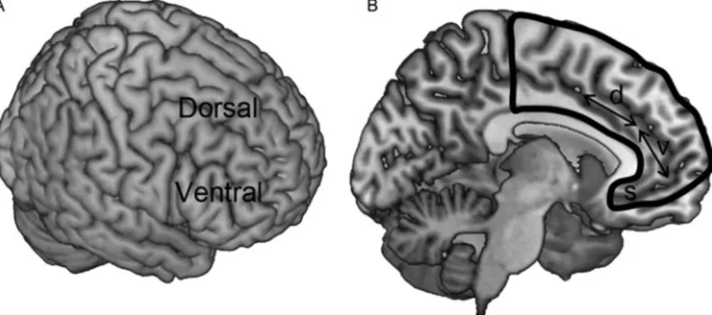
useful to consider the regions outlined in
Figure 1
.
3.1. The“Classical” view: Emotion-cognition push-pull
In an important paper, Drevets and Raichle (
1998
) noted
that regional blood
flow during attentionally demanding
cognitive tasks decreased in regions such as the amygdala,
orbitofrontal cortex, and ventral-medial PFC, whereas
blood
flow increased in these regions during specific
emotion-related tasks. Conversely, blood
flow during
ex-perimentally induced and pathological emotional states
(Mayberg et al.
1999
) decreased in regions such as the
dorsal-medial and dorsal-lateral PFC, whereas blood
flow
increased in these regions during cognitive tasks. These
re-ciprocal patterns of activation suggested to Drevets &
Raichle (
1998
) that emotion and cognition engage in
com-petitive interactions.
This insight led to a wealth of studies pursuing the notion
of a dorsal-cognition versus ventral-emotion axis of
organi-zation in the human brain. For example, Dolcos and
col-leagues investigated emotional distraction during working
memory tasks (see also Anticevic & colleagues
2010
).
Sub-jects were shown sample stimuli that had to be
remem-bered during a subsequent delay period during which
they saw distracting stimuli, including neutral and
emotion-al pictures. The
findings of one of their studies (Dolcos &
McCarthy
2006
) are illustrated in
Figure 2
. During the
delay period, responses in dorsal-lateral PFC (
Fig. 2B
)
were highest for the
“scrambled” (digitally scrambled
ver-sions of pictures), intermediate for neutral, and lowest for
Pessoa: Précis on The Cognitive-Emotional Brain
emotional distractors
– a pattern of responses also observed
in parietal cortex. Behavioral performance mirrored this
and was worst for emotional distractors. Viewing emotional
distractors during the delay period appeared to interfere
with neural activity normally observed in these sites
–
activ-ity that supports working memory performance (e.g.,
Pessoa et al.
2002
). Responses in the ventral-lateral PFC
(
Fig. 2C
) followed the opposite pattern, namely, the
stron-gest responses were observed during the viewing of
emo-tional distractors, suggesting that ventral-lateral PFC
contributed to inhibiting the distracting effects of stimuli
presented during the delay period. Overall, several
studies are consistent with the dorsal-cognition versus
ventral-emotion segregation (both along the lateral
surface of the brain and its medial sector), including
those probing emotional distraction, emotional conflict,
and emotion regulation (Ch. 5).
The organization of the medial PFC, a complex brain
region involved in diverse functions (Vogt
2008
), has
strongly fueled the dorsal versus ventral view of emotion
and cognition organization in the brain, particularly
follow-ing another influential paper (Bush et al.
2000
; see also
Devinsky et al.
1995
). In the next section, I argue against
the dorsal versus ventral framework in the medial PFC in
particular, and in the subsequent section against the
dorsal versus ventral view in the PFC more generally.
3.2. Beyond the dorsal versus ventral-medial dichotomyin the prefrontal cortex
Results from several individual studies challenge the
dichotomy. For example, Mobbs and colleagues (
2010
)
ex-amined how brain responses vary as a function of perceived
threat proximity. In an unusual experimental manipulation,
each participant inside the MRI scanner placed a foot into a
custom-built box containing multiple compartments, while
watching a video of a live tarantula placed into one of the
compartments at varying distances from the foot (actually
prerecorded). Increases in responses as a function of
prox-imity were observed in several brain regions; notably in the
dorsal-medial PFC.
The
“attentional network” involves fronto-parietal
regions, including the dorsal-medial PFC (Corbetta &
Shulman
2002
; Kastner & Ungerleider
2000
). To assess
brain regions that are sensitive to high levels of threat, I
re-viewed activation sites reported in aversive conditioning
studies (Pessoa
2009
). Surprisingly, activation was
repeat-edly reported not only in the amygdala but also in frontal
Figure 1. Frontal cortex anatomy.“Prefrontal cortex” refers to cortex “in front of motor areas,” typically anterior to Brodmann area 6. (A) Lateral surface of cortex, showing dorsal and ventral sectors. (B) Medial surface of cortex, outlined in black, showing approximate locations of dorsal (d) and ventral (v) sectors. In the paper, dorsal parts of medial prefrontal cortex also include parts posterior to the“d” arrow (such as presupplemantary and supplementary motor areas). S, subgenual anterior cingulate cortex.Figure 2. Emotional distraction during a working memory task. Subjects were shown scrambled, negative, or neutral distractor images during the delay period of the task. (A) Schematic representation of differential responses in brain. Regions where responses were stronger to scrambled than to emotional images are shown in light gray; regions where they were stronger to emotional than to scrambled images, in dark gray. (B) Time course data for dorsal-lateral prefrontal cortex. (C) Time course data for ventral-lateral prefrontal cortex. Horizontal bars in panels B and C correspond to onset and duration of sample stimuli, distractors, and probes, respectively. Time series plots kindly provided by Florin Dolcos, adapted with permission from Dolcos and McCarthy (2006).
Pessoa: Précis on The Cognitive-Emotional Brain
sites overlapping with those in the attentional network,
in-cluding the dorsal-medial PFC
– consistent with findings
from formal meta-analyses (Etkin & Wager
2007
;
Mechias et al.
2010
). To understand the organization of
the medial PFC and its role in emotion, Etkin and
col-leagues (
2011
) reviewed both the human and nonhuman
animal literatures. They surmise that sites in both
dorsal-and ventral-medial PFC make prominent contributions to
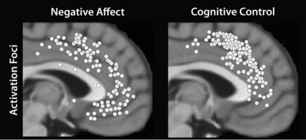
emotional processing. Finally, an extensive formal
meta-analysis of human neuroimaging studies (Shackman et al.
2011
) further demonstrates the considerable overlap of
sites in the medial PFC engaged during negative affect
and cognitive control (
Fig. 3
).
In summary, although it is still influential, the
segrega-tion model of medial PFC organizasegrega-tion is no longer
viable, as different research groups now argue (e.g., Etkin
et al.
2011
; Pessoa
2009
; Shackman et al.
2011
). Large
por-tions of the PFC are engaged during emotional processing,
including both dorsal and ventral portions of the medial
PFC. Indeed, when large numbers of studies are
consid-ered jointly, the weight of their
findings strongly favors
an organization of the medial PFC that is not segregated
into affective and cognitive compartments but instead is
shared by cognitive and affective domains in a way that
allows the medial PFC to support the adaptive control of
complex behaviors (Pessoa
2008
; Shackman et al.
2011
).
3.3. Beyond push-pull: When emotion and cognition worktogether
Now, I will turn to the broader issue of the frequently held
view of emotion-cognition organized as push-pull, or
antag-onistic, systems. Consider once more the study by Dolcos
and McCarthy (
2006
) that showed that emotional
distrac-tors produced decreased responses in parts of the
dorsal-lateral PFC that are important for cognitive tasks. This
type of response, which favors the antagonistic
organiza-tion, is far from universal, however. For example, also
during conditions of emotional distraction, Erk et al.
(
2007
) observed increased responses to emotional stimuli
in the dorsal-lateral PFC. They also observed increased
re-sponses when they increased the load of a separate
non-emotional working memory task. In other words, both
the emotional and cognitive manipulations produced
enhanced responses in the dorsal-lateral PFC. Conversely,
emotional manipulations do not always generate decreased
responses in frontal-parietal areas that are recruited by
ef-fortful, cognitive tasks. For example, in one of our studies,
when subjects viewed a
“threat cue” that signaled a
poten-tial upcoming shock, deactivation was observed in
emotion-related regions (Choi et al.
2012
).
In all, cognitive-emotional interactions take diverse
forms that go beyond a straightforward antagonistic
rela-tionship (Ch. 5). Instead, I suggest that lateral PFC, in
par-ticular, is a convergence site for cognitive and emotional
signals where they are integrated.
3.3.1. The basic“direction” of brain responses and their interpretation
. As discussed above, a key question during
cognitive-emotional interactions is whether emotional
in-formation decreases or enhances a region’s responses
during cognitive tasks
– to decide if the relationship is
push-pull. Unfortunately, the direction (increases vs.
de-creases) of brain responses does not uniquely determine
their functional significance. Consider again the working
memory study by Dolcos and McCarthy (
2006
), which
showed decreased responses in dorsal-lateral PFC during
emotional distraction. More important, this condition was
linked with impaired task performance, matching the
pattern of an antagonistic cognitive-emotional interaction.
But how should we interpret these
findings? Because it is
unknown whether increased responses reflect greater
ca-pacity to utilize the region, neural inefficiency, or increased
effort, the interpretation of the results is equivocal. The
dif-ficulty here is not about problems of interpreting functional
MRI responses given their indirect relationship with
neuro-nal activity. The same issues would arise with cell recordings,
because disentangling, say, neural efficiency, increased effort,
and so on, is again far from simple.
A potential strategy is to interpret response changes in
terms of behavior and brain responses during neutral
tasks. During working memory, we know that dorsal
frontal and parietal regions are important from both
monkey and human work. In these regions, response
mag-nitude even tracks performance on a trial-by-trial basis
(Pessoa et al.
2002
). Thus, when emotional distractors
lead to decreased responses in dorsal-lateral PFC and
im-paired task performance (Anticevic et al.
2010
; Dolcos &
Figure 3. Cognition and emotion in medial frontal cortex. Foci of activation across studies of negative affect and cognitive control. Extensive overlap between emotion and cognition was observed in dorsal-medial prefrontal cortex. Figure kindly provided by Alex Shackman and adapted with permission from Shackman et al. (2011).
McCarthy
2006
), it is possible to more strongly interpret
the
findings in terms of underlying antagonistic
interac-tions. Although in this case the original interpretation
holds, the example underscores the need to ground the
re-sponses during emotional manipulations by building on
closer ties between a brain region’s responses and
associat-ed behavior during nonemotional tasks.
3.3.2. Anxiety, executive function, and prefrontal cortex responses
. A closely related issue arises in the context of
studies of brain responses in anxious individuals: Are
cogni-tive control areas in the PFC, including the dorsal-lateral
PFC, under- or overactivated in these individuals? This
question is relevant given the belief that anxiety is
particu-larly associated with reduced processing efficiency. Thus, to
maintain comparable levels of task performance, anxious
individuals must exert greater cognitive effort (Eysenck
et al.
2007
), which is linked to increased responses in
brain regions involved with cognitive control.
But some studies have reported that anxiety is associated
with underactivation in cognitive control circuits (Bishop
2007
;
2009
; Bishop et al.
2004
; see also Basten et al.
2011
;
for additional discussion, see Eysenck & Derakshan
2011
).
Examining under- or overutilization of a brain area,
however, does not lead to an unequivocal interpretation of
cognitive processing in anxious subjects, as argued by Fales
and colleagues (
2008
). This is because either reduced or
en-hanced neural recruitment may reflect differences in a host
of factors, including efficiency, motivation, effort, or the
ca-pacity to activate regions when needed. The difficulties
sur-rounding the issue of under- versus overactivation are
mirrored by those encountered in the human developmental
literature, where changes in the responses of a brain region
with age are hard to interpret (Somerville & Casey
2010
).
The upshot of section 3 is as follows: The effects of
emotion on cognition, and vice versa, are best viewed not
as a simple push-pull mechanism, but as interactions that
result in processes and signals that are neither purely
cog-nitive nor emotional. Instead, their
“cognitive” or
“emo-tional” nature is blurred in a way that highlights the
integration of these domains in the brain (Pessoa
2008
).
4. Motivation: Interactions between motivation and
cognition
According to traditional psychological models, motivation
relies on a global, rather blunt energization factor to
influ-ence the vigor and frequency of behavioral output, though
without specific effects (e.g., Duffy
1962
; Hull
1943
).
Current progress in understanding the mechanisms of
reward and motivation challenges this view, which has
renewed interest in motivational effects on perception
and cognition. Chapter 6 contains discussion of the selective
ways motivation affects task performance, some of which I
briefly review here.
Jan Engelmann and I investigated the impact of changes in
incentive value on behavior during a difficult spatial
localiza-tion task (Engelmann & Pessoa
2007
). Participants
per-formed the task under conditions in which they could earn
extra monetary rewards, avoid losing money, or, during a
baseline condition, neither gain nor lose. In theory,
motiva-tion could lead to indiscriminate responding increasing the
number of both correct detections and false alarms.
Instead, detection performance improved as a function of
ab-solute incentive value (gains and losses produced similar
results) independent of unspecific influences, such as
general activation (e.g., purely faster response times) or
re-sponse bias (e.g., more conservative rere-sponses). We
observed increases in visual sensitivity (d-prime) in both
en-dogenous and exogenous attention tasks (see also Engelman
et al.
2009
).
In an event-related potential (ERP) study, Hickey and
colleagues (
2010
) sought to dissociate
“strategic” (such as
paying more attention) and
“incidental” (such as undesired)
effects of reward. To that end, they probed how reward in
one trial affected visual processing in the next. Following a
high-reward trial, the P1 ERP response component, which
occurs approximately 100 ms after stimulus onset, was
found to be stronger contralateral to targets of the same
(task-irrelevant) color rewarded on the previous trial,
re-vealing facilitated responses based on previous-trial
reward. So-called N2pc responses were found to be
stron-ger as well, indicating that target processing was enhanced.
Notably, P1 and N2pc effects were observed on trials
fol-lowing high reward when a salient distractor was shown
in the reward-paired color, showing that reward has an
impact that can be independent of its role in establishing
goal-driven attention (e.g., when a subject deliberately
in-creases attention in anticipation of reward). In a related
monkey cell-recording study, Peck and colleagues (
2009
)
showed that cues signaling reward biased attention in a
value-specific fashion, even though they were
“maladap-tive” (they interfered with the required behavior). They
proposed that posterior parietal cortex in the monkey
con-tains a visuospatial map
– a salience map – that takes into
account reward expectations when guiding attention.
Does motivation influence the selection of information?
To answer this question, Srikanth Padmala and I investigated
the effects of reward during a response-conflict task (
Fig. 4
)
Figure 4. Response-conflict paradigm. In the reward condition shown here, a cue stimulus (“$20”) signaled that subjects would be rewarded for fast and correct performance; in the control condition (not shown here), a cue stimulus (“$00”) signaled that there would be no reward. During the target phase, a stimulus picture of a house or building was presented together with a task-irrelevant word (an incongruent condition is illustrated here). After the target stimulus, subjects were informed about the reward and about the total number of points accrued. Reproduced with permission from Padmala and Pessoa (2011).
Pessoa: Précis on The Cognitive-Emotional Brain
(Padmala & Pessoa
2011
). Based on previous studies, we
an-ticipated that motivation would enhance engagement of
fronto-parietal attentional regions and, consequently, that
these regions would be better positioned to exert
goal-direct-ed control influencing visual processing (
Fig. 5
).
Behavior-ally, we observed response interference: Performance was
slower on incongruent trials than on neutral ones. But
reward reduced response interference. Given that reward
also decreased response facilitation (i.e., the beneficial
effect of a congruent task-irrelevant item), the results
sup-ported the inference that motivation enhanced attentional
filtering, thereby reducing the influence of the
task-irrele-vant word item. Our brain imaging results revealed that,
during the cue phase when subjects were told whether a
reward was possible, responses in fronto-parietal regions
were stronger with reward
– consistent with increased
attention. Notably, larger cue-related responses were
associ-ated with larger decreases in interference-relassoci-ated responses
in the dorsal-medial PFC during the subsequent task
phase. This suggested that upregulation of control during
the cue phase led to decreased interference during the
task phase.
We also observed responses to the cue in several
subcort-ical sites that are engaged during reward-related processing,
including the caudate and putamen in the dorsal striatum,
nucleus accumbens in the ventral striatum, and midbrain.
We reasoned that, if motivationally salient cues engage
fronto-parietal regions more robustly during the cue
phase, these regions should exhibit increased
“coupling”
with some of the above regions, which are sensitive to the
motivational significance of the cues (
Fig. 6A
). Indeed, in
the reward condition, we observed increased trial-by-trial
functional connectivity between the intraparietal sulcus in
parietal cortex and the putamen, caudate, and nucleus
accumbens (
Fig. 6B
; see also Harsay et al.
2011
). More
in-teresting, the strength of the differential coupling (reward
minus nonreward) between cortical and subcortical areas
was linearly related to individual differences in reward
sen-sitivity, showing that the functional interaction between
these regions was stronger for subjects who scored higher
in this dimension. See also Krebs et al. (
2010
;
2011
).
Interactions between motivation and working memory
have been the target of several neuroimaging studies
(e.g., Beck et al.
2010
; Gilbert & Fiez
2004
; Jimura et al.
2010
; Pochon et al.
2002
; Taylor et al.
2004
). In the study
by Jimura and colleagues (
2010
), reward did not simply
in-crease activation; it also shifted the timing of working
memory responses (an effect that correlated with individual
differences in reward sensitivity). They suggested that, in
the reward condition, subjects may have adopted a more
proactive control strategy to aid performance instead of a
just-in-time reactive strategy
– and thus increase their
chance of reward (Braver
2012
; Braver et al.
2007
).
Inter-actions between motivation and working memory have
been studied in monkey cell-physiology studies, too. Not
only do cells in the lateral PFC hold information of an
object’s shape and location, but they are also modulated
by reward expectancy (Watanabe
1990
;
1996
; see also
Leon & Shadlen
1999
). In fact, studies demonstrate that
cognition and motivation signals are integrated. For
example, during the delay period of a delayed–eye
saccade task, some lateral prefrontal cells increased their
firing if the monkey was initially cued to make a saccade
to the preferred versus the opposite direction; these cells
also exhibited increased
firing during rewarded versus
un-rewarded trials (Kobayashi et al.
2002
). Importantly,
during rewarded trials of saccades to the preferred
direc-tion, there was an increase of the amount of transmitted
in-formation with respect to target position, as quantified by
information theory; reward information increased the
dis-criminability of target positions, leading to enhanced
per-formance (see also Kobayashi et al.
2007
).
4.1. Energizing force versus selective effects
Traditional accounts describing motivation as a global
activa-tion independent of particular control demands have been
echoed by a functional MRI study in which Kouneiher and
colleagues (
2009
) argue that motivation and cognitive
control can be regarded as two separate and additive
–
instead of interactive
– factors. Although there is little
ques-tion that motivaques-tion can have generalized, activating
contri-butions to behavior (see Robbins & Everitt
2007
; Salamone
et al.
2009
), current
findings (Ch. 6) underscore the ability
of motivation to shape behavior selectively, whether by
reduc-ing response conflict or task-switch costs, via selective effects
on working memory, or by improving long-term memory (for
the latter, see the work of Adcock and colleagues; e.g., Adcock
et al.
2006
). Another body of research demonstrating
selec-tive effects of motivation has investigated attentional effort,
as described by Sarter and colleagues (e.g., Sarter et al.
2006
).
5. Dual competition model
Here, I describe a framework in which both emotional and
motivational signals are integrated with perception and
cognition so as to effectively incorporate value into the
Figure 5. Hypothesized network interactions. (A) Predictedmediation by target/distractor processing in visual cortex of the relationship between attentional control implemented in fronto-parietal cortex during the cue phase and conflict-related activity in medial prefrontal cortex during the subsequent target phase (see white arrow). (B) Predicted effect of motivational context on functional interactions between fronto-parietal cortex and subcortical regions involved in reward processing. Reproduced with permission from Padmala and Pessoa (2011).
unfolding of behavior (Pessoa
2009
; Pessoa & Engelmann
2010
). To reflect the central idea that both emotion and
motivation influence competition at both the perceptual
and the executive levels, the framework is termed the
dual competition model (thus
“dual” spans both “emotion
and motivation” and “perceptual and executive”).
Follow-ing general remarks in the next paragraph, I will describe
how the framework applies to emotion (sect. 5.1, focusing
on emotion-laden negative stimuli) and then motivation
(sect. 5.2, focusing on task manipulations involving reward).
Competition for neural resources exists in the sensory
cortex (Grossberg
1980
). To understand the
flow of
infor-mation processing more generally, we need to go beyond
perceptual competition and explicitly incorporate the role
of executive control. Behavioral research indicates that
ex-ecutive control is not unitary and that different functions
have their own limited processing capacities, or resources.
Neuropsychological research also supports the dissociation
of cognitive operations, consistent with the
“fractionation”
of the central executive (Norman & Shallice
1986
; Stuss
& Knight
2002
). Yet ample evidence also suggests at least
some unity of executive functions
– certain mechanisms
are shared across functions (Duncan et al.
1996
; Miyake
et al.
2000
). Capacity sharing has implications for
informa-tion processing because it implies executive competiinforma-tion:
Subcomponents of executive control are mutually
interact-ing, such that multiple functions cannot be independently
executed simultaneously. This competition can be cast in
terms of resources. Accordingly, even though some
execu-tive processes rely on partly independent mechanisms, they
share a common pool of resources. Therefore, when a given
function is needed, resources devoted to one operation will
not be available for other operations, and behavioral
inter-ference will ensue.
5.1. Emotion
5.1.1. Perceptual competition
. How does affective
signifi-cance influence visual processing? Researchers have
de-scribed a projection system emanating from the amygdala
that reaches nearly all levels of the ventral visual system.
Al-though this system is often highlighted as the sole
modula-tory mechanism for visual processing, I propose that at least
five other mechanisms need to be investigated as well.
These mechanisms, which include both cortical and
sub-cortical structures, involve network interactions that
sculpt how visual signals evolve in response to the
behavio-ral and affective significance of sensory stimuli.
One mechanism through which emotion may affect
per-ception involves other valuation regions, most notably
orbi-tofrontal cortex (Barrett & Bar
2009
) and possibly the
insula. The orbitofrontal cortex is important for the
evalu-ation of sensory stimuli (Zald & Rauch
2007
) and is
recip-rocally interconnected with visual cortex, especially the
more anterior portions of the ventral stream (Barbas
1995
; Cavada et al.
2000
; Rempel-Clower & Barbas
2000
;
Saleem et al.
2008
). This region is thus capable of
influenc-ing responses in visual cortex based on affective value. A
second mechanism involves the basal forebrain, whose
ter-minals influence visual processing through the release of
Figure 6. Functional connectivity during reward trials. (A) Regions exhibiting stronger functional connectivity with the right intraparietal sulcus (IPS) during the cue phase for reward trials. (B) Scatter plot showing the trial-by-trial relationship between right IPS and right nucleus accumbens (NAcc) signals during reward (black dots and line) and no-reward (gray dots and line) trials. Data are illustrated for a sample subject. (A-B) Reproduced with permission from Padmala and Pessoa 2011. (C) The polar plot shows increases in functional connectivity of the right caudate with nearly all regions belonging to the “other” community. Line lengths represent the relative strength of the functional connectivity between regions. Key: _L, left; _R, right; Caud, caudate; FEF, frontal eye field; IPL, inferior parietal lobe; aIns, anterior insula; IPS, intraparietal sulcus; PCG, precentral gyrus; MFG, middle frontal gyrus; MPFC, medial prefrontal cortex; SMA, supplementary motor area. (C) Reproduced with permission from Kinnison et al. (2012).Pessoa: Précis on The Cognitive-Emotional Brain
acetylcholine. For example, cholinergic mechanisms affect
the competition between attended and unattended stimuli
(Furey et al.
2000
;
2008
). Several regions that participate in
the evaluation of incoming inputs project to the basal
fore-brain, which is then able to modify information processing
in visual cortex. Third, regions in lateral frontal cortex and
parietal cortex are suggested to modulate visual processing
according to an item’s affective significance. In particular,
both the frontal eye
field and parietal cortex contain priority
maps (Fecteau & Munoz
2006
; Serences & Yantis
2006
).
To embed affective significance into priority maps,
fronto-parietal regions work closely with regions such as
the hypothalamus, amygdala, orbitofrontal cortex, and
ante-rior insula, to pante-rioritize processing based on the emotional
value of a sensory stimulus (note that anatomical
connectiv-ity will not be direct in some cases; see Ch. 9). A fourth
mechanism involves the pulvinar complex of the thalamus,
whose importance for affective processing is a result not of
its putative role as part of a subcortical pathway, but instead
of its connectivity with other cortical regions (Pessoa &
Adolphs
2010
). I have proposed that the pulvinar amplifies
responses to stimuli of potential value to the organism
during challenging sensory conditions (Padmala et al.
2010
). A
fifth potential mechanism was recently reported
by Zikopoulos and Barbas (
2012
), who described a
pathway from the amygdala to the reticular nucleus of
the thalamus and suggested that the connection is
impor-tant for the capture of attention by emotion-laden
stimuli. I anticipate that additional mechanisms beyond
those described here will need to be considered, too.
5.2. Executive competitionBecause emotion can either enhance or impair cognitive
performance, to see how emotional content impacts
execu-tive control, we must consider at least two factors: the
strength or arousal of the stimulus (or manipulation) and
task relevance (see also Mather & Sutherland
2011
).
When arousal is
“low” and affective significance is task
irrel-evant, some interference with the main task may be
ob-served and the behavioral effect will be typically small.
When, however, arousal is
“high” and the
stimulus/manip-ulation is task irrelevant, resources are more fully diverted
toward the processing of the emotional item and, because
the mobilization of resources is more pronounced, the
effects on behavior are greater (Lang et al.
2000
; Panksepp
1998
). For example, in our investigation of
cognitive-emo-tional interactions, Choi, Padmala, and I (
2012
) observed
that response conflict increased on trials with the possibility
of shock, suggesting that the impact of emotion on behavior
comes in part from the more vigorous recruitment of
atten-tional/effortful control required to prioritize the processing
of high-arousal items. Naturally, attentional/effortful control
involves executive control resources and, because situations
associated with high levels of arousal are expected to recruit
some of these resources (see also Bishop
2007
; Eysenck
et al.
2007
; Mathews & Mackinstosh
1998
), interference
with executive functions will ensue (
Fig. 7A
). The impact
of emotion on performance thus occurs because of limited
processing capacity and competition for common-pool
resources.
What about the situation when the emotional stimulus is
task relevant? Here, two outcomes are possible. If the
af-fective intensity is
“low,” task performance might improve
because control will be mobilized in the service of handling
the task at hand, and the executive functions needed for task
completion will more effectively compete for resources. In
all, task performance will be enhanced. If, however, the
af-fective intensity is sufficiently high, task performance might
be compromised. Thus, in a study of response inhibition, my
colleagues and I asked participants to perform a simple
dis-crimination task but to withhold responding when they saw
Figure 7. Executive control, competition, and processing resources. (A-C) Processes are proposed to share resources called “common-pool resources” (smaller ellipses in gray), such that the engagement of one will detract from the processing of the other. Common-“common-pool resources are necessary for general functions of attentional/effortful control. (A) High-arousal emotional stimuli recruit common-pool resources that allow their processing to be prioritized, thus detracting from other mechanisms sharing those resources. (B) These stimuli also trigger executive functions, such as updating, shifting, and inhibition, to handle the challenges to the organism, as indicated by the arrows emanating from attentional/effortful control. (C) Competition for resources during cognitive and emotional manipulations can, at times, produce push-pull–like interactions. Reproduced with permission from Pessoa (2009).
a stop signal (Pessoa et al.
2012
). We found that, when we
used both fearful and happy faces as low-arousal stop
signals, response inhibition was enhanced relative to
neutral faces, but when we employed high-arousal
emotion-al stimuli (previously paired with mild shock) as stop signemotion-als,
response inhibition was impaired relative to neutral stimuli.
Thus, inhibition performance was degraded even though
emotional content was task relevant. We conjectured that
processing the emotional stimulus consumed resources
needed for inhibition.
5.3. Processing resources
Although the concept of resources invoked in accounts of
the limits of information processing has been criticized in
the past (e.g., Logan
1988
; Navon
1984
; Neisser
1976
) and
has not been mechanistically specified, further insight into
it can be gained by examining brain regions sensitive to
changes in task load, including the attentional network.
Accordingly, researchers have probed attentional
bottle-necks observed during tasks such as the attentional blink
and the phenomenon known as the
“psychological
refrac-tory period.” Based on these paradigms, Marois and
colleagues have proposed the existence of a
“unified”
at-tentional bottleneck that involves several regions of the
fronto-parietal attentional network (Tombu et al.
2011
).
If robust emotional manipulations indeed consume
pro-cessing resources, then they should engage sites
implicat-ed as
“bottleneck areas.” As described in section3, a
compilation of activation peaks in aversive conditioning
functional MRI studies revealed sites throughout the
lateral and medial PFC, in addition to the anterior insula
(Pessoa
2009
). Thus, attentional bottleneck regions are
con-sistently recruited during emotion processing. If this
re-cruitment prevents them from being adequately engaged
when neutral task-related processing is required, we
should expect to see behavioral impairments (see also
Bishop et al.
2004
).
5.4. Triggering additional functions
A distinct impact of emotion is the result of its influence on
specific resources. Dealing with an emotional stimulus
re-quires the types of behavioral adjustments that characterize
executive functions. For example, to refresh the contents of
working memory, to switch the current task set, and to
cancel previously planned actions might require updating,
shifting, and inhibition, respectively. Such adjustments
recruit specific resources required for emotional processing
(
Fig. 7B
) and, if these resources are temporarily
unavail-able for the task at hand, behavioral performance will be
compromised
– the more so, the stronger the emotional
manipulation (see below). An example may help to
illus-trate. Suppose a subject is performing a cognitive task
and a change in background color signals that she or he
will receive a shock sometime in the next 30 seconds.
The subject might update the contents of working
memory to include the
“shock possible” information, shift
between the execution of the cognitive task and
“monitor-ing for shock” every few seconds, and, if another cue
indi-cated that the shock would be delivered in the next second,
inhibit a response to the cognitive task to prepare for the
shock. In other words, dealing with the emotional situation
necessitates the same types of executive functions that are
considered to be the hallmark of cognition.
5.5. Cognitive-emotional interactions versus push-pull
The dual competition framework suggests that brain
regions important for executive control are actively
engaged by emotion. In contrast, push-pull studies have
demonstrated reduced signals in some of these regions
when emotional stimuli are shown. Hence, the two
frame-works appear to make opposite predictions. The
findings of
Anticevic and colleagues (
2010
) provide a potential clue as
to when we might expect antagonistic interactions.
Whereas, relative to neutral, negative distractors decreased
responses in the dorsal-lateral PFC during the delay period
of the working memory task, task-related distractors
(stimuli similar to items to be remembered) actually
in-creased responses, in much the way increases in working
memory demand would. What explains this difference?
Dealing with the negative stimuli during the delay period
produced a momentary
“neglect” of the memory
mainte-nance (Anticevic et al.
2010
). In contrast, because neutral
task-related distractors were so similar to the
to-be-remem-bered items, participants may in effect have also held them
in memory so as to avoid matching the
final probe stimulus
to a distractor. Consequently, the distractors may actually
have increased working memory load. I therefore suggest
that cognitive-emotional push-pull interactions are related
to a type of competition that directs processing away from
the concurrently executed main task, thereby producing
de-creased activation (in relative terms) in some of the key
frontal and parietal regions underlying the task at hand
(
Fig. 7C
). Which is to say, deactivations are the result of
com-petitive interactions between resources required for executive
functions. As such, they should be understood not in terms of
a mutually suppressive relationship between emotion and
cognition, but in terms of executive competition.
5.6. Neural interactions
Cognitive-emotional interactions rely on the
communica-tion between
“task networks” (e.g., the attentional
network during attention tasks) and
“valuation networks,”
which involve both subcortical regions, such as
hypothala-mus and amygdala, and cortical ones, such as orbitofrontal
cortex, anterior insula, and medial PFC. These interactions
are suggested to take place via multiple forms of
communi-cation (
Fig. 8
).
First, direct pathways connect task and valuation
net-works. One example is the pathway between orbitofrontal
and lateral PFC (Barbas & Pandya
1989
). Other examples
are the pathways between the extensively interconnected
lateral surface of the PFC (including dorsal-lateral PFC)
and all cingulate regions (Morecraft & Tanji
2009
). A
second type of communication relies on
“hub” regions at
the intersection of task and valuation networks
– hubs are
highly connected and central regions that play a key role
in information communication between different parts of
a network.
What are some of the hub regions? Dorsal-medial PFC
plays a prominent role as
“common node” of executive
and emotional networks because of its participation in
inte-grating inputs from diverse sources, notably cognitive and
affective ones (e.g., Devinsky et al.
1995
;
Fig. 8
). This
Pessoa: Précis on The Cognitive-Emotional Brain
region is involved in multiple executive functions, such as
conflict detection, error likelihood processing, and error
monitoring (Alexander & Brown
2011
). As reviewed in
section3, the dorsal-medial PFC is also reliably engaged
during conditions involving negative affect (see
Fig. 3
), as
are all sectors of the anterior-medial PFC.
A second hub region, the anterior insula, is important for
interoception (Craig
2002
;
2009
). Moreover, threat,
uncer-tainty, and risk are all factors that engage the anterior
insula (Singer et al.
2009
), which is also reliably recruited
by cognitive processes (Craig
2009
; Van Snellenberg &
Wager
2010
). Indeed, in a recent analysis of the functional
diversity of brain regions (see sect. 7.4 and
Fig. 14
), the
an-terior insula emerged as one of the most diverse in the brain
(Anderson et al.
2013
; see also Uddin et al.
2013
). In all, the
dorsal-medial PFC and anterior insula provide substrates for
ample cognitive-emotional integration that, in broad terms,
include both bodily
“input” and “output” signals (roughly,
via anterior insula and dorsal-medial PFC, respectively). Of
course, these regions do not work in isolation. During
cogni-tive-emotional interactions, they interact with the lateral
PFC and parietal cortex, for example (
Fig. 8
).
A third type of communication depends on the diffuse
action of neuromodulatory systems, including the action
of dopamine and norepinephrine. Widespread modulatory
connections originating from these systems reach large
portions of the cortical surface and multiple subcortical
areas, from which they are able to rapidly influence brain
responses during emotional situations (Arnsten
2009
;
Panksepp
1998
).
6. Motivation
The framework of the dual competition model described
thus far for the case of negative emotion also describes
how motivation influences perceptual and executive
com-petition. This applies to situations in which individuals
work for a potential reward, as well as paradigms in
which an item acquires motivational significance by being
paired with reward.
6.1. Perceptual competition