HAL Id: inserm-00251503
https://www.hal.inserm.fr/inserm-00251503
Submitted on 29 Feb 2008
HAL is a multi-disciplinary open access archive for the deposit and dissemination of sci-entific research documents, whether they are pub-lished or not. The documents may come from teaching and research institutions in France or abroad, or from public or private research centers.
L’archive ouverte pluridisciplinaire HAL, est destinée au dépôt et à la diffusion de documents scientifiques de niveau recherche, publiés ou non, émanant des établissements d’enseignement et de recherche français ou étrangers, des laboratoires publics ou privés.
object-based?
Federica Rastelli, Maria-Jesus Funes, Juan Lupiáñez, Christophe Duret, Paolo
Bartolomeo
To cite this version:
Federica Rastelli, Maria-Jesus Funes, Juan Lupiáñez, Christophe Duret, Paolo Bartolomeo. Left visual neglect: is the disengage deficit space- or object-based?. Experimental Brain Research, Springer Verlag, 2008, 187 (3), pp.439-46. �10.1007/s00221-008-1316-x�. �inserm-00251503�
Left visual neglect: Is the disengage deficit
space- or object-based?
Federica Rastelli1,2, Maria-Jesus Funes3, Juan Lupiáñez3, Christophe Duret4, and Paolo Bartolomeo1,2,5
1. INSERM UMR_S 610, IFR 70, Hôpital Salpêtrière, Paris, France
2. UPMC Univ Paris 06
3. Departamento de Psicología Experimental y Fisiología del Comportamiento, Facultad
de Psicología, Universidad de Granada, Granada, Spain
4. Médecine Physique et Réadaptation, Unité de Rééducation Neurologique CRF "Les
Trois Soleils", Boissise le Roi, France
5. Fédération de Neurologie, AP-HP, IFR 70, Hôpital Salpêtrière, Paris, France
Address correspondence to: Or to:
Federica Rastelli Paolo Bartolomeo
INSERM-UPMC UMRS 610 Pavillon Claude Bernard Hôpital Salpêtrière 47 bd de l'Hôpital F-75013 Paris - France phone +33 (0)1 42 16 00 25 or 58 FAX +33 (0) 1 42 16 41 95 Email: [email protected] INSERM-UPMC UMRS 610 Pavillon Claude Bernard Hôpital Salpêtrière 47 bd de l'Hôpital F-75013 Paris - France phone +33 (0)1 42 16 00 25 or 58 FAX +33 (0) 1 42 16 41 95 Email: [email protected] Web: http://marsicanus.free.fr/
HAL author manuscript inserm-00251503, version 1
HAL author manuscript inserm-00251503, version 1
Experimental Brain Research 2008;:epub ahead of print
HAL author manuscript inserm-00251503, version 1
Experimental Brain Research 2008;187(3):439-46
HAL author manuscript inserm-00251503, version 1
Experimental Brain Research 2008;187(3):439-46
HAL author manuscript inserm-00251503, version 1
Abstract
Attention can be directed to spatial locations or to objects in space. Patients with left
unilateral spatial neglect are slowed to respond to a left-sided target when it is preceded by a
right-sided “invalid” cue, particularly at short cue-target intervals, suggesting an impairment
in disengaging attention from the right side in order to orient it leftward. We wondered
whether this deficit is purely spatial, or it is influenced by the presence of a right-sided visual
object. To answer this question, we tested 10 right brain-damaged patients with chronic left
neglect and 41 control participants on a cued response time (RT) detection task in which
targets could appear in either of two lateral boxes. In different conditions, non-informative
peripheral cues either consisted in the brightening of the contour of one lateral box (onset cue
condition), or in the complete disappearance of one lateral box (offset cue condition). The
target followed the cue at different stimulus-onset asynchronies (SOAs). If the disengagement
deficit is purely space-based, then it should not vary across the two cueing conditions. With
onset cues patients showed a typical disengagement deficit at short SOAs. With offset cues,
however, the disengagement deficit disappeared. Thus, patients did not show any
disengagement deficit when there was no object from which attention must be disengaged.
These findings indicate that the attentional bias in left neglect does not concern spatial
locations per se, but visual objects in space.
Key words: Spatial Attention, Brain Damage, Response Time
Introduction
Patients with right brain damage and visual neglect fail to orient and respond to left-sided
visual stimuli. A large amount of neuropsychological evidence suggests that neglect is a
heterogeneous syndrome (Bartolomeo 2007), but some of its underlying mechanisms may be
understood as an association of disorders of visual attention (Bartolomeo and Chokron 2002).
For example, Posner and his colleagues (Posner et al. 1984; Posner et al. 1987) have
proposed an influential model of attentional disorders in neglect/extinction. In Posner et al’s
framework, at least three operations are involved in normal attentional orienting (Posner
1980): First, attention is disengaged from its actual focus of fixation; then it is moved towards
the target location, and finally there is a new engagement at the target location. This
hypothesis was based on evidence from a speeded visual detection visual orienting paradigm
(Posner 1980). In this paradigm, following a central (e.g. a left- or right-pointing arrow) or a
peripheral cue (e.g. a luminance increase in one of the possible target locations), a target
appears either at the cued location (i.e. valid cue condition) or at an uncued location (i.e.
invalid cue condition). Normal individuals usually show an advantage for valid trials as
compared to invalid ones (the facilitation effect). This suggests that the cue prompts attention
to be oriented towards the cued location, which speeds up processing of targets appearing at
that region and slows down responses to targets appearing at other locations. When a target is
presented at a cued location, attention is already engaged at this location, which results in
quick responses. In contrast, when the cue and the target appear at different locations (invalid
trials), attention must be disengaged from the wrong location, moved towards the actual target
location and then engaged on the target. These additional steps would be responsible for the
delay in response times (RTs) observed on invalid trials. Facilitation effects can be found with
both central symbolic cues signalling the most likely target location (known as endogenous
cues), and with peripheral, abrupt onset cues, that may not predict the target location (known
as exogenous cues). This result is consistent with the view that there are two modes by which
attention can be oriented; a voluntary or endogenous mode, which is responsive to internally
developed expectancies, and a reflexive or exogenous mode, which is related to the perceptual
saliency of external stimuli. Endogenous orienting is long lasting, whereas exogenous
attentional orienting quickly disappears, leading to a reversion in the effect at longer
cue-target Stimulus Onset Asynchronies (SOA), i.e., slower RTs on valid trials. This phenomenon
is often labelled Inhibition of Return (IOR; Posner et al. 1985), and its mechanisms are
currently object of intense debate (Bartolomeo and Lupiáñez 2006). Using the cued detection
paradigm, Posner and co-workers (Posner et al. 1984; Posner et al. 1987) reported that
patients with parietal lobe damage exhibit disproportionally slow RTs to contralesional targets
preceded by ipsilesional cues, and interpreted this pattern of results as reflecting a difficulty in
disengaging attention from an invalidly precued location in the ipsilesional hemifield when
the target is presented in the contralesional field. This “disengagement deficit” (DD) can be
observed after damage to either hemisphere (Posner et al. 1984), and in patients with or
without signs of spatial neglect (Posner et al. 1984; Friedrich et al. 1998; Siéroff et al. 2007).
However, the DD is particularly evident after right hemisphere lesions, with peripheral cues,
with short SOAs, and in patients with left neglect (Morrow and Ratcliff 1988; Losier and
Klein 2001; Bartolomeo and Chokron 2002; Siéroff et al. 2007). Taken together, these
features suggest an impairment in exogenous orienting towards targets in contralesional space
as an important component deficit of left visual neglect (Smania et al. 1998; Bartolomeo et al.
2001; Siéroff et al. 2007). In contrast, endogenous orienting seem to be relatively preserved, if
slowed, in left unilateral neglect (Smania et al. 1998; Bartolomeo et al. 2001).
This response delay to contralateral stimuli preceded by ipsilateral exogenous cues
appears to be a stable marker of neglect. Indeed, even if the DD is greater in neglect patients
with right hemisphere damage, it is also present in patients with left brain damage, but only if
they show sings of right neglect (Losier and Klein 2001). Therefore, a causal relationship
between the magnitude of the DD and the severity of neglect has been suggested (Morrow
and Ratcliff 1988; but see Siéroff et al. 2007), despite the fact that DD can also be observed in
patients without clinical signs of neglect (Posner et al. 1984). Thus, the DD can be a valuable
marker for clinical assessment of neglect patients, for example in evaluating the therapeutic
effect of rehabilitation strategies (Striemer and Danckert 2007).
The DD was originally conceived as a difficulty in disengaging attention “from a
location other than the target” (Posner et al. 1984, p. 1872). However, attention can be
directed not only to a region of space, but also (and perhaps more importantly) to visual
objects in space (Egly et al. 1994; Valdes-Sosa et al. 1998). This raises important issues
concerning of the nature of the DD. Does the DD reflect a directional deficit of disengaging
attention from an ipsilesional to a contralesional location (Posner et al. 1987), or could it
better be conceived as an impaired disengagement from visual objects presented on the
ipsilesional side?
To address this issue, we asked normal controls and neglect patients to perform a
speeded detection task in which targets were preceded by non-informative peripheral cues. In
one condition, the cue consisted on the brightening of one of two lateral boxes (onset cues),
whereas in the other condition the cue consisted on the disappearance of one box (offset
cues). It has been shown that both types of cue can attract spatial attention and produce
standard facilitation effects at short SOAs (Pratt and McAuliffe 2001). Therefore, if neglect
patients’ DD is exclusively space-based, it should occur even with offset cues. If, on the
contrary, the DD concerns not space per se, but objects in space, then the DD should occur
only in, or be increased by, the onset condition.
A further issue of interest concerns the question of how onset and offset cues influence
the IOR phenomenon. Among the several controversies concerning the nature and
mechanisms of IOR (see Bartolomeo and Lupiáñez 2006), it has been suggested that right
brain-damaged patients can show asymmetric IOR, which may decrease (Vivas et al. 2006) or
even revert to facilitation on the ipsilesional side (Bartolomeo et al. 1999). If abnormal IOR
depends only on the side of presentation, then it should not vary with the nature of the cue
(onset or offset). If, on the other hand, abnormal IOR on the right side results from the
abnormal persistence of attention on right-sided cues, then the abnormal advantage for cued
trials should be increased in the onset condition.
Method
Participants
Ten patients with right unilateral hemispheric lesions and chronic left neglect and 41
participants without neurological impairment consented to participate in the study, which was
carried out by following the guidelines of the Ethics Committee of the Salpêtrière Hospital in
Paris. Patients were included on the basis of showing signs of left visual neglect, as assessed
by means of tests of letter and shape cancellation and line bisection (see Bartolomeo and
Chokron 1999, for a detailed description of the tests). All participants were right-handed and
reported normal or corrected-to-normal vision. No patient had hemianopia (which was an
exclusion criterion), but 4 showed visual extinction for left targets on double simultaneous
visual stimulation. Patients’ mean age was 65.3 years (SD, 11.58; range, 41-81). Control
participants was divided in two subgroups, an “old” control group (N=15; mean age, 66.4
years; SD, 12.63; range, 49-87), which matched in age the patient group, and a “young”
control group (N=26; mean age, 28.8 years; SD, 4.1; range, 23-37), in order to explore
possible age-based differences in performance. Table1 shows the demographic and clinical
characteristics of patients, as well as their performance on the neglect battery.
---Insert table 1 about here---
Apparatus and stimuli
Stimulus presentation and response collection were controlled by SuperLab Pro (version
2.0.4; www.superlab.com). Three empty black square boxes, with a 1.4° long, 0.5° thick side,
were displayed on a white background. The boxes were horizontally arranged, with the central
box being located at the center of the screen. The central box contained a small rectangular
black fixation point (0.15° x 0.2°). Distance between boxes was 4.1°. In different tests, cues
either consisted in the thickening of the contour of one lateral box (from 0.1° to 0.2°; hereafter
"onset" cues), or in the disappearance of one lateral box ("offset" cues). Cues remained on or
off until the end of each trial. The target was an asterisk 0.6° wide appearing inside one of the
lateral boxes, at a retinal eccentricity of about 4.8°. The target followed the cue at 100, 500, or
1,000-ms SOA. Targets appeared with equal probabilities at the cued or at the uncued
location, thus cues were not informative about target location.
Procedure
Participants sat in front of a computer monitor at a distance of approximately 57 cm. Each
trial began with the appearance of the three boxes for 500 ms. After that time the cue
appeared in one of the two peripheral boxes. Then the target appeared and remained visible
for 5 seconds or until a response was made. After an intertrial interval of 1,000 ms, a new trial
began. Participants were asked to respond to the target as soon as possible, by pressing the
space bar of the computer keyboard. Two different cue-target combinations were presented in
each recording run. In the valid condition the cue correctly indicated the position of the target.
In the invalid condition the cue appeared or disappeared at the lateral box opposite the
location of the subsequent target. These cue conditions were equiprobable and the targets
appeared equally to the left and right of fixation. Each participant received 12 practice trials
followed by 192 trials intermingled randomly within two blocks. A brief period of rest was
allowed between blocks. The onset and offset tests were blocked, and administered in
counterbalanced order across participants.
Participants were instructed to maintain fixation and to respond to the target as quickly
and accurately as possible, by pressing the space bar on a standard keyboard with their right
index finger. Participants were told that the side of appearance of the cue was not informative
about the side of the upcoming target, and were instructed to respond exclusively to the
targets. Eye movements were controlled by one of the experimenters, who sat in front of the
participants during the practice block and if a saccade took place, gave feedback to the
participants together with further instructions to fix the central cross on the remaining trials.
Patients unable to maintain fixation throughout the remaining practice trials were excluded
from the study. The procedure is summarized graphically in Figure 1.
---Insert Fig. 1 about here---
Results
Patients who were unable to maintain the fixation or who had no signs of neglect at the time
of test were excluded from analysis. This led to the exclusion of 24 patients out of the 34
originally recruited. Trials with RTs slower or greater than 2.5 SD per participant per side
were eliminated from the analysis (2.6% of trials on average; range, 0.56% - 5.29%). Mean
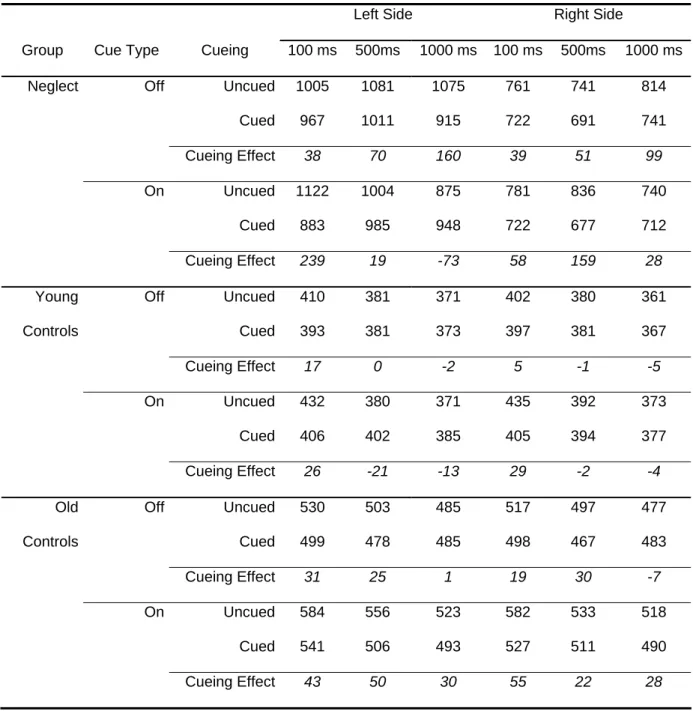
RTs were computed for each experimental condition (Table 2) and introduced in a
repeated-measures analysis of variance (ANOVA) with the following factors: Group (Young Controls,
Old Controls and Neglect Patients), Test (On, Off), Side (Left, Right), SOA (100, 500, 1,000
ms) and Cueing (Cued, Uncued). The Group variable was manipulated between participants,
whereas the other variables were manipulated within participants. The α level was set to 0.05. ---Insert Table 2 about here---
Neglect patients were much slower than controls, F(2, 48)=37.806, p < 0.0001,
especially for left targets (Group x Side interaction, F(2, 48)=16.454, p < 0.0001). Given this
substantial difference in mean RTs, and in order to be able to compare the size of the effects
shown by neglect patients to that of the control groups, a further ANOVA was performed with
the same factors on the proportional RTs, i.e. the RT on each specific experimental condition
per participant, divided by the average RT for that participant (see Lupiáñez et al. 2004).
The interaction between the five factors was significant (F(4, 96)=3.95, p = 0.006). To
explore this complex pattern of interaction, and following our a priori predictions, we
performed four different Group x Side x Cueing ANOVAs, two on the data from the short,
100-ms SOA (one for the On Test, and the other for the Off Test), where facilitation is
predicted, and two on the data from the longest, 1,000-ms SOA, where IOR is predicted
instead.
The Side x Group interaction was significant in all cases, showing the already
described pattern (neglect patients’ longer RTs for left-sided targets than for right-sided
targets), and will no longer be reported.
The ANOVA performed on the short SOA, On Test condition revealed a significant
three way interaction, F(2, 48)=9.5257, p < 0.001, resulting from a substantial slowing in
neglect patients for uncued left targets, i.e. a typical DD (Fig. 2A).
---Insert Fig. 2 about here---
This cueing effect for left-sided targets was significantly larger than that for right-sided
targets in neglect patients, F(1, 9)=7.978, p = 0.0199, as well as than that for left-sided target
in the two groups of controls, F(2, 49)=11.505, p < 0.0001, which did not differ between each
other F<1.
In contrast, the ANOVA performed on the short SOA, Off Test condition (Fig. 2B) showed a highly significant Cueing effect, F(1, 48)=15.529, p = 0.0003, which was independent of Side and Group (all ps > 0.12).
---Insert Fig. 3 about here---
Turning now to the 1,000-ms long SOA, the ANOVA performed on the On Test
condition showed again a significant interaction between cueing and Group, F(2, 48)=4.715, p
= 0.014 (Fig. 3A). However, in this case the interaction was independent of side, F< 1, and
resulted from the opposite cueing effect shown by old and young controls, F(1, 39)=13.652, p
= 0.0007. In support of this interpretation, the interaction between cue and group resulted far
from significance when the controls were analyzed as a single group without age
differentiation (F<1). Interestingly, whereas young controls showed an IOR effect (slower RT
for cued than for uncued trials), which just failed to reach significance, F(1, 25)=4.041, p =
0.055, old controls showed significant facilitation (faster RT for cued than for uncued trials,
F(1, 14)=9.157, p = 0.0091). Neglect patients showed no significant effect at all, although the
tendency was to show IOR for left targets and facilitation for right targets (both Fs < 1),
consistent with previous reports (Bartolomeo et al. 1999; Vivas et al. 2006).
The ANOVA performed on the data from the long SOA, Off Test condition showed a
significantly larger cueing effect for neglect patients than for controls, F(2, 48)=5.442, p =
0.0074. However, in this case the effect was independent of side, F <1 (Fig. 4B), and
therefore it cannot be interpreted as reflecting a DD.
Discussion
Attention is a heterogeneous set of processes whose aim is to maintain coherent behavior in
the face of irrelevant distractions, while allowing the agent to respond rapidly to novel and
important stimuli. Brain damage can severely disrupt these processes. For example, neglect
patients’ attention can be captured by ipsilesional stimuli (Gainotti et al. 1991; D'Erme et al.
1992; Bartolomeo et al. 2004), even if they are irrelevant to the current task. After ipsilesional
capture, patients’ attention may remain, as it were, “stuck” on non-neglected items, so that its
reorienting to other portions of the visual space is slowed (DD). The aim of the present study
was to determine whether the locus of the DD is purely spatial, as it is commonly assumed, or
whether visual objects in space are in fact crucial to capture and confine patients’ attention.
Concerning this issue, our results in neglect patients are clear-cut. A typical DD was
obtained with onset cues at short SOA, but the DD completely disappeared with offset cues.
Except for the overall slowed RTs, neglect patients demonstrated a similar pattern of results
as both young and old controls in the offset condition at short SOA. Thus, in the absence of a
visual stimulus capable of holding their attention on the non-neglected side, neglect patients
are able to redirect attention to the neglected side in a relatively fast manner.
Previous studies using paper-and-pencil tasks have provided abundant analogous
evidence of normal or near-normal performance in neglect patients in the absence of
attention-capturing stimuli on the right side. For example, neglect patients performance can
improve when visual feedback is minimal or absent (Chokron et al. 2004; Loetscher and
Brugger 2007; Urbanski and Bartolomeo 2008). When patients had to detect multiple targets
on a paper sheet, either by drawing over them with a pencil stroke or by erasing them, they
omitted more left-sided target in the “draw” than in the “erase” condition, as if the already
detected right-sided targets continued to capture their attention when still present on the sheet
(Mark et al. 1988; see also Làdavas et al. 1993). Even when the right-sided objects are not
targets, but distractors, they can nevertheless exacerbate neglect-related behaviour
(Bartolomeo et al. 2004). Finally, more relevant to the present context, in a Posner-like RT
task patients demonstrated more DD when the targets appeared in placeholder boxes than
when they appeared in a blank space (D'Erme et al. 1992). D’Erme et al interpreted their
findings as suggesting that right-sided box exogenously captured patients’ attention and kept
it confined. The present results directly support this interpretation. The privileged status of
stimulus onsets, as compared to stimulus offsets, to capture and maintain attention is also
consistent with evidence from visual search tasks in normal participants (see, e.g. Yantis and
Jonides 1990).
In a study devoted to discriminate space-based from object-based neglect, Behrmann
and Tipper (1999) asked left neglect patients to respond to targets appearing inside one of two
horizontally aligned circles of different colors. As expected, patients responded faster to right
than to left targets (space-based neglect). However, in most (∼80%) patients this effect was reversed when the two circles were connected by a line, like a barbell (thus forming a single
perceptual object), and the barbell rotated by 180° just before the target appeared. In this case,
RTs for the targets now on the left side, but appearing in a previously right-sided circle, were
faster than RTs for the targets appearing on the right, thus suggesting object-based neglect. A
possible account of these results is that the circle originally on the right side captured patients’
attention and generated a cost (DD) for targets appearing on the left-sided circle. Patients’
attention then followed the circle as it changed its location. Thus, neglect was not only
modulated by the absolute spatial location of the target, but also by the target being an object
capable of holding attention "stuck". On the other hand, when the two circles were treated as a
single object, the right side of space by itself was insufficient to retain patients’ attention and
to generate a DD, in agreement with the present results.
However, in our results the confinement of attention to the attended objects
disappeared with time. Thus, at long SOA, the strong DD demonstrated by neglect patients
with onset cues disappeared. This dependency of DD on SOA has repeatedly been reported in
the literature (see Losier and Klein 2001, for review), and is also consistent with the
prevalently exogenous nature of the attentional bias in neglect, because, as already mentioned,
exogenous orienting typically occurs at short SOAs (Müller and Rabbitt 1989).
A general caveat in interpreting the present results at long SOA is that we cannot
completely rule out the possibility of contamination from occasional eye movements. Despite
the fact that all participants were able to maintain fixation and complied with the instructions,
with long SOA we cannot exclude the possibility that eye movements occurred in occasional
trials and went undetected by the examiner. Thus, while the lack of IOR for right-sided targets
in neglect patients with the onset test confirmed previous similar results (Bartolomeo et al.
1999; Vivas et al. 2006), we cannot exclude that an advantage for cued trials might have
resulted from patients occasionally looking a right-sided cues, and consequently receiving the
target at the fovea.
A similar caveat applies to the results shown by control participants, who showed an
intriguing pattern of performance with the onset test al long SOA. While young controls
showed a marginally significant IOR, old controls had significant facilitation. Despite the
possibility of eye movements, we note that this pattern is in agreement with previous reports
in the literature showing a lack of IOR in older adults (Faust and Balota 1997). The fact that
no cue back was presented between the cue and the target (see Posner and Cohen 1984), and
the presence of a temporal overlap between cues and targets might explain why IOR was only
marginally significant for young controls, and completely absent for old controls, who
demonstrated instead significant facilitation. However, this pattern was not observed with
offset cues, where controls showed no difference between cued and uncued trials. The
discrepancy between onset and offset cues might suggest that these cueing effects are related
to object (cue-target) integration processes. It has indeed been proposed that IOR might be
due to the lack of novelty induced by an old object (i.e., the cue) in the processing of the
target when it appears in the same location, leading the system to treat the target as less novel,
and thus less able to capture attention (Lupiáñez et al. 2007). Note that this tendency to
integrate the target within the object file of the cue might be clearly reduced or eliminated
when the cue disappears, as in the off condition. Contrary to this hypothesis, previous studies
(e.g., Riggio et al. 1998) did demonstrate IOR with offset cues. However, in these previous
studies a visual object (the placeholder box) remained in the location signalled by the offset
cue, and the target appeared inside the box. In the present case, instead, the cue offset
consisted in the disappearance of the box, leaving a blank location on the screen. Thus, in the
present experiment the perceptual discrimination between off cues and targets was probably
much easier.
An unexpected finding was the large facilitation for cued trials in the offset test for
neglect patients at long SOA. Although we cannot offer a plausible explanation for this effect,
we note that it was clearly independent of side, and therefore must be considered as different
from standard DD.
In conclusion, the present results demonstrate an important characteristic of the
disengagement deficit in neglect patients: The presence of a visual object in which the target
appears is a necessary condition for the DD to emerge. As a consequence, the DD cannot
simply be considered as a directional spatial deficit (Posner et al. 1987). Any future models of
orienting of attention in neglect must take into account the relationship of the DD to visual
objects in space.
References
Bartolomeo P (2007) Visual neglect. Curr Opin Neurol 20:381-386
Bartolomeo P, Chokron S (1999) Egocentric frame of reference: Its role in spatial bias after right hemisphere lesions. Neuropsychologia 37:881-894
Bartolomeo P, Chokron S (2002) Orienting of attention in left unilateral neglect. Neurosci Biobehav Rev 26:217-234
Bartolomeo P, Chokron S, Siéroff E (1999) Facilitation instead of inhibition for repeated right-sided events in left neglect. NeuroReport 10:3353-3357
Bartolomeo P, Lupiáñez J (2006) Inhibitory after-effects in spatial processing: Experimental and theoretical issues on Inhibition of Return. Cognitive Neuropsychol 23
Bartolomeo P, Siéroff E, Decaix C, Chokron S (2001) Modulating the attentional bias in unilateral neglect: The effects of the strategic set. Exp Brain Res 137:424-431
Bartolomeo P, Urbanski M, Chokron S, Chainay H, Moroni C, Siéroff E, Belin C, Halligan P (2004) Neglected attention in apparent spatial compression. Neuropsychologia 42:49-61
Behrmann M, Tipper SP (1999) Attention accesses multiple reference frames: evidence from visual neglect. J Exp Psychol Hum Percept Perform 25:83-101
Chokron S, Colliot P, Bartolomeo P (2004) The role of vision on spatial representations. Cortex 40:281-290
D'Erme P, Robertson IH, Bartolomeo P, Daniele A, Gainotti G (1992) Early rightwards orienting of attention on simple reaction time performance in patients with left-sided neglect. Neuropsychologia 30:989-1000
Egly R, Driver J, Rafal RD (1994) Shifting visual attention between objects and locations: Evidence from normal and parietal lesion patients. J Exp Psych Gen 123:161-177
Faust ME, Balota DA (1997) Inhibition of return and visuospatial attention in healthy older adults and individuals with dementia of the Alzheimer type. Neuropsychology 11:13-29
Friedrich FJ, Egly R, Rafal RD, Beck D (1998) Spatial attention deficits in humans: a comparison of superior parietal and temporal-parietal junction lesions.
Neuropsychology 12:193-207
Gainotti G, D'Erme P, Bartolomeo P (1991) Early orientation of attention toward the half space ipsilateral to the lesion in patients with unilateral brain damage. J Neurol Neurosurg Psychiatry 54:1082-1089
Làdavas E, Umiltà C, Ziani P, Brogi A, Minarini M (1993) The role of right-sided objects in left side neglect: A dissociation between perceptual and directional motor neglect. Neuropsychologia 31:761-773
Loetscher T, Brugger P (2007) A disengagement deficit in representational space. Neuropsychologia 45:1299-1304
Losier BJ, Klein RM (2001) A review of the evidence for a disengage deficit following parietal lobe damage. Neurosci Biobehav Rev 25:1-13
Lupiáñez J, Decaix C, Siéroff E, Chokron S, Milliken B, Bartolomeo P (2004) Independent effects of endogenous and exogenous spatial cueing: Inhibition of return at
endogenously attended target locations. Exp Brain Res 159:447-457
Lupiáñez J, Ruz M, Funes MJ, Milliken B (2007) The manifestation of attentional capture: facilitation or IOR depending on task demands. Psychological Research 71:77-91
Mark VW, Kooistra CA, Heilman KM (1988) Hemispatial neglect affected by non-neglected stimuli. Neurology 38:640-643
Morrow LA, Ratcliff G (1988) The disengagement of covert attention and the neglect syndrome. Psychobiology 16:261-269
Müller HJ, Rabbitt PM (1989) Reflexive and voluntary orienting of visual attention: time course of activation and resistance to interruption. J Exp Psychol Hum Percept Perform 15:315-330
Posner MI (1980) Orienting of attention. Q J Exp Psychol 32:3-25
Posner MI, Cohen Y (1984) Components of visual orienting. In: Bouma H, Bouwhuis D (eds) Attention and Performance X. Lawrence Erlbaum, London, pp 531-556
Posner MI, Rafal RD, Choate LS, Vaughan J (1985) Inhibition of return: Neural basis and function. Cognitive Neuropsychol 2:211-228
Posner MI, Walker JA, Friedrich FA, Rafal RD (1987) How do the parietal lobes direct covert attention? Neuropsychologia 25:135-145
Posner MI, Walker JA, Friedrich FJ, Rafal RD (1984) Effects of parietal injury on covert orienting of attention. J Neurosci 4:1863-1874
Pratt J, McAuliffe J (2001) The effects of onsets and offsets on visual attention. Psychological Research 65:185-191
Riggio L, Bello A, Umiltà C (1998) Inhibitory and facilitatory effects of cue onset and offset. Psychological Research 61:107-118
Siéroff E, Decaix C, Chokron S, Bartolomeo P (2007) Impaired orienting of attention in left unilateral neglect: A componential analysis. Neuropsychology 21:94-113
Smania N, Martini MC, Gambina G, Tomelleri G, Palamara A, Natale E, Marzi CA (1998) The spatial distribution of visual attention in hemineglect and extinction patients. Brain 121:1759-1770
Striemer C, Danckert J (2007) Prism adaptation reduces the disengage deficit in right brain damage patients. Neuroreport 18:99-103
Urbanski M, Bartolomeo P (2008) Line bisection in left neglect: The importance of starting right. Cortex
Valdes-Sosa M, Bobes MA, Rodriguez V, Pinilla T (1998) Switching attention without shifting the spotlight object-based attentional modulation of brain potentials. J Cogn Neurosci 10:137-151
Vivas AB, Humphreys GW, Fuentes LJ (2006) Abnormal inhibition of return: A review and new data on patients with parietal lobe damage. Cognitive Neuropsychol 23:1049-1064
Yantis S, Jonides J (1990) Abrupt visual onsets and selective attention: Voluntary versus automatic allocation. J Exp Psychol Hum Percept Perform 16:121-134
Tables
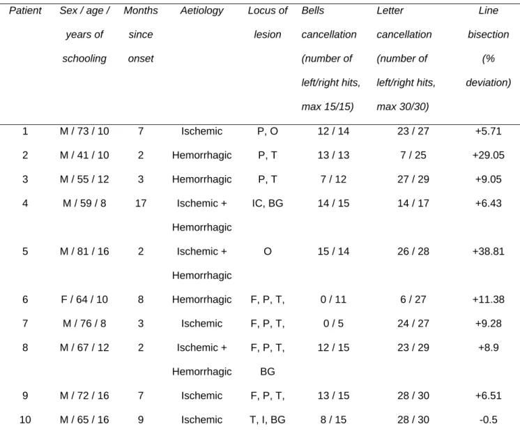
Table 1. Demographical and clinical characteristics of neglect patients
Patient Sex / age / years of schooling Months since onset Aetiology Locus of lesion Bells cancellation (number of left/right hits, max 15/15) Letter cancellation (number of left/right hits, max 30/30) Line bisection (% deviation) 1 M / 73 / 10 7 Ischemic P, O 12 / 14 23 / 27 +5.71 2 M / 41 / 10 2 Hemorrhagic P, T 13 / 13 7 / 25 +29.05 3 M / 55 / 12 3 Hemorrhagic P, T 7 / 12 27 / 29 +9.05 4 M / 59 / 8 17 Ischemic + Hemorrhagic IC, BG 14 / 15 14 / 17 +6.43 5 M / 81 / 16 2 Ischemic + Hemorrhagic O 15 / 14 26 / 28 +38.81 6 F / 64 / 10 8 Hemorrhagic F, P, T, 0 / 11 6 / 27 +11.38 7 M / 76 / 8 3 Ischemic F, P, T, 0 / 5 24 / 27 +9.28 8 M / 67 / 12 2 Ischemic + Hemorrhagic F, P, T, BG 12 / 15 23 / 29 +8.9 9 M / 72 / 16 7 Ischemic F, P, T, 13 / 15 28 / 30 +6.51 10 M / 65 / 16 9 Ischemic T, I, BG 8 / 15 28 / 30 -0.5
See Bartolomeo and Chokron (1999) for detailed test description. For the line bisection test, the cumulated percentage of deviation from the true centre of all the lines was calculated, with rightward deviations carrying a positive sign and leftward deviations having a negative sign. For the
cancellation tests, the number of items cancelled on the left / right halves of the page is reported. Locus of lesion: P, parietal; O, occipital; T, temporal; F, frontal; IC, internal capsule; BG, basal ganglia; I, insula.
Table 2. Mean RTs per experimental condition.
Left Side Right Side
Group Cue Type Cueing 100 ms 500ms 1000 ms 100 ms 500ms 1000 ms Neglect Off Uncued 1005 1081 1075 761 741 814
Cued 967 1011 915 722 691 741
Cueing Effect 38 70 160 39 51 99
On Uncued 1122 1004 875 781 836 740
Cued 883 985 948 722 677 712
Cueing Effect 239 19 -73 58 159 28
Young Off Uncued 410 381 371 402 380 361
Controls Cued 393 381 373 397 381 367
Cueing Effect 17 0 -2 5 -1 -5
On Uncued 432 380 371 435 392 373
Cued 406 402 385 405 394 377
Cueing Effect 26 -21 -13 29 -2 -4
Old Off Uncued 530 503 485 517 497 477
Controls Cued 499 478 485 498 467 483
Cueing Effect 31 25 1 19 30 -7
On Uncued 584 556 523 582 533 518
Cued 541 506 493 527 511 490
Cueing Effect 43 50 30 55 22 28
Figure Legends
Figure 1. Outline of the experimental procedure depicting the sequence of events in each of
the two experimental conditions. The onset cue test consisted in the brightening of the
contour of one of the boxes, which remained present until the end of the trial. The offset
cue test consisted on the disappearance of one of the lateral boxes, which remained
absent until the end of the trial.
Figure 2: RTs for the onset test (A) and the offset test (B) at short (100-ms) SOA.
Figure 3: RTs for the onset test (A) and the offset test (B) at long (1,000-ms) SOA.
Fig. 1
Fig. 2
Short SOA, On Test
300 400 500 600 700 800 900 1000 1100 1200
Left Right Left Right Left Right
Neglect Young Old
Group Me a n R T (i n m s ) Uncued Cued
Short SOA, Off Test
300 400 500 600 700 800 900 1000 1100 1200
Left Right Left Right Left Right
Neglect Young Old
Group M ean RT ( in m s ) Uncued Cued B A
Fig. 3
Long SOA, On Test
300 400 500 600 700 800 900 1000 1100 1200
Left Right Left Right Left Right
Neglect Young Old
Group Me a n R T (i n m s ) Uncued Cued
Long SOA, Off Test
300 400 500 600 700 800 900 1000 1100 1200
Left Right Left Right Left Right
Neglect Young Old
Group Me a n R T (i n m s ) Uncued Cued B A