HAL Id: hal-02924786
https://hal.archives-ouvertes.fr/hal-02924786
Submitted on 31 Aug 2020
HAL is a multi-disciplinary open access archive for the deposit and dissemination of sci-entific research documents, whether they are pub-lished or not. The documents may come from teaching and research institutions in France or abroad, or from public or private research centers.
L’archive ouverte pluridisciplinaire HAL, est destinée au dépôt et à la diffusion de documents scientifiques de niveau recherche, publiés ou non, émanant des établissements d’enseignement et de recherche français ou étrangers, des laboratoires publics ou privés.
Reciprocal regulation of Aurora kinase A and ATIP3 in
the control of metaphase spindle length
Anne Nehlig, Cynthia Seiler, Yulia Steblyanko, Florent Dingli, Guillaume
Arras, Damarys Loew, Julie Welburn, Claude Prigent, Marin Barisic, Clara
Nahmias
To cite this version:
Anne Nehlig, Cynthia Seiler, Yulia Steblyanko, Florent Dingli, Guillaume Arras, et al.. Reciprocal regulation of Aurora kinase A and ATIP3 in the control of metaphase spindle length. Cellular and Molecular Life Sciences, Springer Verlag, 2021, 78 (4), pp.1765-1779. �10.1007/s00018-020-03614-8�. �hal-02924786�
UN
C
ORRECTED PR
OOF
https://doi.org/10.1007/s00018-020-03614-8ORIGINAL ARTICLE
Reciprocal regulation of Aurora kinase A and ATIP3 in the control
of metaphase spindle length
Anne Nehlig1,2,3 · Cynthia Seiler1,2,3 · Yulia Steblyanko4 · Florent Dingli5 · Guillaume Arras5 · Damarys Loew5 · Julie Welburn6 · Claude Prigent7 · Marin Barisic4,8 · Clara Nahmias1,2,3
Received: 30 January 2020 / Revised: 18 July 2020 / Accepted: 7 August 2020 © Springer Nature Switzerland AG 2020
Abstract
Maintaining the integrity of the mitotic spindle in metaphase is essential to ensure normal cell division. We show here that depletion of microtubule-associated protein ATIP3 reduces metaphase spindle length. Mass spectrometry analyses identi-ied the microtubule minus-end depolymerizing kinesin Kif2A as an ATIP3 binding protein. We show that ATIP3 controls metaphase spindle length by interacting with Kif2A and its partner Dda3 in an Aurora kinase A-dependent manner. In the absence of ATIP3, Kif2A and Dda3 accumulate at spindle poles, which is consistent with reduced poleward microtubule lux and shortening of the spindle. ATIP3 silencing also limits Aurora A localization to the poles. Transfection of GFP-Aurora A, but not kinase-dead mutant, rescues the phenotype, indicating that ATIP3 maintains Aurora A activity on the poles to control Kif2A targeting and spindle size. Collectively, these data emphasize the pivotal role of Aurora kinase A and its mutual regulation with ATIP3 in controlling spindle length.
Keywords Aurora A · Dda3 · Kif2A · MTUS1 · Mitotic kinase · Mitotic spindle · Poleward microtubule lux
Introduction
The size of the mitotic spindle in metaphase varies among diferent cell types and species but remains constant for a given cell type, despite high dynamics of spindle micro-tubules [1–3]. The spindle in metaphase must reach an optimal size to capture and bi-orient all duplicated chro-mosomes prior to cell division, and to allow separation of
the asters beyond a minimal distance for proper cleavage by the cytokinesis machinery at late stages of cell division [3]. Defects in spindle size and architecture can lead to mitotic defects and promote aneuploidy, which is a hall-mark of cancer. Metaphase spindle length is controlled by intrinsic and extrinsic factors [3]. Poleward microtubule lux, which consists of translocation of tubulin heterodi-mers towards the poles, coupled to a balance between polymerization of microtubule plus ends at kinetochores and depolymerization of minus ends at the poles [4, 5], is a major mechanism to maintain the spindle at a constant
Cellular and Molecular Life Sciences
Electronic supplementary material The online version of this
article (https ://doi.org/10.1007/s0001 8-020-03614 -8) contains supplementary material, which is available to authorized users. * Clara Nahmias
clara.nahmias@inserm.fr
1 Inserm U981, Department of Molecular Medicine, Gustave
Roussy Cancer Center, Institut Gustave Roussy, 114 rue Edouard Vaillant, 94800 Villejuif, France
2 LabEx LERMIT, Université Paris Saclay,
92296 Châtenay-Malabry, France
3 Institut Gustave Roussy, Inserm, Biomarqueurs prédictifs et
nouvelles stratégies thérapeutiques en oncologie, Université Paris-Saclay, 94800 Villejuif, France
4 Cell Division Laboratory, Danish Cancer Society Research
Center, 2100 Copenhagen, Denmark
5 Centre de Recherche, Laboratoire de Spectrométrie de
Masse Protéomique, Institut Curie, PSL Research University, 75248 Paris Cedex 05, France
6 Wellcome Trust Centre for Cell Biology, School
of Biological Sciences, University of Edinburgh, Edinburgh, Scotland, UK
7 Institut de Génétique et Développement de Rennes
(IGDR), Unité CNRS, UMR 6290, Université de Rennes, 35043 Rennes, France
8 Department of Cellular and Molecular Medicine,
Faculty of Health Sciences, University of Copenhagen, 2100 Copenhagen, Denmark AQ1 AQ2 1 2 3 4 5 6 7 8 9 10 11 12 13 14 15 16 17 18 19 20 21 22 23 24 25 26 27 28 29 30 31 32 33 34 35 36 A1 A2 A3 A4 A5 A6 A7 A8 A9 A10 A11 A12 A13 A14 A15 A16 A17 A18 A19 A20 A21 A22 A23 A24 A25 A26 A27
Author Proof
UN
C
ORRECTED PR
OOF
length in metaphase [2, 6]. Microtubule-associated pro-teins and depolymerizing kinesins, that regulate micro-tubule dynamics at both ends, thus appear as important regulators of poleward lux and spindle length [3, 7–9].
The Kif2A kinesin is a key regulator of poleward micro-tubule lux and spindle size in drosophila, xenopus and human cells [6, 10–13]. Kif2A belongs to the kinesin-13 family of depolymerizing kinesins also including Kif2B and MCAK/Kif2C [14]. During mitosis, Kif2A local-izes to spindle poles where it depolymerlocal-izes microtubule minus ends [10, 15, 16], thereby controlling the rate of poleward microtubule lux. Transport of Kif2A to spin-dle poles is ensured by minus-end-directed motor protein dynein/dynactin [10] and is increased by interaction with microtubule-associated Dda3 protein, whose expression and phosphorylation are markedly elevated in mitosis [17,
18]. Depletion of either Dda3 or Kif2A increases mitotic spindle size [10, 17]. The microtubule depolymerizing activity of Kif2A and its recruitment to the poles are inhib-ited by Aurora kinase A (Aurora A) [19], a major mitotic kinase that localizes on spindle poles. Aurora A fulills numerous functions [20, 21] including the regulation of spindle size [13, 22]. However, the mechanisms by which this kinase controls metaphase spindle length are poorly deined. Whether the maintenance of correct spindle size in metaphase depends on Aurora A-mediated regulation of Kif2A recruitment to the poles remains to be investigated.
ATIP3 is a microtubule-associated protein that deco-rates the centrosome and microtubule cytoskeleton in interphase, and the mitotic spindle and spindle poles dur-ing mitosis [23]. Previous studies have shown that ATIP3 is a potent microtubule stabilizer [24] that reduces micro-tubule dynamics by interacting with End-Binding protein EB1 in interphase [25, 26]. ATIP3 is the product of can-didate tumor suppressor gene MTUS1 whose expression is markedly down-regulated in aggressive breast tumors [23,
24]. We have recently shown that ATIP3 depletion is asso-ciated with increased aneuploidy and mitotic defects [27]. In this study, we show that ATIP3 depletion leads to shortening of the metaphase spindle. Mass spectrometry analysis identiied Kif2A as an intracellular partner of ATIP3. We provide evidence that ATIP3 forms a complex with Kif2A and Dda3 and limits their recruitment to the poles, thereby contributing to maintaining constant spindle size in metaphase. The interaction between ATIP3, Dda3 and Kif2A is increased in mitosis and requires phosphoryl-ation by Aurora kinase A. Conversely, the localizphosphoryl-ation of Aurora A at spindle poles is regulated by ATIP3. Overall, we show that reciprocal regulation of ATIP3 and Aurora kinase A during mitosis ensures the timely assembly of a molecular ATIP3-Dda3-Kif2A complex that controls metaphase spindle length.
Materials and methods
Cell lines and synchronization
Human cancer cell lines MCF-7, MCF-7 cells stably expressing moderate levels of GFP-ATIP3 (HC6 clone), HCC1143 and HeLa were described previously [23, 24]. MCF-7 cells express undetectable levels of ATIP3 whereas HCC1143 and HeLa cells express endogenous ATIP3 [23,
24]. MCF-7 cells were cultured in DMEM-F12 supple-mented with 5% bovine calf serum. HCC1143 cells were cultured in RMPI supplemented with 10% bovine calf serum and 1% sodium pyruvate. HeLa cells were cultured in DMEM supplemented with 10% bovine calf serum, 100 U/ml penicillin, and 100 U/ml streptomycin (all from Gibco) at 37 °C and 5% CO2. Cells were routinely
authenticated by morphologic observation and tested for absence of mycoplasma contamination using Venor® GeM
Advance Kit (MB Minerva biolabs®).
For synchronization in metaphase, MCF-7 and HCC1143 cells were treated with 50 ng/ml nocodazole for 16 h and released for 45 min.
Plasmid constructs, siRNAs and transfections
Plasmids encoding ATIP3, D1, D2, GFP-D2, GFP-D2N, GFP-D2C, GFP-D3, GFP-ATIP3delCN and GFP-D2delCN were described elsewhere [24, 25]. Deletion mutants GFP-delCN2 and GFP-D2delCN2 (lack-ing amino acid positions 635–816 of ATIP3, accession number NP_001001924) were obtained by PCR ampliica-tion of the ATIP3 and D2 sequence, respectively, using the following oligonucleotides: 5′-GGG TCC GTT TCT GCG TTG TTT TCT GGT AAT GCC GCT GTC-3′ and 5′-GAC AGC GGC ATT ACC AGA AAA CAA CGC AGA AAC GGA CCC-3′ and QuikChange® II XL Site-Directed and
Mutagenesis Kit (Agilent). Plasmids encoding GFP-AurA and GFP-AurA-KD were described elsewhere [28–30]. Plasmid encoding Kif2A-GFP was provided by Jamel Chelly (Institut de Génétique et de Biologie Moléculaire et Cellulaire, Strasbourg, France). All cDNA constructs were transfected (2–4 µg) for 24 h using X-treme Gene 9 (Roche) or Lipofectamine 2000 (Invitrogen) and were expressed at levels similar to those of endogenous proteins.
Specific and control scrambled siRNAs were from Dharmacon (ThermoFisher Scientific). The following sequences were used: ATIP3#1, 5′-UGG CAG AGG UUU AAG GUUA-3′; ATIP3#2, 5′-GCA AAU AGC UGC UCC AAA A-3′; Kif2A, 5′-GGC AAA GAG AUU G ACC UGG -3′; Aurora A, 5′-AUG CCC UGU CUU ACU GUC A-3′; Dda3, 5′-AAG CAA GACU UCA GUA GCAUU-3′. For rescue
37 38 39 40 41 42 43 44 45 46 47 48 49 50 51 52 53 54 55 56 57 58 59 60 61 62 63 64 65 66 67 68 69 70 71 72 73 74 75 76 77 78 79 80 81 82 83 84 85 86 87 88 89 90 91 92 93 94 95 96 97 98 99 100 101 102 103 104 105 106 107 108 109 110 111 112 113 114 115 116 117 118 119 120 121 122 123 124 125 126 127 128 129 130 131 132 133 134 135
Author Proof
UN
C
ORRECTED PR
OOF
experiments of ATIP3 knock-down, HeLa cells were trans-fected with ATIP3#1 siRNA that targets the 5′ untranslated sequence of ATIP3 and allows ectopic expression of the ATIP3 coding sequence [25].
All siRNAs (50 nM) were transfected for 48–72 h using Lipofectamine 2000 (Invitrogen) and silencing eiciency was evaluated by immunoblotting. Antibodies used for immunoblotting were rabbit anti-ATIP3 (anti-MTUS1; ARP44419-P050; Aviva Systems, 1/1000e), rabbit anti-Kif2A (ab37005; Abcam, 1/5000e), mouse anti-Aurora A (ab13824; Abcam, 1/1000e), rabbit anti-Dda3 (GTX128047; GeneTex, 1/1000e), rabbit anti-GFP (6556; Abcam, 1/1000e), mouse anti-GFP (ab1218; Abcam, 1/1000e), mouse alpha-tubulin (T9026; Abcam, 1/5000e), anti-phospho-histone H3 (06-570; Millipore, 1/1000e), mouse gamma-actin (ab123034; Abcam, 1/5000e), mouse anti-vinculin (V9264; Sigma, 1/5000e).
Immunoprecipitations
HeLa or MCF-7 cells were transfected as described above and lysed in lysis bufer containing 50 mM Tris-HCl pH 7.4, 150 mM NaCl, 1 mM EDTA, 2.5 mM MgCl2, 0.2 M PMSF, 1 mM Aprotinin, 5 mg/ml Leupeptin, 2 mg/ml Pepstatin, 0.2 M orthovanadate, 1 M sodium luoride, and 10 µM okadaic acid. Lysates were centrifuged, incubated with uncoupled magnetic agarose beads at 4 °C for 15 min, and then incubated with anti-GFP VHH coupled to magnetic agarose beads (GFP-trap_MA; Chromotek) at 4 °C over-night. Antibody beads were recovered with a bar magnet, washed once with the lysis bufer in the presence of anti-proteases and then twice with the lysis bufer, and analyzed by Western blotting with appropriate antibodies as described above. Results shown are representative of 3–5 independent experiments.
Proteomics and mass spectrometry
MCF-7 cells were transiently transfected with 2 µg of GFP-ATIP3 or GFP for 24 h then lysed as described above. For immuno-isolated samples, 1 mg extract was incubated with 25 μl magnetic beads (GFP-trap_MA; Chromotek) for 2 h at 4 °C.
Proteins on magnetic beads were washed twice with 100 μl of 25 mM NH4HCO3 and on-beads digestion were
performed with 0.2 μg of trypsin/LysC (Promega) for 1 h in 100 µl of 25 mM NH4HCO3. Sample were then loaded
onto a homemade C18 StageTips for desalting (principle by stacking one 3 M Empore SPE Extraction Disk Octadecyl (C18) and beads from SepPak C18 CartridgeWaters into a 200 μl micropipette tip). Peptides were eluted using 40/60 MeCN/H2O + 0.1% formic acid and vacuum concentrated
to dryness.
Online chromatography was performed with an RSLC-nano system (Ultimate 3000, Thermo Scientiic) coupled online to an Orbitrap Fusion Tribrid mass spectrometer (Thermo Scientiic). Peptides were trapped on a C18 col-umn (75 μm inner diameter × 2 cm; nanoViper Acclaim PepMapTM 100, Thermo Scientiic) with bufer A (2/98 MeCN/H2O in 0.1% formic acid) at a low rate of 4.0 µl/min
over 4 min. Separation was performed on a 50 cm × 75 μm C18 column (nanoViper Acclaim PepMapTM RSLC, 2 μm, 100 Å, Thermo Scientiic) regulated to a temperature of 55 °C with a linear gradient of 2–30% bufer B (100% MeCN in 0.1% formic acid) at a low rate of 350 nl/min over 160 min. Full-scan MS was acquired in the Orbitrap analyzer with a resolution set to 120,000 and ions from each full scan were HCD fragmented and analyzed in the linear ion trap.
For identiication the data were searched against the Swis-sProt Homo Sapiens (February 2017, no isoforms) database using Sequest HF through proteome discoverer (version 2.1). Enzyme speciicity was set to trypsin and a maximum of two missed cleavage site were allowed. Oxidized methionine, N-terminal acetylation, and carbamidomethyl cysteine were set as variable modiications. Maximum allowed mass devia-tion was set to 10 ppm for monoisotopic precursor ions and 0.6 Da for MS/MS peaks.
The resulting iles were further processed using myProMS [31] v3.6 (work in progress). FDR calculation used Perco-lator and was set to 1% at the peptide level for the whole study. The label free quantiication was performed by pep-tide Extracted Ion Chromatograms (XICs) computed with MassChroQ version 2.2.2 [32]. For protein quantiication, XICs from proteotypic peptides shared between compared conditions (TopN matching) with no missed cleavages were used. Median and scale normalization was applied on the total signal to correct the XICs for each biological replicate. To estimate the signiicance of the change in protein abun-dance, a linear model (adjusted on peptides and biological replicates) was performed and p-values were adjusted with a Benjamini–Hochberg FDR procedure with a control thresh-old set to 0.05. Fthresh-old change-based GO enrichment analysis was performed as described [33].
The mass spectrometry proteomics data have been deposited to the ProteomeXchange Consortium via the PRIDE [34] partner repository with the dataset identiier PXD009182 (username: reviewer03478@ebi.ac.uk, pass-word: UFFpYuYZ).
Immunoluorescence
Cells were ixed on cover glasses with ice-cold methanol for 5 min and washed with PBS. Coverslips were subsequently incubated at room temperature for 1 h in primary antibodies and for 1 h in Alexa Fluor 488-, Alexa Fluor 549- and Alexa Fluor 647-coupled secondary antibodies diluted in PBS/
136 137 138 139 140 141 142 143 144 145 146 147 148 149 150 151 152 153 154 155 156 157 158 159 160 161 162 163 164 165 166 167 168 169 170 171 172 173 174 175 176 177 178 179 180 181 182 183 184 185 186 187 188 189 190 191 192 193 194 195 196 197 198 199 200 201 202 203 204 205 206 207 208 209 210 211 212 213 214 215 216 217 218 219 220 221 222 223 224 225 226 227 228 229 230 231 232 233 234 235
Author Proof
UN
C
ORRECTED PR
OOF
Triton 0.1%, BSA 0.2%. The following primary antibodies were used: rabbit anti-Kif2A (ab37005; Abcam, 1/250e), rabbit pericentrin (ab4448; Abcam, 1/500e), rabbit anti-Dda3 (GTX128047; GeneTex, 1/500e), mouse anti-Aurora A (ab13824; Abcam, 1/200e), rabbit Phospho-Aurora A
(Thr288) (3079S; Cell Signaling, 1/200e), mouse anti-GFP (ab1218; Abcam, 1/250e), rat anti-alpha-tubulin (ab6160; Abcam, 1/500e), mouse anti-gamma-tubulin (T5326; Sigma, 1/500e) and mouse anti-alpha- tubulin (T9026; Sigma, 1/100e). Coverslips were ixed with Glycergel Mounting
236 237 238 239 240 241 242 243 244 245
Author Proof
UN
C
ORRECTED PR
OOF
Medium (Dako) coupled with DAPI. Images were acquired with a confocal laser scanning microscope Dmi8- SP8 with an HC PL APO CS2 40x-1.3 Oil (Leica). Linescan analyses of pole-to-pole distance and luorescence intensities were done with Image J. At least 30 cells in 4–10 ields per condi-tion were analyzed.
Results shown are representative of 3–5 independent experiments.
Fluorescence recovery after photobleaching (FRAP)
HeLa cells were transfected with control or ATIP3 siRNA and co-transfected with Kif2A-GFP. For the experi-ment, cells were seeded in 35 mm glass-bottomed dishes (14 mm, No. 1.5, MatTek Corporation). The siRNA trans-fection was done 72 h prior to ilming using Lipofectamine 2000 (Invitrogen). Kif2A-GFP was co-transfected 48 h after siRNA transfection. 10 nM SiR-DNA (Spirochrome)
was added to the cell culture medium 30 min before start-ing the live-cell imagstart-ing. Time-lapse imagstart-ing was per-formed at 37 °C using a Plan-Apochromat 63x/1.4NA with diferential interference contrast oil objective mounted on an inverted Zeiss Axio Observer Z1 microscope (Mari-anas Imaging Workstation from Intelligent Imaging and Innovations Inc. (3i), Denver, CO, USA), equipped with a CSU-X1 spinning-disk confocal head (Yokogawa Corpo-ration of America). Images were acquired using an iXon Ultra 888 EM-CCD camera (Andor Technology). After identiication of spindle poles in metaphase cells, one of the two poles was photobleached with a 5 ms laser pulse using 100% power of the 488 nm solid state laser, and the luorescence recovery was recorded for 90 s at 500 ms intervals. For FRAP calculations, the luorescence sity of the bleached area was normalized using the inten-sity of the whole cell, after background correction. Then a full-scale normalization was performed and the mean values were plotted and used for exponential curve itting using GraphPad Prism 7.01 to determine the half-life and eiciency of the recovery.
Poleward microtubule lux rate measurement
Photo-conversion of alpha-tubulin was performed using a stable U2OS-mEOS-tubulin cell line [35] cultivated on 35 mm glass-bottomed dishes (14 mm, No. 1.5, MatTek Corporation). Transfection of control or ATIP3 siRNA was done 48 h prior to ilming using Lipofectamine 2000 (Invitrogen). Time-lapse imaging was performed at 37 °C using a Plan-Apochromat 63x/1.4NA oil objective with diferential interference contrast mounted on an inverted Zeiss Axio Observer Z1 microscope (Marianas Imaging Workstation from Intelligent Imaging and Innovations Inc. (3i), Denver, CO, USA), equipped with a CSU-X1 spinning-disk confocal head (Yokogawa Corporation of America) and a photo-activation system with 405 nm laser line. Images were acquired using an iXon Ultra 888 EM-CCD camera (Andor Technology). Late prometaphase/ metaphase cells were identiied upon green luorescent tubulin signals and two line-shaped regions of interest were placed perpendicular to the main spindle axis on both sides of the metaphase plate to be photo-activated. Photo-conversion was performed by a 5-ms 405-nm laser pulse. Photo-converted red signals were then followed over time together with green luorescence signal using 561- and 488-nm lasers and images were acquired every 5 s for 3 min. Microtubule lux was quantiied by tracking photo-converted mEos-tubulin over time using sum-projected kymographs generated by a MATLAB-based algorithm [36].
Fig. 1 ATIP3 interacts with Kif2A. a Immunoblotting with
anti-ATIP3 (MTUS1) antibodies shows silencing eiciency of two dif-ferent ATIP3-speciic siRNAs (siA#1 and siA#2) transfected in HeLa cells. Blots were reprobed with anti-vinculin antibodies for internal control. b Immunoluorescence imaging of HeLa cells trans-fected with control (siC), and two diferent ATIP3-speciic siRNAs (siA#1 and siA#2) as indicated. Cells were stained with anti-peri-centrin (red), anti-alpha-tubulin (gray) antibodies and DAPI (blue). Scale bar, 10 µm. Pole-to pole distance (white arrow) was meas-ured by drawing a line with ImageJ software between the two poles stained with pericentrin. Right panel shows quantiication of spindle length in metaphase. Number of cells analyzed is under brackets. ***p < 0.001. c Volcano plot showing distribution of the 9 proteins related to the microtubule cytoskeleton and/or mitosis present in GFP-ATIP3 compared with GFP and identiied by mass spectrom-etry. x axis = log2(fold-change), y axis = − log10(p-value). Position of the ATIP3 partner Kif2A is indicated in red. d MCF-7 cells were transfected with GFP-ATIP3 or GFP and immunoprecipitation was performed using anti-GFP antibodies. Western blots were probed with anti-Kif2A, anti-Kif2B, anti-ATIP3 and anti-GFP antibodies to reveal Kif2A, Kif2B, GFP-ATIP3 and GFP, respectively. A green asterisk in the INPUT panel indicates endogenous ATIP3. e HeLa cells were transfected with Kif2A-GFP or GFP and immunopre-cipitation was performed using anti-GFP antibodies. Western blots were probed with anti-ATIP3 (MTUS1) and anti-GFP antibodies to reveal ATIP3, Kif2A-GFP and GFP. f Schematic representation of the ATIP3 protein sequence illustrating the position of D1, D2 and D3 regions and ATIP3 deletion mutants and their ability (+) or not (−) to bind Kif2A. Kif2A-binding regions are in red. CC: coiled coil region. Amino acid numbering is from accession number NP_001001924. Right panel: immunoprecipitation assay of MCF-7 cell lysates expressing GFP, GFP-ATIP3 or GFP-fused D1, D2, D3 regions. Blots were probed with anti-Kif2A and anti-GFP antibodies. A green aster-isk indicates non-speciic band. A black star indicates the position of GFP-D1 which migrates at the same apparent molecular weight as endogenous Kif2A revealed in previous blotting. A red asterisk indi-cates cleavage product of GFP-D2. g MCF-7 cells were transfected with GFP-D2 fragments (left panel) or GFP-ATIP3 deletion mutants (right panel) and immunoprecipitation was performed using anti-GFP antibodies. Blots were probed as in d. A green asterisk shows endog-enous ATIP3. Red asterisks indicate cleavage products of fusion pro-teins ◂ AQ3 246 247 248 249 250 251 252 253 254 255 256 257 258 259 260 261 262 263 264 265 266 267 268 269 270 271 272 273 274 275 276 277 278 279 280 281 282 283 284 285 286 287 288 289 290 291 292 293 294 295 296 297 298 299 300 301 302 303 304 305 306 307 308 309 310
Author Proof
UN
C
ORRECTED PR
OOF
Statistical analysis
Statistical analyses were done using GraphPad Prism 6.0 software. Data in bar graphs (mean ± SD) and dot plots were analyzed using two-tail unpaired t test and ANOVA test.
p < 0.05 was considered statistically signiicant.
Results
ATIP3 depletion reduces metaphase spindle length
To evaluate the consequences of ATIP3 depletion in mito-sis, HeLa cells were transfected with two independent ATIP3 siRNAs (siA#1 and siA#2) (Fig. 1a) and mitotic cells were analyzed by immunoluorescence. By measuring pole-to-pole distance in metaphase cells using pericentrin as a marker, we observed that ATIP3 depletion with each siRNA reduces spindle length (Fig. 1b). Similar results were obtained using both ATIP3 siRNAs in HCC1143 breast cancer cells (Fig. S1A). Conversely, moderate expression of GFP-ATIP3 in ATIP3-negative MCF-7 cells led to an increase in spindle size compared to control cells expressing GFP (Fig. S1B).
Other mitotic defects such as multipolar spindles (Fig. S1C) and spindle mispositioning (Fig. S1D) were observed in ATIP3-depleted cells but spindle orientation, symmetry and cell size remained unchanged (Fig. S1E).
ATIP3 interacts with Kif2A
In a first step to elucidate the molecular mechanisms by which ATIP3 controls spindle size, we undertook a
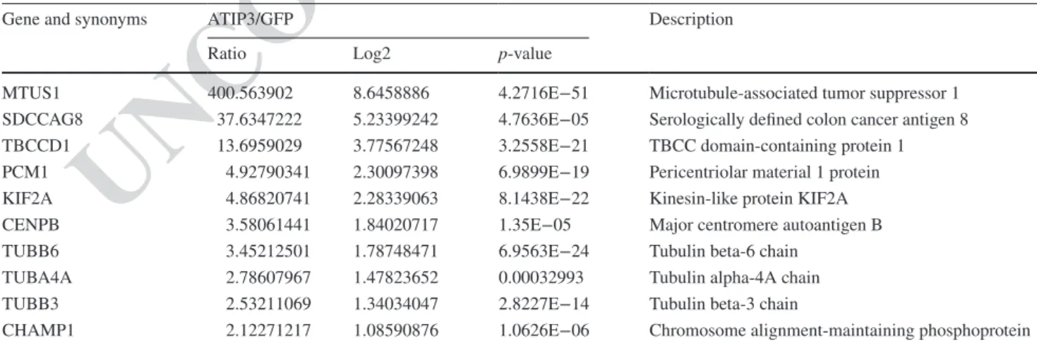
proteomic approach to identify novel intracellular ATIP3 binding partners. Anti-GFP antibodies were used to immu-noprecipitate molecular complexes from MCF-7 breast cancer cells expressing either GFP or GFP-ATIP3 fusion protein. Mass spectrometry analysis allowed us to identify 145 proteins that selectively interact with ATIP3 (Table S1), nine of which were related to the microtubule cytoskeleton and/or mitosis (Fig. 1c, Table 1). These include 3 tubulin chains (beta6, alpha4 and beta3), 2 centromere/kinetochore associated proteins (CENPB, CHAMP1) and 4 centroso-mal/spindle pole proteins (SDCCAG8, TBBCD1, PCM1 and Kif2A). The Kif2A kinesin was among the best can-didate ATIP3 partners in terms of fold change (4.86) and
p value (8.14e−22). This depolymerizing kinesin is also a
well-known regulator of mitotic spindle length [10] and was therefore selected for further studies.
The interaction between ATIP3 and Kif2A was con-irmed by co-immunoprecipitation analyses. GFP-ATIP3 fusion protein expressed in MCF-7 cells was immuno-precipitated using anti-GFP antibodies and found to bind endogenous Kif2A (Fig. 1d). Kinesin Kif2B was not retained in GFP-ATIP3 immunocomplexes, indicating speciicity. Conversely, endogenous ATIP3 was found to interact with Kif2A-GFP in HeLa cells (Fig. 1e), conirming the exist-ence of ATIP3-Kif2A molecular complexes. As shown in Fig. S1F, ATIP3 accumulates along the entire length of the spindle, including the spindle poles where Kif2A is localized.
To better characterize the interaction, we analyzed three truncated constructs designated D1, D2 and D3 (Fig. 1f) encompassing the N-terminal acidic region, the central MT-binding domain and the C-terminal coiled-coil region of ATIP3, respectively [24]. After transient transfection of
Table 1 ATIP3-interacting partners related to microtubule cytoskeleton and/or mitosis
MCF-7 cells were transfected with GFP or GFP-ATIP3 and cell lysates were precipitated using GFP-trap magnetic agarose beads. Proteins retained in immunocomplexes were analyzed by quantitative label-free mass spectrometry analysis performed on four replicates. Shown are the fold changes of 9 proteins related to microtubules and/or mitosis that were selected among 145 proteins quantiied by label-free quantitative anal-ysis of GFP-ATIP3 versus GFP according to absolute fold change ≥ 2, adjusted p-value of ratio signiicance ≤ 0.05 and more than 3 peptides. Proteins are listed by ratio order
Gene and synonyms ATIP3/GFP Description
Ratio Log2 p-value
MTUS1 400.563902 8.6458886 4.2716E−51 Microtubule-associated tumor suppressor 1
SDCCAG8 37.6347222 5.23399242 4.7636E−05 Serologically deined colon cancer antigen 8
TBCCD1 13.6959029 3.77567248 3.2558E−21 TBCC domain-containing protein 1
PCM1 4.92790341 2.30097398 6.9899E−19 Pericentriolar material 1 protein
KIF2A 4.86820741 2.28339063 8.1438E−22 Kinesin-like protein KIF2A
CENPB 3.58061441 1.84020717 1.35E−05 Major centromere autoantigen B
TUBB6 3.45212501 1.78748471 6.9563E−24 Tubulin beta-6 chain
TUBA4A 2.78607967 1.47823652 0.00032993 Tubulin alpha-4A chain
TUBB3 2.53211069 1.34034047 2.8227E−14 Tubulin beta-3 chain
CHAMP1 2.12271217 1.08590876 1.0626E−06 Chromosome alignment-maintaining phosphoprotein 1
311 312 313 314 315 316 317 318 319 320 321 322 323 324 325 326 327 328 329 330 331 332 333 334 335 336 337 338 339 340 341 342 343 344 345 346 347 348 349 350 351 352 353 354 355 356 357 358 359 360 361 362 363 364 365 366 367 368 369
Author Proof
UN
C
ORRECTED PR
OOF
GFP-tagged constructs followed by co-immunoprecipitation, results showed that the central region of 410 amino-acids (D2 region) interacts with Kif2A (Fig. 1f, right panel). Trun-cated mutants of the D2 region (D2N, D2C) were then ana-lyzed. The D2C sequence encompassing amino acid residues 705–874 of ATIP3 was suicient to bind Kif2A (Fig. 1g, left panel). Deletion of amino acids 705–816 (GFP-delCN) and 635–816 (GFP-delCN2) in the ATIP3 sequence led to a marked decrease in Kif2A interaction (Fig. 1g, right panel). Deleting the same sequences from the D2 polypeptide also impaired the interaction (Fig. S2A), therefore indicating that a minimal sequence of 112 amino acids (CN sequence) is required for ATIP3 interaction with Kif2A.
ATIP3-Kif2A complex controls metaphase spindle length
To investigate the functional impact of ATIP3-Kif2A com-plex in the control of mitotic spindle length, we investi-gated whether GFP-ATIP3 construct and deletion mutants were able to rescue the ATIP3 depletion phenotype. RNAi-resistant GFP-ATIP3 expressed at moderate levels in ATIP3-depleted cells indeed restored a spindle size similar to that observed in control cells (Fig. 2a). In contrast, deletion mutants GFP-DelCN and GFP-DelCN2—that are unable to interact with Kif2A—failed to rescue the phenotype (Fig. 2a, S2B). Furthermore, expressing the Kif2A-interacting region D2 was suicient to restore a correct spindle length in ATIP3-depleted cells whereas Kif2A-binding defective mutants D2delCN and D2delCN2 were not (Fig. S2C). Together these data strongly suggest that ATIP3 may control metaphase spindle size by interacting with Kif2A.
ATIP3 depletion increases Kif2A targeting to the spindle poles
Immunoluorescence studies revealed that ATIP3 depletion markedly increases Kif2A luorescence intensity at spindle poles (Fig. 2b) with no efect on the cytosolic pool of Kif2A (Fig. S2D). In contrast, ATIP3 depletion did not signiicantly modify luorescence intensity of pericentrin, a major spindle pole protein (Fig. S2E). Expression of RNAi-resistant GFP-ATIP3 in GFP-ATIP3-depleted cells rescued the phenotype and decreased Kif2A intensity at the poles (Fig. 2c). Of note, ATIP3 silencing had no efect on total Kif2A protein levels detected by western blotting (Fig. 2b, left panel), suggesting that increased luorescence intensity of Kif2A may relect increased recruitment to the poles rather than increased expression of the protein.
We then evaluated whether increased Kif2A accumula-tion at spindle poles may contribute to the phenotype of ATIP3 deiciency. Silencing of Kif2A in ATIP3-depleted cells (Fig. S2F) indeed rescued the phenotype and restored
a correct spindle size (Fig. 2d), indicating that increased Kif2A recruitment to the poles is responsible for spindle length shortening in ATIP3-depleted cells.
It has been previously shown that Kif2A regulates meta-phase spindle length by controlling poleward microtubule lux via its microtubule depolymerizing activity at the spin-dle poles [6, 10, 11]. We reasoned that ATIP3 deiciency may disrupt the balance of microtubule dynamics in the spindle by allowing increased Kif2A depolymerizing activ-ity at the poles, resulting in decreased poleward lux and shorter spindle length. To address that question, microtu-bule lux rates were analyzed following ATIP3 silencing in metaphase U2OS cells stably expressing alpha-tubulin fused to the photo-convertible luorescent protein mEos2 [35]. Results indicate that poleward microtubule lux is signii-cantly decreased in ATIP3-depleted cells (0.53 ± 0.13 µm/ min) compared to control cells (0.67 ± 0.08 µm/min,
p < 0.0001) (Fig. 2e), supporting the notion that ATIP3 may
control spindle size by regulating the recruitment—and thus, the depolymerizing activity—of Kif2A to the poles. Col-lectively, these data indicate that ATIP3 controls metaphase spindle length by interacting with the Kif2A kinesin and reducing its recruitment to the poles.
ATIP3-Kif2A interaction involves Dda3
How does ATIP3 silencing increase Kif2A targeting at the spindle poles? We hypothesized that ATIP3 may sequester Kif2A in the cytosol and slower the exchange rate between spindle pole-associated Kif2A and a difuse cytosolic pool of the protein. We thus explored the dynamic behavior of Kif2A turnover on spindle poles in the presence or absence of ATIP3. Fluorescence recovery after photobleaching (FRAP) experiments were performed on metaphase HeLa cells expressing endogenous ATIP3 and transfected with Kif2A-GFP. Bleaching was done on one of the two spindle poles. We found that in control cells, Kif2A-GFP association with spindle poles is dynamic. Ninety percent of the signal was recovered after photobleaching, with a half-recovery time of 11.6 s (Fig. 3a) which is faster than that (30.7 s) reported for NuMA, another major spindle pole-associated protein [37]. In ATIP3-depleted cells, Kif2A-GFP luores-cence recovery parameters remained unchanged (half-recov-ery time of 11.62 s), ruling out the hypothesis that ATIP3 depletion may modify the exchange rate between cytosolic and spindle pole-associated pools of Kif2A.
We then investigated whether ATIP3 may regulate Kif2A localization by a mechanism involving Dda3, a microtubule-associated protein known to interact with and increase Kif2A targeting to spindle poles [17]. As shown in Fig. 3b, Dda3 silencing in ATIP3-depleted cells rescued Kif2A luorescence intensity at spindle poles, indicating that Dda3 contributes to the regulatory efects of ATIP3
370 371 372 373 374 375 376 377 378 379 380 381 382 383 384 385 386 387 388 389 390 391 392 393 394 395 396 397 398 399 400 401 402 403 404 405 406 407 408 409 410 411 412 413 414 415 416 417 418 419 420 421 422 423 424 425 426 427 428 429 430 431 432 433 434 435 436 437 438 439 440 441 442 443 444 445 446 447 448 449 450 451 452 453 454 455 456 457 458 459 460 461 462 463 464 465 466 467 468 469
Author Proof
UN
C
ORRECTED PR
OOF
UN
C
ORRECTED PR
OOF
on Kif2A recruitment. Accordingly, Dda3 silencing also restored a correct spindle length in ATIP3-depleted cells (Fig. 3b), indicating that Dda3 is essential to the ATIP3 phenotype. Immunoluorescence analyses revealed that, as for Kif2A, Dda3 luorescence intensity at the poles is increased upon ATIP3 depletion (Fig. 3c). Conversely, expression of GFP-ATIP3 led a decrease of Dda3 recruit-ment to the poles compared with GFP (Fig. 3d). Of note, silencing of either Kif2A or Dda3 had no signiicant efect on ATIP3 luorescence intensity (Fig. S3), pointing to ATIP3 as an upstream regulator of Kif2A and Dda3 tar-geting to the poles.
Dda3 is a known partner of Kif2A, raising the possibility that it may be part of the ATIP3-Kif2A complex. Immu-noprecipitation studies revealed that Dda3 indeed inter-acts with ATIP3 (Fig. 3e). To evaluate whether Dda3 may compete with, or contribute to, ATIP3-Kif2A interaction, Dda3 was silenced using siRNA prior to immunoprecipita-tion with GFP-ATIP3. Results showed that Dda3 depleimmunoprecipita-tion impaired Kif2A binding to GFP-ATIP3 immunocomplexes (Fig. 3f) indicating that Dda3 is required for ATIP3-Kif2A interaction.
The interaction between ATIP3, Kif2A and Dda3 is regulated in mitosis
Dda3 levels have been shown to be elevated in mitosis [17] leading us to investigate whether ATIP3-Dda3 interaction may also be regulated in mitosis. Co-immunoprecipitation studies performed in synchronized MCF-7 cells expressing GFP-ATIP3 indeed revealed that the interaction between ATIP3 and Dda3 is increased in mitosis (Fig. 3g). Impor-tantly, Kif2A binding to GFP-ATIP3 was also markedly increased in synchronized cells although Kif2A levels remained unchanged (Fig. 3g), which further supports the notion that Dda3 is essential to the ATIP3-Kif2A complex. Data showing Dda3 hyperphosphorylation in mitosis [38] prompted us to examine whether the interaction between ATIP3, Dda3 and Kif2A may be regulated by phosphoryla-tion. Treatment with lambda phosphatase prior to immuno-precipitation with GFP-ATIP3 totally abrogated both Dda3- and Kif2A-binding to GFP-ATIP3 (Fig. S4A), indicating that protein phosphorylation is required for ATIP3-Dda3-Kif2A interaction.
Aurora kinase A plays a pivotal role in the ATIP3 phenotype
Mitosis is tightly regulated by mitotic kinases among which Aurora kinase A (Aurora A), that is localized to the poles of the spindle and whose expression is markedly increased in G2/M. Aurora A has been shown to phosphorylate both Dda3 [18, 38] and Kif2A [19] and to decrease the recruit-ment and microtubule-depolymerizing activity of Kif2A at the spindle poles [19]. We thus investigated whether Aurora A may be a master kinase regulating ATIP3-Dda3-Kif2A interaction.
Treatment with Aurora A kinase inhibitor MLN8054 impaired both ATIP3-Kif2A and ATIP3-Dda3 interactions (Fig. 4a), indicating that Aurora A kinase activity is required for ATIP3 interaction with Kif2a and Dda3. Aurora A depletion by siRNA also markedly decreased ATIP3-Kif2A interaction (Fig. S4B). Co-immunoprecipitation studies revealed that Aurora A binds to GFP-ATIP3 (Fig. S4C). Of note, ATIP3/Aurora A complexes were not always clearly
Fig. 2 ATIP3 regulates Kif2A recruitment to spindle poles. a
Immu-noluorescence imaging of HeLa cells transfected for 48 h with con-trol (siC) or ATIP3-speciic (siA) siRNA as indicated, then for 24 h with GFP, GFP-ATIP3, or ATIP3 deletion mutants (GFP-delCN and GFP-delCN2) as indicated. Cells were stained with anti-GFP (green) and anti-pericentrin (red) antibodies and DAPI (blue). Scale bar, 10 µm. Spindle length was measured as in Fig. 1b. Right panel shows quantiication of spindle length in metaphase HeLa cells. Number of cells analyzed is under brackets. ***p < 0.001, ns: not signiicant. b Left panel: Immunoblotting of HeLa cell lysates following transfec-tion with control (siC) or ATIP3-speciic (siA) siRNA as indicated. Blots were probed with MTUS1 (ATIP3), Kif2A and anti-tubulin antibodies. Middle panel: Immunoluorescence imaging of HeLa cells transfected with control (siC) or ATIP3-speciic (siA) siRNA as indicated. Cells were ixed and stained with anti-Kif2A (green) antibodies and DAPI (blue). Scale bar, 10 µm. Kif2A luores-cence intensity was measured by drawing a line with ImageJ software between the two poles and the maximum intensity values at the poles were collected and normalized to the intensity of gamma-tubulin. Right panel: Quantiication of Kif2A intensity in metaphase HeLa cells. Number of poles analyzed is under brackets. ***p < 0.001. c Immunoluorescence imaging of Hela cells transfected with control or ATIP3-speciic siRNA for 48 h, then were transfected for 24 h with GFP or GFP-ATIP3, ixed and stained with anti-Kif2A (red) and anti-GFP (green) antibodies and DAPI (blue). Scale bar, 10 µm. Right panel shows quantiications of Kif2A intensity at poles in metaphase HeLa cells. Number of poles analyzed is under brackets. ***p < 0.001. d Immunoluorescence imaging of HeLa cells trans-fected with control (siC), ATIP3-speciic (siA) and/or Kif2A-speciic (siK) siRNA as indicated, and stained as in b. Scale bar, 10 µm. Mid-dle panel: SpinMid-dle length (pole-to-pole distance) was measured as in Fig. 1b. Number of metaphase cells analyzed is under brackets. Right panel: Quantiication of Kif2A intensity in metaphase HeLa cells as in b. Number of poles analyzed is under brackets. *p < 0.05, **p < 0.01, ***p < 0.001. e Upper left panel: Representative luores-cence images of U2OS-mEOS-tubulin cells in late prometaphase/ metaphase transfected with control (siC) and ATIP3 (siA) siRNAs. Images were captured before (pre) and after photo-conversion of mEOS-tubulin. Microtubule lux was measured by photoconversion of line-like regions from both half-spindles in mEos-tubulin cells and tracking of photoconverted mEOS-tubulin over a time interval of 3 min. Scale bar, 10 μm; time, min:s. Lower left panel: Representa-tive sum-projected kymographs of photoactivated regions used for quantiication of lux rates in control (siC) and ATIP3-depleted (siA) cells. Red dashed line indicates the slope of mEOS tubulin translo-cation over time. Scale bar, 30 s. Upper right panel: Quantiitranslo-cation of microtubule lux in control versus ATIP3-depleted cells. Number of cells analyzed is under brackets. ***p < 0.001. Lower right panel: Immunoblotting-based validation of ATIP3 siRNA eiciency ◂ 470 471 472 473 474 475 476 477 478 479 480 481 482 483 484 485 486 487 488 489 490 491 492 493 494 495 496 497 498 499 500 501 502 503 504 505 506 507 508 509 510 511 512 513 514 515 516 517 518 519 520 521 522 523 524 525 526 527 528 529 530
Author Proof
UN
C
ORRECTED PR
OOF
detectable, suggesting that this interaction may be weak and/ or transient.
In addition to disrupting the interaction of ATIP3 with Kif2A and Dda3, Aurora A inactivation by silencing or pharmacological inhibition mimics the phenotype of ATIP3 depletion regarding Kif2A recruitment to the pole
and regulation of spindle size (Fig. S4D, S4E). This led us to investigate whether Aurora A itself may be regulated by ATIP3. Immunoluorescence analyses revealed that ATIP3 silencing decreases Aurora A fluorescence intensity at spindle microtubules and poles (Figs. 4b, S4F), with no sig-niicant change in total protein levels detected by western
531 532 533 534 535 536 537 538 539 540 541 542
Author Proof
UN
C
ORRECTED PR
OOF
blot (Fig. 4b). Fluorescence intensity of the phosphorylated (Thr288) active form of the kinase was also reduced upon ATIP3 depletion (Fig. 4b), suggesting that ATIP3 contrib-utes to maintaining an active pool of Aurora A at the poles of the mitotic spindle.
As shown in Fig. 4c, expression of RNAi-resistant GFP-ATIP3 into ATIP3-depleted cells was able to fully restore Aurora A luorescence intensity at the spindle poles. Importantly, ATIP3-deletion mutants delCN and delCN2, that do not interact with Kif2A, were also able to rescue
the phenotype, indicating that ATIP3-mediated control of Aurora targeting to spindle poles is independent of the ATIP3/Kif2A complex.
We next examined the possibility that Aurora kinase A activity may be involved in the ATIP3 phenotype. To investi-gate whether the short spindle phenotype induced by ATIP3 depletion may be due to decreased Aurora A kinase activ-ity at the poles, we ectopically expressed a GFP-Aurora A construct (GFP-AurA), or kinase-dead mutant (GFP-AurA-KD) in ATIP3-silenced cells and analyzed mitotic cells. As shown in Fig. 4d, active GFP-AurA expressed at moderate levels in ATIP3 deicient cells was able to restore a correct mitotic spindle size whereas the kinase dead GFP-AurA-KD mutant remained ineicient. Expression of GFP-AurA construct, but not the kinase dead GFP-AurA-KD mutant, also reverted the efects of ATIP3 silencing on Kif2A luo-rescence intensities at the poles (Fig. 4e). Together, these results demonstrate that Aurora A kinase activity rescues mitotic spindle defects induced by ATIP3 deiciency, and highlight the central role of Aurora A in the control of meta-phase spindle length by ATIP3.
Discussion
Tight control of mitotic spindle length during metaphase is essential to ensure correct cell division. Results presented here indicate that microtubule-associated protein ATIP3 controls metaphase spindle length by interacting with the microtubule depolymerizing kinesin Kif2A to negatively regulate its association with spindle poles. ATIP3 depletion causes increased Kif2A targeting to the poles, reduced pole-ward microtubule lux and shorter spindle size. Increased accumulation of Kif2A on spindle poles, and subsequent excessive depolymerization of spindle microtubule minus ends, may disrupt the balance between microtubule polym-erization at kinetochores and depolympolym-erization at spindle poles in metaphase, thereby leading to mild changes in poleward microtubule lux. Moderate negative regulation of microtubule lux observed upon ATIP3 depletion may also be due to its negative efect on Aurora kinase A, which was shown to regulate poleward lux via TPX2 and CLASP1 [13].
We show here by fluorescence recovery after pho-tobleaching (FRAP) analyses that the binding of Kif2A on spindle poles is dynamic. Cytosolic pools of GFP-Kif2A were found to exchange in and out of spindle poles with half-recovery time around 10 s, which is a little faster than for other spindle pole-associated proteins such as NuMA [37]. Of note, ATIP3 silencing has no signiicant efect on Kif2A turnover at the poles, ruling out the hypothesis that ATIP3 may regulate the dynamic exchange between cytosolic and spindle pole pools of Kif2A proteins. The efect of ATIP3
Fig. 3 ATIP3-Kif2A interaction involves Dda3. a Left panel:
Rep-resentative spinning-disk confocal live-cell images of HeLa cells in metaphase, expressing Kif2A-GFP and co-stained with SiR-DNA. Red circle indicates the bleaching area used for luorescence recov-ery after photobleaching (FRAP). Scale bar, 5 µm. Time shown in seconds. Right panel: FRAP data points represent means obtained from 28 control cells (siC) and 21 siATIP3-transfected cells (siA), collected from 3 independent experiments. The mean values of dou-ble normalized pixel intensities were plotted as a function of time
and single exponential curves were itted (R2 > 0.99). Kif2A-GFP
luorescence was recovered to 89% in control cells and to 86% in siATIP3-transfected cells. The half-lives of Kif2A-GFP luorescence recovery were calculated for control (11.62 s) and siATIP3-trans-fected cells (11.6 s). Error bars represent standard deviation. Rep-resentative immunoblotting data obtained from the indicated total cell extracts used in FRAP experiment is shown below the curves. b Left panel: HeLa cells were transfected with control (siC), ATIP3-speciic (siA) and/or Dda3-ATIP3-speciic (siD) siRNA as indicated and analyzed by immunoblotting with anti-MTUS1 (ATIP3), anti-Kif2A and anti-Dda3 antibodies. Middle panel: Immunoluorescence imag-ing of HeLa cells transfected as in left panel, then ixed and stained with anti-Kif2A (green) antibodies and DAPI (blue). Scale bar, 10 µm. Right panel: Quantiication of Kif2A luorescence intensity at the poles as in Fig. 2b and quantiication of spindle length as in Fig. 1b. Number of poles (for Kif2A staining) and cells (for spindle measurement) analyzed is under brackets. **p < 0.01, ***p < 0.001.
c Immunoluorescence imaging of HeLa cells transfected with
con-trol (siC) or ATIP3-speciic (siA) siRNA, then ixed and stained with anti-Dda3 antibodies (magenta) and DAPI (blue). Scale bar, 10 µm. Dda3 luorescence intensity at the poles was measured using ImageJ software. Lower panel: Quantiication of maximal Dda3 intensity in metaphase HeLa cells. Number of poles analyzed is under brackets. ***p < 0.001. d Immunoluorescence imaging of MCF-7 cells sta-bly expressing GFP or GFP-ATIP3 as indicated. Cells were ixed and stained with anti-GFP (green), anti-Dda3 (magenta) antibod-ies and DAPI (blue). Scale bar, 10 µm. Lower panel: Dda3 luo-rescence intensity at the poles was analyzed and quantiied as in c. Number of poles analyzed is under brackets. *p < 0.05. e MCF-7 cells were transfected with GFP-ATIP3 or GFP and immunoprecipitation was performed using anti-GFP magnetic beads. Western blots were probed with anti-Dda3 and anti-GFP antibodies. f MCF-7 cells were transfected for 24 h with GFP-ATIP3 or GFP after transfection for 48 h with control (siC), Kif2A-speciic (siK) or Dda3-speciic (siD) siRNA, and immunoprecipitation was performed using anti-GFP magnetic beads. Western blots were probed with Kif2A, anti-Dda3 and anti-GFP antibodies. Green asterisks indicate cleavage products of ATIP3. g MCF-7 cells were transfected with GFP-ATIP3 or GFP and left asynchronous (AS) or synchronized in mitosis (M). Immunoprecipitation was performed using anti-GFP antibodies. Blots were probed with Dda3, Kif2A, GFP and anti-PhosphoH3 antibodies. Red asterisks indicate cleavage products of GFP-ATIP3 ◂ 543 544 545 546 547 548 549 550 551 552 553 554 555 556 557 558 559 560 561 562 563 564 565 566 567 568 569 570 571 572 573 574 575 576 577 578 579 580 581 582 583 584 585 586 587 588 589 590 591 592 593 594 595 596 597 598 599 600 601 602
Author Proof
UN
C
ORRECTED PR
OOF
UN
C
ORRECTED PR
OOF
rather involves Dda3, a microtubule-associated protein that binds Kif2A and facilitates its recruitment to the poles [17]. As for Kif2A, ATIP3 depletion increases Dda3 accumulation on the poles. Dda3 silencing in ATIP3-deicient cells rescues the phenotype and restores basal levels of Kif2A luores-cence intensity on the poles as well as a correct size of the spindle. These indings strongly suggest that Dda3 contrib-utes to ATIP3 efects on spindle length by regulating Kif2A recruitment to the poles. While ATIP3 regulates both Kif2a and Dda3 targeting to the poles, silencing of either Kif2A or Dda3 has no efect on ATIP3 localization, suggesting that ATIP3 is upstream of Kif2A/Dda3 and orchestrates the tar-geting of both proteins to the poles to control spindle length. ATIP3 interacts with Kif2A and Dda3 in a molecular complex that is increased in mitosis and requires phospho-rylation by the mitotic kinase Aurora A. In return, ATIP3 is necessary to maintain an active pool of Aurora A at spindle poles, indicating a positive regulatory loop between Aurora A and ATIP3.
In the absence of ATIP3, Aurora A levels at spindle poles are decreased whereas those of Kif2A and Dda3 are increased. Expression of GFP-Aurora A kinase—but not a
kinase-dead mutant—in ATIP3-depleted cells restored basal levels of Kif2A localized at the poles as well as a correct spindle size, conirming the requirement of Aurora kinase activity in the mitotic efects of ATIP3. Together these stud-ies favor a model by which ATIP3 regulates the localiza-tion of active Aurora kinase A during mitosis to limit Kif2A recruitment to the poles and control spindle length.
The negative regulation of Kif2A accumulation at spin-dle poles during metaphase is essential to prevent exces-sive depolymerization of microtubule minus ends that may lead to mitotic abnormalities. Besides ATIP3, other negative regulators of the Dda3/Kif2A complex have been reported. Mdp3 (microtubule-associated protein 7 domain-containing 3) forms a complex with Dda3 and Kif2A in mitosis and counteracts Dda3-mediated Kif2A recruitment to the pole [39]. Another Dda3 partner, the E3 ubiquitin ligase ASB7 (Cullin 5-interacting suppressor of cytokine signaling box 7) was shown to degrade Dda3 through the ubiquitin–protea-some degradation pathway, thereby reducing Kif2A recruit-ment to the pole [40]. Our data reveal a novel mechanism for negative regulation of Kif2A/Dda3 complex recruitment to the poles, which involves Aurora kinase A and its recipro-cal regulation with ATIP3. Of note, ATIP3 weakly interacts with Aurora A and its amino acid sequence presents several potential Aurora A phosphorylation sites, raising the pos-sibility that ATIP3 may also be a substrate of Aurora A. TPX2, a well-known substrate and regulator of Aurora A, also interacts with Kif2A [13] and its phosphorylation by Aurora A contributes to the regulation of poleward micro-tubule lux and spindle size [13, 22]. Future studies should investigate potential involvement of TPX2 in the ATIP3 phenotype.
In conclusion, results presented here provide the irst evi-dence for the role of Aurora kinase A in the formation of a molecular complex comprising ATIP3, Kif2A and Dda3, highlighting the pivotal role of ATIP3 and Aurora A kinase cross-regulation on spindle length. This study extends our knowledge on the control of the mitotic spindle size in metaphase.
Acknowledgements We thank Dr. Sylvie RODRIGUES-FERREIRA
(Inovarion SAS, Paris, France) for helpful discussion. We are grateful to Dr Jamel CHELLY (Institut de Génétique et de Biologie Molécu-laire et CelluMolécu-laire, Strasbourg, France) for the kind gift of Kif2A-GFP construct and Dr Stephan GELEY (Innsbruck Medical University) for U2OS-mEOS-tubulin cells. We wish to thank Dr Vasily OGRYZKO and Aline VOUILLON (Proteomics Platform of the Gustave Roussy Cancer Campus, Villejuif, France) and Fred LEEUW and Corinne LAPLACE (Plate-forme d’Imagerie et Cytométrie of the Gustave Roussy Cancer Campus, UMS 23/3655, Villejuif, France) for techni-cal assistance and helpful discussion.
Funding This work was supported by the Inserm, the CNRS, the
Ligue Nationale Contre le Cancer (Grant no. LNCC94s), the Institut Gustave Roussy, the Labex LERMIT, the GEFLUC association, the Fonds de Dotation Agnès b., the Fondation Janssen Horizon, AG2R
Fig. 4 Aurora kinase A plays a pivotal role in the ATIP3 phenotype.
a MCF-7 cells were transfected with GFP-ATIP3 or GFP, then treated
with 500 nM MLN8054 (MLN) (s1100, Selleckchem) or vehicle (NT) for 30 min at 37 °C, and immunoprecipitation was performed using anti-GFP antibodies. Blots were probed with anti-GFP antibod-ies and anti-Kif2A (left panel) or anti-Dda3 (right panel) antibodantibod-ies. A black star indicates weakly detectable immunoprecipitated GFP-ATIP3. b Left panel: HeLa cells were transfected with control (siC) or ATIP3-speciic (siA) siRNA as indicated. Immunoblotting was performed using anti-MTUS1 (ATIP3), anti-Aurora kinase A (AurA) and anti-tubulin antibodies, and indicates that ATIP3-silencing does not modify total expression levels of Aurora A. Middle panel: cells were ixed and stained with anti-Aurora A (AurA, red), anti-phospho-Aurora A (P-AurA, green) antibodies and DAPI (blue). Scale bar, 10 µm. Right panels: Quantiication of the area occupied by total Aurora A luorescence on spindles and poles, measured with ImageJ software. Phospho-Aurora A maximal luorescence intensity at the poles was measured and quantiied as in b. Number of poles analyzed is under brackets. ***p < 0.001. c Immunoluorescence imaging of HeLa cells transfected with control (siC) or ATIP3- speciic (siA) siRNA for 48 h and then with GFP, GFP-ATIP3 or deletion mutants GFP-ATIP3delCN and GFP-ATIP3delCN2 for 24 h as indicated. Cells were ixed and stained with anti-Aurora A (AurA, red) antibod-ies and DAPI (blue). Scale bar, 10 µm. Right panel: quantiication of Aurora A kinase luorescence intensity as in b. Number of poles ana-lyzed in under brackets. ***p < 0.001. d Immunoluorescence imag-ing of HeLa cells transfected with control (siC) or ATIP3- speciic (siA) siRNA for 48 h and then with GFP, GFP-AurA or GFP-AurA-KD constructs for 24 h as indicated. Cells were ixed and stained with anti-GFP (green), anti-pericentrin (red) antibodies and DAPI (blue). Note that all GFP-fusion constructs are expressed at similar levels. Right panel: Quantiication of pole-to-pole distance. Number of cells analyzed is under brackets. *p < 0.05, ***p < 0.001. e Immunoluo-rescence imaging of HeLa cells transfected as in d, then ixed and stained with anti-Kif2A (red) antibody and DAPI (blue). Right panel: Quantiication of Kif2A intensity at poles as in b. Number of poles analyzed is under brackets. **p < 0.01, ***p < 0.001
◂ AQ4 603 604 605 606 607 608 609 610 611 612 613 614 615 616 617 618 619 620 621 622 623 624 625 626 627 628 629 630 631 632 633 634 635 636 637 638 639 640 641 642 643 644 645 646 647 648 649 650 651 652 653 654 655 656 657 658 659 660 661 662 663 664 665 666 667 668 669 670 671 672 673 674 675 676 677 678
Author Proof
UN
C
ORRECTED PR
OOF
La Mondiale, the associations Odyssea and Proliic (grants to C.N), bythe “Région Ile-de-France” and Fondation pour la Recherche Médicale (grants to D.L.), by grants from Taxe d’apprentissage TA2015 and TA2018 (University Paris-Saclay, France) (grants to A.N) and grants from Lundbeck Foundation (R215-2015-4081) and Danish Cancer Society Research Council (KBVU—R146-A9322) (grants to MB) and Fondation ARC pour la Recherche sur le Cancer.
Compliance with ethical standards
Conflict of interest The authors declare that they have no conlict of
interest.
References
1. Dumont S, Mitchison TJ (2009) Force and length in the mitotic spindle. Curr Biol 19:R749–R761. https ://doi.org/10.1016/j. cub.2009.07.028
2. Goshima G, Scholey JM (2010) Control of mitotic spin-dle length. Annu Rev Cell Dev Biol 26:21–57. https ://doi. org/10.1146/annur ev-cellb io-10010 9-10400 6
3. Prosser SL, Pelletier L (2017) Mitotic spindle assembly in ani-mal cells: a ine balancing act. Nat Rev Mol Cell Biol 18:187– 201. https ://doi.org/10.1038/nrm.2016.162
4. Mitchison TJ (1989) Polewards microtubule lux in the mitotic spindle: evidence from photoactivation of luorescence. J Cell Biol 109:637–652
5. Mitchison TJ, Salmon ED (1992) Poleward kinetochore iber movement occurs during both metaphase and anaphase-A in newt lung cell mitosis. J Cell Biol 119:569–582
6. Ganem NJ, Compton DA (2006) Functional roles of poleward microtubule lux during mitosis. Cell Cycle 5:481–485. https :// doi.org/10.4161/cc.5.5.2519
7. Goshima G, Wollman R, Stuurman N et al (2005) Length con-trol of the metaphase spindle. Curr Biol 15:1979–1988. https :// doi.org/10.1016/j.cub.2005.09.054
8. Tillement V, Remy M-H, Raynaud-Messina B et al (2009) Spin-dle assembly defects leading to the formation of a monopolar mitotic apparatus. Biol Cell 101:1–11. https ://doi.org/10.1042/ BC200 70162
9. Young S, Besson S, Welburn JPI (2014) Length-dependent ani-sotropic scaling of spindle shape. Biol Open 3:1217–1223. https ://doi.org/10.1242/bio.20141 0363
10. Gaetz J, Kapoor TM (2004) Dynein/dynactin regulate meta-phase spindle length by targeting depolymerizing activities to spindle poles. J Cell Biol 166:465–471. https ://doi.org/10.1083/ jcb.20040 4015
11. Ganem NJ, Upton K, Compton DA (2005) Eicient mitosis in human cells lacking poleward microtubule lux. Curr Biol 15:1827–1832. https ://doi.org/10.1016/j.cub.2005.08.065 12. Rath U, Rogers GC, Tan D et al (2009) The Drosophila
kine-sin-13, KLP59D, impacts Pacman- and Flux-based chromo-some movement. Mol Biol Cell 20:4696–4705. https ://doi. org/10.1091/mbc.e09-07-0557
13. Fu J, Bian M, Xin G et al (2015) TPX2 phosphorylation main-tains metaphase spindle length by regulating microtubule lux. J Cell Biol 210:373–383. https ://doi.org/10.1083/jcb.20141 2109 14. Manning AL, Ganem NJ, Bakhoum SF et al (2007) The kine-sin-13 proteins Kif2a, Kif2b, and Kif2c/MCAK have distinct roles during mitosis in human cells. Mol Biol Cell 18:2970– 2979. https ://doi.org/10.1091/mbc.E07-02-0110
15. Desai A, Verma S, Mitchison TJ, Walczak CE (1999) Kin I kine-sins are microtubule-destabilizing enzymes. Cell 96:69–78. https ://doi.org/10.1016/S0092 -8674(00)80960 -5
16. Cameron LA, Yang G, Cimini D et al (2006) Kinesin 5-independ-ent poleward lux of kinetochore microtubules in PtK1 cells. J Cell Biol 173:173–179. https ://doi.org/10.1083/jcb.20060 1075 17. Jang C-Y, Wong J, Coppinger JA et al (2008) DDA3 recruits
microtubule depolymerase Kif2a to spindle poles and controls spindle dynamics and mitotic chromosome movement. J Cell Biol 181:255–267. https ://doi.org/10.1083/jcb.20071 1032
18. Jang C-Y, Coppinger JA, Yates JR, Fang G (2011) Mitotic kinases regulate MT-polymerizing/MT-bundling activity of DDA3. Biochem Biophys Res Commun 408:174–179. https :// doi.org/10.1016/j.bbrc.2011.04.004
19. Jang C-Y, Coppinger JA, Seki A et al (2009) Plk1 and Aurora A regulate the depolymerase activity and the cellular localiza-tion of Kif2a. J Cell Sci 122:1334–1341. https ://doi.org/10.1242/ jcs.04432 1
20. Barr AR, Gergely F (2007) Aurora-A: the maker and breaker of spindle poles. J Cell Sci 120:2987–2996. https ://doi.org/10.1242/ jcs.01313 6
21. Magnaghi-Jaulin L, Eot-Houllier G, Gallaud E, Giet R (2019) Aurora A protein kinase: to the centrosome and beyond. Biomol-ecules. https ://doi.org/10.3390/biom9 01002 8
22. Bird AW, Hyman AA (2008) Building a spindle of the correct length in human cells requires the interaction between TPX2 and Aurora A. J Cell Biol 182:289–300. https ://doi.org/10.1083/ jcb.20080 2005
23. Rodrigues-Ferreira S, Di Tommaso A, Dimitrov A et al (2009) 8p22 MTUS1 gene product ATIP3 is a novel anti-mitotic protein underexpressed in invasive breast carcinoma of poor prognosis. PLoS ONE 4:e7239. https ://doi.org/10.1371/journ al.pone.00072 39
24. Molina A, Velot L, Ghouinem L et al (2013) ATIP3, a novel prognostic marker of breast cancer patient survival, limits can-cer cell migration and slows metastatic progression by regulating microtubule dynamics. Cancer Res 73:2905–2915. https ://doi. org/10.1158/0008-5472.CAN-12-3565
25. Velot L, Molina A, Rodrigues-Ferreira S et al (2015) Negative regulation of EB1 turnover at microtubule plus ends by inter-action with microtubule-associated protein ATIP3. Oncotarget 6:43557–43570. https ://doi.org/10.18632 /oncot arget .6196 26. Nehlig A, Molina A, Rodrigues-Ferreira S et al (2017) Regulation
of end-binding protein EB1 in the control of microtubule dynam-ics. Cell Mol Life Sci 74:2381–2393. https ://doi.org/10.1007/ s0001 8-017-2476-2
27. Rodrigues-Ferreira S, Nehlig A, Moindjie H et al (2019) Improv-ing breast cancer sensitivity to paclitaxel by increasImprov-ing ane-uploidy. Proc Natl Acad Sci USA 116:23691–23697. https ://doi. org/10.1073/pnas.19108 24116
28. Meraldi P, Honda R, Nigg EA (2002) Aurora-A overexpression reveals tetraploidization as a major route to centrosome
ampliica-tion in p53−/− cells. EMBO J 21:483–492
29. Rannou Y, Troadec M-B, Petretti C et al (2008) Localization of aurora A and aurora B kinases during interphase: role of the N-ter-minal domain. Cell Cycle 7:3012–3020. https ://doi.org/10.4161/ cc.7.19.6718
30. Courtheoux T, Diallo A, Damodaran AP et al (2018) Aurora A kinase activity is required to maintain an active spindle assembly checkpoint during prometaphase. J Cell Sci 131:jcs191353. https ://doi.org/10.1242/jcs.19135 3
31. Poullet P, Carpentier S, Barillot E (2007) myProMS, a web server for management and validation of mass spectrometry-based pro-teomic data. Propro-teomics 7:2553–2556. https ://doi.org/10.1002/ pmic.20060 0784 AQ5 679 680 681 682 683 684 685 686 687 688 689 690 691 692 693 694 695 696 697 698 699 700 701 702 703 704 705 706 707 708 709 710 711 712 713 714 715 716 717 718 719 720 721 722 723 724 725 726 727 728 729 730 731 732 733 734 735 736 737 738 739 740 741 742 743 744 745 746 747 748 749 750 751 752 753 754 755 756 757 758 759 760 761 762 763 764 765 766 767 768 769 770 771 772 773 774 775 776 777 778 779 780 781 782 783 784 785 786 787 788 789 790 791 792 793 794 795 796 797 798 799 800
Author Proof
UN
C
ORRECTED PR
OOF
32. Valot B, Langella O, Nano E, Zivy M (2011) MassChroQ: aversatile tool for mass spectrometry quantiication. Proteomics 11:3572–3577. https ://doi.org/10.1002/pmic.20110 0120 33. Kowal J, Arras G, Colombo M et al (2016) Proteomic
compari-son deines novel markers to characterize heterogeneous popula-tions of extracellular vesicle subtypes. Proc Natl Acad Sci USA 113:E968–E977. https ://doi.org/10.1073/pnas.15212 30113 34. Vizcaíno JA, Csordas A, del-Toro N et al (2016) 2016 update
of the PRIDE database and its related tools. Nucleic Acids Res 44:D447–D456. https ://doi.org/10.1093/nar/gkv11 45
35. Wandke C, Barisic M, Sigl R et al (2012) Human chromokine-sins promote chromosome congression and spindle microtubule dynamics during mitosis. J Cell Biol 198:847–863. https ://doi. org/10.1083/jcb.20111 0060
36. Pereira AJ, Maiato H (2010) Improved kymography tools and its applications to mitosis. Methods San Diego Calif 51:214–219. https ://doi.org/10.1016/j.ymeth .2010.01.016
37. Stenoien DL, Sen S, Mancini MA, Brinkley BR (2003) Dynamic association of a tumor amplified kinase, Aurora-A, with the
centrosome and mitotic spindle. Cell Motil Cytoskelet 55:134– 146. https ://doi.org/10.1002/cm.10120
38. Jang C-Y, Coppinger JA, Yates JR, Fang G (2010) Phospho-regu-lation of DDA3 function in mitosis. Biochem Biophys Res Com-mun 393:259–263. https ://doi.org/10.1016/j.bbrc.2010.01.115 39. Kwon HJ, Park JE, Song H, Jang C-Y (2016) DDA3 and Mdp3
modulate Kif2a recruitment onto the mitotic spindle to control minus-end spindle dynamics. J Cell Sci 129:2719–2725. https :// doi.org/10.1242/jcs.18010 9
40. Uematsu K, Okumura F, Tonogai S et al (2016) ASB7 regulates spindle dynamics and genome integrity by targeting DDA3 for proteasomal degradation. J Cell Biol 215:95–106. https ://doi. org/10.1083/jcb.20160 3062
Publisher’s Note Springer Nature remains neutral with regard to
jurisdictional claims in published maps and institutional ailiations.
801 802 803 804 805 806 807 808 809 810 811 812 813 814 815 816 817 818 819 820 821 822 823 824 825 826 827 828 829 830 831 832 833 834 835
Author Proof
Article:
3614
Author Query Form
Please ensure you fill out your response to the queries raised below and return this form along
with your corrections
Dear Author
During the process of typesetting your article, the following queries have arisen. Please check your typeset proof carefully against the queries listed below and mark the necessary changes either directly on the proof/online grid or in the ‘Author’s response’ area provided below
Query Details Required Author’s Response
AQ1 Author details: Kindly check and conirm whether the corresponding author is correctly identiied.
AQ2 Kindly check and conirm the organization name in ailiation 1.
AQ3 Please conirm all the section headings are correctly identiied.
AQ4 Kindly check and conirm the inserted grant number (LNCC94) and funder name ‘Fondation ARC pour la Recherche sur le Cancer’ in funding statement.
AQ5 Kindly check and conirm the page number for the reference [30].