HAL Id: hal-00552679
https://hal.archives-ouvertes.fr/hal-00552679
Submitted on 6 Jan 2011HAL is a multi-disciplinary open access archive for the deposit and dissemination of sci-entific research documents, whether they are pub-lished or not. The documents may come from teaching and research institutions in France or abroad, or from public or private research centers.
L’archive ouverte pluridisciplinaire HAL, est destinée au dépôt et à la diffusion de documents scientifiques de niveau recherche, publiés ou non, émanant des établissements d’enseignement et de recherche français ou étrangers, des laboratoires publics ou privés.
The high frequency of Complement Factor H-Related
CFHR1 Gene Deletion is restricted to specific subgroups
of patients with atypical Haemolytic Uraemic Syndrome
Marie-Agnès Dragon-Durey, Caroline Blanc, Florence Marliot, Chantal Loirat,
Jacques Blouin, Catherine Sautes-Fridman, Wolf Herman Fridman, Véronique
Frémeaux-Bacchi
To cite this version:
Marie-Agnès Dragon-Durey, Caroline Blanc, Florence Marliot, Chantal Loirat, Jacques Blouin, et al.. The high frequency of Complement Factor H-Related CFHR1 Gene Deletion is restricted to specific subgroups of patients with atypical Haemolytic Uraemic Syndrome. Journal of Medical Genetics, BMJ Publishing Group, 2009, 46 (7), pp.447. �10.1136/jmg.2008.064766�. �hal-00552679�
The high frequency of Complement Factor H-Related CFHR1 Gene Deletion is restricted to specific subgroups of patients with atypical Hemolytic Uremic
Syndrome
Marie-Agnès Dragon-Durey, MD, PhD1,2,3,4; Caroline Blanc 2,3,4, F. Marliot1, Chantal Loirat, MD5, Jacques Blouin1, Catherine Sautes-Fridman, PhD2,3,4, Wolf Herman Fridman, MD,
PhD1,2, 3,4 and Véronique Frémeaux-Bacchi, MD, PhD1,2, 3,4.
1
Assistance Publique-Hôpitaux de Paris, Hôpital Européen Georges Pompidou, Service d’Immunologie Biologique, Paris, France.
2
Université Paris Descartes, Faculté de médecine, Paris, France
3
Unité INSERM UMRS 872, Centre de Recherche des Cordeliers, Paris, France.
4
Centre de Recherche des Cordeliers, Université Pierre et Marie Curie, Paris, France.
5
Assistance Publique-Hôpitaux de Paris, Hôpital Robert Debré, Service de Néphrologie, Université Paris 7, Paris, France.
Address for correspondence:
Dr. Marie-Agnès Dragon-Durey, Service d'Immunologie Biologique, Hôpital Européen Georges Pompidou, 20 rue Leblanc, 75 015 Paris, France. Phone: (33) 1 56 09 39 41, Fax: (33) 1 56 09 20 80. email: marie-agnes.durey@egp.ap-hop-paris.fr
The Corresponding Author has the right to grant on behalf of all authors and does grant on behalf of all authors, an exclusive licence (or non-exclusive for government employees) on a worldwide basis to the BMJ Publishing Group Ltd and its Licensees to permit this article (if accepted) to be published in JMG and any other BMJPGL products to exploit all subsidiary rights, as set out in our licence .
Key Words : Hemolytic Uremic Syndrome – Complement - Auto-antibodies - Facteur H -
CFHR1 gene. Words count : 2219
Funding : This work was supported in part by grants from the Direction de la Recherche
Clinique (DRC) of the Assistance Publique-Hôpitaux de Paris (APHP) (PHRC AOM05130; AOM08198; CIRC 06037) and from GIS-Institut des maladies rares (Maladies Rares et Anomalies de structure du génome).
No potential conflict of interest relevant to this article is reported.
Abbreviations:
AHUS : atypical hemolytic uremic syndrome CFHR : Complement Factor H Related
MLPA :Multiplex Ligation-dependant Probe Amplification FH : Factor H
CFH : Complement Factor H CFI : Complement Factor I
MCP : Membrane Cofactor Protein CFB : Complement Factor B
RCA : Regulators of Complement Activation AMD : Age-related macular degeneration IgG : Immunoglobulin G
ABSTRACT
Background: Deletion of the Complement Factor H Related 1 (CFHR1) gene is a
consequence of non-allelic homologous recombination and has been reported to be more frequent in atypical hemolytic uremic syndrome (aHUS) patients than in the normal population. Therefore, it is considered a susceptibility factor for the disease. Atypical HUS is associated with hereditary or acquired abnormalities that lead to uncontrolled alternative pathway complement activation. We tested the CFHR1 deletion for association with aHUS in a population of French aHUS cases and controls. Furthermore, we examined the effect of the deletion in the context of known aHUS risk factors.
Methodology and findings: 177 aHUS patients and 70 healthy donors were studied. The
number of CFHR1 alleles was quantified by Multiplex Ligation-dependant Probe Amplification (MLPA). The frequency of the deleted allele was significantly higher in aHUS patients than in controls (22.7% versus 8.2%, P<0.001). The highest frequency was in the subgroup of patients exhibiting anti-Factor H (FH) auto-antibodies (92.9%, P<0.0001 versus controls) and in the group of patients exhibiting a Factor I (CFI) gene mutation (31.8%,
P<0.001 versus controls). The CFHR1 deletion was not significantly more frequent in the
cohort of aHUS patients when patients with anti-FH IgG or CFI mutation were excluded. Conclusions: The high frequency of CFHR1 deletion in aHUS patients is restricted to the
subgroups of patients presenting with anti-FH auto-antibodies or, to a lesser degree, CFI mutation. These results suggest that the CFHR1 deletion plays a secondary role in susceptibility to aHUS.
INTRODUCTION
Atypical, or non-Shigatoxin-associated haemolytic uremic syndrome (aHUS,
OMIM#235400), is a rare form of thrombotic microangiopathy that principally affects the
kidneys’ microvasculature, leading to severe acute renal failure. Genetic susceptibility factors
have been identified in almost 60% of aHUS cases. These factors are genetic abnormalities leading to loss-of-function proteins encoded by genes involved in the complement alternative
pathway regulation: Factor H (CFH), Membrane Cofactor Protein (MCP or CD46) and Factor I (CFI) [1]. Recently, other mutations have been found that lead to gain-of-function proteins
encoded by genes for the C3 convertase components, C3 or factor B (CFB) [2, 3]. In addition, a genetic abnormality has been identified that is due to unequal recombination in the RCA (Regulators of Complement Activation) locus, which comprises several genes encoding
proteins implicated in regulation of complement activation. CFH and the genes encoding the five Complement Factor H Related proteins (CFHR1-CFHR5) exhibit a high degree of
sequence homology. There are two highly homologous sequences with long interspersed nuclear elements (retrotransposons) within this locus, which favours the occurrence of
unequal recombinations [4]. This mechanism leads to the production of a hybrid protein secondary to the fusion of the CFH gene encoding factor H and CFHR1 gene, leading to a
Factor H functional deficiency [5].
Finally, aHUS may occur in the context of an auto-immune disease, with the development of
anti-FH auto-antibodies leading to an acquired FH deficiency [6, 7]. All these abnormalities lead to a hyperactive alternative pathway C3 convertase.
In addition to these causative factors, several studies have identified additional susceptibility factors that consist of genetic polymorphisms more frequently observed in aHUS patients than
the CFH, MCP or C4BP genes [8, 9] or a deletion of CFHR1 and CFHR3 genes resulting from an unequal recombination occurring outside the coding sequences of CFH and CFHR1
[4]. This deletion of CFHR1-CFHR3 has been reported to be associated with aHUS and also
to be protective against age-related macular degeneration (AMD; OMIM#603075) [10, 11]. Recently, Jozsi et al. reported a high frequency of lack of circulating CFHR1 and CFHR3
proteins in a group of patients with the auto-immune form of aHUS [7], which could be due to the same genetic mechanism.
The aim of our study was to determine the frequency of CFHR1 deletion in a French cohort of aHUS patients and to correlate the deleted allele and the deletion homozygosity frequencies
with other susceptibility factors. We show that the high frequency of CFHR1-CFHR3 deletion found in aHUS patients is due to a particularly high frequency of the homozygous deletion in the subgroup of patients with anti-FH auto-antibody – associated aHUS and, to a lesser
degree, a CFI mutation.
MATERIALS AND METHODS
Participants
All patients of the French cohort of aHUS were studied between 2002 and 2007. The criteria
for the diagnosis of HUS were thrombocytopenia (<150 G/l), renal dysfunction associated with acute anemia and fragmented red cells on blood film. None of the patients exhibited biological criteria of a shigatoxin productive bacteria infection. Patients who developed the illness in a context of infectious disease (HIV, Mycoplasma pneumoniae, Borderellia
pertussis, Varicellavirus), hemopathy, solid neoplasia or organ transplantation (except renal
graft) were excluded. The cohort consisted of 86 children and 91 adults. Informed consent was obtained from each patient (or parents in the case of children), and the study was
approved by the Ethics Committee of the Assistance Publique-Hôpitaux de Paris. The control group comprised 70 French healthy blood donors.
Complement assays
Measurement of CH50 activity in EDTA plasma samples was performed as previously described. Plasma concentrations of the complement components C4, C3 and Factor B (FB) antigens were measured by nephelometry (Dade Behring, Paris La Defense, France). Factor H and Factor I antigen concentrations were measured by sensitive ELISA methods and CD46 membrane expression was determined by flux cytometry as previously described [12]. Presence of anti-FH IgG was detected by using an ELISA method as previously described [6].
Genomic CFH, CFI and MCP DNA sequencing
For genetic analysis, genomic DNA was extracted from peripheral blood cells and amplified by Polymerase Chain Reaction (PCR) using oligonucleotides flanking each exon of the CFH,
FI and MCP genes. Primer sequences, length of the PCR-amplified fragments and
temperatures of hybridization used for each reaction and direct DNA sequencing procedure have been previously described [12]. Samples were run on the ABI PRISM 3730 Analyser
Capillary electrophoresis system (Applied Biosystems) and sequence analyses were
performed using the Sequencher® software.
Multiplex Ligation-dependant Probe Amplification (MLPA)
The MLPA reaction was performed as previously described [13]. Briefly, 50 ng DNA was incubated with 2 fmol of each set of 2 synthetic probes that hybridize immediately adjacent
targets at the sequences of interest. Sequences of probes were designed to determine dosage for exon 5 of CFHR1 and exon 23 of CFH along with control probe C1INH exon 8. In some patients, dosage for exon 3 of CFHR3 was determined. Hybridization sequences were :
CFHR1a : GACTGACTGAGGACAGCCAAACAGAAGCTTTATTT-3’, CFHR1b :
5’-GAGAACAGGTGAATCAGCTGAATTTG-3’, CFHex23a : 5’-GGACAGCCAAACAGAAGCTTTATTC-3’, CFHex23b : 5’-GAGAACAGGTGAATCAG TTGAATTTG-3’, C1Iex8a : 5’-CTGAAGGGCTTCACGACCAAAGGTGT-3’, C1Iex8b : 5’-CACCTCAGTCTCTCAGATCTTCCCAC-3’, CFHR3a : 5’-GTTTGTACAGGGT AACTCTAC-3’, CFHR3b : 5’-AGAAGTTGCCTGCCATCCTGGC-3’. Probes contained binding sites for primers used for MLPA and a stuffer sequence used to determine a unique length of each amplified probe product. Right-hand probes were 5' phosphorylated (MWG, Roissy, France). MLPA reagents were purchased from MRC Holland (MRC Holland, Amsterdam, The Netherlands), and the reaction was carried out according to the manufacturer's recommended protocol. Amplified products were diluted 1/10 in deionised formamide (Applied Biosystems, Courtaboeuf, France) with a ROX 400HD (Applied Biosystems, Courtaboeuf,France) internal size standard. Samples were run on the ABI PRISM 3730Analyser Capillary electrophoresis system (Applied Biosystems, Courtaboeuf, France). Peaks and areas for each sample were determined using Genemapper v4.0 Software (Applied Biosystems, Courtaboeuf, France) and dosage quotients were calculated.
Statistical analysis
The statistical analysis used the Chi square methods.
RESULTS
We used MLPA (Multiplex Ligation-dependant Probe Amplification) reaction to quantify the number of allele copies of CFHR1 gene (Figure 1) in a French cohort of 177 patients with aHUS.
A genetic susceptibility factor was identified in 117 (66%) of patients. A genetic abnormality implicating CFH (comprising one CFH/CFHR1 hybrid gene), CFI or MCP was found in
21.5%, 12.4% and 9.6% of patients, respectively. In addition, sixteen (9%) patients had a C3 mutation and seven (4%) patients had combined mutations implicating two or three genes. Some of the mutations identified have been reported previously [2, 12, 14, 15, 16, 17]. Finally, fourteen (7.9%) patients presented with an auto-immune form of aHUS with anti-Factor H auto-antibodies.
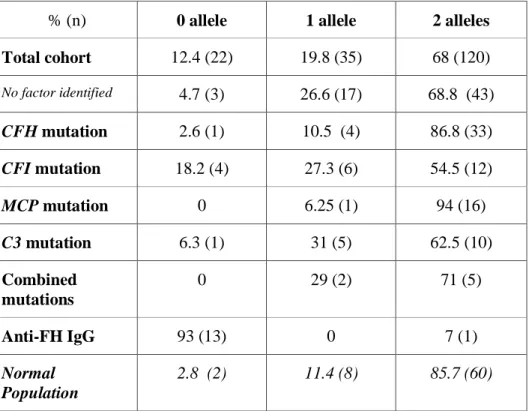
A genomic deletion of CFHR1 was found in 56 (31.6%) patients. This deletion was homozygous in 12.4 % (n=22) or heterozygous in 19.8% (n=35) of patients, as determined by the number of CFHR1 copies (Table 1).
Table 1 : Determination of the number of CFHR1 alleles according the previously identified susceptibility factor in the French aHUS cohort (n=177 patients).
% (n) 0 allele 1 allele 2 alleles Total cohort 12.4 (22) 19.8 (35) 68 (120) No factor identified 4.7 (3) 26.6 (17) 68.8 (43) CFH mutation 2.6 (1) 10.5 (4) 86.8 (33) CFI mutation 18.2 (4) 27.3 (6) 54.5 (12) MCP mutation 0 6.25 (1) 94 (16) C3 mutation 6.3 (1) 31 (5) 62.5 (10) Combined mutations 0 29 (2) 71 (5) Anti-FH IgG 93 (13) 0 7 (1) Normal Population 2.8 (2) 11.4 (8) 85.7 (60)
Thus, the calculated frequency of the deleted allele was 22.7%. In the control population, a genomic deletion was found in ten controls (14.3%, homozygous in two and heterozygous in
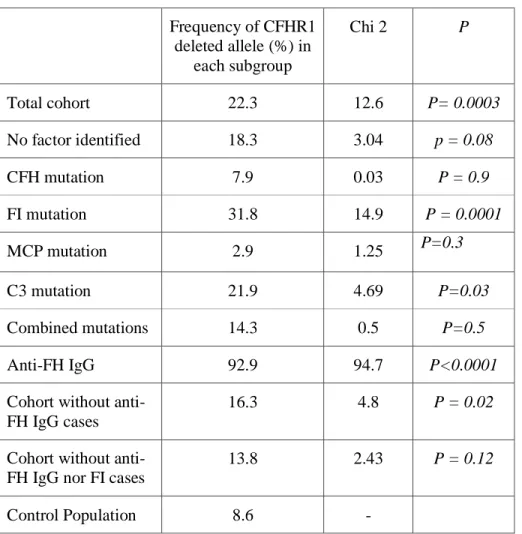
eight), giving an allele frequency of 8.2%. Thus, the CFHR1 deleted allele frequency was significantly higher in aHUS patients as compared to controls (22.7% versus 8.2%, P<0.001). The distribution of the deleted allele in our cohort of aHUS patients, according to the susceptibility factor(s) identified in each patient, is depicted in Table 2.
Table 2 : Frequency of a CFHR1 deleted allele according to the susceptibility factor and comparison of each subgroup with the control population using a χ2
test. Frequency of CFHR1 deleted allele (%) in each subgroup Chi 2 P Total cohort 22.3 12.6 P= 0.0003 No factor identified 18.3 3.04 p = 0.08 CFH mutation 7.9 0.03 P = 0.9 FI mutation 31.8 14.9 P = 0.0001 MCP mutation 2.9 1.25 P=0.3 C3 mutation 21.9 4.69 P=0.03 Combined mutations 14.3 0.5 P=0.5 Anti-FH IgG 92.9 94.7 P<0.0001
Cohort without anti-FH IgG cases
16.3 4.8 P = 0.02
Cohort without anti-FH IgG nor FI cases
13.8 2.43 P = 0.12
Control Population 8.6 -
The frequency of CFHR1 deletion was particularly high in the subgroup of patients presenting with an auto-immune form of aHUS, in which the allele frequency was 92.9% (χ2 = 94.7,
those exhibiting a CFI mutation (31.8 %, P<0.001) or a C3 mutation (21.9%, P=0.03) showed a significantly higher frequency of the deleted allele compared to controls. In the other subgroups, no significant difference was observed compared to controls. In patients in whom no genetic factor was identified, the frequency of the deleted allele was significantly higher than in the control population (18.3%, P=0.02). On the contrary, the frequency of the deleted allele was lower in subgroups of patients exhibiting a genetic abnormality in the CFH and
MCP genes than in the control group (7.9% and 2.9% respectively, versus 8.6% in controls)
but did not confer a significant protective status (OR=0.75, 95% CI= 0.25-2.21, P>0.1) (Figure 2). After exclusion of patients with anti-FH IgG and CFI mutation, the frequency of the CFHR1 deletion in the cohort of aHUS patients was not significantly different from the frequency in the control group.
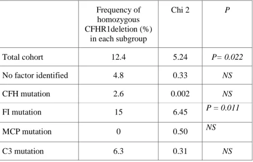
We then hypothesized that CFHR1 deletion homozygosity alone could be a susceptibility factor. Therefore, we studied the distribution of homozygous CFHR1 deletion in the different subgroups of patients according to susceptibility factor(s) (Table 3).
Table 3 : Frequency of a homozygous CFHR1 deletion according to the susceptibility factor and comparison of each subgroup with the control population using a χ2test.
Frequency of homozygous CFHR1deletion (%) in each subgroup Chi 2 P Total cohort 12.4 5.24 P= 0.022 No factor identified 4.8 0.33 NS CFH mutation 2.6 0.002 NS FI mutation 15 6.45 P = 0.011 MCP mutation 0 0.50 NS C3 mutation 6.3 0.31 NS
Combined mutations 0 0.20 NS
Anti-FH IgG 92.9 64.45 P<0.0001
Cohort without anti-FH IgG cases
5.5 0.77 NS
Cohort without anti-FH IgG nor FI cases
3.5 0.07 NS
Control Population 2.9 -
The frequency of homozygous CFHR1 deletion was higher than in the control population only in the subgroup of patients with CFI mutation (15% versus 2.9% in controls, P = 0.011) and in patients exhibiting anti-FH auto-antibodies (92.9% versus 2.9% in controls, P<0.0001). In this last subgroup, 13 of 14 patients exhibited homozygous CFHR1 deletion. The frequency of the deletion homozygosity in the subgroup of patients with no known susceptibility factor was not significantly higher than in controls (5.5% versus 2.9%, OR = 1.96, 95% CI 0.32-11.8, P>0.1) (Figure 2).
We then performed CFHR3 allele quantification by MPLA in the subgroup of patients with anti-FH IgG auto-antibodies. With one exception, a complete absence of the CFHR3 gene was observed in all CFHR1 deletion patients.
DISCUSSION
In our study, the deleted allele’s frequency is 8.2% and 22.7% in the normal and the study population, respectively, with a deletion homozygosity ‘s frequencies of 2.9% and 12.4%. These results are in accordance with the previously reported frequencies in healthy controls and in aHUS patients. The frequency of the CFHR1 homozygous deletion in different control
frequency of homozygous deletion of CFHR1 in 16% and 10.6% of aHUS patients from the Jena and Newcastle cohorts, respectively [4]. In addition, Hughes et al. have reported the presence of the deleted allele in 20% of chromosomes in a normal but elderly population (n=170). The frequency was significantly lower (8%) in an age-matched population (n=173) affected by AMD, suggesting that the CFHR1 deletion confers protection against AMD (OR 0.4 (95%CI,0.3-0.5) [11]
The fact that the CFHR1 deletion occurred at a higher frequency in the subgroups of patients exhibiting a mutation in a gene located outside the RCA locus (CFI, C3) suggests that a defect affecting one gene in the RCA locus might be necessary for the development of the disease or play a role for its severity. These roles remain to be elucidated.
The deletion homozygosity is particularly high in the subgroup of patients exhibiting anti-FH auto-antibodies. This group comprises eleven children (age at disease onset : 7 months –13 years, median : 9 years old, five males and six females) and three adults (median age at disease onset : 28 years old, all males). The deletion affects the CFHR1 and CFHR3 genes. These results confirm that the homozygous deletion of CFHR1 and CFHR3 genes is the genetic mechanism responsible for the absence of circulating CFHR1 and CFHR3 observed in the particular group of patients exhibiting an auto-immune form of aHUS [7].
The particular correlation that exists between the presence of a homozygous deletion of
CFHR1 and CFHR3 genes and the development of anti-FH auto-antibodies reveals that most,
if not all, patients with an “acquired” form of aHUS share the same homozygous genetic polymorphism. This genetic predisposition seems to be necessary but not sufficient for developing the disease, as this homozygous deletion is also observed in controls. The mechanisms responsible for development of auto-antibodies against FH are not yet understood. Our results suggest a particular role of CFHR1 and CFHR3 proteins in the development of this auto-immunity.
In the group of patients without genetic or acquired abnormality affecting the alternative pathway (n = 55), the frequency of the deletion homozygosity is not different than in the control population conferring a non significant risk for the disease. These results suggest that in our study population, the CFHR1 deletion does not represent a susceptibility factor for aHUS by itself.
In conclusion, the high frequency of CFHR1 deletion observed in our aHUS patients is due to the presence of CFHR1 homozygous deletion in only two categories of patients: those with anti-FH auto-antibodies associated HUS and, to a lesser degree, those with a CFI mutation. The results highlight the link between the CFHR1 and CFHR3 homozygous deletion and the development of anti-FH auto-antibodies.
FIGURE LEGEND:
Figure 1 : Risk for developing aHUS associated with CFHR1 deletion homozygosity.
The Odds Ratio values are represented by circles. The risk was significant in the subgroups of patients with anti-FH auto-antibodies and CFI mutation. In 2 groups of patients, no Odds Ratio may be calculated (N/A: no available) as no CFHR1 homozygous deletion was found in these groups.
REFERENCES
1 Jokiranta TS, Zipfel PF, Fremeaux-Bacchi V, Taylor CM, Goodship TJ, Noris M. Where next with atypical hemolytic uremic syndrome? Mol Immunol 2007;44(16):3889-900.
2 Fremeaux-Bacchi V, Miller EC, Liszewski MK, Strain L, Blouin J, Brown AL, Moghal N, Kaplan BS, Weiss RA, Lhotta K, Kapur G, Mattoo T, Nivet H, Wong W, Gie S, Hurault de Ligny B, Fischbach M, Gupta R, Hauhart R, Meunier V, Loirat C, Dragon-Durey MA, Fridman WH, Janssen BJ, Goodship TH, Atkinson JP. Mutations in complement C3 predispose to development of atypical hemolytic uremic syndrome.
Blood 2008.
3 Goicoechea de Jorge E, Harris CL, Esparza-Gordillo J, Carreras L, Arranz EA, Garrido CA, Lopez-Trascasa M, Sanchez-Corral P, Morgan BP, Rodriguez de Cordoba S. Gain-of-function mutations in complement factor B are associated with atypical hemolytic uremic syndrome. Proc Natl Acad Sci U S A 2007;104(1):240-5. 4 Zipfel PF, Edey M, Heinen S, Jozsi M, Richter H, Misselwitz J, Hoppe B, Routledge
D, Strain L, Hughes AE, Goodship JA, Licht C, Goodship TH, Skerka C. Deletion of complement factor H-related genes CFHR1 and CFHR3 is associated with atypical hemolytic uremic syndrome. PLoS Genet 2007;3(3):e41.
5 Venables JP, Strain L, Routledge D, Bourn D, Powell HM, Warwicker P, Diaz-Torres ML, Sampson A, Mead P, Webb M, Pirson Y, Jackson MS, Hughes A, Wood KM,
Goodship JA, Goodship TH. Atypical haemolytic uraemic syndrome associated with a hybrid complement gene. PLoS Med 2006;3(10):e431.
6 Dragon-Durey MA, Loirat C, Cloarec S, Macher MA, Blouin J, Nivet H, Weiss L, Fridman WH, Fremeaux-Bacchi V. Anti-Factor H autoantibodies associated with atypical hemolytic uremic syndrome. J Am Soc Nephrol 2005;16(2):555-63.
7 Jozsi M, Licht C, Strobel S, Zipfel SL, Richter H, Heinen S, Zipfel PF, Skerka C. Factor H autoantibodies in atypical hemolytic uremic syndrome correlate with CFHR1/CFHR3 deficiency. Blood 2008;111(3):1512-4.
8 Fremeaux-Bacchi V, Kemp EJ, Goodship JA, Dragon-Durey MA, Strain L, Loirat C, Deng HW, Goodship TH. The development of atypical haemolytic-uraemic syndrome is influenced by susceptibility factors in factor H and membrane cofactor protein: evidence from two independent cohorts. J Med Genet 2005;42(11):852-6.
9 Blom AM, Bergstrom F, Edey M, Diaz-Torres M, Kavanagh D, Lampe A, Goodship JA, Strain L, Moghal N, McHugh M, Inward C, Tomson C, Fremeaux-Bacchi V, Villoutreix BO, Goodship TH. A Novel Non-Synonymous Polymorphism (p.Arg240His) in C4b-Binding Protein Is Associated with Atypical Hemolytic Uremic Syndrome and Leads to Impaired Alternative Pathway Cofactor Activity. J Immunol 2008;180(9):6385-91.
10 Hageman GS, Hancox LS, Taiber AJ, Gehrs KM, Anderson DH, Johnson LV, Radeke MJ, Kavanagh D, Richards A, Atkinson J, Meri S, Bergeron J, Zernant J, Merriam J, Gold B, Allikmets R, Dean M. Extended haplotypes in the complement factor H (CFH) and CFH-related (CFHR) family of genes protect against age-related macular
degeneration: characterization, ethnic distribution and evolutionary implications. Ann
Med 2006;38(8):592-604.
11 Hughes AE, Orr N, Esfandiary H, Diaz-Torres M, Goodship T, Chakravarthy U. A common CFH haplotype, with deletion of CFHR1 and CFHR3, is associated with lower risk of age-related macular degeneration. Nat Genet 2006;38(10):1173-7.
12 Sellier-Leclerc AL, Fremeaux-Bacchi V, Dragon-Durey MA, Macher MA, Niaudet P, Guest G, Boudailliez B, Bouissou F, Deschenes G, Gie S, Tsimaratos M, Fischbach M, Morin D, Nivet H, Alberti C, Loirat C. Differential impact of complement mutations on clinical characteristics in atypical hemolytic uremic syndrome. J Am Soc
Nephrol 2007;18(8):2392-400.
13 Schouten JP, McElgunn CJ, Waaijer R, Zwijnenburg D, Diepvens F, Pals G. Relative quantification of 40 nucleic acid sequences by multiplex ligation-dependent probe amplification. Nucleic Acids Res 2002;30(12):e57.
14 Fremeaux-Bacchi V, Dragon-Durey MA, Blouin J, Vigneau C, Kuypers D, Boudailliez B, Loirat C, Rondeau E, Fridman WH. Complement factor I: a susceptibility gene for atypical haemolytic uraemic syndrome. J Med Genet 2004;41(6):e84.
15 Dragon-Durey MA, Fremeaux-Bacchi V, Loirat C, Blouin J, Niaudet P, Deschenes G, Coppo P, Herman Fridman W, Weiss L. Heterozygous and homozygous factor H deficiencies associated with hemolytic uremic syndrome or membranoproliferative glomerulonephritis: report and genetic analysis of 16 cases. J Am Soc Nephrol 2004;15(3):787-95.
16 Fremeaux-Bacchi V, Moulton EA, Kavanagh D, Dragon-Durey MA, Blouin J, Caudy A, Arzouk N, Cleper R, Francois M, Guest G, Pourrat J, Seligman R, Fridman WH, Loirat C, Atkinson JP. Genetic and functional analyses of membrane cofactor protein (CD46) mutations in atypical hemolytic uremic syndrome. J Am Soc Nephrol 2006;17(7):2017-25.
17 Fremeaux-Bacchi V, Sanlaville D, Menouer S, Blouin J, Dragon-Durey MA, Fischbach M, Vekemans M, Fridman WH. Unusual clinical severity of complement membrane cofactor protein-associated hemolytic-uremic syndrome and uniparental isodisomy. Am J Kidney Dis 2007;49(2):323-9.
18 Spencer KL, Hauser MA, Olson LM, Schmidt S, Scott WK, Gallins P, Agarwal A, Postel EA, Pericak-Vance MA, Haines JL. Deletion of CFHR3 and CFHR1 genes in age-related macular degeneration. Hum Mol Genet 2008;17(7):971-7.