HAL Id: tel-01231859
https://tel.archives-ouvertes.fr/tel-01231859
Submitted on 21 Nov 2015HAL is a multi-disciplinary open access
archive for the deposit and dissemination of sci-entific research documents, whether they are pub-lished or not. The documents may come from teaching and research institutions in France or abroad, or from public or private research centers.
L’archive ouverte pluridisciplinaire HAL, est destinée au dépôt et à la diffusion de documents scientifiques de niveau recherche, publiés ou non, émanant des établissements d’enseignement et de recherche français ou étrangers, des laboratoires publics ou privés.
Generation Potential : an epidemiological approach
Maria-Ares Rocanin-Arjo
To cite this version:
Maria-Ares Rocanin-Arjo. Genetic and Epigenetic Determinants of Thrombin Generation Potential : an epidemiological approach. Génétique humaine. Université Paris Sud - Paris XI, 2014. Français. �NNT : 2014PA11T067�. �tel-01231859�
UNIVERSITÉ PARIS-SUD
ÉCOLE DOCTORALE 420 :
SANTÉ PUBLIQUE PARIS SUD 11, PARIS DESCARTES
Laboratoire :
Equip 1 de Unité INSERM UMR_S1166
Genomics & Pathophysiology of Cardiovascular Diseases
THÈSE DE DOCTORAT
SANTÉ PUBLIQUE - GÉNÉTIQUE STATISTIQUE
par
Ares ROCAÑIN ARJO
Genetic and Epigenetic Determinants of Thrombin
Generation Potential: an epidemiological approach.
Date de soutenance : 20/11/2014
Composition du jury :
Directeur de thèse : David Alexandre TREGOUET DR, INSERM U1166, Université Paris 6, Jussieu
Rapporteurs : Guy MEYER PU_PH, Service de pneumologie. Hôpital européen Georges Pompidou
Richard REDON DR, Institut thorax, UMR 1087 / CNRS UMR 6291 , Université de Nantes
Examinateurs : Laurent ABEL DR, INSERM U980, Institut Imagine Marie Aline CHARLES DR, INSERM U1018, CESP
Al meu pare
(to my father
/à mon père)
Your genetics load the gun. Your lifestyle pulls the trigger.
Mehmet Oz
Reserve your right to think, for even to think wrongly is better than not to think at all.
Hypatia
If you want to know about thrombin- measure thrombin"
C'est incroyable, mais malgré ce que je craignais au début, ces trois ans ont passé très vite ! Il y a trois ans, déjà, j'ai commencé ma première journée dans l'unité U937 (maintenant UMR_S1166). Tout juste 5 jours avant, j'arrivais avec mes deux valises. Je m’en souviens encore : Moi, attendant le bus 351 à l’extérieur du terminal de CDG. Une dame m'avait alors demandé comment se rendre au centre de Paris. Puis elle m'avait dit: "Je viens pour visiter Paris, vous aussi ?". Et je lui avais répondu : "Non, je viens pour y vivre, je vais faire un doctorat.". Deux heures plus tard et une bonne route à travers les banlieues parisiennes, je débarquais sur la place Gallieni pour retrouver celui qui serait mon colocataire et que je ne connaissais pas encore. Quels premiers jours ! Que d’émotions ! La veille de commencer ma thèse, à l’occasion d’Halloween, j'étais allée au cinéma Le Champo, où j’avais passé la nuit devant 3 films d’horreur. En sortant au petit matin, avec mon premier croissant et mes nouvelles connaissances, nous avions décidé de rentrer à pied à travers les rues de Paris. Quelle belle promenade. Il n’y avait personne et en arrivant à la Seine nous avions découvert Notre Dame illuminée par le soleil levant et toute pour nous. Que Paris est belle !
J’ai aussi des souvenirs de mes premiers jours au labo et notamment des 3-4 alertes incendie que nous avions eues en trois jours. Je me souviens de la première fois que j'ai entendu la sirène des pompiers, en pensant, "C'est grave ? Qu'est-ce qu'il faut faire ? C'est une alerte ? Que font les autres ? Rien. Tranquilles... Donc, ok c'est rien ". Je me rappelle aussi de la petite souris qui habitait dans la cuisine. Maintenant, à la fin de ma thèse, nombre d’expériences et de moments vécus, qui m’ont appris aussi bien professionnellement qu’humainement, se rappellent à moi.
Je tiens d’abord à remercier François Cambien et Laurence Tiret pour m'avoir accueillie tout au début au sein de l'ancienne unité U937 intitulée "Génomique Cardiovasculaire".
Je voudrais aussi remercier l'ensemble du jury d’avoir accepté de consacrer de leur temps pour évaluer ce travail : mes deux rapporteurs, le Professeur Guy Meyer et Monsieur Richard Redon, pour leur relecture profonde et leurs critiques ; je remercie aussi mon examinateur Monsieur Laurent Abel et mon examinatrice et présidente de jury Madame Marie-Aline Charles.
Je remercie le professeur Pierre Emmanuel Morange pour son important apport dans ce travail et pour avoir mis en place l'étude MARTHA, une étude très puissante sans laquelle je n'aurais pu faire ma thèse. De même, je remercie France Gagnon et les members de l'étude des "Three City" pour m'avoir permis d'utiliser des données issues de leur cohorte.
Votre sympathie, votre complicité et votre soutien constant m'ont permis de me sentir intégrée et m’ont donné la sensation d’appartenir à une petite "famille" : Merci aux filles, aux nouvelles Beata et Maguelonne ainsi qu’aux deux Marie-Laure. Merci Carole pour ta joie et ton effort pour que je fasse du sport. Merci Claire pour m’avoir fait découvrir le reblochon et pour m’avoir permis de me maintenir en contact avec la biologie. Vielen danke Ulli (ma belle!!) pour tout: les fêtes, les instants de complicité et pour avoir partagé avec moi tous ces moments. Zahia, pour ta sympathie constante et pour partager avec moi le goût pour le café crème. Grazie mille Veronica pour ton soutien constant. Je vous remercie Ewa, Laurence et Hervé pour nos conversations dans la cuisine. Merci Badreddine pour tes agréables "salut" dans le couloir et "bon appétit" en passant par la cuisine. Merci Nadjim et Lyamine pour être toujours si galants. Merci François (le roux, juif, irlandais,...) pour tes cours de "français des jeunes". Merci Nathalie pour nos discussions politiques et éthiques et pour rendre le monde administratif moins effrayant et plus accessible. Merci Henri, revenu juste pour la fin, pour tes blagues provocatrices et ton ironie. Merci beaucoup à mon compagnon de "fatigue" et bon ami, Dylan (Methylman), qui est toujours dispo pour faire un petit "break" pour se détendre et discuter mais surtout pour m'aider avec les machines. Merci à Maxime même si nous n’avons jamais été en même temps dans l'unité. Merci à Raphaele, Ricardo, Vinhou, Nico, Marie et Guillaume (Boby), pour leur sympathie, leur joie et leur bonne humeur, pour les activités "afterwork chez Jimmy", pour les questions "Nico", pour les "hotdogs chilenos", pour l'hippodrome, pour le cours d'escalade ou le poulet au curry thaï de Vinh! Et aussi merci aux stagiaires (oompa loompas) Lynn, Bathou, Yasmine, Evangelia, Francesca, "Doudou", Pauline, Bastien, pour leurs croissants, Romain qui est passé au coté "compagnon de fatigues" et merci aussi aux canadiens avec qui je garde une amitié précieuse, Martin et Jessica.
Je remercie vivement mes copines de bureau: Marine et Sophie, pour avoir égayé mon quotidien, pour avoir répondu à toutes mes questions, pour votre précieuse aide en relisant ce mémoire, pour m’avoir "supportée" dans les derniers mois difficiles, pour nos moments
Enfin je veux remercier plus particulièrement mon directeur de thèse, David-Alexandre Trégouët pour sa confiance en moi, surtout au début quand il ne me connaissait pas, en m'aidant pour ma première inscription à la faculté, pour la recherche d'appartement et pour les démarches administratives. Merci beaucoup d’avoir accordé ta confiance à une biologiste sans expérience pour réaliser ce projet. Merci pour ton très bon encadrement, pour ton aide en tout moment au cours de ces trois ans et pendant l'écriture de ma thèse. Merci pour avoir été patient avec ma façon de faire (lui très organisé et moi j’ai une tendance au désordre), et pour ton exigence, qui m’a certes valu certains moments difficiles à endurer, mais m’a permis de me surpasser, de m'organiser et d’avoir le travail que je présente ici.
Je vais désormais retourner à mes origines et continuer dans ma langue natale, le Catalan, pour mieux exprimer mes remerciements à mes amis et à ma famille: Sou moltes persones que també heu aportat el vostre granet de sorra i m'heu permès arribar fins aquí.
Tots els meus companys de grups anteriors: Magda, Georgios, Marc, Robert, Esther, Mireia, i Pedro, els primers en ensenyar-me i formar-me com a investigadora i amb els que encara conservo una boníssima amistat. Després, les pipetes Raquel, Sonia, Biel i Miquel Soute, pels nostres dinars, esmorçars, cafès, birres i moments a la cabanya i els tecles Angel, Alfonso, Anna i Leonor que em van començar a mostrar en els "lab-meetings" el món dels bioinformàtics i estadístics. Moltíssimes gràcies, José Manuel Soria, per dornar-me l'oportunitat de fer la tesi a Paris, interessant-te i demanant per beques als teus col·laboradors. Encara recordo el moment en que m'ho vas comunicar: em vas trucar perquè m'havia demanat festa per poder saltar en paracaigudes. Dues emocions fortes en un mateix dia. Recordo com vaig estar pensant el què hauria de fer. Vull reiterar les gràcies a Sonia per la temporada que va estar a Paris, en la qual em va ajudar molt al inici de la meva estada que sempre és dura, 6 mesos on vam compartir converses post dinar, moments explosius de desesperació,fins i tot cases. Em van ajudar molt.
parat, coincidint com podíeu quan era, o ja més tard érem, a Barcelona. Han arribat a convertir-se com en un grup de teràpia per a doctorants amb anècdotes, converses profundes sobre la ciència, sopars, birretes, braves,...les braves són mooolt importants! Moltes Gràcies Cris D per la correcció d'una part del treball, sobretot per les molèsties en fer-ho ràpid.
Moltes gràcies també a les "nenes del cole": Marta, Mireia, Delia, Laura, Patri, Neus i consorts per interessar-vos sempre per com m'anava i pel vostre suport. Sembla mentida que després de 10 anys, quan vàrem deixar l'escola, i dels diferents camins que hem agafat cadascuna, continuem encara juntes! Moltes gràcies també als "frifris", per donar-me suport amb el seu humor constant.
Moltes gràcies a la meva petita família parisina: alguns presents des del principi acollint-me des dels priacollint-mers dies i als que ens hem anat trobat de la manera acollint-menys probable. Vosaltres feu possible viure a Paris amb menys enyorança; Marina, totes les valencianetes, Jacob, Belen, Chantal, Valentine, Andrea, Yaiza, Jordi i Jerôme (français mais entre catalans, merci pour ton bon humeur), gràcies.
Finalment, els més importants per a mi. Moltes gràcies a la meva família pel seu suport en aquests tres anys, interessant-se sempre i venint-me a visitar quan podien. No puc expressar en paraules l'agraïment profund que sento per ma germana, Anaïs, en Guifré (si, ja ets de la família) i ma mare, Rosamari, els quals han fet gests titànics per ajudar-me en tot aquest procés. Venint-me a veure al preu que fos, o traslladant-se a Paris, deixant la preuada mar per venir a viure amb mi. Gràcies també per corregir moltes parts d'aquest treball i per donar-me forces i consell en tot el camí, per estar-hi sempre. Sense ells no crec que hagués arribat tan lluny. El seu recolzament en tot moment ha estat molt valuós i clau en molts moments. Guardo pel final, una persona molt especial per mi, que malauradament ja no és aquí. Ell sempre va tenir la il·lusió de que jo fes un doctorat i per què negar-ho, la seva voluntat ha estat una part important en la empresa d'aquesta tesi. Ell també, sense voler-ho, em va donar el rumb, fent-me interessar per la trombosi i sé que a ell, amant de tot allò francès, li hagués agradat compartir amb mi aquest procés. Moltes gràcies papa!
I. Scientific formation and contribution
Main publications
Ares Rocanin-Arjo, William Cohen, Laure Carcaillon, Corinne Frère, Noémie Saut, Luc Letenneur, Martine Alhenc-Gelas, Anne-Marie Dupuy, Marion Bertrand, Marie-Christine Alessi, Marine Germain, Philipp S. Wild, Tanja Zeller, Francois Cambien, Alison H. Goodall, Philippe Amouyel, Pierre-Yves Scarabin, David-Alexandre Trégouët, Pierre-Emmanuel Morange and and the CardioGenics Consortium. Blood. 2014;123:777-85.
A. Rocanin-Arjo, J. Dennis, P. Suchon, D. Aïssi, V.Truong, D-A. Trégouët, F. Gagnon, P-E. Morange. Thrombin Generation Potential and Whole-Blood DNA methylation. Thrombosis Research.
Collaborations
Aïssi D, Dennis J, Ladouceur M, Truong V, Zwingerman N, Rocanin-Arjo A, et al. Genome-Wide Investigation of DNA Methylation Marks Associated with FV Leiden Mutation. PLoS One 2014;9:e108087. Oral communications
Rocanin-Arjo A, Carcaillon L, Cohen W, Saut N, Germain M, Letenneur L,Alhenc-Gelas M, Dupuy AM, Bertrand M, Amouyel P, Scarabin PY, Trégouët DA, Morange PE. A meta-analysis of genome-wide association studies identifies a novel locus associated with Thrombin generation potential. American Society of Human Genetics Conference 2013 Boston October 22nd-26th.
Rocanin-Arjo A, Carcaillon L, Cohen W, Saut N, Germain M, Letenneur L,Alhenc-Gelas M, Dupuy AM, Bertrand M, Amouyel P, Scarabin PY, Trégouët DA, Morange PE. A meta-analysis of genome-wide association studies identifies a novel locus associated with Thrombin generation potential.CORDDIM.12 septembre.
Poster communications
Rocanin-Arjo A, Carcaillon L, Cohen W, Saut N, Germain M, Letenneur L,Alhenc-Gelas M, Dupuy AM, Bertrand M, Amouyel P, Scarabin PY, Trégouët DA, Morange PE. A meta-analysis of genome-wide association studies identifies a novel locus associated with Thrombin generation potential. European Human Genetics Conference 2013 Paris June 8th-13th
ii
Morange PE. A meta-analysis of genome-wide association studies identifies a novel locus associated with Thrombin generation potential. ICAN conferences 2013 Paris December 12th-14th
Rocañín-Arjó A, Pierre Suchon, Noémie Saut, David-Alexandre Trégouët,Pierre-Emmanuel Morange. Association of Orosomucoid plasma levels with thrombin generation potential and cardiometabolic biomarkers. ICAN 2014 22th September, Paris, France, (Poster).
Courses and formation
"Leena Peltonen Summer School of Human Genomics", Welcome Trust Conference Centre, 18 – 22 August 2013, Hinxton, Cambridgeshire, United Kingdom.
"2nd Non-coding genome", Institute Curie, 10-14 December 2012, Paris, France.
IV. List of principal abbreviations
TGP: Thrombin generation potential.
MARTHA: MARseille THrombosis Analysis Study.
3C: Three City Study.
FII: factor II or prothrombin.
FIIa: active factor II or thrombin.
FVIII: factor VIII(FVIIIa active form).
FVL: factor V Leiden (FVLa active form).
VT: Venous Thrombosis.
vWF: von Willebrand factor.
FDR: False Discovery Rate.
GWAS: GenomeWide Association Study.
EWAS: Epigenetic Wide Association Study.
IBD: Identical By Descent.
MAF: Minor Allele Frequency.
MCMC: Monte-Carlo Markov Chain Method.
QTL: Quantitative Trait Locus.
Résumé en français
Contexte du projet et motivation
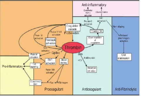
La thrombine, aussi connue sous le nom "facteur de coagulation II", est synthétisée dans le foie sous forme de zymogène: la prothrombine (ou FII), qui, une fois relâchée, est clivée, en exposant ses domaines et en activant ses fonctions (FIIa). Sa fonction la plus connue et la plus importante est celle de de catalyseur de la formation de fibrine qui constituera le caillot, d'activateur d'autres facteurs pro-coagulants mais également de déclencheur d'enzymes anti-coagulantes8.
L'activation de la thrombine est le résultat de la cascade de coagulation à laquelle participent de nombreuses protéines (Tableau 1.1, page 6) s'activant les unes les autres (Figure 1.2., page 7). Le début de la coagulation a lieu quand le facteur tissulaire (TF) est exposé au sang, à cause d'une lésion des vaisseaux. Ensuite TF active le facteur VII (FVIIa, a pour activé) devenant le complexe TF-FVIIa qui active alors deux autres facteurs: le facteur IX et le facteur X. Le FXa seul est peu efficace et ne protéolyse qu’une petite quantité de prothrombine4. Cette petite, mais suffisante, quantité de thrombine activée (2 nmol / L)
commence à interagir avec des cofacteurs et substrats en provoquant le début de réactions en chaîne menant à la coagulation1,3,4,9,15. La thrombine protéolyse le fibrinogène en formant
des unités de fibrine (Figure 1.3., flèche 1, page 10) qui vont être assemblées en fibres à fin de stabiliser le caillot. En outre, elle protéolyse les facteurs V et VIII (Figure 1.3., flèche 4, page 10) qui vont aider à amplifier l'activation de la thrombine. La thrombine va également activer des facteurs anticoagulants qui vont aider à contrôler et finaliser la coagulation.
La thrombine participe également, au-delà de l'hémostase, à d'autres systèmes physiologiques, comme par exemple les systèmes immunitaire, nerveux, gastro-intestinal, et musculo-squelettique. Elle interagit avec des protéines et des récepteurs, en activant les plaquettes et les cellules endothéliales, et en stimulant l'adhésion, l'angiogénèse, la croissance cellulaire, la différenciation, la prolifération, la vasoconstriction et l'inflammation. Tout ce large éventail de fonctions est modulé grâce à une combinaison complexe de substrats et de cofacteurs4 (Tableau 1.1. et 1.2., pages 6-7).
Pour toutes les fonctions expliquées ci-dessus, la régulation de la thrombine est fondamentale pour une physiologie normale. Des niveaux déséquilibrés de thrombine se traduisent par différents types d'anomalies. Les plus connues et étudiées sont la thrombose/l'hémophilie, l'inflammation et l'athérosclérose. Il est alors essentiel de pouvoir
reflètent seulement 5% de l'ensemble du processus1 de l'activation et de l'activité de la
thrombine.
Le potentiel de génération de thrombine (TGP, en anglais) est un test qui a été mis à point pour mesurer la quantité potentielle de thrombine qui est capable d'être activée au moment de coagulation45. Il peut être vu comme un reflet l'ensemble du processus de la
coagulation allant de son l'initiation, sa propagation et sa terminaison-amplification47. Il est
plus sensible aux déficits de facteurs de coagulation: activateurs (FVII, FV, FX) et inhibiteurs (antithrombine, protéine C, protéine S) et à de nombreux troubles de la coagulation associés à une résistance à la protéine C activée (APC, en anglais). En outre, il est sensible à tous les types d'anticoagulants ou d'autres médicaments.
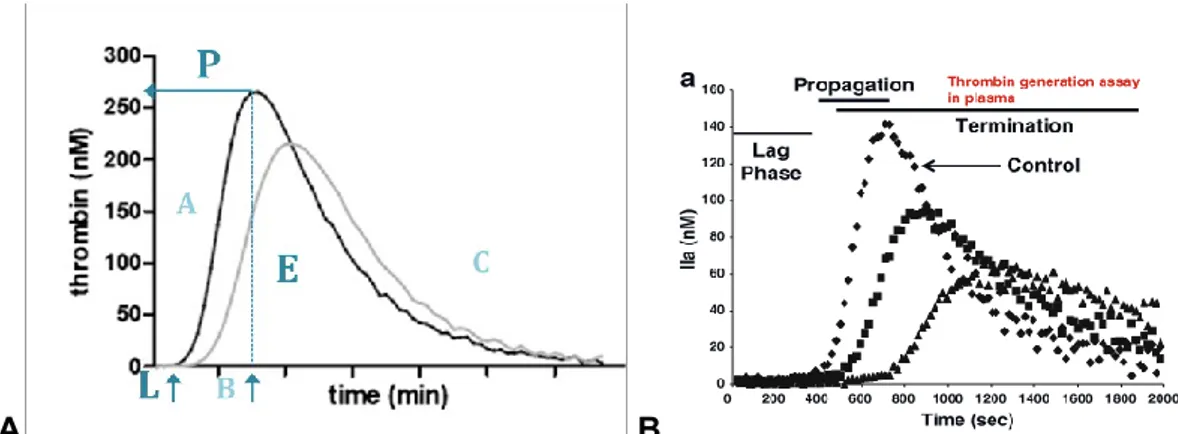
Le TGP est mesuré à partir d'une méthode, communément appelée thrombogramme calibré automatisé (CAT, en anglais), qui consiste en deux mesures simultanées de l'activation de la thrombine avec un substrat fluorochromé pour un même échantillon de plasma. L'une mesure la génération de thrombine (TG) et l'autre sert de calibrage pour corriger le biais entre le signal fluorochrome et l'activation de la thrombine. Dans le test TG, la thrombine est produite dans une réaction de coagulation activée par du facteur tissulaire, des phospholipides et du calcium. La quantité de thrombine générée est alors mesurée en temps réel par la capture du signal de fluorescence qui émet le substrat consommé par la thrombine. Ce signal est capturé et corrigé simultanément pour être affiché sous la forme du courbe (appelée thrombogramme) (Figure 2.1., page 24).
A partir de cette courbe, il est possible d'extraire plusieurs paramètres quantitatifs (Figure 2.2., page 25). Les plus utilisés sont : 1- le temps de latence (Lagtime en anglais) qui mesure le temps écoulé depuis le moment où la coagulation est déclenchée jusqu'à le début de formation du caillot; 2 - le potentiel endogène de thrombine (ETP pour Endogeneous Thrombin Potential en anglais) qui représente la totalité de thrombine activée (l'aire sous la courbe du thrombogramme) et permettant de représenter plus précisément l'état de coagulation et toute l'activée enzymatique autour la génération de la thrombine; 3- le hauteur du pic (Peak, P) qui mesure la quantité maximale de thrombine activée à un moment donné du processus de coagulation.
Le test TGP est sensible aux variations des facteurs FX, FIX, FVII et FVIII, du fibrinogène, des D-dimères (produits de la formation des unités de fibrine à partir de fibrinogène) et de protéines anticoagulantes. Le TGP est également associé à l'indice de masse corporelle (IMC), l'âge et le sexe. En ce qui concerne les facteurs génétiques connus
pour influencer la variabilité interindividuelle du TGP, il en existait deux au moment où je commençais mon projet de thèse: les polymorphismes rs1799963 (20210G> A) et rs3136516 (19911A> G) du gène codant pour la prothrombine (F2).
Des niveaux élevés de TGP, en particulier d' ETP, ont été associés au risque de thrombose veineuse80–82, aux accidents vasculaires cérébraux (AVC) ischémiques aigus223 et
à l'infarctus du myocarde35. En revanche, des niveaux bas d'ETP sont associés à des
troubles de la coagulation46,86 (Figure 2.6.). Le TGP a également été trouvé associé à
diverses désordres cardiométaboliques comme l'athérosclérose87,224, l'obésité60, le diabète
de type 288, la néphropathie diabétique89, et des troubles hépatiques comme la cirrhose225.
Des troubles inflammatoires ont également été associés aux TGP tels que la maladie de Crohn91, la septicémie92 et la drépanocytose93.
Objectifs:
Comme mentionné ci-dessus, seuls deux polymorphismes génétiques, tous les deux situés dans le gène F2, étaient connus pour influencer les taux plasmatiques de TGP. Cependant, ils n'expliquent que 11,3% de la variabilité interindividuelle de ce biomarqueur. Mon projet de thèse avait pour objectifs d'identifier de nouveaux facteurs génétiques et épigénétiques modulant ce phénotype et ses biomarqueurs associés.
Partie I Identification de nouveaux déterminants génétiques contrôlant la variabilité interindividuelle plasmatique du potentiel de génération de thrombine
Dans le cadre de mon projet de thèse, j'ai mené la toute première étude d'association génome-entier (GWAS pour Genome Wide Association Study en anglais) sur les biomarqueurs du TGP (à savoir ETP, Peak et Lagtime), afin d'identifier de nouveaux gènes participants à la variabilité du TGP.
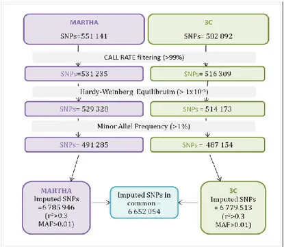
Dans une première étape de découverte, j'ai testé l'association entre 6 652 054 de polymorphismes génétiques, plus précisément des polymorphismes de substitution d'un seul nucléotide (SNP pour Single Nucleotide Polymorphism in anglais), et la variabilité plasmatique des trois biomarqueurs du TGP dans deux échantillons rassemblant 1967 sujets. Outre les deux polymorphismes du gène F2 déjà connus et mentionnés ci-dessus, j'ai identifié un polymorphisme au sein du gène ORM1, rs150611042, montrant une association statistique très forte (p = 3.36 10-7) avec le temps de latence (Lagtime). J'ai ensuite cherché
à répliquer cette association dans deux autres échantillons indépendants rassemblant 1254 sujets. Dans cette deuxième étape de validation, le polymorphisme rs150611042 a été
l'autre de 1374 individus, l'allèle du polymorphisme rs150611042 qui diminuait le temps de latence était également associé à une diminution de l'expression monocytaire du gène ORM1 (p = 8.70 10-10 et p = 5.21 10-16, respectivement). Des expériences fonctionnelles ont
été réalisées pour compléter ce travail et ont confirmé in vitro l'association entre la molécule codée par le gène ORM1 et la génération de thrombine.
En conclusion, cette partie de mon travail de thèse a permis d'identifier un nouveau gène participant à la régulation de la production de thrombine. Les mécanismes précis de cette régulation restent cependant à élucider.
Partie II Marques de méthylation d'ADN associées au potentiel de génération de thrombine
La méthylation de l'ADN est une marque épigénétique qui consiste en l'addition enzymatique d'un groupement méthyle (CH3, une molécule de carbone et trois d'hydrogène) à la position 5 de carbone de la cytosine principalement dans le contexte de la séquence 5'-cytosine-guanine (communément appelé dinucléotide CpG). Ce groupe méthyle est transféré par une ADN méthyltransférase à partir d'une autre molécule appelée S-adenylmethylcisteine (SAM), qui devient une S-adenylhomocysteine (SAH). Dans le même temps, l'ADN méthyltransférase attire des protéines qui vont se lier l'ADN et réguler l'expression et la structure de la chromatine161.
Ces dernières années, de plus en plus d'études ont démontré le rôle de la méthylation de l'ADN dans le développement des maladies humaines. Les premiers résultats ont été observés dans le domaine du cancer, mais étendus rapidement à d'autres maladies humaines complexes telles que la sclérose en plaques, le diabète, les maladies inflammatoires et cardiovasculaires166,175,176,226,227.
Dans la deuxième partie de mon travail de thèse, j'ai utilisé des données de méthylation mesurées dans des échantillons d'ADN sanguin à partir d'une technologie haut-débit, la puce HumanMethylation450K développée par la société Illumina, pour rechercher des marques de méthylation associées à la variabilité plasmatique des mêmes 3 biomarqueurs du TGP que ceux utilisés dans mon projet GWAS (Partie I). Cette puce permet de mesurer les niveaux de méthylation de l'ADN d'environ 480 000 sites CpG répartis tout au long du génome. Son utilisation dans de grandes cohortes épidémiologiques fait l'objet d'un enthousiasme très important en raison des récents succès qu'elle a permis d'obtenir pour détecter des marques
de méthylation associés à des facteurs environnementaux, génétiques et biologiques185,228.
Dans le cadre de mon projet, j'ai eu accès à deux échantillons, l'un de 238 sujets, l'autre de 187, dans lesquels cette puce de méthylation avait été utilisée à partir de l'ADN issu du sang périphérique des sujets, et pour lesquels les biomarqueurs du TGP avaient aussi été mesurés. J'ai ainsi réalisé la première étude de recherche agnostique d'associations entre des niveaux de méthylation de sites CpG et la variabilité plasmatique du TGP. Ce type de recherche est communément appelée MWAS pour Methylome-Wide Association Study en anglais. J'ai suivi une stratégie de recherche assez similaire à celle que j'avais appliquée pour mon étude GWAS. Malheureusement, je n'ai identifié aucune association robuste entre des niveaux de méthylation de l'ADN sanguin et les biomarqueurs de la génération de thrombine.
Conclusions
L'étude GWAS que j'ai menée sur les taux plasmatiques de 3 marqueurs de la génération de thrombine a permis d'identifier un nouveau gène, ORM1, participant à la modulation du temps de latence. Le polymorphisme que j'ai identifié n'est vraisemblablement pas le variant fonctionnel et les mécanismes d'action d'ORM1 sur la génération de thrombine ne sont clairement pas caractérisés. Mon travail n'a donc ouvert qu'une petite porte vers un champ de recherches beaucoup plus vaste qui reste à explorer. D'autres pistes mériteraient d'être également explorées pour mieux disséquer les mécanismes génétiques associés à la génération de thrombine. La recherche que j'ai effectuée ne s'est concentrée que sur des associations univariées entre SNPs et TGP alors qu'il serait également intéressant d'étudier si des phénomènes d'interaction entre SNPs peuvent également contribuer à influencer la génération de thrombine. De plus, alors que je me suis concentrée sur 3 biomarqueurs du TGP, il existe d'autres biomarqueurs qu'il serait également possible d'étudier dans le contexte d'une étude GWAS, ce qui pourrait mener à l'identification d'autres mécanismes de régulation.
L'étude MWAS que j'ai conduite sur les taux plasmatiques de TGP s'est avérée moins fructueuse que l'étude GWAS. Plusieurs explications peuvent être relevées. Tout d'abord, la taille des échantillons que j'ai analysés dans ce projet était relativement modeste par rapport à ceux dont je disposais pour mon étude GWAS. Je ne peux donc pas exclure un manque de puissance pour détecter des effets épigénétiques modérés, et disposer d'autres échantillons mesurés à la fois pour la méthylation de l'ADN et le TGP serait alors le bienvenu. De plus, cette étude MWAS a été réalisée à partir de l'ADN du sang périphérique qui n'est peut-être pas le bon modèle pour étudier des mécanismes de méthylation de l'ADN
épidémiologique, cela semble un peu plus compliqué.
Enfin, j'ai mené de manière indépendante mon étude GWAS et mon étude MWAS sans avoir intégré les résultats de l'une pour augmenter la puissance de l'autre. De nombreux travaux indiquent que les niveaux de méthylation d'un site CpG peuvent également être sous le contrôle génétique de polymorphisme(s). Il serait donc tout à fait intéressant de combiner les résultats que j'ai obtenus dans mes 2 projets pour essayer d'identifier des polymorphismes génétiques influençant les taux de TGP via des mécanismes de régulation de la méthylation d'ADN. Affaire à suivre...
TABLE OF CONTENTS
Acknowledgements ... Scientific contribution ... i List of principal abbreviations ... iii Résumé substantiel en français ... v Table of contents ... xii Index of Figures ... xv Index of Tables ... xviii Introduction ... 1 PROJECT MOTIVATIONS AND BACKGROUND ... 3 Chapter 1 Thrombin ... 5 1.1. Function and Physiology ... 5 1.1.1. Thrombin in Haemostasis ... 7 1.1.2. Thrombin and Inflammation ... 13 1.1.3. Other thrombin functions ... 14 1.2. Related diseases and disorders ... 16 1.3 Thrombin tests and measurements ... 19 Chapter 2. Thrombin generation potential ... 21 2.1 Short history of the thrombin generation measurements ... 21 2.2, Method of measurement: calibrated automated thrombogram
(CAT) ... 22
2.3. The parameters (biomarkers) ... 24 2.4. Known biological, environmental and genetic determinants of
2.5. TGP related diseases ... 28 2.6. Main objectives ... 30 PART I: identification of novel genetic determinants controlling the
inter-individual plasma variability of thrombin generation potential ... 31 Chapter 3. Genome-Wide Association Studies: concepts ... 33 Chapter 4. Genome-Wide Association Studies "for dummies":
step-by-step analysis ... 37
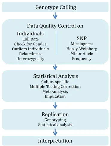
4.1. Genotype calling ... 37 4.2. Data quality control ... 39 4.3. Statistical testing for SNP-phenotype association ... 44 4.4. Correction for multiple testing ... 47 4.5. Meta-analysis of GWAS datasets ... 48 4.6. Imputation analysis ... 50 4.7. Replication of the GWAS findings ... 52 Chapter 5. A "GWAS Study" on Thrombin Generation Potential ... 53 5.1. Discovery GWAS cohorts ... 53 5.2. Replication cohorts ... 57 5.3. Main GWAS findings ... 59 5.4. Replication of GWAS findings ... 63 5.5. Further analysis at the identified ORM1 locus ... 65 5.6. Discussion ... 68 Publication 1 ... 71 PARTII: Investigations of DNA methylation marks associated to the
Thrombin generation potential. ... 81
Chapter 6. DNA methylation, an epigenetic mechanism ... 83 6.1. Histone epigenetic code ... 84
6.3. DNA methylation and human diseases ... 86 6.4. Peripheral blood DNA methylation, a good epidemiological tool ... 86 Chapter 7. How to perform an Methylation Wide Association step by
step ... 87
7.1. Methylation determination ... 87 7.2. Data quality control ... 93 7.3. Bias and corrections ... 99 7.4 Statistical testing for methylation-phenotype associations ... 103 Chapter 8. The MWAS strategy applied to TGP biomarkers ... 104 8.1. Cohort studies ... 104 8.2. Strategy 1 MWAS findings ... 108 8.3. Strategy 2 MWAS findings ... 111 8.4. Further analysis ... 111 8.5. Discussion ... 114 Publication 2 ... 116 GENERAL CONCLUSIONS ... 124 Chapter 9. General discussion, balance and perspectives ... 126 BIBLIOGRAPHY ... 130 Annexes ... 150 Annex 1 ... 152
Index of Figures
Figure 1.1. Image of a vein section where a thrombus is being formed. 8 Figure 1.2. Coagulation cascade and comparison of the two nomenclatures. 9
Figure 1.3. Amplification phase of coagulation. 10
Figure 1.4. Inhibitors of thrombin and its activation. 12 Figure 1.5. The Cross talk between the coagulation, fibrinolysis and inflammation
responses. 14
Figure 1.6. Thrombin physiology functions. 15
Figure 1.7. Arterial versus venous thrombosis and their different possible causes. 17 Figure 1.8. Cross-talk between Atherosclerosis, inflammation and thrombosis. 18 Figure 2.1 Diagram of the CAT method measures for a plasma sample. 24 Figure 2.2. Parameters of Thrombin Generation Potential measured by
Thrombogram. 25
Figure 2.3. Comparison of thrombin generation curves of (left) controls, (right A) an individual with antithrombin deficiency and (right B) an individual with deficiency of protein C.
26
Figure 2.4. Comparison of thrombin generation curves by sex (A), age (B), BMI (C)
and oral contraceptives (D). 27
Figure 2.5. Thrombin generation in females vs males. 28 Figure 2.6. Thrombin generation curves from individuals with different levels of FXI
deficiency. 29
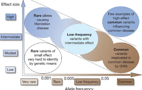
Figure 3.1. Relation between the frequency of the alleles and their effect (or causal
relation) upon the trait. 33
Figure 3.2. Linkage analysis vs Association analysis. 34 Figure 4.1. Diagram of the steps to follow to perform a GWAS. 37 Figure 4.2. Representation of a microarray genotyping using as example Illumina
images. 38
Figure 4.3. Examples of genotype calling results of a SNP. 39 Figure 4.4. Two plots representing the relation between missingness (y-axis) and
mean heterozygosity in a GWAS sample (3C study samples). 41 Figure 4.5. Multidimensional scaling graphics to detect outliers in a group of
samples from Three City Study. 42
Figure 4.6. Graphic representation of a PCA. 43
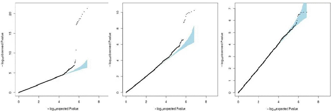
Figure 5.1. Diagram of the filters applied in MARTHA and 3C studies. 54 Figure 5.2. Density distributions of TGP biomarkers. 55 Figure 5.3. Diagram of the analysis procedure of the discovery step. 56 Figure 5.4. Quantile-Quantile plots of the meta-analysis p-values combining
MARTHA and 3C. 59
Figure 5.5: Manhattan plot representing the p-values of the GWAS meta analysis
on ETP. 60
Figure 5.6. Manhattan plot representing the p-values of the meta-analyzed GWAS
for ETP conditioning on the F2 rs1799963. 61
Figure 5.7. Manhattan plot representing the p-values of the GWAS meta analysis
on Peak height. 62
Figure 5.8. Manhattan plot representing the p-values of the GWAS meta analysis
on Lagtime. 62
Figure 5.9. Association of ORM1 rs150611042 with ORM1 monocyte and
macrophage expression. 67
Figure 5.10. Correlation between ORM1 plasma levels and Lagtime in a sample of
10 healthy individuals. 67
Figure 5.11. Diagram of the position of the rs150611042 SNP. 69 Figure 6.1. Image of the two main epigenetic types. 83
Figure 6.2. Scheme of histone cores. 84
Figure 6.3. Methylation process. 85
Figure 6.4. DNA methylation tissue specificity. 85
Figure 7.1. Methylation-wide association study step by step. 87 Figure 7.2. Different methods of DNA methylation measurement. 88
Figure 7.3. Methylation determination protocol. 89
Figure 7.4. Example of the Infinium I probes display on the microarray for 3
methylation sites. 90
Figure 7.5. Scheme of the functioning of Infinium I (Illumina images). 90 Figure 7.6. Scheme of the functioning of Infinium II (Illumina images). 91 Figure 7.7. Density distribution of the intensity signals obtained from the Illumina
methylation microarray. 91
Figure 7.8. Distribution of β values. 92
Figure 7.9. Density distribution of M value. 93
Figure 7.11. Density destribution of the Negative control probes. 95
Figure 7.12. MDS of the 577 individuals. 98
Figure 7.13. Green intensity of Y chromosome probes in females (left) and males
(right). 98
Figure 7.14. Negative controls and out-of-band intensities. 100 Figure 7.15. Distribution of the mean β before (a) and after (b) correction with
SWAN. 101
Figure 7.16. Distribution of M value of the Infinium II probe intensities before (a)
and (b) after correction. 102
Figure 7.17. Distribution of β of each one of the 7 plates. 103 Figure 8.1. Diagram of the filters used for the quality control of the probes. 105 Figure 8.2. Distribution of TGP biomarkers in MARTHA (purple) and F5L pedigree
study (green). 107
Figure 8.3. Quantile-Quantile graphics. 108
Figure 8.4. Manhattan plots from the MWAS results observed in MARTHA. 109 Figure 8.5. Quantile-Quantile graphics from the Meta analysis. 111 Figure 8.6. Manhattan plots of the results from the meta-analysis of the MARTHA
Table 1.1. Thrombin actions and substrates 6 Table 1.2. Interactions where thrombin is the substrate 7 Table 1.3. List of the principal coagulation factors. 9 Table 5.1. Characteristics of the studied populations 55 Table 5.2. Characteristics of the MARTHA12 and FITENAT populations. 58 Table 5.3. Association of ORM1 rs150611042 with TGP biomarkers in four
independent studies 64
Table 5.4. Association of RPL7AP69 rs55724737 with TGP biomarkers . 65 Table 5.5. Genotype frequencies of ORM1 rs150611042 depending on the
type of VT in MARTHA and MARTHA12 66
Table 7.1. Types and number of control probes. 94
Table 8.1. Population characteristics 106
Table 8.2. CpG sites showing evidence for association at p <10-4 with both
ETP and Peak in the MARTHA MWAS. 110
Table 8.3. Replication in the F5L pedigree study of the Table II.XX results
Introduction
Thrombin is a crucial protein that is mostly known for its central role in the haemostasis but that also takes part in a wide variety of other physiological functions, such as inflammatory response, cell differentiation and proliferation, vasoconstriction and cell adhesion. Anomalies in its activity, due to either lost or excess of function, can onset different diseases. Thrombosis is one of the most important ones able to trigger myocardial infarction, atherosclerosis, inflammation or stroke. To study any disease, it is highly recommended to have access to a proper, reliable and easy method to measure and detect as many phenotypic and symptomatic shades as possible in order to use it in routine clinical practice. In case of thrombin-related anomalies, the used procedures are composite measurements of coagulation protein concentrations, of platelet counts and of function/activity assays (e.g., time to clot formation). However, the majority of those methods are not specifically measuring thrombin or they get a reduced information of its activity ("scratch the surface") because they do not measure the total amount of thrombin activated in the coagulation reaction.
Recently, a measure called Thrombin Generation Potential (TGP) has been proposed to get a much closer representation of the in vivo thrombin activation all along the coagulation response of an individual. This measure brings the opportunity to better investigate the process of thrombin activation in plasma. TGP has already been used for studying thrombin-associated disorders in human and these works have confirmed that this measure is sensible to the already known thrombin-associated environmental and genetic factors. Being such a solid method, it is interesting to further study new factors that might influence thrombin generation and that could be later used in clinical diagnosis or prevention of diseases.
To contextualize my research, I will first start reviewing what is known about the thrombin protein, its functions and implications in human physiology. As it is the protein that is assessed by the TGP method, I want to stress its importance in human (also in animal) physiology and, hence, supporting the interest of the main subject of my PhD project: the identification of new genetic and epigenetic factors influencing the inter-individual plasma variability of TGP. I will also introduce some description about the technical method used for measuring TGP and summarize the main discoveries and works carried out so far on this subject. All along this document, I will refer to TGP as the measurement used for
investigating thrombin generation in a given individual. Hence, I might also define it as a phenotype because it characterizes a trait or state of the physiology of that individual.
After introducing these general descriptions, my work will be divided in two main parts: first, I will focus on the search for new genetic factors associated to TGP I have conducted in the framework of a genome-wide association study; second, I will analyze epigenetic data with the aim of looking for DNA methylation patterns associated to the TGP inter-individual variability in plasma.
The result of these works constitute my PhD project entitled: Genetic and Epigenetic Determinants of Thrombin Generation Potential: an epidemiological approach.
MOTIVATIONS
AND
Chapter 1. Thrombin
‘The living enzyme of my blood’ is how Walter Seegers, one of the pioneers studying thrombin, defined this protein ten years ago1.
Thrombin, also known as coagulation factor II, is a 36000 Da serine protease composed of two chains: a light one (or A) and a heavy one (or B); unified by a covalent disulfide bound. His precursor, the zymogen Prothrombin (or FII), is synthesized in the liver and, once released, it is cleaved, exposing the domains and activating its functions (FIIa). Like others clotting factors, prothrombin is part of the vitamin K dependent family and is characterized by a γ-carboxyglutamic acid domain (Gla-domain) that depends on the union of the vitamin K and calcium (calcium-binding domain). Both molecules allow prothrombin to easily anchor into the cell membrane where it is functionally more active2,3. Other
important domains for thrombin activities are the Sodium-binding site, and the exosites I and II3–5. The first is essential to modulate the ambivalent main functions. In normal
physiological conditions, 60% of thrombin has this site occupied by Na+ making it more efficient to interact with pro-coagulation factors. On the other hand, without Na+, thrombin is more anti-coagulant. The two other thrombin epitopes are the binding sites of cofactors and proteins. In a general manner, the exosite I binds to fibrinogen, factor V, factor VIII, factor FXI, PAR1, thrombomodulin and protein C; while exosite II binds to heparin, glycoproteins and other cofactors that make thrombin more efficient in its functions4,6,7 (Table 1.1.).
1.1. The functions of thrombin
The most fundamental and studied function of thrombin is his central and pivotal role in haemostasis: catalyzing the formation of Fibrin that will form the thrombus (or fibrin clot), activating other pro-coagulant factors and also triggering anti-coagulation enzymes8.
Thrombin also participates, beyond the haemostasis, in other physiological mechanisms , such as the immune, nervous, gastrointestinal, and musculoskeletal systems. Thrombin interacts with proteins and receptors, activating platelets and endothelial cells, and stimulating cell adhesion, angiogenesis, cell growth and differentiation, proliferation, vasoconstriction and inflammation. All this wide variety of
functions is modulated by a complex combination of substrates, cofactors and their plasma levels4 (Table 1.1. and 1.2.).
Table 1.1. Thrombin actions and substrates3–5. Thrombin's substrate Cofactor
Thrombin-Domain Function Na
++/Ca++
Fibrinogen - exosite I Activation of fibrinogen -> fibrin +
FVIII - exosite I & II Activation of FVIII -> FVIIIa +
FV - exosite I & II Activation of FV -> FVa +
FXIII fibrin exosite I Plus fibrin activation of FXIII ->
FXIIIa = stabilization of fibrin + FXI GpIbα exosite I Activation of FXI -> FXIa =
activation of intrinsic pathway + Thrombin activatable
fibrinolysis inhibitor (TAFI) TM
Activation of TAFI=TAFIa->
anti-fibrinolysis Glycoprotein V GpIbα_exositeII exosite I Activation of platelets
Thrombomodulin (TM) exosite I Physical inhibition to
procoagulant actions -
Protein C (PC) TM (exosite I and
sometimes II) exosite I
Activation of PC resulting in: Inactivation of Va ->Vi and VIIIa->VIIIi
-
Heparin cofactor II (HCII) glycosaminoglycans Acts like antithrombin
plasminogen activator
inhibitor (PAI-1)
Inhibition of plasmin formation;
fibrinolysis Glycoprotein GpIb-IX-V
platelet receptor complex exosite II
Cofactor. Helps to cleavage PAR and glycoprotein V by thrombin, FXI,.
protease activator inhibitor (PAR) receptor present in
almost all cell types
exosite I
Healing tissue injuries
Activates platelet activation
factor (PAF) Cell growth and differentiation
Smooth muscle, macrophage and endothelial cell
proliferation Angiogenesis Pro-inflammatory Leukocyte adhesion Lymphocytes mitogenesis
Activation: IL-6, IL-8
Vasoconstriction and vascular
procoagulant Anticoagulant
Activation thrombin Thrombin-PAR functions
Table 1.2. Interactions where thrombin is the substrate. Enzyme Cofactor
Thrombin-Domain Function
FX -
First activator of thrombin from prothrombin. Exposure active
domains
FVa-FXa complex -
Activation of thrombin from prothrombin. Exposure active
domains
Thrombomodulin (TM) exosite I Cofactor
Antithrombin (AT) Non or glycosaminoglycans: heparin, heparan sulfate proteoglycans-(exosite II) -
Without cofactor: AT inhibits thrombin reversibly. With a cofactor is irreversible. Also Inactivates of Xa
-> Xi and IXa->Ixi; Heparin cofactor II (HCII) glycosaminoglycans Acts like AT
Tissue factor pathway
inhibitor (TFPI) Fibrinolysis-anticoagulant
1.1.1.
Thrombin in Haemostasis
Haemostasis is the main physiological regulatory mechanism of the blood flow and of its integrity. It includes both the formation of the thrombus (i.e coagulation) and its
dissolution (i.e fibrinolysis) 9. The dynamics of the coagulation can be explained in three
phases10.
1) The initiation of coagulation. 2) The propagation and amplification. 3) The termination.
This last step and the fibrinolysis are both the other pan of the haemostasis balance, dissolving the formed thrombus. Thrombin is key for haemostasis balance due to its paradoxical main functions. First, it triggers the formation of clots when the vessels are
damaged and then helps to stop the process to avoid their progress into the normal vasculature4.
Figure 1.1. Image of a vein section where a thrombus is being formed9. The
collagen and the tissue factor are components of the vessel wall located under the first layer of cells (endothelium). When the vessel is injured, collagen (yellow arrows) and tissue factor (blue arrows) will get in the blood flow and trigger the coagulation.
1.1.1.1. Blood coagulation: Initiation. Platelet plug formation
At the beginning, when a vessel is damaged, collagen and von Willebrand factor become exposed to the blood (Figure 1.1.) and attract the platelets to the injured point starting to build the clot. Both collagen and vWF activate platelets through glycoprotein (GP) membrane receptors such as the GPIIb-V-IX and GP1bα respectively4,9, and that
causes i.e. the releasing of P-selectins in the cell surface to help the cell-cell adhesion11.
This step is called primary haemostasis. But this first plug of platelets is unstable and needs the fibrin fibbers to consolidate it.
On the surface membrane of these first aggregated platelets takes place the thrombin production and then its activity will contribute to transform fibrinogen into fibrin4. That is why
the anchoring of thrombin to the cell surface through the Gla-domain and calcium binding domain is very important (page 5). Thrombin activation is the result of the coagulation cascade, a mechanism very efficient where many proteins are involved (Table 1.3.) and ones activate the following others (Figure 1.2.). The cascade can also be described based on two pathways: the intrinsic and the extrinsic. At the beginning, they were considered as equal part of the initiation process of coagulation. Nowadays, it is suggested that the coagulation initiation step would correspond to the extrinsic pathway and the intrinsic one would be part of the amplification and prolongation12 (Figure 1.2.).
The coagulation cascade is triggered by the tissue factor (TF), which is a membrane receptor. It is normally present in extravascular tissues but when it is exposed to blood, meaning there is some kind of vessel damage, the coagulation starts4. Its first action is to
activate factor VII (FVIIa, activated) becoming the TF-FVIIa complex that then activates two other factors: factor IX and factor X. The extrinsic and intrinsic pathways both lead to the
activation of FX, reason why FX is established as the connection between the two pathways and thereafter the common coagulation pathway starts13. FXa alone is scarcely
efficient and only proteolyses a small amount of prothrombin to its active state thrombin, plus other secondary fragments: fragment F1+2 and meizothrombin4. Together, the
meizothrombin and thrombin join to adrenergic receptors in the nearby smooth muscle cells resulting in a vasoconstriction of the area of clotting helping to stop the bleeding7.
Table 1.3 List of the principal coagulation factors14.
Figure 1.2. Coagulation cascade (Adapted from Esmon 201315) and comparison of
the two nomenclatures. The two different pathways, extrinsic and intrinsic, allow
thrombin activation. Then, with dot-lined square (left) the initiation phase, which starts with the presence of tissue factor in the blood (possibly a vessel damage). Afterwards, with the apparition of the first amount of thrombin, the amplification phase (the right dot-lined square) is activated and helps to amplify its own activation (positive feedback).
1.1.1.2. Blood coagulation: Propagation and amplification
Once that small, but sufficient, amount of prothrombin is activated (2nmol/L), thrombin starts to interact with cofactors and substrates causing the burst of coagulation1,3,4,9,15. In
this part of the process, the presence of sodium ions (Na++) is essential, as it will give to
thrombin the suitable allosteric conformation to be bound easily with pro-coagulant substrates. Besides, calcium, also called coagulation factor IV, is important for the activity of thrombin and different proteic complexes.
The most important thrombin's substrate is fibrinogen that is proteolysed to form fibrin units (Figure 1.3., arrow 1). These units are assembled in fibers that tangled the platelets and other necessary cells and molecules, and all together help to fix and stabilize the clot. Factor XIII is also activated (FXIIIa) by thrombin (Figure 1.3., arrow 2) and it acts cross-linking the fibrin fibers and helping to form the net of the thrombus (Figure 1.3., arrow 3).
Furthermore, thrombin proteolyses the factors V and VIII (Figure 1.3., arrows 4) that are both important to ameliorate the efficiency of the factors Xa and IXa, respectively. FVIIIa with FIXa form the IXa-VIIIa complex (tenasa complex) that activates more factor Xa. This conjugation makes FIXa 105-106 folds more active (Figure 1.3., arrow 5). Then, FXa
combined with factor Va, become the Xa-Va complex (the prothrombinase complex). This complex also increases by 3·105 folds the thrombin activation1,16 (Figure 1.3., arrow 6).
Figure 1.3. Amplification phase of coagulation. Scheme of the main thrombin activations
to form fibrin and to increase its own formation (the burst). The 1st, 2nd and 3rd arrows are implicated in the fibrin formation; and the others arrows in the coagulation amplification.
Thrombin also activates factor XI, which becomes FXIa (Figure 1.3., arrow 7) helped by FXIIa whose activation in vivo is still controversial9,17. This part of the cascade is equivalent
to the intrinsic pathway (Figure 1.2.), also known as the contact pathway (or even kallikrein/kinin system) because FXII in vitro is activated by the negative charges present in the surface of sampling tubes8. Once FXI is activated into FXIa, it triggers the coagulation
activating even more FIXa (Figure 1.3., arrow 8). From here starts the common pathway again. FIXa complexes with FVIIIa to boost the activation of FXa and together activate more FXa (Figure 1.2., and 1.3.). This intrinsic pathway is very important to amplify the coagulation and the density of the fibrin. Deficiencies (mild or total) of FXI produce a very variable bleeding disease also called Haemophilia C18. Instead, FXII deficiencies are not
associated to bleeding disorders, which makes it interesting as a possible anti-coagulation treatment17
.
Concurrently with all the reactions described above, thrombin activates more platelets and endothelial cells allowing increased cell aggregation by P-selectin and mobilization of more receptors to anchor more prothrombin3.
1.1.1.3. Blood coagulation: Termination
A tightly and accurate regulation of coagulation is very important to ensure that it do not pass beyond to a physiologically normal vessel. Antithrombin, tissue factor pathway inhibitor (TFPI) and protein C are the main proteins controlling the amounts of thrombin generated. In this situation, a lack of sodium ions gives thrombin the ability to attract more anti-coagulant factors and cofactors.
There are two kinds of inhibitors: the indirect and the direct (Figure 1.4.). Indirect inhibitors
The indirect inhibitors are those that cleave o inactivate the necessary proteins for the thrombin activation. These include TFPI, protein Z, ADAMTS13 and thrombomodulin.
TFPI is one of the most important and principal inhibitors of the coagulation19 with big
affinity for TF-FVIIa and FXa (Figure 1.3.). It is widely distributed in healthy arteries, in the endothelial cells, in smooth muscular cells and in macrophages, and is colocalized with TF8. It only takes place in early stages of the coagulation and TFPI inactivates the TF-VIIa
complex and also the factor Xa (FXa) forming a TF-VIIa-Xa-TFPI complex. The latter blocks the formation of the efficient prothrombinase complex (FXa-FVa). TFPI has also been proposed to inhibit FXa, when it is complexed with protein S20. A dysfunction of TFPI
protein in the system is related to risk of thrombosis and disseminated intravascular coagulation19.
Figure 1.4. Inhibitors of thrombin and its activation11. In the two panels are
represented some of the coagulation inhibitors: antithrombin (AT), Heparin, tissue factor pathway inhibitor (TFPI), the Z-Z protein inhibitor (Z-ZPI), and protein C (PCI or APC).
As an activator of thrombin, FXa is also the target of other inhibitor proteins such as antithrombin (see below) and serpin PZ-dependent protease inhibitor. PZ corresponds to protein Z, which is a member of the vitamin K-dependent family (FII, VII, IX, X, protein C and protein S) but has no proteolytic activity. However, it acts as a cofactor of the serpin PZ-dependent protein (Z-ZPI, or SERPINA)11.
ADAMTS13 also acts as an indirect inhibitor in very early stages of coagulation. ADAMTS13 overbreaks von Willebrand factor and makes impossible its interaction with platelets, hence avoiding their aggregation. Mutations in vWF or ADAMTS13 genes that can modify the efficiency of ADAMT13 for easily tagging vWF are responsible for bleeding disorders known as von Willebrand disease (vWD).
Finally, thrombomodulin (TM) is a protein located in the endothelium surface that connects to thrombin as a cofactor by the exosite I and anchors thrombin to the cell membranes. Therefore, it helps and increases the probability of union between thrombin (now T-TM) and the anticoagulant protein C (PC). PC also bound to endothelial cell protein C receptor (EPCR), making it easier the interaction4. The complex T-TM activates protein C
(APC) and once liberated from its membrane receptor, APC can interact with its cofactor protein S, and together inactivate the factors Va and VIIIa to Vi and VIIIi (i for inactivate).
Direct inhibitors
The direct inhibitors are those who bind "directly" to free activated thrombin and inactivate it. These include thrombomodulin, again, together with antithrombin, heparin cofactor II and alpha 2 macroglobulin.
Thrombomodulin (TM) is additionally considered as direct inhibitor because it interacts with thrombin by the same domain as fibrinogen does, reducing the free thrombin. Hence, it is a physical inhibitor, avoiding the activation of fibrin, platelets and endothelial cells.
Antithrombin (AT) inactivates thrombin by cleaving it. This action can be reversible if AT acts alone but if thrombin is bound to a glycosaminoglycan molecule, for instance heparin, the attraction between AT and thrombin increases and the inactivation is irreversible. AT also inhibits the coagulation factors IXa, Xa and XIa stopping the thrombin generation. From the same AT family, heparin cofactor II (HCII) acts similarly binding to thrombin when there are glycosaminoglycans acting as cofactors.
Finally, alpha2 macroglobulin (a2M) is not really a specific thrombin inhibitor but better known for its relation with Alzheimer disease. It is a blood protease inhibitor that bounds to free thrombin that avoids thrombin functions21.
1.1.1.4. Thrombin and (Anti-) Fibrinolysis
Part of the haemostasis, the fibrinolysis is the process that dissolves the thrombus. It starts few days after the formation of the thrombus. One of the main proteins in this process is plasmin, which breaks the fibrin net connections. Plasmin is activated by tissue
plasminogen activator (t-PA) from its precursor, the plasminogen.
As already mentioned, thrombin also activates FXIII helping to stabilize the thrombus. Additionally, when thrombin is bound to thrombomodulin, it activates thrombin activatable fibrinolysis inhibitor (TAFI) that inhibits the t-PA, ergo TAFI inhibits the fibrinolysis3,11,22.
However, when the complex thrombin-TM activates APC, it also promotes the fibrinolysis because APC inhibits the plasminogen activator inhibitor (PAI-1, SERPINE1). Thus it helps to produce plasmin, and that results with the dissolution of fibrin3.
1.1.2. Thrombin and Inflammation
Inflammation is part of the protective mechanisms of the body as a response of the immunity system to pathogens. The relation between thrombin and inflammation is very
tight, is a cross-talk where they activate each other (Figure 1.5.). Inflammation and sepsis are defence mechanisms against bacterial spreading and lead their destruction.
Thrombin activates platelets by interacting with the protease-activated receptors (PAR). This stimulates the G-protein pathway which can result in the production of chemokines (as monocyte chemoattractant protein-1) that will attract leukocytes on platelets and endothelial cells. At the same time, platelets express protein P-selectin, CD40 ligands and binding receptors favoring the aggregation of those leukocytes. These cell-cell interactions produce the releasing of cytokines such as Interleukin-6 and 8 (IL-6, IL-8), tumour necrosis factor-α (TNFα) and growth factors (as vascular endothelial cell GF) by monocytes and endothelial cells. Thrombin also proteolyses complement components, e.g. activating C3 and C5. All these reactions together result in a inflammatory response11 by thrombin and normally, it
can be stopped by the action of anticoagulant factors such TFPI, APC and AT and also TAFI11,23.
Figure 1.5. The Cross talk between the coagulation, fibrinolysis and inflammation responses4.
1.1.3. Other thrombin functions
Since the discovery of PAR (PAR 1,3 and 4) in 1991 by Coughlin et al.24, thrombin
started to be considered more than a coagulation protein. These receptors are membrane proteins present in many cell types not only from the vascular and immune systems but also from nervous, gastrointestinal, and musculoskeletal systems (Figure 1.6.).
On the vascular cells, the thrombin-PAR (T-PAR) stimulus activates the protein G resulting in angiogenesis, cell growth and differentiation, and smooth muscle cell, macrophage and endothelial cell proliferation. T-PAR stimulates different growth factors
such as connective tissue growth factor (GTGF) and vascular endothelial GF (VEGF). GTGF stimulates the fibroblast mitogenesis and chemotaxis that produce procollagen and fibronectin3. VEGF affects the endothelial cells inducing cellular migration, endothelial cell
proliferation, and vascular tube formation promoting angiogenesis. These responses permit to heal and repair the damaged tissue of the vessel. Besides, VEGF also promotes the generation of more thrombin.
Figure 1.6. Thrombin physiology functions25.
Additionally, T-PAR interaction stimulates the secretion of nitric oxide (NO). On one hand, NO produces vasoconstriction at the site of the injury contributing to stop the loss of blood. On the other hand, it leads to vasodilation of the normal endothelium to permit the blood flow beyond the injury3.
In the musculoskeletal system, T-PAR promotes osteoblasts, myoblasts and chondrocytes proliferation, which might be important in cases of bone injuries and fractures. Thrombin also has been found highly expressed in neonatals and embryos having important influences in muscle denervation and during brain development3.
Neurons, astrocytes and olygodendrocytes express also PAR and release thrombin. Their interaction leads to brain development, synaptic plasticity and neuroprotection3,26.
Every cell that express PAR, could be stimulated by thrombin, therefore there could be still many thrombin actions to investigate. Although PAR can be activated by other proteases (e.g. FVIIa or FXa), thrombin seems to be the most efficient one3.
1.2. Related diseases and disorders
For all the functions explained above, a careful thrombin regulation is fundamental for normal physiology and unbalanced levels of thrombin result in different kind of disorders. The most known and studied are: thrombosis, haemophilia, inflammation and atherosclerosis.
1.2.1. Thrombosis and haemophilia
Regarding the main functions of thrombin, two diseases are related to excess or deficiency of thrombin: thrombosis and haemophilia (respectively).
Thrombosis is a very critical disorder that consists in a tendency to form thrombus, increasing the risk of vessel obstruction and interruption of the blood flow. There are two kinds of thrombosis: arterial and venous (Figure 1.7.). The arterial thrombosis is characterized by an excess of platelet activation, generally due to the rupture of an atherosclerotic plaque, whereas venous thrombosis is more related to an excess of activation of the clotting factors and/or decrease of the blood flow27.
Therefore, all factors that increase the normal amount of thrombin in plasma may increase the risk of arterial and venous thrombosis when mutated8. Some of the most well
known factors are blood group and the gene mutations: prothrombin G20210A or FV Leiden. However, the risk can be also caused by: up-regulation of FVII, FX, FV, FVIII, FXI, FXIII, TAFI and logically FII; and/or down-regulation of PC, PS, TM, AT, TFPI among others19,22,28,29. Total deficiencies in these latter proteins are either incompatible with life or