HAL Id: hal-02682518
https://hal.inrae.fr/hal-02682518
Submitted on 1 Jun 2020
HAL is a multi-disciplinary open access
archive for the deposit and dissemination of
sci-entific research documents, whether they are
pub-lished or not. The documents may come from
teaching and research institutions in France or
abroad, or from public or private research centers.
L’archive ouverte pluridisciplinaire HAL, est
destinée au dépôt et à la diffusion de documents
scientifiques de niveau recherche, publiés ou non,
émanant des établissements d’enseignement et de
recherche français ou étrangers, des laboratoires
publics ou privés.
Copyright
Expression of prion protein increases cellular copper
binding and antioxidant enzyme activities but not
copper delivery
Walid Rachidi, Didier Vilette, Pascale Guiraud, Marie Arlotto, Jacqueline
Riondel, Hubert Laude, Sylvain Lehmann, Alain Favier
To cite this version:
Walid Rachidi, Didier Vilette, Pascale Guiraud, Marie Arlotto, Jacqueline Riondel, et al.. Expression
of prion protein increases cellular copper binding and antioxidant enzyme activities but not copper
delivery. Journal of Biological Chemistry, American Society for Biochemistry and Molecular Biology,
2003, 278 (11), pp.9064-9072. �10.1074/jbc.M211830200�. �hal-02682518�
Expression of Prion Protein Increases Cellular Copper Binding and
Antioxidant Enzyme Activities but Not Copper Delivery*
Received for publication, November 20, 2002 Published, JBC Papers in Press, December 23, 2002, DOI 10.1074/jbc.M211830200 Walid Rachidi‡§, Didier Vilette¶, Pascale Guiraud‡, Marie Arlotto储, Jacqueline Riondel‡,
Hubert Laude¶, Sylvain Lehmann储, and Alain Favier**
From the ‡Laboratoire de Biologie du Stress Oxydant (LBSO), Faculte´ de Pharmacie, Domaine de La Merci, 38706 La Tronche-Grenoble cedex 9, France, the¶Unite´ de Virologie Immunologie Mole´culaires,
Institut National de la Recherche Agronomique (INRA), 78350 Jouy-en Josas, France, the储Institut de Ge´ne´tique Humaine, CNRS U.P.R. 1142, 141, rue de la Cardonille, 34396 Montpellier Cedex 5, France, and the **Laboratoire des Le´sions des Acides Nucle´iques, UMR CNRS/CEA/UJF, 5046, Avenue des Martyrs, 38000 Grenoble, France
The N-terminal region of the prion protein PrPC
con-tains a series of octapeptide repeats. This region has been implicated in the binding of divalent metal ions, particularly copper. PrPChas been suggested to be
in-volved in copper transport and metabolism and in cell defense mechanisms against oxidative insult, possibly through the regulation of the intracellular CuZn super-oxide dismutase activity (CuZn-SOD) or a SOD-like ac-tivity of PrPCitself. However, up to now the link
be-tween PrPCexpression and copper metabolism or SOD
activity has still to be formally established; particularly because conflicting results have been obtained in vivo. In this study, we report a link between PrPC, copper
binding, and resistance to oxidative stress. Radioactive copper (64Cu) was used at a physiological concentration
to demonstrate that binding of copper to the outer plasma cell membrane is related to the level of PrPC
expression in a cell line expressing a doxycycline-induc-ible murine PrPC gene. Cellular PIPLC pretreatment
indicated that PrPCwas not involved in copper delivery
at physiological concentrations. We also demonstrated that murine PrPCexpression increases several
antioxi-dant enzyme activities and glutathione levels. Prion protein may be a stress sensor sensitive to copper and able to initiate, following copper binding, a signal trans-duction process acting on the antioxidant systems to improve cell defenses.
Prion diseases form a group of fatal neurodegenerative dis-orders including Creutzfeldt-Jakob diseases, Gerstmann-Stra¨ussler Syndrome, Kuru and Fatal Familial Insomnia in humans, and scrapie and bovine spongiform encephalopathy in animals (1). All these disorders are characterized by the accu-mulation of an abnormally folded isoform of the cellular prion
protein PrPC,1denoted PrPSc, which represents the major
com-ponent of infectious prion diseases (2). The formation of PrPSc
from PrPCis accompanied by profound changes in structure
and biochemical properties. PrPCrich in ␣-helical regions is
converted into a molecule with highly-sheeted structures and
partial resistance to proteolytic digestion (2, 3). The conversion
of PrPCinto PrPScremains enigmatic. Biosynthesis of PrPCis
necessary for PrPScformation, as mice lacking PrPCare
resist-ant to scrapie infection (4).
Human PrPChas 253 amino acids and is mainly expressed
on neurons (5, 6). In its N-terminal region, a repeated sequence of five octapeptides can be found, which was shown to bind copper and zinc (7–9). The protein may have some superoxide dismutase-like activity and therefore a possible protective function against oxidative stress (10). Wild-type mouse brains have a significantly higher level of membrane-associated
cop-per than PrPC-deficient mice. Treatment with
phosphatidyl-inositol phospholipase C (PIPLC) specifically reduced the cop-per content from wild type mice but had no effect on the copcop-per
content of PrPCknockout mice (8). However, these results have
not been confirmed (11). Incorporation of radioactive-labeled copper into CuZn-SOD was found to be proportional to the level of PrP expression (12). Pattison and Jebbett (13) noticed more than 30 years ago the similarity between prion histopathology to the histopathology induced by a copper chelator, cuprizone. The incidence of chronic Wasting disease (CWD), a sporadic prion disease of deer and elk, was observed to be higher in regions where the soil had a low copper content (14). Therefore, prion diseases may be related to an alteration of copper trans-port and a loss of copper-enzyme activities.
In a previous work, we demonstrated that neuronal cells infected with prion strains resulted in an alteration of the molecular mechanism promoting cellular resistance to ROS (15). The same alteration of antioxidant enzymes was shown in infected animals (16, 17). In the present study, we used a transfected transgenic cell line with a doxycycline-inducible
murine PrPCgene to investigate the involvement of PrPCin
copper metabolism and in the resistance mechanism to toxic stress.
EXPERIMENTAL PROCEDURES
Cell Culture and Construction—Murine PrPC was cloned in the
pTRE plasmid (Clontech), and the resulting plasmid was transfected by the LipofectAMINE method (Invitrogen) into rabbit kidney epithelial cells (RK13) (18, 19). Stable transfectants were selected in the presence of puromycin (1g/ml), and one (A74) was amplified for further study. RK13 and A74 cells were grown at 37 °C in a 5% CO2-enriched
atmo-* This work was supported by the European Community QRT-2000-02353. The costs of publication of this article were defrayed in part by the payment of page charges. This article must therefore be hereby marked “advertisement” in accordance with 18 U.S.C. Section 1734 solely to indicate this fact.
§ Supported by le centre evian pour l’eau. To whom correspondence should be addressed: Laboratoire de Biologie du Stress Oxydant (LBSO), Faculte´ de Pharmacie, Domaine de la Merci, 38706 la Tronche, France. Tel.: 33-4-76-63-74-56; Fax: 33-4-76-63-74-85; E-mail: walid. rachidi@ujf-grenoble.fr.
1The abbreviations used are: PrPC, cellular isoform of prion protein;
SOD, superoxide dismutase; dox, doxycycline; PIPLC, phosphoinositol phospholipase C; ROS, reactive oxygen species; GPX, glutathione per-oxidase; GR, glutathione reductase; MDA, malondialdehyde acid; SIN-1, 3-morpholinosydnonimine; PrPSc, scrapie isoform of prion
protein; MTT, (4,5-dimethyl-thiazol-2-yl)-2,5-diphenyl-tetrazolium bro-mide; TBARS, thiobarbituric acid reactants.
THEJOURNAL OFBIOLOGICALCHEMISTRY Vol. 278, No. 11, Issue of March 14, pp. 9064 –9072, 2003 © 2003 by The American Society for Biochemistry and Molecular Biology, Inc. Printed in U.S.A.
This paper is available on line at http://www.jbc.org
9064
at INRA Institut National de la Recherche Agronomique on June 15, 2018
http://www.jbc.org/
sphere in minimal essential medium supplemented with 10% heat-inactivated fetal calf serum and were usually split at one-fourth dilu-tion each week.
Immunofluorescence and Western Blot
Analysis—Immunofluores-cence analysis on living A74 cells was performed at 4 °C, with rabbit polyclonal antibody P45– 66, raised against synthetic peptide encom-passing mouse PrPC(MoPrP) residues 45– 66. Fluorescein-conjugated
IgG was used as second antibody.
For Western blot analysis, confluent cells were washed twice with cold phosphate-buffered saline, calcium- and magnesium-free, and ly-sed for 30 min at 4 °C in Triton-deoxycholate lysis buffer (1⫻ buffer is 150 mMNaCl, 0.5% Triton X-100, 0.5% sodium deoxycholate, and 50 mM
Tris-HCl, pH 7.5) plus protease inhibitors. After 1 min of centrifugation at 10,000⫻ g, the supernatant was collected, and its protein concen-tration was measured by the BCA assay (Pierce). The equivalent of 20 g of total protein in SDS loading buffer was subjected to 12% SDS-PAGE electrophoresis followed by electroblotting on polyvinylidene di-fluoride in Tris-glycine buffer containing 20% methanol. The membrane was blocked with 5% nonfat dry milk in TBST (0.1% Tween 20, 100 mM
NaCl, 10 mMTris-HCL, pH 7.8) for 1 h at room temperature, and MoPrP was detected by immunoblotting with P45– 66 antibody as pre-viously described (20). After adding the second antibody (horseradish peroxidase-coupled rabbit IgG), immunoreactive proteins were detected with ECL Western blot system. Quantification was achieved by densi-tometric scanning.
PrPCanalysis in culture medium was immunodetected by Western
blot. To release cell surface PrPC, cultures were treated with PIPLC (0.2
units/ml) in opti-MEM serum-free medium at 37 °C for 2 h. Proteins were precipitated from the PIPLC incubation medium with at least 4 volumes of methanol at⫺20 °C, collected by centrifugation, and immu-noblotted with P45⫺66 antibody using ECL visualization.
Cellular 64Cu Binding—Cells (RK13 and A74) were cultured in
35-mm Petri dishes. Culture medium was replaced by 2 ml of fresh complete medium containing different concentrations of dox (0 –500 ng/ml) to stimulate murine PrPCexpression in A74 cells, and 1.6
M
64Cu (CIS biointernational, Gif-sur-Yvette, France; specific activity 20
mCi/mg) to evaluate copper binding to cells as a function of the level of murine PrPC expression. Non-transfected RK13 cells were used as
control and treated under the same conditions. Cells were incubated at 37 °C under 5% CO2. The radioactive medium was removed after 30 – 40
min, 2, 4, 8, 10, 24, and 26 h. Cells were rinsed twice with 2 ml of diluted Puck’s saline A solution (Invitrogen), and harvested after addition of 1 ml of 0.25% trypsin. After harvesting, each dish was rinsed with 1 ml of Puck’s saline A solution. The final 2 ml obtained for each dish were counted for 2 min using a Packard Cobra III, mono well gamma counter (Packard Instrument Company, Meriden, CT). Protein content was assayed with the BCA protein assay reagent kit. Data were analyzed using a “self made” computer half-life calculation program, to obtain results asCi of64
Cu incorporated or retained per mg of protein.
Cellular Copper Determination—Stimulated (500 ng/ml dox for 24 h)
or unstimulated A74 cells were cultured in the presence or absence of 100MCuSO4for 1 h. For intracellular copper determination, cells
were trypsinized, washed three times in Ca/Mg-free phosphate-buffered saline, and lysed by three cycles of freeze-thawing. Lysates (total ex-tract) were then centrifuged at 13,000 rpm for 10 min to obtain the soluble fraction. Copper concentration was determined by electrother-mal atomic absorption spectrophotometry (PerkinElmer Life Sciences). Their levels were normalized to the protein content, measured with a protein assay kit.
Cell Viability Assay—Cell viability was determined by a modified
3-(4,5-dimethyl-thiazol-2-yl)-2,5-diphenyl-tetrazolium bromide (MTT) assay (21). Briefly 3000 cells per well were plated in 96-well microtiter plates with 100l of complete medium. The next day, the medium was changed, and the cells were challenged for 24 h with different drugs. The medium was then changed, and the cells were incubated for an additional 24 h without drugs. For the MTT assay, 10l of MTT (5 mg/ml stock in phosphate-buffered saline) were added to each well for 3 h at 37 °C. 100l of dimethyl sulfoxide (Me2SO) were added to
dissolve the formazan crystals, and plates were shaken for 5 min on a plate shaker to ensure adequate solubilization. The absorbance read-ings for each well were performed at 570 nm using Multiscan ascent plate reader (Labsystems). The absorbance is proportional to viable cell number, and survival was calculated as the percentage of the staining values of untreated cultures.
Lipid Peroxidation—Lipid peroxidation was evaluated using an
as-say based on fluorescence of thiobarbituric acid reactants measured after extraction with n-butyl alcohol (22). Subconfluent cells were trypsinized in 75-cm2flasks, washed three times by 10 ml of isotonic,
trace element-free Tris-HCl buffer (400 mM, pH 7.3), and then lysed in hypotonic Tris-HCl buffer (20 mM) by five freeze-defrost cycles. 750l of
a mixture of thiobarbituric acid at 8 g/7% perchloric acid (2:1) were added to a 100-l sample. After agitation the mixture was placed in a 95 °C water bath for 60 min and then cooled in an ice bath. The fluorescent compound was extracted by mixing with n-butyl alcohol for 2 min. After centrifugation the fluorescence in the n-butyl alchohol phase was determined with an Aminco-Bowman fluorimeter (PerkinElmer Life Sciences) with excitation at 532 nm and emission at 553 nm. A blank was run for each sample. The calibration curve was created with a stock solution of 1,1,3,3-tetraethoxypropane prepared in alcohol. The results were expressed as TBARS,mol/g of protein.
Superoxide Dismutase Activity—For SOD activity, subconfluent cells
in 75-cm2flasks, were washed three times and collected in 10 ml of
isotonic, trace element-free Tris-HCl buffer (400 mM, pH 7.3), and lysed in hypotonic Tris-HCl buffer (20 mM) by five freeze-defrost cycles. After
10 min of centrifugation at 4000 rpm, 4 °C, the lysate was assayed for metalloenzyme activities and soluble protein content. Total SOD, Mn-SOD, and CuZn-SOD were determined using the pyrogallol assay fol-lowing the procedure described by Marklund and Marklund (23), based on the competition between pyrogallol oxidation by superoxide radicals and superoxide dismutation by SOD, and spectrophotometrically read at 420 nm. Briefly, 50l of the sample were added to 1870 l of Tris (50 mM)-DTPA (1 mM)-cacodylic acid buffer, pH 8.3 and to 80l of
pyrogal-lol (10 mM) in order to induce an absorbance change of 0.02 in the absence of SOD. The amount of SOD inhibiting the reaction rate by 50% in the given assay conditions was defined as one SOD unit. The specific CuZn-SOD inhibition by KCN (60l of KCN, 54 mM) added to 300l of
lysate allowed the Mn-SOD determination under the same conditions. Each sample was assayed twice, and results were expressed as SOD units and normalized to the cell protein content.
Analysis of Glutathione-dependent Antioxidant System—For the
de-termination of total glutathione levels, confluent cells in 25-cm2flasks
were washed three times in phosphate-buffered saline and collected in 10 ml of isotonic, trace element-free Tris-HCl buffer (400 mM, pH 7.3), and lysed in hypotonic Tris-HCl buffer (20 mM) by five freeze-defrost
cycles. Samples of whole lysate were deproteinized by adding metaphos-phoric acid (6%) (lysate-metaphosmetaphos-phoric 5:1, v/v). After 10 min at 4 °C the solutions were spun at 4000 rpm for 10 °C at 4 °C, and the super-natants were assayed for total glutathione content according to the Akerboom and Sies method (24).
The glutathione peroxidase (GPX) activity was assayed by the method of Gunzler et al. (25). GPX was measured in a coupled reaction with glutathione reductase (GR), using tert-butylhydroperoxide as sub-strate. Briefly, 25l of the sample were added to 900 l of Tris (50 mM), EDTA, sodium azide buffer, pH 7.6 (azide was included in the assay mixture to inhibit interference of catalase) and 20l of glutathione (0.15M), 20l of glutathione reductase (200 units/ml), 20 l of NADPH2
(8.4 mM) in order and incubated for 1 min for mixture equilibrium. Then 20l of tert-butylhydroperoxide were added, and the decrease of the absorbance was monitored at 340 nm for 200 s. The difference in absorbance per minute was used to calculate the enzyme activity, and results were expressed as GPX units/g of protein.
GR activity was determined by following the oxidation of NADPH to NADP⫹ during the reduction of oxidized glutathione (GSSG) (26). The main reagent was prepared by combining 18 ml of KH2PO4(139 mM),
EDTA (0.76 mM), pH 7.4, and 2 ml of NADPH2(2.5 mM). 20l of sample
were added with 220l of the main reagent, and then 30 l of GSSG (22 mM) plus 10l of KH2PO4were added to start the reaction; the
absorb-ance was followed at 340 nm for 175 s. The difference in absorbabsorb-ance per min was used to calculate the activity of the enzyme. The results were expressed as glutathione reductase units/g of protein.
RESULTS
Doxycycline-inducible Expression of Murine PrP in A74 Cells—We used the tetracycline-inducible (tet-on) system (18,
27) to achieve regulated high-level expression of the murine
PrPC. After transfection of several cell lines, a strong inducible
expression of murine PrPCwas obtained in most of the clones
derived from a rabbit kidney epithelial cell line (RK13). Data obtained with a representative clone (A74) are presented in
this article. Expression of murine PrPC was related to dox
concentration in the culture medium, detectable at 10 ng/ml dox and reaching a maximum at 500 ng/ml of dox (Fig. 1). No
PrPCwas detected in either unstimulated A74 cells (Fig. 1) or
non-transfected RK13 (data not shown), confirming that
ex-Prion Expression Increases PrP
Copper Binding
9065
at INRA Institut National de la Recherche Agronomique on June 15, 2018
http://www.jbc.org/
pression of endogenous, rabbit PrP was undetectable in these
cells (18). We also studied the induction kinetics of PrPC
ex-pression in A74 cells stimulated with 500 ng/ml dox.
Expres-sion of PrPCcan be detected 8 h after induction, and a plateau
was obtained at 24 h (Fig. 2). Not all, although up to 32% of
cells, produced PrPCat a high level and expressed it on the
outer face of the plasma membrane (Fig. 3).
64Cu Binding Is Correlated to PrPCExpression—No signifi-cant difference in copper binding was observed during the first
4 h following induction of PrPC expression (Fig. 4A). This
apparent lack of effect might be due to the lack of detectable
PrPC 8 h following addition of dox (Fig. 2). Then, the 64Cu
binding increased proportionately to dox concentration. 26 h following the addition of 500 ng/ml of dox, copper binding was 2.7-fold higher in stimulated cells compared with the
unstimu-lated cells. This increase was due to PrPCexpression and not to
an effect of dox on copper binding because dox had no effect in non-transfected RK13 control cells (Fig. 4B).
The copper binding curve obtained with the non-transfected control cells RK13 was similar to that obtained with unstimu-lated A74 (Fig. 4, A and B). This corresponds to the copper uptake via the classical transport systems (CTR1 and other potential transporters), while the increase observed in dox-stimulated A74 corresponds to the binding or transport activity
of PrPC.
To study the influence of PrPCcleavage on copper binding,
the cells were treated for 2 h with 0.2 units/ml PIPLC at 37 °C before measuring copper binding. PIPLC pretreatment
dra-matically decreases64Cu binding in stimulated A74 cells (500
ng/ml dox) but had no effect on unstimulated cells (Fig. 4C). However, there was a small difference in the kinetics of copper binding between stimulated cells treated with PIPLC and un-stimulated cells presumably because PIPLC might not have
cleaved all PrPCfrom membranes. PIPLC pretreatment had no
effect on stimulated or unstimulated non-transfected RK13 cell controls (Fig. 4D).
PrPCBinds Copper at the Outer Side of the Cell Membrane at Physiological Concentrations—To investigate the location of
bound copper and check for a real entry, cells were rinsed twice with 2 ml of diluted Puck’s saline A solution (Invitrogen) 24 h
after PrPCinduction with 500 ng/ml of dox and incubated in a
radioactive medium containing64Cu. Treatment with PIPLC
decreased the amount of PrPC in stimulated cells (Fig. 5A)
concomitantly with a high decrease in cell-associated
radioac-tivity (Fig. 5C). Immunoblotting demonstrated that PrPCwas
found only in the medium when stimulated cells (500 ng/ml
dox) were treated with PIPLC (Fig. 5B). The release of PrPC
was correlated with a high increase of radioactivity in the medium (Fig. 5D). These findings indicate that PrP binds cop-per at the outer side of the cell membrane and that cleavage of
PrPCliberates copper into the medium. The difference in
cop-per binding between stimulated and unstimulated cells was abolished after treatment with PIPLC indicating that no
cop-per had been taken up by the PrPC. Therefore, PrPCdoes not
transport copper inside the cell at physiological concentrations. These results are confirmed by measuring cellular copper
con-FIG. 1. PrPC
induction is doxycycline-dependent. Dox was
added at different concentrations (0, 10, 25, 50, 100, 500, and 1000 ng/ml) to medium for 24 h, and PrPCexpression was determined in A74
cells by Western blot. The equivalent of 20g of protein (as determined with the BCA protein assay kit) were loaded to a 12% polyacrylamide gel, transferred onto polyvinylidene difluoride membrane, and PrPC
was detected with antibody P45– 66 raised against the N terminus of the protein. PrPCexpression reaches a maximum at 500 ng/ml of dox;
after this concentration we have a plateau. Specific murine PrPC
bands were quantified by densitometry and plotted as a percentage of maxi-mum signal of PrPCexpression in A74 cells. Molecular mass markers
are indicated on the left in kDa.
FIG. 2. Time course of PrPC
expression in A74-stimulated cells.
500 ng/ml of dox was added at time 0, and cells were harvested and lysed at different times (0, 1, 2, 4, 6, 8, 10, 24, and 48 h). 20g of protein were Western blotted and analyzed with the same antibody P45– 65. Expression of PrPC
can be detected 8 h after induction with a plateau at 24 h. Specific murine PrPCbands were quantified by densitometry and
plotted as a percentage of maximum signal of PrPCexpression in A74
cells. Molecular mass markers are indicated on the left in kDa.
FIG. 3. A74 cells expressed PrPC
on the cell membrane. A,
fluorescence microscopy photography of stimulated (500 ng/ml dox for 24 h) or unstimulated A74 cells. Fluorescein-conjugated IgG were used as second antibody. B, phase contrast microscopy for the same cells. 32% of stimulated cells produced PrPC
at the outer side of cell membrane.
Prion Expression Increases PrP
CCopper Binding
9066
at INRA Institut National de la Recherche Agronomique on June 15, 2018
http://www.jbc.org/
tent by electrothermal atomic absorption spectrophotometry.
Copper content was increased up to⬃2-fold in the total but not
the soluble fraction of stimulated cells (see Table II). Thus, at
physiological concentrations PrPC did not transport copper
from the extracellular medium to cytoplasm since there was no
difference in64Cu content between the cytosolic fractions of
stimulated and unstimulated cells.
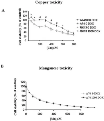
Effect of PrPCExpression on Transition Metal Toxicity—As
induction of PrPCexpression did not increase the incorporation
of physiological levels of copper, we decided to investigate
whether PrPC-dependent bound copper could play a role in the
protection of cells toward copper toxicity. Since it was sug-gested that manganese can compete with copper for the binding sites (28), the resistance to this metal was also tested. The data presented in Fig. 6, A and B clearly show that cells
overex-pressing PrPC (1000 dox) withstood higher copper (but not
manganese doses, Fig. 6B) than unstimulated (0 dox) cells or stimulated RK13 cells. This resistance to copper was more
related to stimulated cells, with an LC50 of 540 and 341M
CuSO4 when they were compared with unstimulated cells.
Therefore PrP increases cellular resistance to copper but not to manganese toxicity.
PrPCOverexpression Increases Resistance to Oxidative Stress and Antioxidant Enzyme Activities—Because it has been
sug-gested that one of the physiological functions of PrPCcould be
in the protection of cells toward an oxidative stress (10), we investigated both the resistance to an oxidative stress and the
activities or levels of the main antioxidant systems in cells
overexpressing PrPC. To study the relationship between prion
protein expression and resistance to oxidative stress, MTT assays were performed following 3-morpholinosydnoni-mine (SIN-1) treatments, which generates different free
radi-cals: O2., NO䡠, and/or NOx, and other potent oxidants such as
ONOO⫺ (46). Stimulated cells (500 ng/ml dox) presented
higher resistance (cell viability, 95%) to this oxidative stress when compared with unstimulated cells (0 dox) (cell viability, 59%) or control cells (RK13 0 or 500 dox) (Fig. 7A). In contrast,
cells overexpressing PrPCwere surprisingly more susceptible
to hydrogen peroxide than unstimulated cells. Treatment for
3 h with different concentrations of H2O2 induced a more
se-vere decrease in viability for doses exceeding 200M(Fig. 7B)
in stimulated cells as compared with unstimulated cells. At 500
MH2O2-stimulated cells revealed⬃50% lower viability than
unstimulated cells. These data demonstrate that PrPC
expres-sion decreases resistance to peroxide toxicity.
We also evaluated the involvement of PrPCexpression in the
cellular defense against oxidative stress by measuring different antioxidant activities such as SOD, GPX, GR, and glutathione
levels. Induction of PrP increases total SOD (⬃21%),
CuZn-SOD (⬃27%), GR (⬃64%) activities, and GSH levels (⬃78%),
while the activities of GPX and mitochondrial Mn-SOD remain unchanged (see Table I). Interestingly, Western blot detection of CuZn-SOD in A74 cells indicates that the total level of this protein was unchanged in stimulated and unstimulated cells
FIG. 4. Relationship between PrPCexpression and64Cu binding. A,64Cu binding increases in proportion to PrPCexpression in A74 cells.
0.1g of64Cu was added to A74 cell culture medium containing different concentrations of dox (0, 25, 100, and 500 ng/ml). Cells were incubated
for 0.6, 2, 4, 8, 10, 24, and 26 h in radioactive media and harvested, and64Cu binding was measured. Copper binding was correlated to murine PrPC
expression. B, dox treatment did not change copper binding in non-transfected control cells RK13. 0.1g of64Cu was added to RK13 cell culture
medium containing 0 or 500 ng/ml of dox. Cells were incubated for 0.75, 10, and 24 h in radioactive media, harvested, and64Cu binding was
measured and normalized to protein cell lysates. C, the experiments described above were repeated, but cells were treated with 0.2 units/ml PIPLC before measuring radioactivity in stimulated and unstimulated A74 cells (C) or in stimulated and unstimulated non-transfected RK13 cells (D). Controls were at times (2.5, 10, 18, and 24 h). Copper binding was quantitated and normalized to the protein of cell lysates. Data represent the mean of three experiments⫾ S.D.
Prion Expression Increases PrP
Copper Binding
9067
at INRA Institut National de la Recherche Agronomique on June 15, 2018
http://www.jbc.org/
(Fig. 8). This may reflect an increase in CuZn-SOD activity of stimulated cells resulting from increased copper incorporation into SOD or from SOD-like activity of PrP-copper complexes. Finally to detect if PrP expression decreases oxidative damage, lipid peroxidation was evaluated by measuring the formation of TBARS as a stress biomarker. The basal level of oxidative damage was significantly lower in stimulated cells as compared with unstimulated and control cells. These data indicate that
PrPC expression increases resistance to basal as well as
in-duced oxidative stress by increasing cellular defenses.
DISCUSSION
In this study, we used a cellular model derived from a heter-ologous epithelial cell line (RK13) in which the expression of
murine PrPCwas regulatable in a dose-dependent manner by a
doxycycline treatment. Actually most epithelial cell lines we have
tested, unlike, RK13, do express PrP.2 The RK13 cells were
chosen because they express no detectable levels of endogenous PrP. The risk that endogenous PrP could interfere with the function of transfected PrP is therefore reduced. This may be a reason why we succeeded in a previous study to infect RK13 cells transfected with ovine PrP (18). It was then logical to generate a clone of cells overexpressing murine PrP, which is used for cell biology and transmission studies. RK13 cells are the only avail-able cell lines allowing a PrP expression from zero to high levels.
In the present work we used radioactive copper (64Cu) to study
the effect of PrPCexpression on copper binding and uptake.
There is an increasing amount of data supporting a
func-tional role for PrPCin copper metabolism. First the N-terminal
half of PrPCcontains five or six highly conserved octapeptide
tandem motifs of the general form PHGGGWGQ, which are capable of binding copper ions with micromolar affinity (7, 8).
Indeed, PrPCisolated from hamster brain can bind a copper
affinity column (29). Second, copper content of
membrane-en-riched brain extract from PrP⫺/⫺mice is 10 –15-fold lower than
in wild type controls while no significant difference was ob-served for other metals (8). Third, neuronal CuZn-SOD from
PrP⫺/⫺ mice showed decreased activity linked to decreased
copper incorporation by the enzyme (10, 30). Neurons cultured
from PrP⫺/⫺mice were also more sensitive to oxidative stress,
perhaps because of the alteration of CuZn-SOD (10). However,
the significance of copper binding on PrPCfunctions or the role
of PrPC copper metabolism has yet to be clarified. Several
hypotheses have been proposed. Copper could have a role in the
conformation of the protein (31). PrPC could have a role in
copper transport across the cell membrane, and this could follow different processes. Copper needs to be mobilized from its extracellular ligands (albumin and histidine), and Cu(II) is reduced to Cu(I) by unknown metalloreductases at the mem-brane surface prior to its delivery across the plasma memmem-brane by the high affinity transporter CTR1 (32). These processes are still unclear. It is also possible that copper may be transported by more than one transport system, at least in some tissues.
The cooperative copper-binding mode of PrPCwithin the
phys-iological concentration range suggests a role in copper
trans-port (33). So, the contribution of PrPC, if any, in copper uptake
2F. Archer and H. Laude, unpublished data.
FIG. 5. Effect of PIPLC treatment on stimulated and unstimulated A74 cells. A, after 24 h of PrPCinduction with 500 ng/ml dox in
radioactive media, cells were treated with 0.2 units/ml PIPLC for 2 h at 37 °C, lysed in lysis buffer and loaded onto a 12% polyacrylamide gel, transferred onto polyvinylidene difluoride membrane, and PrPCwas detected with antibody P45-66 raised against the N terminus of the protein.
B, proteins in culture were precipitated with 4 volumes of methanol, and PrPCwas detected with P45-66 antibody. C, 24 h after PrPCinduction
with or without 500 ng/ml of dox in a radioactive medium containing64Cu, cells were treated with PIPLC (0.2 units/ml) for 2 h at 37 °C, and
radioactivity was measured. D, radioactivity was measured in the opti-MEM medium of the same cells and normalized to protein cell lysates and protein in the media. Results are expressed as the mean of three experiments⫾ S.D. of radioactivity (Ci of64Cu/mg of protein). *, p⬍ 0.01; #,
p⬍ 0.005.
Prion Expression Increases PrP
CCopper Binding
9068
at INRA Institut National de la Recherche Agronomique on June 15, 2018
http://www.jbc.org/
by cells could be a direct transport across the plasma mem-brane or a binding step allowing either the mobilization of copper from its ligand or its reduction prior to its effective transport by other systems.
PrPC is normally attached to the cell membrane via a
phosphatidylinositol anchor (34). It has been shown that the
enzyme PIPLC releases PrPCfrom the cells into media (35).
Our ex vivo experiments confirm the copper binding activity
of the PrPC protein, because we established a correlation
between copper binding and PrPC expression. This finding
was further confirmed when PrPC was cleaved with PIPLC
prior to64Cu labeling. However, our work does not support
that PrPCcould be involved in the copper transport across the
membrane, as suggested by studies reporting
histidine-de-pendent uptake of 67Cu proportional to PrPC expression in
cerebellar cells derived from three lines of mice expressing
various amounts of PrPC (36). However, Pauly and Harris
(37) have reported that copper stimulates endocytosis of both mouse PrP and chicken PrP on the cell surface of N2a mouse neuroblastoma cells via clathrin-coated pits. They suggested
that PrPCcould serve as a recycling receptor for the uptake of
copper ions from the extracellular milieu. Also, it has been
shown that 100Mcopper resulted in the rapid endocytosis of
biotinylated murin PrPCexpressed in human neuroblastoma
SH-SY5Y cells (38). In these two studies the minimum
con-centration of CuSO4 required to produce an observable
in-crease in PrPCinternalization was⬃100
M, which is 15-fold
greater than the estimated Kdfor binding to synthetic PrP
peptides and recombinant PrP (7, 9, 39). In our work we used very low levels of copper, which we believe renders our re-sults much closer to physiological conditions. In any case
when high concentrations of copper (100M) are used in our
cultures the intracellular copper level increased up to 2-fold in both total and soluble fractions in stimulated cells (see
Table II). So, only under high copper concentrations PrPC
expression increases copper uptake in the cell. However, our results clearly demonstrated that at physiological
concentra-tions of copper, murine PrPCbinds copper at the outer side of
the cell membrane but also indicates that PrPC does not
function as a copper transporter from the extracellular me-dium to the cytoplasm. This supports the hypothesis that
PrPCmay rather be an extracellular copper sensor (33).
Murine PrPCmay serve as a copper chelating or buffering
agent in the outer side of the cell membrane, and this may serve to protect cells against toxicity of free copper ions or a copper and reactive oxygen species-dependent cleavage of PrP into the octapeptide repeat region. This process may be related to the function of the molecule in the response to oxidative stress and suggests that the binding of copper is important for its processing (40).
The link between copper and PrPCmay explain the
mecha-nism of neurodegeneration in prion diseases because copper and other transition metals play an important role in the neuropathology of neurodegenerative disorders such as Parkin-son’s disease (PD), Alzheimer’s disease (AD), and Amyotrophic lateral sclerosis (ALS) (41). Copper is an important component of various redox enzymes because of its ability to readily adopt two ionic states Cu(I) and Cu(II). Free copper is also a toxic ion, as exemplified by its ability to inactivate proteins through tyrosine nitration, and both deficiency and excess lead to dis-orders such as Menkes syndrome or Wilson’s disease (42), illustrating its physiological importance and duality in the central nervous system. In the absence of copper chelating agent on the cell surface, free copper could react with peroxides such as hydrogen peroxide produced by superoxide dismutation or directly by many enzyme catabolites such as monoamine
oxidase, urate oxidase, glucose oxidase,D-amino acid oxidase,
and others to form the highly reactive hydroxyl radical (䡠OH), which can initiate lipid peroxidation as well as protein oxida-tion and cause apoptosis. It has been shown that in the brain,
highest concentrations of PrPC are found at synapses, and
copper binding by PrPCin the synaptic cleft has a significant
influence on synaptic transmission (43). Changes in electro-physiological properties such as long-term potentiation (LTP),
circadian rhythm between PrP⫺/⫺and wild-type mice could be
related to a disturbed copper uptake in PrP⫺/⫺mice (43).
Stim-ulated A74 cells undergo high resistance to copper but not to manganese or cadmium toxicity when compared with unstimu-lated or control cells. This specific protection against copper toxicity may be due to the chelating or buffering effect of
murine PrPCon the cell surface. Previously PC12, cells selected
for resistance to copper toxicity and oxidative stress showed
high levels of PrPC (44). Primary cerebellar granule culture
derived from PrP knockout mice were significantly more
sus-ceptible to H2O2toxicity than wild type; this toxicity was
re-lated to a significant decrease in glutathione reductase activity (45) Moreover, increased oxidative damage to proteins and
lipids was observed in the brain lysates from Prnp⫺/⫺as
com-pared with wild type mice of the same genetic background (46, 47). As oxidative stress has been frequently implicated in neu-rodegeneration it was very interesting to test the influence of PrP expression in stimulated A74 on antioxidant enzymes
ac-tivities and resistance to oxidative stress. PrPC induction in
stimulated cells increases significantly CuZn-SOD, catalase, glutathione reductase activities, and glutathione levels in cells. In addition stimulated cells were more resistant to oxidative stress caused by SIN-1. This active metabolite of the vasodila-tatory drug molsidomine is frequently used as a model for the
continuous release of different free radicals: O2., NO䡠, and/or
NOx, and other potent oxidants such as ONOO⫺ (48). The
FIG. 6. PrPCexpression increases resistance to copper but not
to manganese toxicity. Cell lines were incubated with the indicated
concentration of copper (A) or manganese (B) for 24 h, and viability was then measured as described under “Experimental Procedures.” Results are expressed as mean percentage⫾ S.D. of viable cells, assuming 100% viability for untreated A74 cells. *, p⬍ 0.01; #, p ⬍ 0.005.
Prion Expression Increases PrP
Copper Binding
9069
at INRA Institut National de la Recherche Agronomique on June 15, 2018
http://www.jbc.org/
relationship between PrPC and oxidative stress arose from
results showing an alteration in cellular response to stress with
the decrease in PrPCexpression or conversion to the infectious
form. PrP⫺/⫺mouse brains have reduced CuZn-SOD, and
cer-ebellar cells derived from these mice were more sensitive to
oxidative stress (10); increased levels of PrPC were linked to
increased levels of CuZn-SOD activity because of an increase in copper incorporation (12). In our model, we believe that in-creased CuZn-SOD activity in stimulated cells is due to SOD-like activity of PrP-Cu complexes in the outer side of the cell membrane. Indeed, we detected no change in the protein levels of CuZn-SOD in stimulated and unstimulated cells (Fig. 8), and
FIG. 7. PrPC expression increases
resistance to oxidative stress pro-duced by SIN-1 but not to H2O2
toxic-ity. Cell viability was evaluated by a
mod-ified MTT assay as described under “Experimental Procedures” in stimulated and unstimulated A74 and non-trans-fected RK13 cells after 24 h of exposure to 1 mMSIN-1 (A) or after 3 h of exposure to different concentrations of H2O2(B).
Re-sults are expressed as mean percentage⫾ S.D. of survival cell; survival was calcu-lated as the percentage of the staining values of untreated cultures. *, p⬍ 0.01; #, p⬍ 0.005.
TABLE I
PrPc
and antioxidant enzymes activities
Antioxydant enzymes activities were determined in unstimulated, and stimulated A74 (500 ng/ml dox), and untransfected stimulated cells (RK13 500 ng/ml dox) as described under “Experimental Procedures.” Data are expressed as the mean⫾ SD for three independent determinations (*, p⬍ 0005; and $, p ⬍ 0.05). SOD and CAT activities are in unit/mg of protein, GPX and GR activities in unit/g of protein, GSH inmol/g of protein, and MDA is in nmol/g of protein.
Parameter A74 0 dox A74 500 dox RK13 500 dox
Total SOD 4.5⫾ 0.65 5.445⫾ 0.55* 4.2⫾ 0.67 CuZn-SOD 2.7⫾ 0.25 3.445⫾ 0.46* 2.42⫾ 0.38 Mn-SOD 1.8⫾ 0.08 2⫾ 0.095 1.78⫾ 0.085 Catalase 7.45⫾ 0.63 8.75⫾ 0.96$ 7.29⫾ 0.42 GPX 45.6⫾ 3.4 49⫾ 5.7 43⫾ 5.9 GSH 64.59⫾ 6.29 114.7⫾ 12.16* 70.45⫾ 11.8 GR 30.86⫾ 2.7 50.58⫾ 1.5* 32.2⫾ 3.8 MDA 64.3⫾ 10.5 40.7⫾ 7.4* 59.4⫾ 5.8
Prion Expression Increases PrP
CCopper Binding
9070
at INRA Institut National de la Recherche Agronomique on June 15, 2018
http://www.jbc.org/
we demonstrated that copper stays at the outer side of the cell membrane. This antioxidant function in the outer of the cell membrane is very important, especially in neurons, to detoxify
free radicals such as O2. and other toxic products like
peroxyni-trite (ONOO⫺), from reactions between NO䡠 and O2., which
results in dose-dependent neuronal damage. Peroxynitrite has been reported to oxidize protein and nonprotein sulfhydryls (49), to induce membrane lipid peroxidation (50), and nitroty-rosine formation (51). This nitrotynitroty-rosine formation markedly inhibits phosphorylation of tyrosine residues in vitro, possibly interfering with normal signal transduction pathways (52).
Recently PrPCwas involved in signal transduction in neuronal
cell cultures (53). In contrast, stimulated cells show decreased
resistance to H2O2toxicity. This may be due to both the
SOD-like activity of murine PrP (Fig. 7B) and to the Fenton reaction via the increased amount of copper bound to the cell mem-brane. Stimulated cells show a large increase in glutathione levels and glutathione reductase activity. Glutathione, a trip-eptide consisting of glycine, cysteine, and glutamic acid moi-eties, is a major antioxidant and functions directly in the elim-ination of ROS. Glutathione acts as a cellular redox buffer and even modest variations in GSH concentrations can strongly modulate redox state. It also may be involved in intracellular copper transport and homeostasis by chelating copper ions and diminishing their ability to generate free radicals. This
rela-tionship between PrPCand GSH may be explained by the high
resistance to copper toxicity in our cell model when we
overex-press murine PrPCand brain metal perturbations observed in
scrapie-infected mice (54).
In conclusion, we have shown that expression of heterologous
PrPC (murine PrPC) in rabbit kidney cells (RK13) increases
copper binding but not uptake, and several antioxidant en-zymes activities confer high resistance to oxidative stress. These findings are of major importance since oxidative stress is implicated in several neurodegenerative diseases. We can
hy-pothesize that in prion diseases the conversion of PrPCto PrPSc
inhibited PrP copper binding. This inhibition could affect, the enzymatic activity of PrP-Cu complexes in the outer of the cell membrane and also, the regulation of the anti-oxidant system,
which is PrP-dependent in neurons. In the future, it will be important to determine the influence of PrP conversion on copper binding in a similar model permissive to prion replica-tion (18) and the influence of trace elements such as copper, zinc, or antioxidant on prion diseases as a new therapeutic agent to re-equilibrate the antioxidant deficiencies in these diseases.
Acknowledgments—We thank David Harris (Washington University,
St. Louis) for antibody P45– 66, Dr. Josiane Arnaud for trace element measurement by atomic spectrophotometry, and P. Schaeffer for helpful technical assistance.
REFERENCES
1. Parchi, P., and Gambetti, P. (1995) Curr. Opin. Neurol. 8, 286 –293 2. Prusiner, S. B. (1998) Proc. Natl. Acad. Sci. U. S. A. 95, 13363–13383 3. Horiuchi, M., and Caughey, B. (1999) Structure Fold Des. 7, R231– 40 4. Bueler, H., Aguzzi, A., Sailer, A., Greiner, R. A., Autenried, P., Aguet, M., and
Weissmann, C. (1993) Cell 73, 1339 –1347
5. Kretzschmar, H. A., Stowring, L. E., Westaway, D., Stubblebine, W. H., Prusiner, S. B., and Dearmond, S. J. (1986) Dna 5, 315–324
6. Kretzschmar, H. A., Prusiner, S. B., Stowring, L. E., and DeArmond, S. J. (1986) Am. J. Pathol. 122, 1–5
7. Hornshaw, M. P., McDermott, J. R., and Candy, J. M. (1995) Biochem. Biophys.
Res. Commun. 207, 621– 629
8. Brown, D. R., Qin, K., Herms, J. W., Madlung, A., Manson, J., Strome, R., Fraser, P. E., Kruck, T., von Bohlen, A., Schulz-Schaeffer, W., Giese, A., Westaway, D., and Kretzschmar, H. (1997) Nature 390, 684 – 687 9. Stockel, J., Safar, J., Wallace, A. C., Cohen, F. E., and Prusiner, S. B. (1998)
Biochemistry 37, 7185–7193
10. Brown, D. R., Schulz-Schaeffer, W. J., Schmidt, B., and Kretzschmar, H. A. (1997) Exp. Neurol. 146, 104 –112
11. Waggoner, D. J., Drisaldi, B., Bartnikas, T. B., Casareno, R. L., Prohaska, J. R., Gitlin, J. D., and Harris, D. A. (2000) J. Biol. Chem. 275, 7455–7458 12. Brown, D. R., and Besinger, A. (1998) Biochem. J. 334, 423– 429
13. Pattison, I. H., and Jebbett, J. N. (1971) Nature 230, 115–117 14. Purdey, M. (2000) Med. Hypotheses 54, 278 –306
15. Milhavet, O., McMahon, H. E., Rachidi, W., Nishida, N., Katamine, S., Mange, A., Arlotto, M., Casanova, D., Riondel, J., Favier, A., and Lehmann, S. (2000) Proc. Natl. Acad. Sci. U. S. A. 97, 13937–13942
16. Lee, D. W., Sohn, H. O., Lim, H. B., Lee, Y. G., Kim, Y. S., Carp, R. I., and Wisniewski, H. M. (1999) Free Radic. Res. 30, 499 –507
17. Choi, S. I., Ju, W. K., Choi, E. K., Kim, J., Lea, H. Z., Carp, R. I., Wisniewski, H. M., and Kim, Y. S. (1998) Acta Neuropathol. (Berl.) 96, 279 –286 18. Vilette, D., Andreoletti, O., Archer, F., Madelaine, M. F., Vilotte, J. L.,
Lehmann, S., and Laude, H. (2001) Proc. Natl. Acad. Sci. U. S. A. 98, 4055– 4059
19. Christofinis, G. J., and Beale, A. J. (1968) J. Pathol. Bacteriol. 95, 377–381 20. Lehmann, S., and Harris, D. A. (1995) J. Biol. Chem. 270, 24589 –24597 21. Hansen, M. B., Nielsen, S. E., and Berg, K. (1989) J. Immunol. Methods 119,
203–210
22. Richard, M. J., Portal, B., Meo, J., Coudray, C., Hadjian, A., and Favier, A. (1992) Clin. Chem. 38, 704 –709
23. Marklund, S., and Marklund, G. (1974) Eur. J. Biochem. 47, 469 – 474 24. Akerboom, T. P., and Sies, H. (1981) Methods Enzymol. 77, 373–382 25. Gunzler, W. A., Kremers, H., and Flohe, L. (1974) Z. Klin. Chem. Klin.
Bio-chem. 12, 444 – 448
26. Spooner, R. J., Delides, A., and Goldberg, D. M. (1981) Biochem. Med. 26, 238 –248
27. Gossen, M., and Bujard, H. (1992) Proc. Natl. Acad. Sci. U. S. A. 89, 5547–5551
28. Brown, D. R., Hafiz, F., Glasssmith, L. L., Wong, B. S., Jones, I. M., Clive, C., and Haswell, S. J. (2000) EMBO J. 19, 1180 –1186
29. Pan, K. M., Stahl, N., and Prusiner, S. B. (1992) Protein Sci. 1, 1343–1352 30. Brown, D. R., Wong, B. S., Hafiz, F., Clive, C., Haswell, S. J., and Jones, I. M.
(1999) Biochem. J. 344, 1–5
31. Wadsworth, J. D., Hill, A. F., Joiner, S., Jackson, G. S., Clarke, A. R., and Collinge, J. (1999) Nat. Cell Biol. 1, 55–59
32. Lee, J., Pena, M. M., Nose, Y., and Thiele, D. J. (2002) J. Biol. Chem. 277, 4380 – 4387
33. Kramer, M. L., Kratzin, H. D., Schmidt, B., Romer, A., Windl, O., Liemann, S., Hornemann, S., and Kretzschmar, H. (2001) J. Biol. Chem. 27, 27 34. Stahl, N., Borchelt, D. R., Hsiao, K., and Prusiner, S. B. (1987) Cell 51,
229 –240
35. Stahl, N., Borchelt, D. R., and Prusiner, S. B. (1990) Biochemistry 29, 5405–5412
36. Brown, D. R. (1999) J. Neurosci. Res. 58, 717–725
37. Pauly, P. C., and Harris, D. A. (1998) J. Biol. Chem. 273, 33107–33110 38. Perera, W. S., and Hooper, N. M. (2001) Curr. Biol. 11, 519 –523
39. Hornshaw, M. P., McDermott, J. R., Candy, J. M., and Lakey, J. H. (1995)
Biochem. Biophys. Res. Commun. 214, 993–999
40. McMahon, H. E., Mange, A., Nishida, N., Creminon, C., Casanova, D., and Lehmann, S. (2001) J. Biol. Chem. 276, 2286 –2291
41. Sayre, L. M., Perry, G., and Smith, M. A. (1999) Curr. Opin. Chem. Biol. 3, 220 –225
42. Mercer, J. F. (2001) Trends Mol. Med. 7, 64 – 69
43. Kretzschmar, H. A., Tings, T., Madlung, A., Giese, A., and Herms, J. (2000)
Arch. Virol. Suppl. 16, 239 –249
44. Brown, D. R., Schmidt, B., and Kretzschmar, H. A. (1997) Int. J. Dev. Neurosci.
15, 961–972
FIG. 8. PrPC expression did not change CuZn-SOD protein
level. Cell lysates of stimulated and unstimulated A74 cells were
pre-pared as in the legend to Fig. 1 and the total amounts measured by using the BCA protein assay kit. Equal amounts of proteins were Western blotted with an anti-CuZn-SOD sheep polyclonal antibody. Molecular mass markers are indicated on the left in kDa.
TABLE II
Determination of cellular copper content under low and high copper concentrations
Copper concentrations have been determined on either whole cells or lysate supernatants by electrothermal atomic absorption spectropho-tometry. Values are expressed as copper concentration normalized to protein content. The standard errors are the standard deviations of the means from several measurements (n⫽ 3). (*, p ⬍ 0.05 versus 0 dox; ⫹,
p⬍ 0.05 versus 0 dox ⫹ Cu).
Cells Whole cells Soluble fraction
0 Dox 8.3⫾ 1.4 8.1⫾ 2.3
500 Dox 15.5⫾ 2.9* 9.4⫾ 2.7 0 Dox⫹ 100MCu 467.2⫾ 18 410.7⫾ 23 500 Dox⫹ 100MCu 1067⫾ 38⫹ 863⫾ 41⫹
Prion Expression Increases PrP
Copper Binding
9071
at INRA Institut National de la Recherche Agronomique on June 15, 2018
http://www.jbc.org/
45. White, A. R., Collins, S. J., Maher, F., Jobling, M. F., Stewart, L. R., Thyer, J. M., Beyreuther, K., Masters, C. L., and Cappai, R. (1999) Am. J. Pathol.
155, 1723–1730
46. Wong, B. S., Liu, T., Li, R., Pan, T., Petersen, R. B., Smith, M. A., Gambetti, P., Perry, G., Manson, J. C., Brown, D. R., and Sy, M. S. (2001) J. Neurochem.
76, 565–572
47. bio Klamt, F., Dal-Pizzol, F., Conte da Frota, M. L., Walz, R., Andrades, M. E., da Silva, E. G., Brentani, R. R., n Izquierdo, I., and Fonseca Moreira, J. C. (2001) Free Radic. Biol. Med. 30, 1137–1144
48. Gergel, D., Misik, V., Ondrias, K., and Cederbaum, A. I. (1995) J. Biol. Chem.
270, 20922–20929
49. Radi, R., Beckman, J. S., Bush, K. M., and Freeman, B. A. (1991) J. Biol. Chem.
266, 4244 – 4250
50. Radi, R., Beckman, J. S., Bush, K. M., and Freeman, B. A. (1991) Arch
Biochem. Biophys. 288, 481– 487
51. Beckman, J. S., Ischiropoulos, H., Zhu, L., van der Woerd, M., Smith, C., Chen, J., Harrison, J., Martin, J. C., and Tsai, M. (1992) Arch. Biochem. Biophys.
298, 438 – 445
52. Brito, C., Naviliat, M., Tiscornia, A. C., Vuillier, F., Gualco, G., Dighiero, G., Radi, R., and Cayota, A. M. (1999) J. Immunol. 162, 3356 –3366 53. Mouillet-Richard, S., Ermonval, M., Chebassier, C., Laplanche, J. L.,
Lehmann, S., Launay, J. M., and Kellermann, O. (2000) Science 289, 1925–1928
54. Wong, B. S., Brown, D. R., Pan, T., Whiteman, M., Liu, T., Bu, X., Li, R., Gambetti, P., Olesik, J., Rubenstein, R., and Sy, M. S. (2001) J. Neurochem.
79, 689 – 698
Prion Expression Increases PrP
CCopper Binding
9072
at INRA Institut National de la Recherche Agronomique on June 15, 2018
http://www.jbc.org/
Hubert Laude, Sylvain Lehmann and Alain Favier
Walid Rachidi, Didier Vilette, Pascale Guiraud, Marie Arlotto, Jacqueline Riondel,
Enzyme Activities but Not Copper Delivery
Expression of Prion Protein Increases Cellular Copper Binding and Antioxidant
doi: 10.1074/jbc.M211830200 originally published online December 23, 2002
2003, 278:9064-9072.
J. Biol. Chem.
10.1074/jbc.M211830200
Access the most updated version of this article at doi:
Alerts:
When a correction for this article is posted
•
When this article is cited
•
to choose from all of JBC's e-mail alerts
Click here
http://www.jbc.org/content/278/11/9064.full.html#ref-list-1
This article cites 54 references, 17 of which can be accessed free at
at INRA Institut National de la Recherche Agronomique on June 15, 2018
http://www.jbc.org/