HAL Id: inserm-00110104
https://www.hal.inserm.fr/inserm-00110104
Submitted on 26 Oct 2006HAL is a multi-disciplinary open access archive for the deposit and dissemination of sci-entific research documents, whether they are pub-lished or not. The documents may come from teaching and research institutions in France or abroad, or from public or private research centers.
L’archive ouverte pluridisciplinaire HAL, est destinée au dépôt et à la diffusion de documents scientifiques de niveau recherche, publiés ou non, émanant des établissements d’enseignement et de recherche français ou étrangers, des laboratoires publics ou privés.
Autotaxin, a secreted lysophospholipase D, is essential
for blood vessel formation during development.
Laurens van Meeteren, Paula Ruurs, Catelijne Stortelers, Peter Bouwman,
Marga van Rooijen, Jean-Philippe Pradère, Trevor Pettit, Michael Wakelam,
Jean-Sébastien Saulnier-Blache, Christine Mummery, et al.
To cite this version:
Laurens van Meeteren, Paula Ruurs, Catelijne Stortelers, Peter Bouwman, Marga van Rooijen, et al.. Autotaxin, a secreted lysophospholipase D, is essential for blood vessel formation during develop-ment.. Molecular and Cellular Biology, American Society for Microbiology, 2006, 26 (13), pp.5015-22. �10.1128/MCB.02419-05�. �inserm-00110104�
MCB 02419-05 Revised
Autotaxin, a secreted lysophospholipase D, is essential for blood vessel
formation during development
Laurens A. van Meeteren1, Paula Ruurs1, Catelijne Stortelers1, Peter Bouwman2, Marga
A. van Rooijen3, Jean Philippe Pradère4, Trevor R. Pettit5, Michael J. O. Wakelam5, Jean
Sébastien Saulnier-Blache4, Christine L. Mummery3, Wouter H. Moolenaar1,*, and Jos
Jonkers2
1Division of Cellular Biochemistry and Center for Biomedical Genetics and 2Division of
Molecular Biology, The Netherlands Cancer Institute, Plesmanlaan 121, 1066 CX Amsterdam, the Netherlands
3Hubrecht Laboratory, Netherlands Institute for Developmental Biology, 3584 CT
Utrecht, the Netherlands
4Inserm U586, Unité de Recherches sur les Obésités, 31432 Toulouse, France
5CRUK Institute for Cancer Studies, Birmingham University, Birmingham, B15 2TT, UK
Running title: Autotaxin is essential for vascular development Word count Materials and Methods: 750
Combined word count (Intro, Results, Discussion): 2400
*Corresponding author. Mailing address: Division of Cellular Biochemistry, The Netherlands Cancer Institute, Plesmanlaan 121, 1066 CX Amsterdam, the Netherlands. Phone: +31-20-5121971. Fax: +31-20-5121989. E-mail: w.moolenaar@nki.nl
1
Hal author manuscript inserm-00110104, version 1
Hal author manuscript
ABSTRACT
Autotaxin (ATX), or nucleotide pyrophosphatase/phosphodiesterase 2 (NPP2), is a secreted lysophospholipase D that promotes cell migration, metastasis and angiogenesis. ATX generates lysophosphatidic acid (LPA), a lipid mitogen and motility factor that acts on several G protein-coupled receptors. Here we report that ATX-deficient mice die at embryonic day 9.5 with profound vascular defects in yolk sac and embryo, resembling the Gα13 knockout phenotype. Furthermore, at E8.5, ATX-deficient
embryos showed allantois malformation, neural tube defects and asymmetric headfolds. The onset of these abnormalities coincided with increased expression of ATX and LPA receptors in normal embryos. ATX heterozygous mice appear healthy but show half-normal ATX activity and plasma LPA levels. Our results reveal a critical role for ATX in vascular development, indicate that ATX is the major LPA-producing enzyme in vivo, and suggest that the vascular defects in ATX-deficient embryos may be explained by loss of LPAsignaling through Gα13.
2
INTRODUCTION
Autotaxin (ATX), also known as ectonucleotide pyrophosphatase/phosphodiesterase 2 (NPP2 or ENPP2), belongs to the NPP family of ecto/exo-enzymes, originally defined by their ability to hydrolyze nucleotides in vitro (8,15,44). Full-length ATX is cleaved along the classical export pathway and secreted as a catalytically active glycoprotein (21,52). ATX was initially isolated as an autocrine motility factor for melanoma cells (45) and later found to promote metastasis and tumor vascularization in nude mice as well as eliciting an angiogenic response in Matrigel assays (31,32). Hence, ATX may contribute to tumor progression by providing an invasive and/or angiogenic microenvironment for both malignant and stromal cells, a notion supported by growing evidence that ATX expression is upregulated in various invasive and metastatic cancers (4,18,22,28,43,55).
The physiological substrate of ATX has long remained elusive until it was discovered that ATX is identical to lysophospholipase D (lysoPLD), a secreted enzyme present in plasma and conditioned media that converts lysophosphatidylcholine (LPC) into bioactive lysophosphatidic acid (LPA) (11,47,48). LPA stimulates cell proliferation, migration and survival by acting on specific G protein-coupled receptors (GPCRs) that are linked to multiple G proteins, including Gq/11, Gi/o and G12/13 (20,30). LPA promotes
wound healing in vivo and has been implicated in tumor progression, inflammation, vascular disease and neural development (5,23,28,42,51). It has now become clear that LPA production, rather than nucleotide metabolism, accounts for the growth factor-like effects of ATX observed in cell culture. Strikingly, the other NPP family members lack intrinsic lysoPLD activity despite the similarity between their catalytic domain and that of ATX (14), implying that ATX/NPP2 is a unique lysoPLD with no functional redundancy within the NPP family.
In addition to converting LPC into LPA, ATX can also hydrolyze sphingosyl-phosphorycholine (SPC) to yield sphingosine 1-phosphate (S1P) (7), a lipid mediator
3
with signaling properties similar to those of LPA while acting on distinct GPCRs. The physiological significance of the SPC-to-S1P conversion is doubtful, however, since plasma levels of SPC are >1000-fold lower than those of LPC (26) and ATX hydrolyzes SPCless efficiently than LPC (7) ; in fact, S1P production can be accounted for entirely by the action of sphingosine kinases, with no need to invoke a role for ATX/lysoPLD activity, as shown by the analysis of sphingosine kinase knockout mice (29).
ATX is widely expressed, with highest mRNA levels detected in brain, placenta, ovary and intestine (12,25,46), but its in vivo functions remain unknown. In development, ATX is prominent in the floor plate of the neural tube at midgestation (3). To assess the biological importance of ATX and its relationship to downstream LPA signaling, we disrupted the gene encoding ATX in mice. We show that ATX deficiency leads to embryonic lethality at midgestation due to impaired vessel formation in the yolk sac and embryo proper, strongly reminiscent of the Gα13 knockout phenotype (34). Our results
suggest a key role for ATX-mediated LPA production and downstream G-protein signaling in vascular development.
4
MATERIALS AND METHODS
Construction of the Enpp2 targeting vector. To generate a conditional Enpp2F
targeting construct, genomic PAC clones encompassing Enpp2 were obtained by screening high-density filters of the RPCI-21 Mouse PAC library with a cDNA probe containing Enpp2 exons 6-8. Primers with AscI, PvuI, and SbfI restriction sites were designed to amplify a 5.2 kb 5’ flanking fragment a 1,4 kb central fragment (containing Enpp2 exons 6-7), and a 5 kb 3’ flanking fragment, respectively. PCR amplification was performed with a proofreading DNA polymerase (Pwo polymerase, Roche) for 12 cycles to prevent introduction of mutations. After cloning of PCR products in a Zero Blunt TOPO cloning vector (Invitrogen), fragments were excised using the appropriate restriction sites and cloned into the pFlexible targeting vector (49).
Generation of Enpp2F/+ ES cells and mice. The targeting construct (Fig. 1A) was linearized with NotI and introduced into 129Ola-derived E14-IB10 embryonic stem (ES) cells by electroporation followed by selection of puromycin-resistant ES clones. Southern blot analysis of
ApaI-digested DNA from 192 drug-resistant colonies with a 3′ external probe (Probe I) yielded 11
correctly targeted ES clones (Fig. 1B). Presence of the 5’ LoxP site was determined by Southern blot analysis of HindIII-digested ES DNA with the 5′ internal probe P2 (data not shown). Out of 7 positive clones, 3 were used to remove the puro∆TK marker by transient FLP recombinase expression (39). Gancyclovir-resistant colonies were analyzed by PCR using primers 1F and 1R to detect deletion of the puro∆TK-cassette and presence of the 3’ LoxP site. Two independent clones with normal karyotypes were injected into C57BL/6 blastocysts. Chimeric mice born from these embryos were crossed to FVB/N females to produce heterozygous mutant F1 offspring. All mouse strains used were maintained on a FVB genetic background.
DNA analysis. DNA was isolated from mouse tissues and tail-tip DNA using the Wizard
Genomic DNA Purification Kit (Promega). Southern analysis was performed with 10 µg of genomic DNA, digested with the appropriate restriction enzymes. Presence of the LoxP sites and deletion of the floxed exons was determined by Southern analysis of PstI-digested DNA with probe 3 (Fig. 1C). PCR analysis of genomic DNA was performed with primers 1F and 1R or primers 2F and 1R. Primer set 1F+1R yields 441 and 540 bp products for the wt and floxed 5
alleles, respectively. Primer set 2F+1R yields a product of 380 bp for the deleted allele (Fig. 1D). Primer sequences were: 1F (5’-CAT TTC CAT TCC CTG CTC C-3’), 1R (5’-ACA GAC TTC TCT GAA GCT GAC-3’), and 2F (5’-GCA CAT ACC TTT AAT TCC AGC AC-3’).
DNA probes. Probe 1 was a 280-bp fragment of Enpp2 intron 8, produced by PCR
amplification with primers GCATCTGCTGATCTCCGGAG-3’), and (5’-CCAAGCATTGTAAAGGCACA-3’). Probe 2 was a 290-bp fragment of Enpp2 intron 5, produced by PCR amplification with primers GCATCTGCTGATCTCCGGAG-3’), and (5’-CCAAGCATTGTAAAGGCACA-3’). Probe 3 was a 425-bp fragment of Enpp2 intron 5, produced by PCR amplification with primers GTGTTTAGATATCTTTATTTTTCC-3’), and (5’-GAATATGTGAGTAATGTATG-3’).
Quantitative RT-PCR. Embryos dissected free of decidua were snap-frozen in liquid
nitrogen and total RNA was extracted. First-strand cDNA was synthesized with Superscript II RT (Invitrogen) and oligo-dT primers. Real-time RT-PCR was carried out using 6.25-12.5 ng cDNA and 300 nM of each oligo in 25 µl of 1x SYBR green PCR mastermix (Applied Biosystems). PCR conditions were: 2 min 50°C, 10 min 95°C, followed by 50 cycles of 15 sec 95°C and 1 min 60°C. Product sizes were verified by collecting a melting curve from 55°C to 95°C after final amplification. Hprt and Gapdh were used for data normalization. Standard curves were produced with serial dilutions of a cDNA mix of E9.5 and E10.5 wt embryos. Primer sequences used are: Atx-F 5’-GACCCTAAAGCCATTATTGCTAA-3’, Atx-R 5’-GGGAAGGTGCTGTTTCATGT-3’; Vegfa-F 5’-TGTACCTCCACCATGCCAAGT-3’, Vegfa-R 5’-TGGAAGATGTCCACCAGGGT-3’; Hprt-F 5’-CTG GTG AAAAGGACCTCTCG-3’; Hprt-R 5’-TGAAGTACTCATTATAGTCAAGGGCA-3’. Primer sequences for mouse LPA receptor genes have been described (17).
Immunohistochemistry. Vascular endothelial cells were visualized by
immuno-histochemistry using rabbit anti-CD31 (PECAM-1) monoclonal antibody (PharMingen) as described (6).
ATX activity and quantification of plasma LPA and S1P levels. Blood was collected
and allowed to clot at 37°C for 1hr. Serum was collected by centrifugation at 1100×g (10 min), followed by centrifugation at 10,000×g (2 min.). Serum was incubated o/n at 37°C with 2µM CPF4 6
and the increase in FRET ratio was measured as described (52). LPA was butanol-extracted from heparin-treatedmouse plasma and quantified using a radioenzymatic assay (38). S1P levels were determined by liquid chromatography/mass spectrometry (LC-MS) as described previously (4).
RESULTS
Generation of conditional ATX knockout mice. ATX is encoded by the Enpp2 gene. We used the Cre-loxP system to generate Enpp2F/+ mice carrying a conditional Enpp2
null allele in which exons 6 and 7, encoding the active center of ATX, are flanked by loxP sites (Fig. 1). Intercrossing of heterozygous Enpp2F/+ mice produced homozygous
Enpp2F/F animals that were phenotypically normal, indicating that insertion of the loxP sites did not disrupt essential functions of ATX. To induce germline inactivation of ATX, Enpp2F/F mice were mated to mice carrying a Cre transgene driven by the β-actin promoter. Cre-mediated deletion of Enpp2 exons 6 and 7 introduces an early stop codon and removes most of the ATX protein sequence.
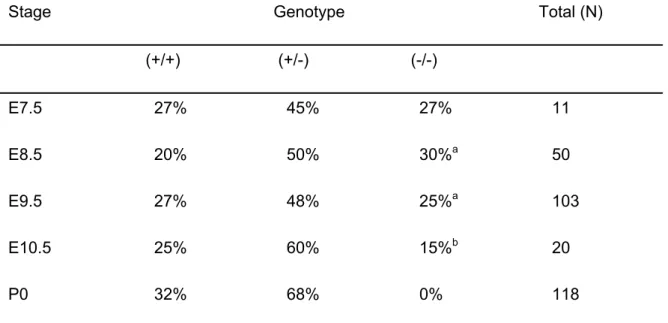
ATX-deficient mice die at midgestation with severe vascular defects. Heterozygous Enpp2+/- knockout mice were healthy and fertile. However, no
homozygous Enpp2-/- offspring was found among 118 newborn mice from heterozygous
intercrosses (Table 1), suggesting that ATX-deficiency is embryonic lethal. To investigate this, embryos were genotyped at different development stages. At embryonic day 9.5 (E9.5), ATX-deficient embryos could be recovered at the expected Mendelian frequency (Table 1), but all of them showed severe vascular defects in the yolk sac and were retarded in their development. By E10.5, most ATX-deficient embryos were resorbed.
Strikingly, blood vessels in the yolk sac of ATX-deficient embryos were poorly developed when compared to their wt and heterozygous littermates at E9.5. Between 7
E8.5 and E9.5, extra-embryonic endothelial cells normally remodel into a vascular network that connects with the embryo proper; the yolk sac then functions as the primary source of nutrients. At E9.5, blood appeared dispersed in the ATX-deficient yolk sac rather than in a vascular network as in wt and ATX heterozygous yolk sacs (Fig. 2A). Mutant yolk sacs showed patched cavities surrounded by endothelial cells and filled with blood cells, while the mesothelial cells on the inner aspect of the yolk sac were rounded rather than flattened (Fig. 2B).
Vessels of ATX-deficient embryos within the non-vascularized yolk sac were strikingly enlarged, particularly in the head region, when compared to wt and heterozygote embryos at E9.5; however, mutant embryos were not hemorrhagic (Fig. 2C,D,E and data not shown). To determine whether endothelial cells had differentiated from early angioblasts, we used an antibody to CD31/PECAM, a marker for mature endothelial cells. CD31-positive cells were readily detected in both yolk sac and embryo proper, indicating that ATX deficiency did not impair the differentiation of progenitor cells into endothelial cells (Fig. 2D). Cardiac development appeared to be normal, as judged from heart beating, but was not examined in detail. We conclude that ATX-deficient mice die around E9.5, with circulatory failure the most likely primary cause of death
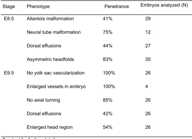
Additional abnormalities in ATX-deficient embryos. A number of additional abnormalities were observed in ATX-deficient embryos at E8.5 and E9.5, as summarized in Table 2. At E9.5, the large majority (85%) of mutant embryos had not initiated axial turning (Fig. 2E), which could reflect generally retarded development. In about 40% of the ATX-deficient embryos analyzed, there was abnormal development of the allantois at E8.5, which appeared swollen and failed to fuse to the chorion (Fig. 2F). Furthermore, in >80% of the E8.5 mutant embryos, the neural headfold (i.e. the future forebrain) was asymmetric due to enlargement of one of the folds, which showed extremely large cavities or effusions (Fig. 3A,B). Further down the neural axis, we 8
observed large effusions on the dorsal side (Fig. 2E), which displayed massive apoptosis as detected by TUNEL assays (data not shown). Such effusions are indicative of osmotic imbalance as a consequence of disrupted circulation.
ATX deficiency also led to malformation of the neural tube in the majority of the mutant embryos analyzed: the neural tube had not closed properly at E9.5 and, furthermore, appeared kinked and undulated over its entire length at E8.5, as opposed to the straight neural tube observed in wt embryos (Fig.2C,D and Fig. 3C,D). Given the prominence of ATX in the floor plate of the neural tube (3), these defects are most likely due to local ATX deficiency and not secondary to circulatory failure or growth retardation. A kinked neural tube is also observed in fibronectin-deficient embryos at this stage (13), suggesting that the neural tube defect associated with ATX deficiency could be due to insufficient extracellular matrix support.
Expression of ATX and LPA receptors during vascular development. We next examined the expression pattern of ATX and the four known LPA receptors (LPA1-LPA4)
in wt, heterozygous and mutant embryos using quantitative RT-PCR (Fig. 4A). In ATX-deficient and heterozygous embryos, the LPA receptor expression pattern was essentially similar to that in wt embryos (E9.5), although LPA3 levels were upregulated
by ~2-fold in the knockouts (Fig. 4A). Of note, ATX expression in the heterozygotes was 50% of that in the wild types. We also examined the temporal expression pattern of ATX and LPA1-4 in wt embryos. ATX and LPA1-4 were expressed during early postimplantation
stages (E6.5-E8.5), prior to yolk sac vascular development (Fig. 4B). Expression increased during the stages of vessel formation and expansion (E8.5-E10.5). ATX, LPA1
and LPA4 mRNA levels reached a maximum by E10.5, whereas LPA3 expression
peaked around E8.5 (Fig. 4B). Expression of LPA1 was significantly higher than that of
LPA2-4 (Fig. 4C). Expression of ATX in conjunction with all four LPA receptors during
E6.5-E10.5 supports the idea that ATX-regulated LPA production and signaling is 9
important for development during midgestation.
Since vasculogenesis is critically dependent on vascular endothelial growth factor (VEGF) (54), and LPA triggers VEGF-A expression in mouse embryonic fibroblasts (C. Stortelers, unpublished), we tested whether the vascular phenotype might involve VEGF deficiency. However, VEGF mRNA levels were increased rather than decreased in the ATX-deficient embryos (Fig. 4A). Because VEGF is a hypoxia target gene, the observed increase in VEGF might be due to oxygen deprivation resulting from circulatory failure. Whatever the precise mechanism, the results show that increased VEGF production does not rescue the vascular defects resulting from loss of ATX.
Half-normal ATX activity and plasma LPA levels in heterozygous mice. Since ATX functions as a lysoPLD, ATX deficiency should lead to loss of LPA production. Determination of LPA levels in interstitial fluids from midgestation embryos is technically quite demanding so we compared serum ATX activity and plasma LPA levels in ATX heterozygous mice (8-12 weeks old) with those in wildtype littermates. As shown in Fig. 5(A,B), if the gene dosage of ATX is reduced by half, a 50% reduction in ATX activity and LPA levels is observed. The average plasma LPA level in wildtype animals was 181 ± 17 nM (n=4), in keeping with previous results (38), versus 86 ± 14 nM (n=4) in their heterozygous littermates (Fig. 5A). Plasma S1P levels in heterozygote and wildtype mice were not significantly different (about 250 nM; Fig. 5C), consistent with the notion that ATX has no major role in generating S1P. Collectively, these findings strongly suggest that ATX is the major LPA-producing enzyme in vivo, and show that there is no physiological compensation for reduced ATX gene expression in the heterozygous animals.
10
DISCUSSION
Our results show that ATX is indispensable for embryonic development, as ATX-deficient embryos die at E9.5 with severely impaired vessel formation in yolk sac and embryo proper as well as other abnormalities (Table 2). Since ATX functions as a lysoPLD, our results suggest that loss of LPA production and downstream GPCR signaling is responsible for the oberved phenotype. While we find that all known LPA receptors are expressed during E6.5-E10.5 (Fig. 4B), the phenotype of individual LPA receptor knockouts (LPA1-3) has not so far been associated with embryonic vascular
defects (20,56). However, an LPA-associated vascular phenotype may only become evident from triple or quadruple receptor knockouts. An alternative or additional possibility is that LPA acts on as-yet-unidentified GPCRs to influence vascular development.
In contrast, a critical role for S1P signaling in vascular development has been well established, but S1P clearly acts at later embryonic stages than ATX. S1P is essential for the stabilization of nascent vessels by smooth muscle cells at around E12.5, rather than for vessel formation per se (27,29). Although ATX can generate S1P from SPC in vitro (7), a physiological role for ATX in sphingolipid metabolism seems unlikely as outlined in the Introduction and supported by our finding that plasma S1P levels, unlike LPA levels, are normal in ATX heterozygote animals (Fig. 5). While it remains formally possible that ATX has some physiological role in nucleotide metabolism or even functions unrelated to its catalytic activity (10), our findings are most consistent with the notion that ATX-mediated LPA production in the microenviroment of endothelial cells and subsequent GPCR signaling is essential for vascular development. Consistent with this, LPA stimulates vessel formation in a GPCR-dependent manner in the chicken embryo CAM assay (C. Rivera-Lopez and K. Lynch, personal
11
communication); futhermore, LPA promotes vascular network formation in murine E8.5 allantois explants, albeit less efficaciously than S1P (1).
How might LPA signaling govern vascular development? Impaired vascular development causing embryonic death around E9.5 is observed in several other mutant mice, including those lacking genes involved in receptor tyrosine kinase signaling, G protein signaling, cell adhesion, migration and oxygen sensing (for review see (2,9)), so comparison to other knockouts may provide a clue. Considering that LPA is a potent upstream activator of Gα13 (and possibly Gα12)(24,30), the most relevant phenotype in
this context is that of Gα13 knockout and Gα12/Gα13 double knockout mice (16,34).
Similar to ATX-deficient mice, the Gα13 knockouts die around E9.5 due to impaired blood
vessel formation in both yolk sac and embryo, with enlarged vessels in the head region (34). This phenotype is rescued by endothelium-specific re-expression of Gα13 (37),
implying that Gα13 signaling in endothelial cells is essential for vascular development.
Combined deficiency of Gα13 with Gα12 yields a somewhat earlier and more severe
phenotype that includes headfold malformation, a short allantois and unclosed and sometimes kinked neural tubes (16). Gα13, probably in cooperation with Gα12, links
GPCRs to guanine nucleotide exchange factors for RhoA, a key regulator of the actin cytoskeleton (40), and to other effectors (36). Through its ability to regulate cell shape and adhesion, RhoA activity is fundamental to cell migration. Indeed, cells deficient in either Gα13 or RhoA activity fail to migrate towards LPA (16,50), underscoring the
importance of the LPA-Gα13-RhoA pathway for cell motility. LPA has multiple effects on
endothelial cells including stimulation of cell migration and invasion, which are critical events during angiogenesis (35,53), and an increase in endothelial monolayer permeability (33,41). LPA also exerts migratory and contractile effects on vascular smooth muscle cells (30). Thus, ATX-mediated LPA production and subsequent LPA
12
signaling through Gα13, probably in cooperation with Gα12 and other G proteins, may
contribute to vascular development by stimulating endothelial cell migration and invasion as well as by regulating adhesive interactions with the extracellular matrix and smooth muscle cells. Consistent with this, the vascular defects observed in ATX- and G(a)13-deficient mice resemble those in mice lacking genes involved in cell migration and adhesion such as fibronectin and focal adhesion kinase (13,19). Further insight into the mechanistic basis of the ATX-deficient phenotype awaits the generation and analysis of endothelium- and/or smooth muscle-specific ATX and LPA receptor knockout mice as well as transgenic rescue studies. Tissue-specific ATX knockouts will also allow to assess how the present findings in the embryo extrapolate to the adult, where ATX and the LPA/LPA receptor axis have been implicated in several disorders including cancer.
In the meantime, an interesting finding of the present study is that ATX heterozygous mice possess half as much plasma LPA as their normal littermates, consistent with ATX being the major LPA-producing enzyme in vivo and, furthermore, indicating that ATX activity is not upregulated to compensate for the null allele. ATX heterozygous mice have not shown any obvious abnormalities until now and thus offer an opportunity to test several potential roles of LPA in vivo, including tumor progression, wound healing and neurophysiological functions.
ACKNOWLEDGMENTS
We thank Junken Aoki, Richard Proia and Kevin Lynch for sharing unpublished results; Stefan Offermanns and Mathieu Bollen for helpful discussions; and Trudi Hengeveld, Jeroen Korving, Rahmen Bin Ali, and John Zevenhoven for experimental assistance. This work was supported by the Dutch Cancer Society.
13
Reference List
1. Argraves, K. M., B. A. Wilkerson, W. S. Argraves, P. A. Fleming, L. M. Obeid, and C. J. Drake. 2004. Sphingosine-1-phosphate signaling promotes critical migratory events in
vasculogenesis. J Biol Chem 279:50580-50590.
2. Argraves, W. S. and C. J. Drake. 2005. Genes critical to vasculogenesis as defined by
systematic analysis of vascular defects in knockout mice. Anat Rec. A Discov Mol Cell Evol Biol 286:875-884.
3. Bachner, D., M. Ahrens, N. Betat, D. Schroder, and G. Gross. 1999. Developmental expression analysis of murine autotaxin (ATX). Mech. Dev. 84:121-125.
4. Baumforth, K. R., J. R. Flavell, G. M. Reynolds, G. Davies, T. R. Pettit, W. Wei, S. Morgan, T. Stankovic, Y. Kishi, H. Arai, M. Nowakova, G. Pratt, J. Aoki, M. J. Wakelam, L. S. Young, and P. G. Murray. 2005. Induction of autotaxin by the
Epstein-Barr virus promotes the growth and survival of Hodgkin lymphoma cells. Blood 106:2138-2146.
5. Boucharaba, A., C. M. Serre, S. Gres, J. S. Saulnier-Blache, J. C. Bordet, J. Guglielmi, P. Clezardin, and O. Peyruchaud. 2004. Platelet-derived lysophosphatidic
acid supports the progression of osteolytic bone metastases in breast cancer. J Clin Invest 114:1714-1725.
6. Carvalho, R. L., L. Jonker, M. J. Goumans, J. Larsson, P. Bouwman, S. Karlsson, P. T. Dijke, H. M. Arthur, and C. L. Mummery. 2004. Defective paracrine signalling by TGFbeta in yolk sac vasculature of endoglin mutant mice: a paradigm for hereditary haemorrhagic telangiectasia. Development 131:6237-6247.
7. Clair, T., J. Aoki, E. Koh, R. W. Bandle, S. W. Nam, M. M. Ptaszynska, G. B. Mills, E. Schiffmann, L. A. Liotta, and M. L. Stracke. 2003. Autotaxin hydrolyzes
sphingosylphosphorylcholine to produce the regulator of migration, sphingosine-1-phosphate. Cancer Res 63:5446-5453.
8. Clair, T., H. Y. Lee, L. A. Liotta, and M. L. Stracke. 1997. Autotaxin is an exoenzyme
possessing 5'-nucleotide phosphodiesterase/ATP pyrophosphatase and ATPase activities. J Biol Chem. 272:996-1001.
9. Copp, A. J. 1995. Death before birth: clues from gene knockouts and mutations. Trends
Genet 11:87-93.
10. Dennis, J., L. Nogaroli, and B. Fuss. 2005. Phosphodiesterase-Ialpha/autotaxin
(PD-Ialpha/ATX): A multifunctional protein involved in central nervous system development and disease. J Neurosci Res 82:737-742.
11. Ferry, G., E. Tellier, A. Try, S. Gres, I. Naime, M. F. Simon, M. Rodriguez, J.
Boucher, I. Tack, S. Gesta, P. Chomarat, M. Dieu, M. Raes, J. P. Galizzi, P. Valet, J. A. Boutin, and J. S. Saulnier-Blache. 2003. Autotaxin is released from adipocytes,
catalyzes lysophosphatidic acid synthesis, and activates preadipocyte proliferation. Up-regulated expression with adipocyte differentiation and obesity. J Biol Chem 278:18162-18169.
14
12. Fuss, B., H. Baba, T. Phan, V. K. Tuohy, and W. B. Macklin. 1997. Phosphodiesterase
I, a novel adhesion molecule and/or cytokine involved in oligodendrocyte function. J Neurosci 17:9095-9103.
13. George, E. L., E. N. Georges-Labouesse, R. S. Patel-King, H. Rayburn, and R. O. Hynes. 1993. Defects in mesoderm, neural tube and vascular development in mouse
embryos lacking fibronectin. Development 119:1079-1091.
14. Gijsbers, R., J. Aoki, H. Arai, and M. Bollen. 2003. The hydrolysis of lysophospholipids
and nucleotides by autotaxin (NPP2) involves a single catalytic site. FEBS Lett. 538:60-64.
15. Goding, J. W., B. Grobben, and H. Slegers. 2003. Physiological and pathophysiological functions of the ecto-nucleotide pyrophosphatase/phosphodiesterase family. Biochim Biophys Acta 1638:1-19.
16. Gu, J. L., S. Muller, V. Mancino, S. Offermanns, and M. I. Simon. 2002. Interaction of
G alpha(12) with G alpha(13) and G alpha(q) signaling pathways. Proc. Natl Acad. Sci. U. S. A 99:9352-9357.
17. Hama, K., J. Aoki, M. Fukaya, Y. Kishi, T. Sakai, R. Suzuki, H. Ohta, T. Yamori, M. Watanabe, J. Chun, and H. Arai. 2004. Lysophosphatidic acid and autotaxin stimulate
cell motility of neoplastic and non-neoplastic cells through LPA1. J Biol Chem 279:17634-17639.
18. Hoelzinger, D. B., L. Mariani, J. Weis, T. Woyke, T. J. Berens, W. S. McDonough, A. Sloan, S. W. Coons, and M. E. Berens. 2005. Gene expression profile of glioblastoma
multiforme invasive phenotype points to new therapeutic targets. Neoplasia. 7:7-16. 19. Ilic, D., Y. Furuta, S. Kanazawa, N. Takeda, K. Sobue, N. Nakatsuji, S. Nomura, J.
Fujimoto, M. Okada, and T. Yamamoto. 1995. Reduced cell motility and enhanced
focal adhesion contact formation in cells from FAK-deficient mice. Nature 377:539-544. 20. Ishii, I., N. Fukushima, X. Ye, and J. Chun. 2004. Lysophospholipid receptors: signaling
and biology. Annu Rev Biochem 73:321-354.
21. Jansen, S., C. Stefan, J. W. Creemers, E. Waelkens, A. Van Eynde, W. Stalmans, and M. Bollen. 2005. Proteolytic maturation and activation of autotaxin (NPP2), a
secreted metastasis-enhancing lysophospholipase D. J Cell Sci. 118:3081-3089. 22. Kehlen, A., N. Englert, A. Seifert, T. Klonisch, H. Dralle, J. Langner, and C.
Hoang-Vu. 2004. Expression, regulation and function of autotaxin in thyroid carcinomas. Int J
Cancer 109:833-838.
23. Kingsbury, M. A., S. K. Rehen, J. J. Contos, C. M. Higgins, and J. Chun. 2003.
Non-proliferative effects of lysophosphatidic acid enhance cortical growth and folding. Nat. Neurosci. 6:1292-1299.
24. Kranenburg, O., M. Poland, F. P. van Horck, D. Drechsel, A. Hall, and W. H. Moolenaar. 1999. Activation of RhoA by lysophosphatidic acid and Galpha12/13
subunits in neuronal cells: induction of neurite retraction. Mol Biol Cell 10:1851-1857. 25. Lee, H. Y., J. Murata, T. Clair, M. H. Polymeropoulos, R. Torres, R. E. Manrow, L. A.
Liotta, and M. L. Stracke. 1996. Cloning, chromosomal localization, and tissue
15
expression of autotaxin from human teratocarcinoma cells. Biochem Biophys. Res Commun. 218:714-719.
26. Liliom, K., G. Sun, M. Bunemann, T. Virag, N. Nusser, D. L. Baker, D. A. Wang, M. J. Fabian, B. Brandts, K. Bender, A. Eickel, K. U. Malik, D. D. Miller, D. M. Desiderio, G. Tigyi, and L. Pott. 2001. Sphingosylphosphocholine is a naturally occurring lipid
mediator in blood plasma: a possible role in regulating cardiac function via sphingolipid receptors. Biochem. J 355:189-197.
27. Liu, Y., R. Wada, T. Yamashita, Y. Mi, C. X. Deng, J. P. Hobson, H. M. Rosenfeldt, V. E. Nava, S. S. Chae, M. J. Lee, C. H. Liu, T. Hla, S. Spiegel, and R. L. Proia. 2000.
Edg-1, the G protein-coupled receptor for sphingosine-1-phosphate, is essential for vascular maturation. J. Clin. Invest 106:951-961.
28. Mills, G. B. and W. H. Moolenaar. 2003. The emerging role of lysophosphatidic acid in
cancer. Nat. Rev. Cancer 3:582-591.
29. Mizugishi, K., T. Yamashita, A. Olivera, G. F. Miller, S. Spiegel, and R. L. Proia.
2005. Essential role for sphingosine kinases in neural and vascular development. Mol Cell Biol 25:11113-11121.
30. Moolenaar, W. H., L. A. van Meeteren, and B. N. Giepmans. 2004. The ins and outs of
lysophosphatidic acid signaling. Bioessays 26:870-881.
31. Nam, S. W., T. Clair, C. K. Campo, H. Y. Lee, L. A. Liotta, and M. L. Stracke. 2000.
Autotaxin (ATX), a potent tumor motogen, augments invasive and metastatic potential of ras-transformed cells. Oncogene 19:241-247.
32. Nam, S. W., T. Clair, Y. S. Kim, A. McMarlin, E. Schiffmann, L. A. Liotta, and M. L. Stracke. 2001. Autotaxin (NPP-2), a metastasis-enhancing motogen, is an angiogenic
factor. Cancer Res 61:6938-6944.
33. Nieuw Amerongen, G. P., M. A. Vermeer, and V. W. van Hinsbergh. 2000. Role of
RhoA and Rho kinase in lysophosphatidic acid-induced endothelial barrier dysfunction. Arterioscler. Thromb. Vasc. Biol 20:E127-E133.
34. Offermanns, S., V. Mancino, J. P. Revel, and M. I. Simon. 1997. Vascular system
defects and impaired cell chemokinesis as a result of Galpha13 deficiency. Science
275:533-536.
35. Panetti, T. S., D. F. Hannah, C. Avraamides, J. P. Gaughan, C. Marcinkiewicz, A. Huttenlocher, and D. F. Mosher. 2004. Extracellular matrix molecules regulate
endothelial cell migration stimulated by lysophosphatidic acid. J Thromb. Haemost.
2:1645-1656.
36. Postma, F. R., K. Jalink, T. Hengeveld, S. Offermanns, and W. H. Moolenaar. 2001.
Galpha(13) mediates activation of a depolarizing chloride current that accompanies RhoA activation in both neuronal and nonneuronal cells. Curr. Biol 11:121-124.
37. Ruppel, K. M., D. Willison, H. Kataoka, A. Wang, Y. W. Zheng, I. Cornelissen, L. Yin, S. M. Xu, and S. R. Coughlin. 2005. Essential role for Galpha13 in endothelial cells
during embryonic development. Proc. Natl Acad. Sci. U. S. A 102:8281-8286.
16
38. Saulnier-Blache, J. S., A. Girard, M. F. Simon, M. Lafontan, and P. Valet. 2000. A
simple and highly sensitive radioenzymatic assay for lysophosphatidic acid quantification. J Lipid Res 41:1947-1951.
39. Schaft, J., R. Ashery-Padan, F. van der Hoeven, P. Gruss, and A. F. Stewart. 2001.
Efficient FLP recombination in mouse ES cells and oocytes. Genesis. 31:6-10.
40. Schmidt, A. and A. Hall. 2002. Guanine nucleotide exchange factors for Rho GTPases:
turning on the switch. Genes Dev 16:1587-1609.
41. Schulze, C., C. Smales, L. L. Rubin, and J. M. Staddon. 1997. Lysophosphatidic acid
increases tight junction permeability in cultured brain endothelial cells. J Neurochem.
68:991-1000.
42. Siess, W. and G. Tigyi. 2004. Thrombogenic and atherogenic activities of
lysophosphatidic acid. J Cell Biochem 92:1086-1094.
43. Stassar, M. J., G. Devitt, M. Brosius, L. Rinnab, J. Prang, T. Schradin, J. Simon, S. Petersen, A. Kopp-Schneider, and M. Zoller. 2001. Identification of human renal cell
carcinoma associated genes by suppression subtractive hybridization. Br J Cancer
85:1372-1382.
44. Stefan, C., S. Jansen, and M. Bollen. 2005. NPP-type ectophosphodiesterases: unity in
diversity. Trends Biochem. Sci. 30:542-550.
45. Stracke, M. L., H. C. Krutzsch, E. J. Unsworth, A. Arestad, V. Cioce, E. Schiffmann, and L. A. Liotta. 1992. Identification, purification, and partial sequence analysis of
autotaxin, a novel motility-stimulating protein. J Biol Chem. 267:2524-2529.
46. Su, A. I., M. P. Cooke, K. A. Ching, Y. Hakak, J. R. Walker, T. Wiltshire, A. P. Orth, R. G. Vega, L. M. Sapinoso, A. Moqrich, A. Patapoutian, G. M. Hampton, P. G. Schultz, and J. B. Hogenesch. 2002. Large-scale analysis of the human and mouse
transcriptomes. Proc Natl Acad Sci U S A 99:4465-4470.
47. Tokumura, A., E. Majima, Y. Kariya, K. Tominaga, K. Kogure, K. Yasuda, and K. Fukuzawa. 2002. Identification of human plasma lysophospholipase D, a
lysophosphatidic acid-producing enzyme, as autotaxin, a multifunctional phosphodiesterase. J Biol Chem. 277:39436-39442.
48. Umezu-Goto, M., Y. Kishi, A. Taira, K. Hama, N. Dohmae, K. Takio, T. Yamori, G. B. Mills, K. Inoue, J. Aoki, and H. Arai. 2002. Autotaxin has lysophospholipase D activity
leading to tumor cell growth and motility by lysophosphatidic acid production. J Cell Biol
158:227-233.
49. van der Weyden, L., D. J. Adams, L. W. Harris, D. Tannahill, M. J. Arends, and A. Bradley. 2005. Null and conditional semaphorin 3B alleles using a flexible puroDeltatk
loxP/FRT vector. Genesis. 41:171-178.
50. Van Leeuwen, F. N., C. Olivo, S. Grivell, B. N. Giepmans, J. G. Collard, and W. H. Moolenaar. 2003. Rac activation by lysophosphatidic acid LPA1 receptors through the
guanine nucleotide exchange factor Tiam1. J Biol Chem. 278:400-406.
51. van Meeteren, L. A., F. Frederiks, B. N. Giepmans, M. F. Pedrosa, S. J. Billington, B. H. Jost, D. V. Tambourgi, and W. H. Moolenaar. 2004. Spider and bacterial
17
sphingomyelinases D target cellular lysophosphatidic acid receptors by hydrolyzing lysophosphatidylcholine. J. Biol. Chem. 279:10833-10836.
52. van Meeteren, L. A., P. Ruurs, E. Christodoulou, J. W. Goding, H. Takakusa, K. Kikuchi, A. Perrakis, T. Nagano, and W. H. Moolenaar. 2005. Inhibition of autotaxin by
lysophosphatidic acid and sphingosine 1-phosphate. J. Biol. Chem. 280:21155-21161. 53. Wu, W. T., C. N. Chen, C. I. Lin, J. H. Chen, and H. Lee. 2005. Lysophospholipids
enhance matrix metalloproteinase-2 expression in human endothelial cells. Endocrinology 146:3387-3400.
54. Yancopoulos, G. D., S. Davis, N. W. Gale, J. S. Rudge, S. J. Wiegand, and J. Holash.
2000. Vascular-specific growth factors and blood vessel formation. Nature 407:242-248. 55. Yang, S. Y., J. Lee, C. G. Park, S. Kim, S. Hong, H. C. Chung, S. K. Min, J. W. Han, H.
W. Lee, and H. Y. Lee. 2002. Expression of autotaxin (NPP-2) is closely linked to
invasiveness of breast cancer cells. Clin Exp Metastasis 19:603-608.
56. Ye, X., K. Hama, J. J. Contos, B. Anliker, A. Inoue, M. K. Skinner, H. Suzuki, T. Amano, G. Kennedy, H. Arai, J. Aoki, and J. Chun. 2005. LPA3-mediated
lysophosphatidic acid signalling in embryo implantation and spacing. Nature 435:104-108.
18
LEGENDS
FIG. 1. Generation of conditional EnppF/+ mice. (A) Schematic representations of the
Enpp2 locus, the targeting construct and the mutated Enpp2 alleles. Exons are indicated by filled boxes, LoxP sites by filled triangles, and FRT sites by open triangles. Probes P1, P2 and P3 are indicated. (H) denotes HindIII sites, (P) PstI sites and (A) ApaI sites. (B) Identification of correctly targeted ES cell clones. Genomic DNA was digested with ApaI and screened with a 3’ flanking probe (P1) located outside the targeting construct. (C,D) Genotyping of adult mice and E9.5 embryos with conditional Enpp2F and/or
deleted Enpp2∆ alleles, using PCR with primers 1F, 1R and 2R or PstI-digested DNA
screened with P3.
FIG. 2. Phenotype of ATX-deficient yolk sacs and embryos. (A) Yolk sacs of heterozygote (+/-) and ATX-deficient (-/-) embryos at E9.5. Note absence of a visible vascular network in (-/-) embryos, whereas blood islands are readily detectable. (B) Sections of yolk sacs (HE-stained) at E9.5, showing primitive vascular structures containing hematopoietic cells. In mutant yolk sacs, the mesothelial cells on the inner aspect of the yolk sac are rounded rather than flattened. (C) Tranverse sections of heterozygous or ATX-deficient embryos, HE-stained, at E9.5. Note enlarged vessels (arrows) and neural tube malformation in the ATX-deficient embryo when compared to the heterozygous control. (D) Tranverse sections of heterozygous or ATX-deficient embryos, HE- and CD31-stained, at E9.5. Note enlargement of CD31-positive vessels in ATX-deficient embryos. (E) Malformation of ATX-deficient embryos at E9.5. Note large effusions in head region and on dorsal side (arrows) and no axial turning of the mutant
19
embryo. (F) Malformation of the allantois at E8.5. Allantois (white arrow) appeared short and swollen, and failed to fuse to the chorion in mutant embryos. (Scale bars: 100 µm)
FIG. 3. Headfold asymmetry and neural tube defects in ATX-deficient embryos. (A) Headfold asymmetry in ATX-deficient embryos (E8.5), showing enlargement of one of the headfolds. (B,C) Transverse sections of the head region (E9.5). Note large cavities in the mutant embryos and malformation of the neural tube, which was often unclosed and kinked. (D) Undulated neural tube in ATX-deficient embryos (E8.5), as opposed to the straight neural tube in wildtype embryos. (Scale bars: 100 µm)
FIG. 4. Expression analysis, as determined by quantitative RT-PCR. (A) Expression patterns of LPA1-4, ATX and VEGF-A in wt, heterozygous and knockout embryos at E9.5.
Expressions were normalized to HPRT. Note increased VEGF-A expression in the ATX-deficient embryos. (B) Temporal expression pattern of LPA1-4 and ATX mRNA levels in
developing wild-type embryos during E6.5-E10.5. Expression levels were normalized to HPRT. (C) Relative LPA receptor expression patterns in wild-type E.9.5 embryos.
FIG. 5. Determination of plasma ATX activity, LPA and S1P levels. (A) Half-normal plasma LPA levels in heterozygous ATX mice (86 ± 14 nmol/L versus 181 ± 17 nmol/L in the wildtypes). LPA levels were determined by the generation of phosphatidic acid, produced by the transfer of a [14C] fatty acyl chain onto LPA (38). Values correspond to
means ± SEM (n=4). (B) ATX activity in serum as determined by the FRET-based ATX sensor, CPF4 (2 µM) (52). Relative ATX activity was calculated as the increase in FRET ratio (+/+, n=7; +/-, n=5). (C) Normal plasma S1P levels in heterozygote mice, as determined by mass spectrometry (LC-MS) (heterozygote, n=10; wildtype, n=13)
20
TABLE 1. Genotypes of progeny from ATX heterozygous intercrosses
Stage Genotype Total (N)
(+/+) (+/-) (-/-) E7.5 27% 45% 27% 11 E8.5 20% 50% 30%a 50 E9.5 27% 48% 25%a 103 E10.5 25% 60% 15%b 20 P0 32% 68% 0% 118
Genotypes were detemined as described in Materials and methods and Fig. 1 (legend).
aEmbryos were morphologically abnormal, as detailed in Table 2.
bAll embryos appeared to be necrotic and were partially resorbed.
21
TABLE 2. Phenotypic abnormalities in ATX-deficient embryos and yolk sacs
Stage Phenotype Penetrance Embryos analyzed (N)
E8.5 Allantois malformation 41% 29
Neural tube malformation 75% 12
Dorsal effusions 44% 27
Asymmetric headfolds 83% 35
E9.5 No yolk sac vascularization 100% 26
Enlarged vessels in embryo 100% 4
No axial turning 85% 26
Dorsal effusions 42% 26
Enlarged head region 54% 26
See text for further details.
22
23.1 9.4 6.6 4.4 Enpp2puro-tk-F Enpp2 * * * ApaI probe (P1) kb Enpp2F 5.0 4.0 3.0 2.0 1.6 1.0 0.85 Enpp2 Enpp2∆ kb PstI probe (P3) +/+ F/+ ∆/+ F/F Adult 1F-1R +/+ F/+ F/F ∆/+ +/+ ∆/+ ∆/∆ Embryo H2O 2F-1R Enpp2∆ Enpp2 Enpp2F Enpp2 locus
Enpp2puro-tk-Fallele
Enpp2Fallele Enpp2∆allele Targeting construct 1F 5 6 7 8 A A P P 5 6 7 8 A A P P A Puro∆TK 5 6 7 A P 8 P P Puro∆TK 5 6 7 A P 8 P P P1 5 8 A P P A P3 P3 P2 P P 1F 2F 2F 2F 1R 1R 1R 5 Exons LoxP-site FRT-site Primer Probe A A 1 kb H H H H H H HH P3 P2 P1
A
B
C
D
Figure 1
+/-A
-/-B
-/-
+/-
-/-
+/-C
+/+
D
-/-
+/-
-/-+/+
F
E
Figure 2
+/+
-/-+/+
-/-
+/-B
D
-/-
+/-A
C
Figure 3
0 1 2 3
lpa1 lpa2 lpa3 lpa4 atx vegfA
F o ld induct io n (n o rm . t o HP RT ) ATX +/+ATX +/-ATX
-/-*
*
LPA4LPA1 LPA2 LPA3 ATX VEGF
A
0 0.5 1 1.5 2 2.5 3LPA1 lpa2 lpa3 lpa4 atx
F o ld induc tion (n o rm . t o HP RT ) E6.5 E8.5 E9.5 E10.5 LPA4
LPA1 LPA2 LPA3 ATX
0 0.05 0.1 0.15 0.2 0.25 0.3 Relat iv e expres sion / H P R T LPA4
LPA1 LPA2 LPA3