HAL Id: hal-01608529
https://hal.archives-ouvertes.fr/hal-01608529
Submitted on 26 May 2020
HAL is a multi-disciplinary open access
archive for the deposit and dissemination of
sci-entific research documents, whether they are
pub-lished or not. The documents may come from
teaching and research institutions in France or
abroad, or from public or private research centers.
L’archive ouverte pluridisciplinaire HAL, est
destinée au dépôt et à la diffusion de documents
scientifiques de niveau recherche, publiés ou non,
émanant des établissements d’enseignement et de
recherche français ou étrangers, des laboratoires
publics ou privés.
Delphine Payros, Ulrich Dobrindt, Patricia Martin-Bizon, Thomas Secher,
Ana-Paula Loureiro-Bracarense, Michèle Boury, Joëlle Laffitte, Philippe
Pinton, Eric Oswald, Isabelle P. Oswald
To cite this version:
Delphine Payros, Ulrich Dobrindt, Patricia Martin-Bizon, Thomas Secher, Ana-Paula
Loureiro-Bracarense, et al.. The food contaminant deoxynivalenol exacerbates the genotoxicity of gut
mi-crobiota. mBio, American Society for Microbiology, 2017, 8 (2), 11 p. �10.1128/mBio.00007-17�.
�hal-01608529�
Microbiota
Delphine Payros,
aUlrich Dobrindt,
b,cPatricia Martin,
d,eThomas Secher,
eAna Paula F. L. Bracarense,
fMichèle Boury,
eJoelle Laffitte,
aPhilippe Pinton,
aEric Oswald,
d,eIsabelle P. Oswald
aToxalim (Research Centre in Food Toxicology), Université de Toulouse, INRA, ENVT, INP-Purpan, UPS, Toulouse, Francea; Institute of Hygiene, University of Münster, Münster, Germanyb; Interdisciplinary Center for Clinical Research (IZKF), University of Münster, Münster, Germanyc; CHU Toulouse, Service de Bactériologie-Hygiène, Institut Fédératif de Biologie, TSA, Toulouse, Franced; Institut de Recherche en Santé Digestive (IRSD), Université de Toulouse, INSERM, INRA, ENVT, UPS, CS, Toulouse, Francee; Laboratorio Patologia Animal, CP, Universidade Estadual de Londrina, Londrina, Paraná, Brazilf
ABSTRACT
An increasing number of human beings from developed countries are
colonized by Escherichia coli strains producing colibactin, a genotoxin suspected to
be associated with the development of colorectal cancers. Deoxynivalenol (DON) is
the most prevalent mycotoxin that contaminates staple food— especially cereal
products—in Europe and North America. This study investigates the effect of the
food contaminant DON on the genotoxicity of the E. coli strains producing
colibac-tin. In vitro, intestinal epithelial cells were coexposed to DON and E. coli producing
colibactin. In vivo, newborn rats colonized at birth with E. coli producing colibactin
were fed a DON-contaminated diet. Intestinal DNA damage was estimated by the
phosphorylation of histone H2AX. DON exacerbates the genotoxicity of the E. coli
producing colibactin in a time- and dose-dependent manner in vitro. Although DON
had no effect on the composition of the gut microbiota, and especially on the
num-ber of E. coli, a significant increase in DNA damage was observed in intestinal
epi-thelial cells of animals colonized by E. coli strains producing colibactin and
coex-posed to DON compared to animals colonized with E. coli strains unable to produce
colibactin or animals exposed only to DON. In conclusion, our data demonstrate that
the genotoxicity of E. coli strains producing colibactin, increasingly present in the
microbiota of asymptomatic human beings, is modulated by the presence of DON in
the diet. This raises questions about the synergism between food contaminants and
gut microbiota with regard to intestinal carcinogenesis.
IMPORTANCE
An increasing number of human beings from developed countries
are colonized by Escherichia coli strains producing colibactin, a genotoxin suspected
to be associated with the development of colorectal cancers. Deoxynivalenol (DON)
is the most prevalent mycotoxin that contaminates staple food— especially cereal
products—in Europe and North America. Our in vitro and in vivo results demonstrate
that the intestinal DNA damage induced by colibactin-producing E. coli strains was
exacerbated by the presence of DON in the diet. This raises questions about the
synergism between food contaminants and gut microbiota with regard to intestinal
carcinogenesis.
T
he gut is colonized by a rich ecological consortium of more than a thousand species
of microorganisms that exert marked effects on basic host physiology, immunity,
and metabolism (1–3). Escherichia coli bacteria are some of the pioneer bacteria that
colonize the guts of mammals within a few days following birth (4). The genetic
structure of the E. coli population is clonal and can be segregated into seven major
Received 6 January 2017 Accepted 13
February 2017 Published 14 March 2017
Citation Payros D, Dobrindt U, Martin P, Secher
T, Bracarense APFL, Boury M, Laffitte J, Pinton P, Oswald E, Oswald IP. 2017. The food contaminant deoxynivalenol exacerbates the genotoxicity of gut microbiota. mBio 8:e00007-17.https://doi.org/10.1128/mBio.00007-17.
Editor Julian E. Davies, University of British
Columbia
Copyright © 2017 Payros et al. This is an
open-access article distributed under the terms of theCreative Commons Attribution 4.0 International license.
Address correspondence to Isabelle P. Oswald, isabelle.oswald@inra.fr.
E.O. and I.P.O. are co-senior authors.
®
phylogenetic groups (A, B1, B2, C, D, E, and F) (5). The prevalence of the B2 group is
increasing among E. coli strains persisting in the microbiota of humans in developed
countries (6, 7). This change in the distribution of phylogenetic groups of the E. coli
population could be a consequence of enriched dietary habits and increased levels of
hygiene in industrialized countries.
E. coli genomes show evidence of a widespread acquisition of functions through
horizontal transfer of genes (8). Up to 50% of E. coli strains from the phylogenetic group
B2 have acquired the pks genomic island (9, 10). This gene cluster encodes a
nonribo-somal peptide synthase-polyketide synthase (NRPS-PKS) assembly line and produces a
genotoxic secondary metabolite called colibactin (9). A short contact between
mam-malian cells and E. coli producing colibactin induces DNA damage, senescence, and
chromosomal abnormalities (9, 11–13). Colonization of the gut by phylogroup B2 E. coli
producing colibactin is associated with the presence of DNA double-strand breaks in
intestinal epithelial cells (14). Phylogroup B2 E. coli bacteria producing colibactin have
an impact on host physiology (14, 15) and contribute to the development of colorectal
cancer in mouse models of colitis (16, 17). Infants are colonized at birth with B2 E. coli
expressing colibactin, and these E. coli strains have a long-term capacity to persist in the
bowel microbiota (14, 18). Except for iron availability, little is known about the
envi-ronmental factors that modulate the genotoxicity of these bacteria in the gut (19).
Mycotoxins are the most frequently occurring natural food contaminants in human
and animal diet (20). Of the mycotoxins, deoxynivalenol (DON) is mainly produced by
Fusarium graminearum and Fusarium culmorum. This toxin frequently develops in
cereals and grains. A survey of 12 European countries indicated that 57% of samples
were positive for DON (21). Analyses of urine samples lead to the estimation that 98%
of adults in the United Kingdom had been exposed to DON, while 80% of children in
the Netherlands exceeded the tolerable daily intake for this mycotoxin (22, 23). DON
targets the intestine (24–26) and interacts with the peptidyl transferase region of the
60S ribosomal subunit, inducing a “ribotoxic stress,” resulting in the activation of
mitogen-activated protein kinases (MAPKs) and their downstream pathways (27, 28).
The consequence of exposure to this food contaminant on the genotoxic potential
of the gut microbiota has never been addressed. In the present study, we investigated
the impact of DON on the genotoxicity of E. coli producing colibactin. Using both in
vitro and in vivo experiments, we demonstrated that DON exacerbates the intestinal
DNA damage induced by genotoxic strains of E. coli.
RESULTS
Genotoxicity of DON and colibactin-producing E. coli on intestinal epithelial
cells. The genotoxicity of DON and colibactin-producing E. coli was first examined in
cultured rat intestinal epithelial cells (IEC-6) using an in-cell Western (ICW) technique
that assessed the phosphorylated form of histone H2AX (
␥H2AX) as a marker of DNA
double-strand breaks. DON alone did not exert detectable genotoxicity on IEC-6 cells,
except at high doses (12.5 to 50
M for at least 8 h) where low levels of ␥H2AX were
observed (2.27
⫾ 0.22 relative fluorescence units [RFU] in control cells versus 4.64 ⫾
1.37 and 5.21
⫾ 1.30 RFU in cells treated with 25
M and 50 M DON, respectively [P ⬍
0.05 and P
⬍ 0.001, respectively]) (Fig. 1A; see Fig. S1A and S2A in the supplemental
material). In contrast to DON-treated cells, IEC-6 cells were highly susceptible to the
genotoxicity of colibactin-producing E. coli (colibactin-producing wild-type E. coli [E. coli
WT]). As shown in Fig. 1B, the infection of IEC-6 cells with E. coli WT induced a
dose-dependent increase in
␥H2AX (2.27 ⫾ 0.22 RFU in control cells versus 9.1 ⫾ 3.92
RFU [P
⬍ 0.05] in E. coli WT with a multiplicity of infection [MOI] of 25 and 29.03 ⫾ 8.19
RFU [P
⬍ 0.001] in E. coli WT with an MOI of 100 compared to noninfected cells).
DON exacerbates the genotoxicity of colibactin-producing E. coli. To determine
whether the genotoxic effect of colibactin can be modulated by exposure to the food
contaminant DON, IEC-6 cells were infected with a low dose of E. coli WT (MOI of 25)
and coexposed to DON (25
M). Exposure of cells to DON exacerbated the genotoxicity
of E. coli producing colibactin on IEC-6 cells compared to cells exposed solely to DON
mbio.asm.org
on June 15, 2017 - Published by
FIG 1 Quantification of DNA double-strand breaks in intestinal epithelial cells in vitro. (A) IEC-6 cells were treated
with increasing doses of DON (0 to 50M) for 8 h, and ␥H2AX was quantified by an in-cell Western (ICW) method. Untreated cells (white bar) and cells exposed to DON (dotted white bars) are indicated. (B) IEC-6 cells were infected with wild-type E. coli producing colibactin (E. coli WT) for 4 h with increasing multiplicities of infection (MOIs [m.o.i. in the figure] of 0 to 100 bacteria per cell) before ICW analysis 4 h after infection (black bars). Mean values plus standard errors of the means (SEM) (error bars) from two independent experiments are shown. Values that are significantly different by one-way ANOVA with Bonferroni’s multiple-comparison correction are indicated by asterisks as follows:*, P⬍ 0.05; **, P ⬍ 0.01; ***, P ⬍ 0.001.
FIG 2 Dose-dependent synergistic genotoxicity of DON and E. coli. (A) IEC-6 cells infected for 4 h with E. coli strains
producing colibactin (E. coli WT) or not producing colibactin (E. coli ΔclbA and E. coli ΔclbP) (MOI of 25) and coexposed to 25M DON 8 h before quantification of ␥H2AX by in-cell Western (ICW). (B) ICW pictures of IEC-6 cells exposed or not exposed to 25M DON and infected (MOI of 25) with E. coli strains producing colibactin (E. coli WT) or not producing colibactin (E. coli ΔclbA or ΔclbP). DNA is artificially colored red, and␥H2AX is shown in green. N.I., not infected. (C) IEC-6 cells infected for 4 h with E. coli WT or not infected with E. coli and treated with increasing doses of DON (0 to 50M) 8 h before ICW. Control (not treated) cells (white bars) and cells infected with
E. coli WT (black bars), E. coli ΔclbP (light gray bars), and E. coli ΔclbA (dark gray bars) are shown. Dotted white, black,
light gray, and dark gray bars represent cells exposed to DON and coinfected with different E. coli strains (dotted black, WT; dotted light gray, ΔclbP; dotted dark gray, ΔclbA) or not coinfected with E. coli strains (dotted white). Mean values plus SEM from three independent experiments are shown. Values that are significantly different from the values for cells infected with colibactin-producing E. coli and exposed to DON with all other groups by one-way ANOVA with Bonferroni’s multiple-comparison correction are indicated by asterisks as follows:*, P⬍ 0.05; **, P ⬍ 0.01;***, P⬍ 0.001.
or infected solely by E. coli WT as demonstrated by the expression of
␥H2AX by an
in-cell Western method (Fig. 2A and B) and Western blotting (data not shown). This
exacerbation was dependent on the dose of DON (Fig. 2C) and was observed only when
DON was present for 8 h during and after infection (Fig. S2B). The DNA double-strand
breaks in intestinal cells was confirmed by the expression and quantification of p53
binding protein 1 (53BP1 [Fig. S3]) and was associated with an increased
phosphory-lation of the MAP kinase extracellular signal-regulated kinase 1 or 2 (ERK1/2) (data not
shown).
Using two independent E. coli mutants (E. coli ΔclbA and E. coli ΔclbP) that did not
produce colibactin, we verified that the genotoxicity observed in the infected cells
depended specifically on the production of colibactin. As shown in Fig. 2A, no
geno-toxicity was observed in IEC-6 cells infected with the mutants and exposed to DON. To
verify that DON did not modulate the physiology or gene expression in E. coli, the
effects of DON on bacterial growth and expression of the pks island genes were
evaluated. Treatment of E. coli with increasing doses of DON for 24 h had no effect on
bacterial growth, nor did it modify the expression of genes of the pks island coding for
enzymes required for the production of colibactin (data not shown).
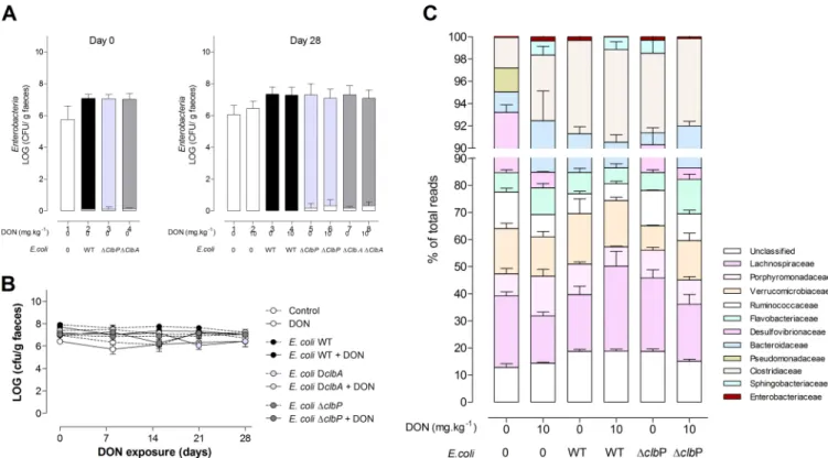
Contamination of the diet with DON does not impair the colonization of the
gut by E. coli strains producing colibactin or not producing colibactin. The next aim
was to assess whether DON could exacerbate the genotoxicity exerted by
colibactin-producing E. coli present in the gut microbiota. To this end, animals were colonized at
birth with E. coli WT or E. coli ΔclbA and E. coli ΔclbP mutants. After weaning, the rats
were fed for 4 weeks with a diet contaminated with DON or not contaminated with
DON (Fig. S1B). We first examined the effect of DON on the colonization of the gut by
E. coli and more generally on the microbiota. Ingestion of a DON-contaminated diet
from weaning to adulthood did not modify the levels of enterobacteria in the feces
FIG 3 Exposure to DON does not impact intestinal colonization by E. coli and the overall composition of the intestinal microbiota. (A) Members of the family Enterobacteriaceae (white bar) and E. coli (E. coli WT [black bar], E. coli ΔclbA [light gray bar], and E. coli ΔclbP [dark gray bar]) were quantified in fecal
homogenates at day 0 (postnatal day [PND] 28, the time of weaning) and day 28 (PND 58). (B) Quantification of E. coli in fecal homogenates after weaning (days 0 to 28) and exposure to a DON-contaminated diet (10 mg · kg⫺1) or no exposure to a DON-contaminated diet. Mean values⫾ SEM are shown (n ⫽ 9 or 10). DclbA, ΔclbA. (C) Evaluation of 16S microbiota diversity in adult animals (PND 58) colonized since birth by E. coli WT or E. coli ΔclbP or in animals in a control group and coexposed or not exposed to a DON-contaminated diet (10 mg · kg⫺1) (n⫽ 4).
mbio.asm.org
on June 15, 2017 - Published by
compared to those in animals fed a normal diet or the fecal E. coli counts (Fig. 3A and
B). A 16S microbiota analysis indicated that ingestion of the DON-contaminated diet
did not significantly alter the composition or diversity of the gut microbiota of rats
colonized at birth with E. coli strains producing colibactin or not producing colibactin
(Fig. 3C).
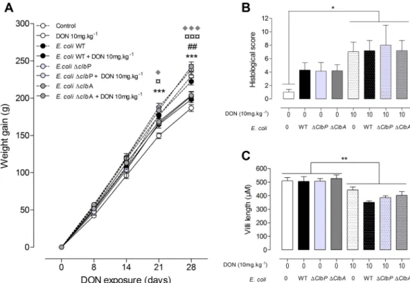
Exposure to DON reduces body weight gain and induces intestinal
modifica-tions in adult rats, independently of neonatal colonization by E. coli strains
producing colibactin or not producing colibactin. As expected, ingestion of the
DON-contaminated diet (10 mg · kg of body weight
⫺1) significantly decreased body
weight compared to animals fed a control diet (Fig. 4A). In addition, animal weight
decreased independently of neonatal colonization with the E. coli strain producing
colibactin or not producing colibactin. Similarly, as previously observed, in the jejuna of
animals fed the DON-contaminated diet, increased histological alterations were
ob-served, demonstrating moderate intestinal lesions and breakdown (Fig. 4B). A
de-creased villus height in the jejunum was also observed in these animals (Fig. 4C). These
histological and morphological modifications occurred independently of colonization
of animals by E. coli strains producing colibactin or not producing colibactin (Fig. 4B
and C). These results suggest that colibactin-producing strains did not impact the
classical effects of DON in relation to weight gain and morphology of the gut.
Exposure to DON exacerbates the intestinal DNA damage induced by
colibactin-producing E. coli in a dose- and time-dependent manner. The in vivo
effect of a diet contaminated with DON and colibactin-producing E. coli on intestinal
DNA damage was evaluated next. To this end, jejunal sections from animals colonized
at birth with E. coli producing colibactin or not producing colibactin that were fed a
FIG 4 Exposure to DON reduces body weight gain and alters the jejunal tissue in adult animals independently of the
colonizing E. coli strain. (A) Body weight gain was evaluated in the progeny from days 0 to 28. (B and C) Histological score (B) and villus length (C) in the jejuna of rats colonized since birth by E. coli strains producing colibactin or not producing colibactin or not colonized by E. coli and coexposed to a DON-contaminated diet (10 mg · kg⫺1) or not coexposed to a DON-contaminated diet. Mean values plus SEM are shown (n ⫽ 8 to 10). Values for animals fed a DON-contaminated diet (10 mg · kg⫺1) that are significantly different for the values for animals fed a control diet by one-way ANOVA with Bonferroni’s multiple-comparison correction are indicated by symbols as follows:* and ¤, P⬍ 0.05; ## and **, P ⬍ 0.01; ***, ¤¤¤, and ⽧⽧⽧,
P⬍ 0.0001. Values for control animals (*), animals colonized with E. coli WT (#), and animals colonized with the E. coli ΔclbP
mutant (¤) or with the E. coli ΔclbA mutant (}) are indicated.
control diet or a DON-contaminated diet for 4 weeks were stained for the
phosphor-ylated form of H2AX. Dietary exposure of animals to 10 mg · kg
⫺1DON alone did not
induce detectable DNA damage in intestinal epithelial cells (Fig. 5A, dotted white bar,
and B). Similarly, adult rats colonized with E. coli strains producing colibactin and fed
the control diet did not exhibit significant intestinal DNA damage (Fig. 5A, black bar).
However, in animals colonized with E. coli WT and fed a diet contaminated with DON
(10 mg · kg
⫺1), a significant increase in
␥H2AX-positive epithelial cells was observed
compared to rats exposed to DON or rats colonized with E. coli WT (Fig. 5A, dotted black
bar, and B). As observed in vitro, no genotoxicity was observed in animals fed a
DON-contaminated diet and colonized with non-colibactin-producing mutants (E. coli
ΔclbA and E. coli ΔclbP) (Fig. 5A and B). The increase in DNA double-strand breaks
observed in intestinal epithelial cells (IECs) of animals colonized since birth by
colibactin-producing E. coli and exposed to a DON-contaminated diet was associated
with a significant increase in activation of phosphorylated MAP kinase ERK p42/p44
(data not shown).
We next investigated the effect of the dose or duration of DON exposure on the
genotoxic effect of colibactin. We first assessed the effect of the dose of DON on DNA
damage in control animals or animals colonized with E. coli WT and fed diets
contam-inated with 2 or 10 mg · kg
⫺1DON (Fig. 6A). In rats exposed for 4 weeks to the lowest
dose of DON contamination, exacerbation of DNA damage induced by the genotoxic
E. coli strain was already observed (Fig. 6A). This effect was significantly less than the
one observed at 10 mg · kg
⫺1DON. We then determined the minimal duration of DON
exposure required to exacerbate the genotoxic effect of colibactin. A significant
increase in DNA damage in intestinal epithelial cells of rats colonized with E. coli WT was
observed from 2-week exposure to 10 mg · kg
⫺1DON, and DNA damage was increased
after a 4-week exposure to the food contaminant (Fig. 6B). Taken together, these
FIG 5 DON exacerbates DNA damage in jejunal epithelial cells of animals colonized by
colibactin-producing E. coli. Immunofluorescence analysis of the jejunal epithelium of adults (PND 58) colonized since birth by E. coli strains (E. coli WT, E. coli ΔclbA, or E. coli ΔclbP strain) or treated with PBS (control group) and coexposed to a DON-contaminated diet (10 mg · kg⫺1) for 4 weeks or not coexposed to a DON-contaminated diet for 4 weeks. (A) Quantification of the percentage of␥H2AX-positive cells in jejunal crypts. Mean values plus⫾ SEM are shown (n ⫽ 8 to 10). Values that are significantly different (P ⬍ 0.0001) by one-way ANOVA with Bonferroni’s multiple-comparison correction are indicated by four asterisks. (B) Representative jejunal frozen sections at PND 58. DNA was stained in blue.␥H2AX foci are shown in green. Bars⫽ 10M.
mbio.asm.org
on June 15, 2017 - Published by
results indicate that ingestion of a DON-contaminated diet exacerbates the
intes-tinal DNA damage induced by colibactin-producing E. coli in a dose- and
time-dependent manner.
DISCUSSION
In the present study, we demonstrated that DON exacerbates the DNA damage
induced by E. coli producing colibactin both in vitro on cultured intestinal epithelial
cells and in vivo in animals colonized with colibactin-producing E. coli and fed
DON-contaminated diets.
Several long-term studies showed that DON is not a carcinogenic compound (29,
30); consequently, this mycotoxin has been classified in group 3 (“not classifiable as to
its carcinogenicity to humans”) by the World Health Organization (WHO) International
Agency for Research on Cancer (IARC). In the present study, DON, used at realistic levels
(31, 32), did not induce detectable DNA damage in the intestine. In vitro, genotoxicity
was observed only upon exposure to very high nonrealistic levels of DON. On the other
hand, our in vitro and in vivo data demonstrated that DON exacerbates the genotoxicity
induced by colibactin-producing E. coli. This raises questions about the synergism
between food contaminants and gut microbiota with regard to intestinal
carcinogen-esis.
There is overwhelming evidence that DON induces a systemic and intestinal
inflam-matory response at both the systemic and intestinal levels (33–35). Through this
inflammatory effect, DON may predispose the gut epithelium to DNA damage. Indeed,
the genotoxic effect of colibactin requires bacterium-host cell contact (9). By decreasing
protective mucins and antimicrobial peptide production (36, 37), the inflammation
induced by DON could also create an environment in which colibactin-producing E. coli
bacteria more readily access the epithelium and express their genotoxic potential. This
hypothesis has already been proposed to explain the genotoxicity and tumorigenicity
of colibactin-producing E. coli in azoxymethane-treated IL-10
⫺/⫺mice (16). Likewise,
numerous studies demonstrated that DON induces oxidative stress (38); it stimulates
the production of reactive oxygen species (ROS) (39) but has no effect on the
produc-tion of nitric oxide (40). The rapid generaproduc-tion of ROS was proposed as one of the
mechanisms for DNA damage in hepatocytes and lymphocytes exposed to high doses
of toxin (41, 42). Secher et al. demonstrated that colibactin-producing E. coli strains also
trigger the production of intracellular and mitochondrial ROS in infected cells (12), and
a recent study shows, in jejunal explants exposed to DON, an increase in the production
FIG 6 DON exacerbates DNA damage in jejunal epithelium in a dose- and time-dependent manner.
Immunoflu-orescence analysis of jejunal epithelium was performed. (A and B) Quantification of␥H2AX-positive cells in jejuna of animals exposed to different doses of DON (2 mg · kg⫺1or 10 mg · kg⫺1) for 4 weeks and colonized or not since birth with E. coli WT (A) or exposed to DON (10 mg · kg⫺1) for 1, 2, or 4 weeks after weaning (B). Mean values plus SEM are shown (n⫽ 6 to 10). Values that are significantly different for control animals (*) or DON-exposed animals (¤) versus E. coli WT-colonized animals exposed to DON by one-way ANOVA with Bonferroni’s multiple comparisons are indicated by symbols as follows:*, P⬍ 0.05; ** and ¤¤, P ⬍ 0.01; **** and ¤¤¤¤, P ⬍ 0.000; ###, P ⬍ 0.001, group exposed to 2 mg · kg⫺1of DON versus group exposed to 10 mg · kg⫺1of DON in panel A; #, P⬍ 0.05, E. coli WT-colonized animals exposed to DON for 2 weeks versus E. coli WT-colonized animals exposed to DON for 4 weeks in panel B.
of COX-2 in the tissue (43). The induction of production of ROS by the mycotoxin and
bacteria could explain the exacerbated DNA double-strand breaks in intestinal
epithe-lial cells exposed to both stressors. Finally, DON is also known to activate MAPKs
through the phosphorylation of protein kinase R (PKR) (44–46). Recent data suggest
that PKR promotes genomic instability by inhibiting DNA damage response signaling
and double-strand break repair (47). Increased expression of PKR has also been
re-ported in patients with colon cancer (48). Thus, DON-induced phosphorylation of PKR
may exacerbate the genotoxicity induced by colibactin and explain the observed
synergism between colibactin and DON. Indeed, in the present study, an increased
activation of a MAP kinase was observed in animals colonized by colibactin-producing
E. coli and exposed to DON.
The prevalence of the specific phylogenetic B2 group, which encompasses E. coli
strains producing colibactin, is increasing among E. coli strains persisting in the
microbiota of humans from developed countries (6). DON is the most prevalent fungal
toxin present in the food chain in Europe and North America (49, 50). The worldwide
incidence of trichothecene contamination and especially of DON has increased in the
last years because of climate change, increased use of no-till farming to prevent soil
erosion, nonoptimal crop rotations, and inadequate fungicide treatments (51). Since
DON is resistant to milling, processing, and heating, this mycotoxin remains present in
final food products, such as bread and pasta, obtained from contaminated grain (52).
The DON concentrations tested in this study are in accordance with the levels plausibly
encountered in the gut after consumption of contaminated food (32). A large
percent-age of the human population can be exposed to both factors.
In conclusion, our results demonstrate that DON exacerbates the genotoxicity of
colibactin. Food contaminants and microbial factors act together to impact host
physiology and especially intestinal epithelial cells. This finding raises questions about
the interaction between food contaminants and gut microbiota in intestinal
carcino-genesis and underlines that the impact of food contaminants, especially mycotoxins,
must be evaluated together with the host microbiota.
MATERIALS AND METHODS
Bacterial strains and toxins. E. coli bacterial strains (E. coli strain M1/5 [14]), bacterial growth
conditions, and the use of toxins used in this study are listed in Text S1 and Table S1 in the supplemental material. Purified DON was purchased from Sigma-Aldrich (Saint-Quentin Fallavier, France).
Cell culture. Nontransformed rat intestinal epithelial IEC-6 cells (ATCC CRL-1592) were cultured as
described before (14). Experimental procedures were described in Text S1 and Fig. S1A. At the end of the treatments, in-cell Western (ICW) procedure and immunofluorescence analysis were performed to analyze DNA damage via phosphorylation of the histone H2AX.
The ICW procedure was performed as previously described (53). The cells were fixed, permeabilized, blocked, and then incubated overnight (ON) with rabbit monoclonal anti-␥H2AX 1/200 (20E3; Cell Signaling, Saint-Quentin en Yvelines, France). An infrared fluorescent secondary antibody (IRDye 800CW; Rockland) (1/500) was applied simultaneously with RedDot2 (1/500) (Biotium, Interchim, Montluçon, France) for DNA labeling. The DNA and␥H2AX were visualized using an Odyssey infrared imaging scanner (LI-COR Science Tec, Les Ulis, France). All experiments were carried out in triplicate.
Experimental animal model. Pregnant Wistar female rats (obtained from Janvier Labs, Le Genest
Saint-Isle, France) were treated with streptomycin (5 g/liter) and inoculated twice with 109bacteria by intragastric gavage (14). Animals were given noncontaminated control food or a DON-contaminated diet (10 mg or 2 mg of DON · kg of body weight⫺1) for 1 to 4 weeks (Fig. S1B and Text S1).
Colonic bacterial load and 16S microbiota analysis. The colonic bacterial load in feces was
analyzed before exposure to a DON-contaminated diet at postnatal day 28 (PND 28) and upon completion of the experiment (PND 58). For 16S microbiota analysis, feces were taken at the end of the experiment. Total DNA was isolated from the individual fecal contents using the QIAamp DNA stool minikit (Qiagen, Courtaboeuf, France) (Text S1).
Histological analysis. The jejuna fixed in 10% buffered formalin were dehydrated and embedded in
paraffin in accordance with standard histological procedures. Sections (5 m) were stained with hematoxylin-eosin for histopathological evaluation and intestinal morphometry. A lesional score was designed to compare histological changes (44). Images were acquired with a Leica DMRB microscope. Analyses were performed using a MOTIC Image Plus 2.0 image analysis system.
Immunofluorescence analysis. Jejunal samples were immediately placed in
optimum-cutting-temperature (OCT; Sakura) compound and snap-frozen at ⫺80°C (14). Sections (5m) were fixed, permeabilized, blocked with phosphate-buffered saline (PBS) containing 0.1% Tween 20 and 5% normal goat serum (NGS) and stained with rabbit anti-phospho-H2AX antibody (Cell Signaling) followed by Alexa Fluor 546-labeled goat anti-rabbit antibodies (Invitrogen). Slides were mounted in Vectashield containing
mbio.asm.org
on June 15, 2017 - Published by
mBio.00007-17
.
TEXT S1, DOCX file, 0.02 MB.
FIG S1, TIF file, 0.5 MB.
FIG S2, TIF file, 0.1 MB.
FIG S3, TIF file, 1.4 MB.
TABLE S1, PDF file, 0.1 MB.
TABLE S2, PDF file, 0.1 MB.
ACKNOWLEDGMENTS
We are grateful to V. Theodorou, M. Olier, C. Cartier, and T. Gauthier (Toxalim) for
their scientific input and help in immunofluorescence on jejunal samples and confocal
microcopy analysis. We thank the staff of the team EZOP for animal care and members
of the histopathology platform of UMS US 006 and imagery platform of CPTP for
excellent technical help. We are grateful to the UPAE (Unité de Préparation des
Aliments Expérimentaux, UE0300, Jouy-en-Josas, France) for the preparation of the
diets. Thank you to J. P. Nougayrede (IRSD) for reading the manuscript. We thank Alice
Darroux Murray for editing the language.
This work was supported by grants from the French National Research Agency
(ANR-10-CESA-0012-05, ANR-15-CE21-0001-02, BSV3-0015-02, and
ANR-13-BSV1-0028-01) and the French Institute of Cancer (grant INCA-PLBIO13-123). U.D. was
supported by the Interdisciplinary Center for Clinical Research (IZKF) Münster (project
Dob2/013/12).
REFERENCES
1. Eckburg PB, Bik EM, Bernstein CN, Purdom E, Dethlefsen L, Sargent M, Gill SR, Nelson KE, Relman DA. 2005. Diversity of the human intestinal microbial flora. Science 308:1635–1638.https://doi.org/10.1126/science .1110591.
2. Rautava S, Luoto R, Salminen S, Isolauri E. 2012. Microbial contact during pregnancy, intestinal colonization and human disease. Nat Rev Gastro-enterol Hepatol 9:565–576.https://doi.org/10.1038/nrgastro.2012.144. 3. Sommer F, Bäckhed F. 2013. The gut microbiota—masters of host
development and physiology. Nat Rev Microbiol 11:227–238.https://doi .org/10.1038/nrmicro2974.
4. Palmer C, Bik EM, DiGiulio DB, Relman DA, Brown PO. 2007. Develop-ment of the human infant intestinal microbiota. PLoS Biol 5:e177. https://doi.org/10.1371/journal.pbio.0050177.
5. Clermont O, Christenson JK, Denamur E, Gordon DM. 2013. The Cler-mont Escherichia coli phylo-typing method revisited: improvement of specificity and detection of new phylo-groups. Environ Microbiol Rep 5:58 – 65.https://doi.org/10.1111/1758-2229.12019.
6. Tenaillon O, Skurnik D, Picard B, Denamur E. 2010. The population genetics of commensal Escherichia coli. Nat Rev Microbiol 8:207–217. https://doi.org/10.1038/nrmicro2298.
7. Massot M, Daubié AS, Clermont O, Jauréguy F, Couffignal C, Dahbi G, Mora A, Blanco J, Branger C, Mentré F, Eddi A, Picard B, Denamur E, The Coliville Group. 2016. Phylogenetic, virulence and antibiotic resistance characteristics of commensal strain populations of Escherichia coli from community subjects in the Paris area in 2010 and evolution over 30 years. Microbiology 162:642– 650.https://doi.org/10.1099/mic.0.000242. 8. Touchon M, Hoede C, Tenaillon O, Barbe V, Baeriswyl S, Bidet P, Bingen E, Bonacorsi S, Bouchier C, Bouvet O, Calteau A, Chiapello H, Clermont O, Cruveiller S, Danchin A, Diard M, Dossat C, Karoui ME, Frapy E, Garry L,
Ghigo JM, Gilles AM, Johnson J, Le Bouguénec C, Lescat M, Mangenot S, Martinez-Jéhanne V, Matic I, Nassif X, Oztas S, Petit MA, Pichon C, Rouy Z, Ruf CS, Schneider D, Tourret J, Vacherie B, Vallenet D, Médigue C, Rocha EP, Denamur E. 2009. Organised genome dynamics in the Esch-erichia coli species results in highly diverse adaptive paths. PLoS Genet 5:e1000344.https://doi.org/10.1371/journal.pgen.1000344.
9. Nougayrède JP, Homburg S, Taieb F, Boury M, Brzuszkiewicz E, Gottschalk G, Buchrieser C, Hacker J, Dobrindt U, Oswald E. 2006. Escherichia coli induces DNA double-strand breaks in eukaryotic cells. Science 313:848 – 851.https://doi.org/10.1126/science.1127059. 10. Putze J, Hennequin C, Nougayrède JP, Zhang W, Homburg S, Karch H,
Bringer MA, Fayolle C, Carniel E, Rabsch W, Oelschlaeger TA, Oswald E, Forestier C, Hacker J, Dobrindt U. 2009. Genetic structure and distribu-tion of the colibactin genomic island among members of the family Enterobacteriaceae. Infect Immun 77:4696 – 4703. https://doi.org/10 .1128/IAI.00522-09.
11. Cuevas-Ramos G, Petit CR, Marcq I, Boury M, Oswald E, Nougayrède JP. 2010. Escherichia coli induces DNA damage in vivo and triggers genomic instability in mammalian cells. Proc Natl Acad Sci USA 107:11537–11542. https://doi.org/10.1073/pnas.1001261107.
12. Secher T, Samba-Louaka A, Oswald E, Nougayrede JP. 2013. Escherichia coli producing colibactin triggers premature and transmissible senes-cence in mammalian cells. PLoS One 8:e77157.https://doi.org/10.1371/ journal.pone.0077157.
13. Cougnoux A, Dalmasso G, Martinez R, Buc E, Delmas J, Gibold L, Sauva-net P, Darcha C, Déchelotte P, BonSauva-net M, Pezet D, Wodrich H, Darfeuille-Michaud A, Bonnet R. 2014. Bacterial genotoxin colibactin promotes colon tumour growth by inducing a senescence-associated secretory
phenotype. Gut 63:1932–1942. https://doi.org/10.1136/gutjnl-2013 -305257.
14. Payros D, Secher T, Boury M, Brehin C, Ménard S, Salvador-Cartier C, Cuevas-Ramos G, Watrin C, Marcq I, Nougayrède JP, Dubois D, Bedu A, Garnier F, Clermont O, Denamur E, Plaisancié P, Theodorou V, Fioramonti J, Olier M, Oswald E. 2014. Maternally acquired genotoxic Escherichia coli alters offspring’s intestinal homeostasis. Gut Microbes 5:313–325. https://doi.org/10.4161/gmic.28932.
15. Secher T, Payros D, Brehin C, Boury M, Watrin C, Gillet M, Bernard-Cadenat I, Menard S, Theodorou V, Saoudi A, Olier M, Oswald E. 2015. Oral tolerance failure upon neonatal gut colonization with Escherichia coli producing the genotoxin colibactin. Infect Immun 83:2420 –2429. https://doi.org/10.1128/IAI.00064-15.
16. Arthur JC, Perez-Chanona E, Muhlbauer M, Tomkovich S, Uronis JM, Fan T-J, Campbell BJ, Abujamel T, Dogan B, Rogers AB, Rhodes JM, Stintzi A, Simpson KW, Hansen JJ, Keku TO, Fodor AA, Jobin C. 2012. Intestinal inflammation targets cancer-inducing activity of the microbiota. Science 338:120 –123.https://doi.org/10.1126/science.1224820.
17. Cougnoux A, Delmas J, Gibold L, Faïs T, Romagnoli C, Robin F, Cuevas-Ramos G, Oswald E, Darfeuille-Michaud A, Prati F, Dalmasso G, Bonnet R. 2016. Small-molecule inhibitors prevent the genotoxic and protumoural effects induced by colibactin-producing bacteria. Gut 65:278 –285. https://doi.org/10.1136/gutjnl-2014-307241.
18. Nowrouzian FL, Oswald E. 2012. Escherichia coli strains with the capacity for long-term persistence in the bowel microbiota carry the potentially genotoxic pks island. Microb Pathog 53:180 –182. https://doi.org/10 .1016/j.micpath.2012.05.011.
19. Tronnet S, Garcie C, Rehm N, Dobrindt U, Oswald E, Martin P. 2016. Iron homeostasis regulates the genotoxicity of Escherichia coli that produces colibactin. Infect Immun 84:3358 –3368. https://doi.org/10.1128/IAI .00659-16.
20. Wu F, Groopman JD, Pestka JJ. 2014. Public health impacts of foodborne mycotoxins. Annu Rev Food Sci Technol 5:351–372.https://doi.org/10 .1146/annurev-food-030713-092431.
21. European Food Safety Authority. 2013. Deoxynivalenol in food and feed: occurrence and exposure. EFSA J 11:3379.
22. Pieters MN, Freijer J, Baars BJ, Fiolet DC, van Klaveren J, Slob W. 2002. Risk assessment of deoxynivalenol in food: concentration limits, expo-sure and effects. Adv Exp Med Biol 504:235–248.https://doi.org/10.1007/ 978-1-4615-0629-4_25.
23. Turner PC, Burley VJ, Rothwell JA, White KL, Cade JE, Wild CP. 2008. Deoxynivalenol: rationale for development and application of a urinary biomarker. Food Addit Contam Part A Chem Anal Control Expo Risk Assess 25:864 – 871.https://doi.org/10.1080/02652030801895040. 24. Pestka JJ, Smolinski AT. 2005. Deoxynivalenol: toxicology and potential
effects on humans. J Toxicol Environ Health B Crit Rev 8:39 – 69.https:// doi.org/10.1080/10937400590889458.
25. Cano PM, Seeboth J, Meurens F, Cognie J, Abrami R, Oswald IP, Guzylack-Piriou L. 2013. Deoxynivalenol as a new factor in the persistence of intestinal inflammatory diseases: an emerging hypothesis through pos-sible modulation of Th17-mediated response. PLoS One 8:e53647. https://doi.org/10.1371/journal.pone.0053647.
26. Pinton P, Oswald IP. 2014. Effect of deoxynivalenol and other type B trichothecenes on the intestine: a review. Toxins (Basel) 6:1615–1643. https://doi.org/10.3390/toxins6051615.
27. Pestka JJ. 2010. Deoxynivalenol: mechanisms of action, human expo-sure, and toxicological relevance. Arch Toxicol 84:663– 679.https://doi .org/10.1007/s00204-010-0579-8.
28. Pierron A, Mimoun S, Murate LS, Loiseau N, Lippi Y, Bracarense A-PFL, Liaubet L, Schatzmayr G, Berthiller F, Moll W-D, Oswald IP. 2016. Intes-tinal toxicity of the masked mycotoxin deoxynivalenol-3-beta-D -glucoside. Arch Toxicol 90:2037–2046.https://doi.org/10.1007/s00204 -015-1592-8.
29. Iverson F, Armstrong C, Nera E, Truelove J, Fernie S, Scott P, Stapley R, Hayward S, Gunner S. 1995-1996. Chronic feeding study of deoxyniva-lenol in B6C3F1 male and female mice. Teratog Carcinog Mutagen 15:283–306.
30. Bondy GS, Coady L, Curran I, Caldwell D, Armstrong C, Aziz SA, Nunnik-hoven A, Gannon AM, Liston V, Shenton J, Mehta R. 2016. Effects of chronic deoxynivalenol exposure on p53 heterozygous and p53 ho-mozygous mice. Food Chem Toxicol 96:24 –34.https://doi.org/10.1016/ j.fct.2016.07.018.
31. Sergent T, Parys M, Garsou S, Pussemier L, Schneider YJ, Larondelle Y. 2006. Deoxynivalenol transport across human intestinal Caco-2 cells and
its effects on cellular metabolism at realistic intestinal concentrations. Toxicol Lett 164:167–176.https://doi.org/10.1016/j.toxlet.2005.12.006. 32. Marin S, Ramos AJ, Cano-Sancho G, Sanchis V. 2013. Mycotoxins:
occur-rence, toxicology, and exposure assessment. Food Chem Toxicol 60: 218 –237.https://doi.org/10.1016/j.fct.2013.07.047.
33. Bracarense APFL, Basso KM, Da Silva EO, Payros D, Oswald IP. 11 October 2016. Deoxynivalenol in the liver and lymphoid organs of rats: effects of dose and duration on immunohistological changes. World Mycotoxin J https://doi.org/10.3920/WMJ2016.2094.
34. Payros D, Alassane-Kpembi I, Pierron A, Loiseau N, Pinton P, Oswald IP. 2016. Toxicology of deoxynivalenol and its acetylated and modified forms. Arch Toxicol 90:2931–2957.https://doi.org/10.1007/s00204-016 -1826-4.
35. Alassane-Kpembi I, Puel O, Pinton P, Cossalter AM, Chou TC, Oswald IP. 3 December 2016. Co-exposure to low doses of the food contaminants deoxynivalenol and nivalenol has a synergistic inflammatory effect on intestinal explants. Arch Toxicol https://doi.org/10.1007/s00204-016 -1902-9.
36. Rhodes JM, Campbell BJ. 2002. Inflammation and colorectal cancer: IBD-associated and sporadic cancer compared. Trends Mol Med 8:10 –16. https://doi.org/10.1016/S1471-4914(01)02194-3.
37. Inaba Y, Ashida T, Ito T, Ishikawa C, Tanabe H, Maemoto A, Watari J, Ayabe T, Mizukami Y, Fujiya M, Kohgo Y. 2010. Expression of the antimicrobial peptide alpha-defensin/cryptdins in intestinal crypts de-creases at the initial phase of intestinal inflammation in a model of inflammatory bowel disease, IL-10-deficient mice. Inflamm Bowel Dis 16:1488 –1495.https://doi.org/10.1002/ibd.21253.
38. Mishra S, Dwivedi PD, Pandey HP, Das M. 2014. Role of oxidative stress in deoxynivalenol induced toxicity. Food Chem Toxicol 72:20 –29. https://doi.org/10.1016/j.fct.2014.06.027.
39. Krishnaswamy R, Devaraj SN, Padma VV. 2010. Lutein protects HT-29 cells against deoxynivalenol-induced oxidative stress and apoptosis: prevention of NF-kappaB nuclear localization and down regulation of NF-kappaB and cyclo-oxygenase-2 expression. Free Radic Biol Med 49: 50 – 60.https://doi.org/10.1016/j.freeradbiomed.2010.03.016.
40. Graziani F, Pujol A, Nicoletti C, Pinton P, Armand L, Di Pasquale E, Oswald IP, Perrier J, Maresca M. 2015. The food-associated ribotoxin deoxyniva-lenol modulates inducible NO synthase in human intestinal cell model. Toxicol Sci 145:372–382.https://doi.org/10.1093/toxsci/kfv058. 41. Awad WA, Ghareeb K, Dadak A, Hess M, Böhm J. 2014. Single and
combined effects of deoxynivalenol mycotoxin and a microbial feed additive on lymphocyte DNA damage and oxidative stress in broiler chickens. PLoS One 9:e88028. https://doi.org/10.1371/journal.pone .0088028.
42. Singh S, Banerjee S, Chattopadhyay P, Borthakur SK, Veer V. 2015. Deoxynivalenol induces cytotoxicity and genotoxicity in animal primary cell culture. Toxicol Mech Methods 25:184 –191.https://doi.org/10.3109/ 15376516.2015.1006743.
43. Olegário E, Gerez JR, Drape C, Bracarense APFRL. 2014. Phytic acid decreases deoxynivalenol and fumonisin B1-induced changes on swine jejunal explants. Toxicol Rep 1:284 –292.https://doi.org/10.1016/j.toxrep .2014.05.001.
44. Lucioli J, Pinton P, Callu P, Laffitte J, Grosjean F, Kolf-Clauw M, Oswald IP, Bracarense AP. 2013. The food contaminant deoxynivalenol activates the mitogen activated protein kinases in the intestine: interest of ex vivo models as an alternative to in vivo experiments. Toxicon 66:31–36. https://doi.org/10.1016/j.toxicon.2013.01.024.
45. Zhou H-R, He K, Landgraf J, Pan X, Pestka JJ. 2014. Direct activation of ribosome-associated double-stranded RNA-dependent protein kinase (PKR) by deoxynivalenol, anisomycin and ricin: a new model for ribotoxic stress response induction. Toxins (Basel) 6:3406 –3425.https://doi.org/ 10.3390/toxins6123406.
46. Pierron A, Mimoun S, Murate LS, Loiseau N, Lippi Y, Bracarense A-PFL, Schatzmayr G, He JW, Zhou T, Moll W-D, Oswald IP. 2016. Microbial biotransformation of DON: molecular basis for reduced toxicity. Sci Rep 6:29105.https://doi.org/10.1038/srep29105.
47. Cheng X, Byrne M, Brown KD, Konopleva MY, Kornblau SM, Bennett RL, May WS. 2015. PKR inhibits the DNA damage response, and is associated with poor survival in AML and accelerated leukemia in NHD13 mice. Blood 126:1585–1594.https://doi.org/10.1182/blood-2015-03-635227. 48. Kim SH, Gunnery S, Choe JK, Mathews MB. 2002. Neoplastic progression
in melanoma and colon cancer is associated with increased expression and activity of the interferon-inducible protein kinase, PKR. Oncogene 21:8741– 8748.https://doi.org/10.1038/sj.onc.1205987.
mbio.asm.org
on June 15, 2017 - Published by