Bioengineered Fascicle-Like Skeletal Muscle Tissue Constructs
The MIT Faculty has made this article openly available.
Please share
how this access benefits you. Your story matters.
Citation
Neal, Devin, et al. “Bioengineered Fascicle-Like Skeletal Muscle
Tissue Constructs.” ASME 2012 Summer Bioengineering
Conference, Parts A and B, 20-23 June, 2012, Fajardo, Puerto Rico,
ASME, 2012, p. 1133.
As Published
http://dx.doi.org/10.1115/SBC2012-80228
Publisher
ASME International
Version
Final published version
Citable link
http://hdl.handle.net/1721.1/118781
Terms of Use
Article is made available in accordance with the publisher's
policy and may be subject to US copyright law. Please refer to the
publisher's site for terms of use.
Copyright © 2012 by ASME 1
INTRODUCTION
Tissue engineered skeletal muscle constructs have and will continue to be valuable in treating, and testing various muscle injuries and diseases. However a significant drawback to numerous methods of producing 3D skeletal muscle constructs grown in vitro is that muscle cell density as a fraction of total volume or mass, is often significantly lower than muscle found in vivo. Therefore a method to increase muscle cell density within a construct is needed.
Skeletal muscle typically begins as myoblasts that proliferate until achieving a high enough local density, then they begin to fuse and form multinucleated myotubes. The myotubes mature into long contractile muscle cells. Alignment of force generating cells is important and has been accomplished with axial stress and geometric constraints. Constructs with unaligned cells do not produce axial force as efficiently.
Skeletal muscles engineered on 2D substrates are limited in thickness, and thus scale. Additionally, substrate grown constructs are not 3D as muscles in vivo are. Numerous 3D muscle constructs are much larger in scale than the 2D tissues; however they lack scalability due largely to diffusion limitations at higher scales. Centrally located cells tend to migrate to the edges, become sick, and/or die resulting in much lower cell density than that found in muscles grown in vivo.
There are two significant advantages to increasing cell density. The first is that there are more cells to produce force, increasing the net force per area, or stress, of the construct. Second, there is less stiff material within the construct to inhibit contraction of the construct, allowing for greater displacement per length, or strain.
We increase muscle cell density by growing constructs in a geometry that is similar to the naturally occurring hierarchical muscle level of the fascicle. A fascicle is a bundle of muscles cells that is small enough in diameter to allow for perfusion of nutrients and
innervation. Essentially, it is an optimized building block that is robust against individual cell failure, and can be combined in a parallel arrangement allowing for scalability.
We are capable of growing numerous fascicle-like structures in a single substrate free, high throughput device. Once the constructs are formed, they may be coated with an ECM-like connective tissue and bundled for scaling to larger dimensions.
This abstract presents a geometrically fascicle-like muscle construct that provides a muscle cell density that is closer to approaching that of muscle tissue grown in vivo.
MATERIALS AND METHODS
C2C12 mouse myoblasts (Ameican Type Culture Collection) are cultured in growth medium (GM) containing DMEM (American Type Culture Collection), supplemented with 10% fetal bovine serum (FBS, Sigma-Aldrich), 1% penicillin-streptomycin 100X (Invitrogen), and 0.1 mg/ml Normacin (Invivogen). Confluence was kept below 70% and cells were seeded into the experimental device at the 5th passage.
Cell/gel suspension is molded such that a uniform cylinder of controlled dimensions is suspended in medium anchored at both ends to the walls of a well. The cell/gel suspension contains GM with C2C12 cells at a concentration of 15e6 cells/ml, fibrinogen (Sigma-Aldrich) at a concentration of 5 mg/ml, and 6-aminocaproic acid (Sigma-Aldrich) at a concentration of 1 mg/ml. The initial fluid surrounding the construct contains thrombin (Sigma-Aldrich) at a concentration of 1 U/ml. The wells are 5 mm in diameter, with walls made of polydimethylsiloxane (PDMS, Dow Corning) plasma bonded to a glass coverslip (VWR) which constitutes the bottom of the well. The length of the molded gels are nominally 5 mm, and the initial nominal diameters of the molded gels range from 250 µm to 500 µm.
BIOENGINEERED FASCICLE-LIKE SKELETAL MUSCLE TISSUE CONSTRUCTS
Devin Neal, Mahmut Selman Sakar, H. Harry Asada
Department of Mechanical Engineering Massachusetts Institute of Technology
Cambridge, MA, 02139, USA
Proceedings of the ASME 2012 Summer Bioengineering Conference SBC2012 June 20-23, 2012, Fajardo, Puerto Rico
SBC2012-80228
Copyright © 2012 by ASME 2
After 24 hours, the initial fluid is changed to GM supplemented with 1 mg/ml 6-aminocaproic acid. This medium is replaced daily for 2 more days, then switched to differentiation medium, DM, containing DMEM supplemented with 10% horse serum (Sigma-Aldrich) 1% pen-strep, 0.1 mg/ml Normacin, and 1 mg/ml 6-aminocaproic acid. DM is changed daily until the termination of the experiment.
350 μm 250 μm
300 μm
350 μm
Figure 1. Phase contrast images of muscle constructs with contracted gel of differing initial diameter (left) 2 weeks after seeding.
(a) (b)
Figure 2. 2 Photon images of F-actin (red) and nuclei (green) from the midplane, a), and bottom plane, b), of a 3 week old construct. Some myotubes are indicated by white arrows in a). Evidence of sarcomeric striations indicated by yellow arrow in b). Scale bar is 50 µm.
RESULTS
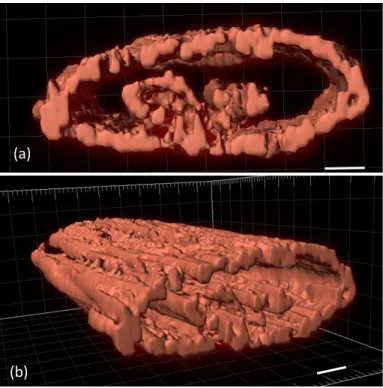
Constructs 5 mm long contract radially and mature over numerous weeks in an axially uniform manner. After initial seeding, the fibrin gel contracts radially a substantial amount within the first 2 days, reducing the diameter by a factor of approximately 2, as shown in Fig. 1. The initial gel contraction does not occur without the presence of cells. After initial contraction, the diameter of the construct remains relatively constant over up to 3 weeks. The initial cross section is a uniform circle. As the construct matures, the thickness along the gravitational axis decreases, as shown in Fig 3 a). Upon seeding, the gel geometry and cell distribution are uniform along the axis of the construct, and this uniformity along the axis is maintained throughout the experiment, as shown in Figs. 1, 2 and 3 b). For given initial gel diameters, the construct geometric properties are uniform and repeatable.
As the construct matures, aligned myotubes form that occupy a substantial fraction of the construct cross sectional area. Actin forms aligned with the axis of the construct, and the nuclei elongate along the same axis as seen in Fig. 2. Myotubes are more mature in cells along the bottom plane (Fig. 2 b)) and in the center (Fig. 2 a)) of the construct as opposed to the less mature cells along the sides (Fig. 2 a)) and top (not shown). The myotubes in the center and along the bottom extend mm in length and evidence of sarcomeric striations are visible (Fig. 2 b)).
The maturity disparity is due to initial settling of cells in the gel, and migration. As the cell/gel solution stiffens, the cells tend to flow due to gravity. This creates a concentration of cells close to the bottom of the gel. The cells proliferate and then some fuse and mature into myotube, while others migrate along the surface of the gel forming a monolayer along the surface. The cells break down the fibrin gel over time. Because there are more cells for a longer period of time along the bottom of the construct, the gel along the bottom is consumed fastest, and results in the decrease in thickness previously noted. The tension in the gel and geometric alignment cues promote alignment as the myoblasts fuse and mature into myotubes. The myotubes along the surface combined with the myotubes deeper within the construct both contribute to increasing the myotube density. The cross sectional area occupied by cells is consistently 30% of the total cross sectional area along the length of the construct and over multiple constructs, much higher than the approximate <5% found in other muscle constructs. Upon contraction, higher muscle cell density will result in higher normalized contractile force.
(a)
(b)
Figure 3. 3-D visualization of F-actin along typical 256 µm section of 3 week old construct. 10 µm scale bar.
ACKNOWLEDGMENTS
This material is based upon work supported by the National Science Foundation under the Science and Technology Center Emergent Behaviors of Integrated Cellular Systems (EBICS) Grant No. CBET-0939511.