Publisher’s version / Version de l'éditeur:
Journal of Thermal Spray Technology, 19, 1-2, pp. 336-343, 2009-12-18
READ THESE TERMS AND CONDITIONS CAREFULLY BEFORE USING THIS WEBSITE. https://nrc-publications.canada.ca/eng/copyright
Vous avez des questions? Nous pouvons vous aider. Pour communiquer directement avec un auteur, consultez la première page de la revue dans laquelle son article a été publié afin de trouver ses coordonnées. Si vous n’arrivez pas à les repérer, communiquez avec nous à PublicationsArchive-ArchivesPublications@nrc-cnrc.gc.ca.
Questions? Contact the NRC Publications Archive team at
PublicationsArchive-ArchivesPublications@nrc-cnrc.gc.ca. If you wish to email the authors directly, please see the first page of the publication for their contact information.
NRC Publications Archive
Archives des publications du CNRC
This publication could be one of several versions: author’s original, accepted manuscript or the publisher’s version. / La version de cette publication peut être l’une des suivantes : la version prépublication de l’auteur, la version acceptée du manuscrit ou la version de l’éditeur.
For the publisher’s version, please access the DOI link below./ Pour consulter la version de l’éditeur, utilisez le lien DOI ci-dessous.
https://doi.org/10.1007/s11666-009-9442-y
Access and use of this website and the material on it are subject to the Terms and Conditions set forth at
HVOF-sprayed nano TiO
₂-HA coatings exhibiting enhanced
biocompatibility
Lima, R. S.; Dimitrievska, S.; Bureau, M. N.; Marple, B. R.; Petiti, A.; Mwale,
F.; Antoniou, J.
https://publications-cnrc.canada.ca/fra/droits
L’accès à ce site Web et l’utilisation de son contenu sont assujettis aux conditions présentées dans le site LISEZ CES CONDITIONS ATTENTIVEMENT AVANT D’UTILISER CE SITE WEB.
NRC Publications Record / Notice d'Archives des publications de CNRC:
https://nrc-publications.canada.ca/eng/view/object/?id=a39c67ce-f097-4211-bea7-784c8ad76161 https://publications-cnrc.canada.ca/fra/voir/objet/?id=a39c67ce-f097-4211-bea7-784c8ad76161HVOF-Sprayed Nano TiO
2
-HA Coatings
Exhibiting Enhanced Biocompatibility
R.S. Lima, S. Dimitrievska, M.N. Bureau, B.R. Marple, A. Petit, F. Mwale, and J. Antoniou (Submitted April 21, 2009; in revised form September 15, 2009)
Biomedical thermal spray coatings produced via high-velocity oxy-fuel (HVOF) from nanostructured titania (n-TiO2) and 10 wt.% hydroxyapatite (HA) (n-TiO2-10wt.%HA) powders have been engineered
as possible future alternatives to HA coatings deposited via air plasma spray (APS). This approach was chosen due to (i) the stability of TiO2in the human body (i.e., no dissolution) and (ii) bond strength
values on Ti-6Al-4V substrates more than two times higher than those of APS HA coatings. To explore the bioperformance of these novel materials and coatings, human mesenchymal stem cells (hMSCs) were cultured from 1 to 21 days on the surface of HVOF-sprayed n-TiO2and n-TiO2-10 wt.%HA coatings.
APS HA coatings and uncoated Ti-6Al-4V substrates were employed as controls. The profiles of the hMSCs were evaluated for (i) cellular proliferation, (ii) biochemical analysis of alkaline phosphatase (ALP) activity, (iii) cytoskeleton organization (fluorescent/confocal microscopy), and (iv) cell/substrate interaction via scanning electron microscopy (SEM). The biochemical analysis indicated that the hMSCs cultured on n-TiO2-10 wt.%HA coatings exhibited superior levels of bioactivity than hMSCs cultured on
APS HA and pure n-TiO2 coatings. The cytoskeleton organization demonstrated a higher degree of
cellular proliferation on the HVOF-sprayed n-TiO2-10wt.%HA coatings when compared to the control
coatings. These results are considered promising for engineering improved performance in the next generation of thermally sprayed biomedical coatings.
Keywords biocompatibility, biomedical coating, human mesenchymal stem cells (hMSCs), HVOF spray-ing, implants, nanostructured TiO2
1. Introduction
1.1 Hydroxyapatite Coatings
Hydroxyapatite (HA) coatings deposited via air plasma spray (APS) have been successfully employed as bio-medical coatings for bio-medical implants (e.g., load bearing bone implants such as hip-joints) (Ref 1). Despite the success of APS HA coatings, there are still concerns regarding their long-term performance related to the HAs
physiologic post-implantation stability, i.e., dissolution and osteolysis (pathologic process involving resorption of bone surrounding the implant) (Ref2-4). Therefore, there is need to improve the performance of the current bio-medical APS HA coatings.
1.2 HVOF-sprayed n-TiO2as a Biomedical Coating
A possible alternative to improve the performance of APS HA coatings is based on the use of nanostructured titania (n-TiO2). Nanostructured agglomerated TiO2
powders produced via spray-drying of individual n-TiO2
particles have been thermally sprayed via the high-velocity oxy-fuel (HVOF) technique as an attempt to produce a new class of biomedical coating (Ref 5,6). This coating was chosen due to the fact that it is a biocompatible material and it is not significantly absorbed/dissolved in the human body. In addition, it exhibits bond strength levels on Ti-6Al-4V substrates higher than 77 MPa (i.e., the epoxy glue breaks before the coating fails during the tensile adhesion test ASTM C633). Bond strength levels of APS HA coatings on Ti-6Al-4V substrates are normally below 30 MPa (Ref5,6). The high bond strength of these HVOF-sprayed n-TiO2 coatings is of pivotal importance
as the ‘‘relatively low’’ bond strength values of standard HA coatings in addition to resorption has led to late delamination and or debonding (Ref 2-4). In addition, previous preliminary studies demonstrated that the HVOF-sprayed n-TiO2coatings support fetal rat calvaria
osteoblast cell growth and matrix production in levels equivalent to those of APS HA coatings (Ref5,6). In vivo experiments were also performed. Coatings were deposited This article is an invited paper selected from presentations at the
2009 International Thermal Spray Conference and has been expanded from the original presentation. It is simultaneously published in Expanding Thermal Spray Performance to New Markets and Applications: Proceedings of the 2009 International
Thermal Spray Conference, Las Vegas, Nevada, USA, May 4-7,
2009, Basil R. Marple, Margaret M. Hyland, Yuk-Chiu Lau, Chang-Jiu Li, Rogerio S. Lima, and Ghislain Montavon, Ed., ASM International, Materials Park, OH, 2009.
R.S. Lima, S. Dimitrievska, M.N. Bureau, and B.R. Marple, National Research Council of Canada, 75 de Mortagne Blvd., Boucherville, QC J4B 6Y4, Canada; andA. Petit, F. Mwale, and J. Antoniou, Lady Davis Institute for Medical Research, Jewish General Hospital, McGill University, 3755 Coˆte Ste-Catherine Road, Montreal, QC H3T 1E2, Canada. Contact e-mail: rogerio. lima@cnrc-nrc.gc.ca. JTTEE5 19:336–343 DOI: 10.1007/s11666-009-9442-y 1059-9630/$19.00 Ó ASM International
Peer
Reviewed
on small Ti-6Al-4V rods that were subsequently implanted in the femurs of rabbits. Grit-blasted Ti-6Al-4V rods without a coating were also implanted and served as a control. After 7 weeks of implantation, the rabbits were euthanized and the contact surface between the bone and implant was measured/evaluated via optical microscopy. On average, the contact surface between the HVOF-sprayed n-TiO2 coating and bone was 1.7 times higher
than that of the uncoated Ti-6Al-4V rods (Ref5). 1.3 Enhanced Biocompatibility of Nanomaterials
Nanostructured zones were observed on the surface of the HVOF-sprayed n-TiO2coatings (Fig. 1). These zones
were formed when semi-molten nanostructured agglom-erated TiO2 particles impinged and re-solidified on the
previously deposited layers during thermal spraying (Ref5,6). The importance of the presence of nanozones on the coating surface comes from the widely accepted hypothesis that nanostructures enhance the biocompati-bility of biomaterials (Ref 7). For example, nanozones may improve the adsorption of the adhesion proteins like vitronectin and fibronectin. These types of proteins mediate the adhesion of anchorage-dependent cells (such as osteoblasts) on substrates and coatings, as observed by Webster et al. (Ref7) and Dalby et al. (Ref8,9). These adhesion proteins spread on the surface of an implant almost immediately upon its implantation in the human body. However, only a fraction of them stick or adsorb onto it. Shi et al. (Ref10) and Lee et al. (Ref11) observed that nano and submicron textured surfaces tend to exhibit a higher likelihood to retain proteins as a result of the interlocking between the proteins and nano-submicron asperities. When the osteoblast cells arrive at the implant surface they ‘‘see’’ a protein-covered surface that will connect with the transmembrane proteins (integrins) of the osteoblast cells, thereby promoting cell adhesion on the surface, as explained by Anselme (Ref12). It should
be noted that these proteins, such as fibronectin, exhibit nanosized lengths and structures. For example, the aver-age size of fibronectin is about 150 nm (Ref 13).
It is important to point out that Webster et al. (Ref7,
14,15) observed higher osteoblast proliferation levels on nanostructured ceramics (including TiO2) when compared
to those of conventional ceramics. This higher osteoblast proliferation on nanostructured ceramics was attributed to the higher probability of protein adsorption caused by nano and submicron asperities on the nanomaterialsÕ surfaces.
1.4 HVOF-Sprayed n-TiO2-10 wt.%HA Coatings
Based on these promising results, it was speculated that this ‘‘good’’ biological performance would be enhanced by adding HA on the coating structure. Therefore, n-TiO2
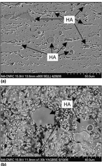
-HA powder mixtures were produced and subsequently thermally sprayed via HVOF (Ref16) (Fig.2a, b). In spite of the HA addition, which is a weaker material, the bond
Fig. 1 Nanostructured zone on the HVOF-sprayed n-TiO2
coating surface (Ref5) Fig. 2 (a) Cross-section and (b) surface of an HVOF-sprayedn-TiO2-10wt.%HA (Ref16)
Peer
strength levels were shown to be higher than 77 MPa for n-TiO2-10 wt.%HA coatings on Ti-6Al-4 V substrates.
Just like the HVOF-sprayed n-TiO2 coatings, the epoxy
glue was breaking before coating failure during the tensile adhesion test ASTM C633. It is important to point out that these HVOF-sprayed coatings exhibited porosity levels lower than 1% (Ref16). Therefore, it is unlikely that the epoxy glue penetrated up to the coating/substrate inter-face, thereby affecting the bond strength data. As previ-ously stated, bond strength levels of APS HA coatings are normally below 30 MPa (Ref 5,6). These coatings have been engineered to exhibit nano and submicron structured surface texture (as those of the pure n-TiO2 coating), in
addition to the chemical character of HA, in an attempt to improve the coating biocompatibility. Therefore, it seems logical as the next step, to carry out biological experiments with these coatings.
1.5 The Interest on Human Mesenchymal Stem Cells
Currently, research works that have the goal of making stem cells to adhere on the surface of a scaffold or coated/ uncoated substrate, and then grow to differentiate into specific cells are gaining importance, as pointed out by Olivier et al. (Ref 17), Zhang et al. (Ref 18), Sjostrom et al. (Ref19), and Kasten et al. (Ref20).
The human mesenchymal stem cells (hMSCs) can be differentiated into a specific group of human cell types, e.g., osteoblasts (bone), chondrocytes (cartilage), myo-cytes (muscle), fibroblasts (connective tissue), and adi-pocytes (fat) (Ref21). The use of hMSCs in the research and development of thermally sprayed biomedical coat-ings represents a very important alternative and a com-plement to the studies in which human osteoblasts cells are used (Ref22). For example, once a prosthetic device (e.g., acetabular cup and femoral stem) is implanted in the human body, hMSCs produced by the bone marrow cover the surface of the implant. Once the hMSCs are well-adhered to the surface of the implant, they begin the process of proliferation and differentiation into osteo-blast cells (Ref 21). Therefore, studying the behavior of hMSCs on the surface of these coatings provides important information and data about the onset of the interaction between the human cells and the implant, which affects both long-term biocompatibility and lon-gevity of the implant.
It is important to point out the major difference between hMSCs and human embryonic stem cells (hESCs). The hESCs can be differentiated into any type of cell of the human body, whereas, as previously stated, hMSCs can differentiate into several but not all types of cells. However, hESCs are obtained from human embryos, which can lead to important ethical issues. hMSCs can be readily obtained from adult human bone marrow, which can be available during hip-joint replacement surgery, such as total hip replacement surgeries. This type of new data will provide valuable information to materials sci-entists, engineers, and biologists on how to engineer the next generation of high performance implants.
2. Experimental Procedure
2.1 Powders, Coatings, and Substrates for Thermal Spraying
The Ti-6Al-4V substrates employed in this work were cut into disk-shaped samples (pucks) of 12.7 mm diam-eter by 2 mm thickness. The substrates for coating deposition and control were grit-blasted with alumina grit #24, as a standard procedure of surface preparation (impurity removal and roughening) for thermal spraying. The average surface roughness (Ra) was 4.6 ± 0.7 lm
(n = 10). The thicknesses of all coatings varied from ~100 to 150 lm.
2.1.1 n-TiO2. The n-TiO2powder [VHP-DCS (5-20 lm),
Altair Nanomaterials Inc., Reno, NV] was HVOF-sprayed (DJ2700-hybrid, Sulzer Metco, Westbury, NY) using propylene as fuel. The average particle temperature and velocity of the powder particles during the deposition step were 1881 ± 162 °C and 686 ± 93 m/s, respectively. The average surface roughness (Ra) of the coating was
2.2 ± 0.3 lm (n = 10). More details about this coating can be found elsewhere (Ref5,6).
2.1.2 HA. The HA powder (Captal 30 SD, Plasma Biotal, Tideswell, North Derbyshire, England) was pure and crystalline. It exhibited a nominal particle size of 15-50 lm. This powder was sprayed via APS (SG100, Praxair, Concord, NH) using only Ar as plasma gas. The average particle temperature and velocity of the powder particles were 2659 ± 234 °C and 189 ± 19 m/s, respec-tively. The coatings produced from this HA powder were used as a control. Crystalline HA and amorphous calcium phosphate were the main phases of the coating, with a minor content of tetracalcium phosphate. The average coating surface roughness (Ra) was 4.1 ± 0.5 lm (n = 10).
More details about this coating can be found in the paper of Auclair-Daigle et al. (Ref 23). The HA coatings were used as a control in this study.
2.1.3 n-TiO2-10 wt.%HA. The feedstock powder
mix-ture was composed of 90 wt.%n-TiO2-10 wt.%HA. The
starting n-TiO2and HA powders were the same materials
employed for producing n-TiO2 and HA coatings. The
n-TiO2-10 wt.%HA powder was prepared through a
mechanical-blending process in a planetary mill. This powder was HVOF-sprayed (DJ2700-hybrid, Sulzer Metco, Westbury, NY) using propylene as fuel. The average particle temperature and velocity of the powder particles were 1875 ± 162 °C and 651 ± 88 m/s, respec-tively. The n-TiO2-10 wt.%HA coating exhibited rutile as
major phase with anatase and HA as minor phases. No significant degradation of HA was observed by means of x-ray diffraction (XRD). The average coating surface roughness (Ra) was 3.0 ± 0.5 lm (n = 10). More details
about the coating can be found elsewhere (Ref16). 2.2 Cell Isolation, Expansion, and Culture
on Samples
2.2.1 Human Mesenchymal Stem Cells. Bone marrow samples were obtained from 15-mL aspirates from the
Peer
intramedullary canal of osteoarthritis patients undergoing total hip replacement surgery (three patients: 1 woman and 2 men, aged 52-76 years). Only tissue that would have been discarded was used, with the approval of the Research Ethics Committee of the Jewish General Hospital. This research also met the requirements of the ethics council committee of the National Research Council of Canada. The hMSCs were isolated and expanded as previously described (Ref24). The medium was changed every 3 days, with all cultures maintained at 37 °C with 5% CO2. Culture-expanded hMSCs were
trypsinized, counted, and seeded separately onto ethylene oxide (EtO) sterilized n-TiO2-10 wt.%HA, n-TiO2, and
HA-coated pucks at a density of 2 9 104cells/cm2. Cells seeded on Ti-6Al-4V were used as references.
2.2.2 hMSC Proliferation on Samples: Alamar Blue. The n-TiO2-10 wt.%HA, n-TiO2, and HA-coated pucks
seeded with the hMSCs were incubated for 1, 7, and 21 days. At each time point, the cell proliferation was monitored using the Alamar BlueÔ assay according to the manufacturer (Biosource, Nivelles, Belgium). The assay is based on a fluorometric/colorometric growth indicator that detects metabolic activity. Numerical data were analyzed statistically using independent Student
t-tests.
2.2.3 Biochemical Analysis: Alkaline Phosphatase Activity. The alkaline phosphatase (ALP) activity of the conditioned media was evaluated as a determinant of osteoblast differentiation. The ALP activity, the driver of bone matrix mineralization, was determined in the hMSC lysates at 1, 7, and 21 days using a commercially available kit (AnaSpec, San Jose, CA) in accordance with the provided instructions. Briefly, the measurements were performed using 100 lL supernatants to which 100 lL of p-nitrophenolphosphate dye was added and the absorbance subsequently measured at 410 nm using a spectrophotometer. For each time point, the activities in three samples were normalized as lmol per mg protein per min.
2.2.4 Cytoskeleton Organization (Confocal Micros-copy): Immunochemistry (Fluorescence). In order to visualize the cytoskeleton and nuclei of the hMSCs, after 1 and 7 days of hMSC seeding, two types of dyes were employed: propidum iodide (PI) for the nuclei and F-actin for the cytoskeleton. Briefly, cells were fixed in formaldehyde (Sigma, St. Louis, MO), permeabilized in 0.1% buffered Triton X-100 (Sigma, St. Louis, MO) and stained for cell cytoskeletal F-actin with 5 U/mL Alexa Fluor 488 phalloidin (Molecular Probes, Burlington, ON, Canada) for 1 h. Cell nuclei were counterstained with 15 M PI (Molecular Probes, Burlington, ON, Canada) for 20 min. Samples were mounted (Vectashield, Vector Laboratories, Burlington, ON, Canada) and examined optically (Cell Observer System, Carl Zeiss, Gottingen, Germany).
2.2.5 hMSC/Substrate Interaction (SEM). The mor-phologies of hMSCs were evaluated using a scanning electron microscope (SEM) (S-4700, Hitachi, Tokyo, Japan). The specimens were sputter coated with palladium and observed at 5009 magnification.
3. Results and Discussion
3.1 Cell Proliferation
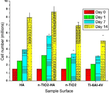
The Alamar Blue assay, which is based on the detection of metabolic activity of living cells, demonstrated that HVOF-sprayed n-TiO2-10 wt.%HA coatings supported
the growth and proliferation of hMSCs (Fig.3). The per-formance levels are equivalent or superior to those of standard APS HA coatings (Fig.3) throughout the time of the study. It is important to point out that the hMSCs are anchorage-dependent cells, i.e., they will die if they do not become well-adhered to the surface of the coatings. Therefore, the results of Fig.3show strong evidence that the surface of the HVOF-sprayed n-TiO2-10 wt.%HA
coating is creating favorable conditions for hMSCs adhe-sion, growth, and proliferation.
At this point, the dominant factor giving rise to the excellent behavior concerning the proliferation of hMSCs on the n-TiO2-10 wt.%HA coatings has not been precisely
identified. However, some hypotheses can be examined. The observations of Shi et al. (10) and Lee et al. (Ref11) on the effect of nano and submicron textured surfaces (like that ofFig. 1) on the likelihood of protein retention (interlocking), as well as the higher osteoblast prolifera-tion on nanostructured ceramics observed by Webster et al. (Ref7), should be taken into account in explaining the experimental results reported in this paper.
In addition, the overall surface roughness of the samples probably also played a role in these results. Comparing the information provided in Section2, the HVOF-sprayed n-TiO2-10 wt.%HA exhibited roughness (Ra) levels 27%
lower than those of APS HA. This difference was caused mainly by the higher average velocity of the HVOF-sprayed particles when compared to that of APS HA particles (651 versus 189 m/s). Anselme et al. (Ref 25) evaluated the adhesion of human osteoblast cells on polished and sand-blasted Ti-6Al-4V surfaces. A higher degree of cell adhesion was observed on the polished sur-faces. In this study, the uncoated Ti-6Al-4V substrates exhibited the highest roughness and the poorest perfor-mance of all samples tested (Fig.3). Therefore, surfaces with lower degree of submicron and micron asperities (i.e., lowerRavalues) seem to facilitate hMSC adhesion.
It is important to point out that the addition of 10 wt.%HA to pure n-TiO2 caused a noticeable
improvement in the bioperformance of the n-TiO2
-10 wt.%HA coating (Fig.3). Consequently, not only the nano-micro topographical effects, but also chemical effects probably played an important role in the improved cell proliferation.
3.2 Biochemical Analysis
The early marker of osteoblastic activity measured in this study, ALP, demonstrated clear differences of cellular behavior among the different coatings (Fig. 4). At day 7, the activity of ALP levels were significantly higher in hMSCs plated on the n-TiO2-10 wt.%HA than in hMSCs
plated on HA and n-TiO2 reference coatings and on
Peer
uncoated Ti-6Al-4V substrates. The increase in ALP activity is a marker of the commitment toward osteo-blastic lineage, i.e., the trend of the hMSCs to differentiate into osteoblast cells. The significantly higher ALP increase on the n-TiO2-10 wt.%HA coatings symbolises the
mes-enchymal cells preference toward an osteoblastic lineage (bone building) when plated on n-TiO2-10 wt.%HA
coatings instead of the diametrically opposite possibility of osteoclasts (bone resorbtion) linage.
3.3 Confocal Microscopy
Confocal microscopy in conjunction with fluores-cence marking was used to investigate the cytoskeleton
Fig. 3 hMSC proliferation profiles during 14 days on HA, n-TiO2-10wt.%HA and n-TiO2coatings and uncoated Ti-6Al-4V substrates
investigated by Alamar Blue
Fig. 4 Normalized alkaline phosphatase activity (ALP), levels in medium of hMSCs cultured on HA, n-TiO2-10wt.%HA and n-TiO2
coatings and uncoated Ti-6Al-4V substrates as a function of time
Peer
organization of the hMSCs at 7 days (Fig.5). In addition, the nuclei staining technique was used to count the cellular number adhered to the various coating as a visual verifica-tion of the metabolic cellular proliferaverifica-tion. In Fig.5, the nuclei are stained with PI (orange-red, disk-shaped) and the cytoskeleton with F-actin (green outline of the cellular body). The cellular growth kinetics among the hMSC plated on the APS HA, HVOF-sprayed n-TiO2-10 wt.%HA, and
n-TiO2 coatings are clearly visible. After 7 days of cell
growth on the various surfaces, the hMSCs exhibited their typical growth phase: elongated or even spindle-shaped morphology similar on all three coatings. The visualization of cellular growth on all coatings confirmed the coatings anticipated biocompatibility. More importantly the F-actin filament deposition was clearly denser and more spread over the entire sample surface (and not only in areas of high cell density) of the HVOF-sprayed n-TiO2-10 wt.%HA
coating when compared to the APS HA and HVOF-sprayed n-TiO2 grown hMSC samples. Also, the
num-ber of cell nuclei (cell numnum-ber), as can be visualized in Fig.7, is significantly higher on the HVOF-sprayed n-TiO2-10 wt.%HA coating attesting to the previously seen
increased growth kinetics on the n-TiO2-10 wt.%HA
nanocomposites when compared to the reference coatings. 3.4 hMSC/Coating Interaction (SEM)
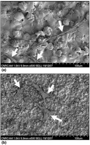
Figure6shows the SEM pictures regarding the inter-action between the hMSCs and the coatings after 2 days of incubation. The hMSCs adhered, spread, and divided on the surface of all coatings. Particularly, at day 2 they exhibit the typical spindle hMSC shape and are elongated as previously seen in the fluorescence assay. Once again as with the fluorescence markings, the hMSC cultured on HA (Fig.6a) seems to exhibit smaller cell bodies, limited spreading and more rounded morphologies when com-pared to the hMSCs cultured on n-TiO2-10 wt.%HA
(Fig.6b) and n-TiO2 (not shown) coatings (the cells are
highlighted by the arrows). This trend is important as cells in rounded configurations divide at a lower rates than those well flattened and spread on a surface, as previously shown by Hunter et al. (Ref26). Therefore, the n-TiO2
-10 wt.%HA (Fig.6b) and n-TiO2 (not shown) coatings
seem to provide improved conditions for cell spreading,
thereby enhancing their proliferation, as shown in the cell proliferation essays (Fig.3 and 5). More information, further biomedical data and discussion on all the results of this paper can be found elsewhere (Ref27).
Fig. 5 Cytoskeleton organization and nuclei morphology of hMSCs at day 7 cultured on HA, n-TiO2-10wt.%HA and n-TiO2coatings.
The scale bar length is 200 lm
Fig. 6 hMSCs on the surface of the (a) APS HA and (b) HVOF-sprayed n-TiO2-10wt.%HA coatings after 2 days of
cell culture—arrows highlight the cells
Peer
3.5 Reaction between TiO2and HA at the Coating
Microstructure
It can be hypothesized that a chemical reaction between TiO2and HA may have occurred in benefit to the
biocompatibility of the n-TiO2-10 wt.%HA system. Li
et al. (Ref 28) deposited powder blends of HA + 10 vol.%TiO2 and HA + 20 vol.%TiO2 via HVOF. They
have observed the presence of a CaTiO3 minor phase in
the structure of the coatings by performing a regular XRD analysis. The XRD of the HVOF-sprayed n-TiO2
-10 wt.%HA coating was previously analyzed (Ref 16); however, no other phases in addition to titania and HA were detected.
Coren˜o and Coren˜o (Ref29) and Manso et al. (Ref30) analysed the performance of CaTiO3as an apatite growth
promoter by using SBF. Both works reported promising results regarding the nucleation and growth of apatite. Consequently, if a similar type of event is occurring in the surface of the composite n-TiO2+ HA coatings, it may be
another factor that helps to explain the biological results of this paper.
Lu et al. (Ref 31) mechanically mixed HA and TiO2
powders and plasma-sprayed a blend of 50 vol.%HA + 50 vol.%TiO2. The composite powder was sprayed into
water. The powders sprayed into water were subsequently dried and evaluated via energy dispersive x-ray (EDX) analysis. No CaTiO3 phase formation was observed via
XRD; however, the EDX analysis confirmed the presence of microregions with coexistence of Ca, P, and Ti in some melted particles, thereby indicating the inter-diffusion of elements from HA and TiO2. Consequently, this type of
reaction (i.e., inter-diffusion) may have occurred in the composite coatings produced for this study.
The same type of inter-diffusion observed when the powder was sprayed into water was also observed in the coating. The HA + TiO2 (50/50) composite powder was
used to produce a bond coat (on a Ti substrate) for an HA topcoat. The composite coating exhibited similar
microstructure to that of the n-TiO2-HA coating of this
work (i.e., a pile up of TiO2 and HA splats). Lu et al.
(Ref31) performed a linear EDX analysis at the HA and TiO2splat interfaces. They also observed the coexistence
of Ca, P, and Ti at those interfacial regions. Therefore, the same type of reaction between TiO2 and HA may have
occurred in the coatings produced in this study.
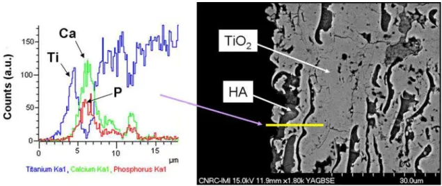
To confirm this hypothesis, an EDX line scan analysis was performed at the upper layers of an HVOF-sprayed n-TiO2-10 wt.%HA coating (Fig.7). It is possible to
observe that for adjacent TiO2 and HA splats there are
microregions containing the coexistence of Ti, Ca, and P atoms. Therefore, there is definitely an inter-diffusion among the elements of TiO2and HA in the coating
micro-structure. However, as previously stated, the XRD spec-trum of this coating did not detect any chemical reaction (e.g., CaTiO3) between these two compositions (Ref16).
It was probably a sensitivity issue of the XRD analysis.
This chemical reaction between the TiO2and HA and
its effect on the biocompatibility of this coating will be one of the most important points to be sought in this ongoing work and presented in future publications.
4. Conclusions
Previous studies have shown that HVOF-sprayed n-TiO2-10 wt.%HA coatings exhibit bond strength levels
higher than 77 MPa, i.e., at least twice those of APS HA coatings deposited on Ti-6Al-4V substrates, which are typically below 30 MPa. In addition, due to the high sta-bility of TiO2 in the human body, longevity related
con-cerns of HA coatings, such as, dissolution and osteolysis, are unlikely to occur. These results provide strong evi-dence that hMSCs exhibit on HVOF-sprayed n-TiO2
-10 wt.%HA coatings (i) growth, proliferation, and attachment, (ii) commitment toward osteoblastic lineage, and (iii) cell/substrate interaction levels/characteristics
Fig. 7 EDX line scan analysis at the upper layers of an HVOF-sprayed n-TiO2-10wt.%HA coating
Peer
superior to those of APS HA coatings. There are no clear explanations regarding this favorable behavior, but it is hypothesized that the nano-micro topography and chem-ical composition of the surface of the HVOF-sprayed n-TiO2-10 wt.%HA coating are playing important roles.
The porosity distribution and residual stresses may also play a role. This is an ongoing work and these biocom-patibility issues will be addressed in future publications. This results demonstrate that surfaces engineered in this fashion have the potential to become the next generation of biomedical thermal spray coatings with enhanced per-formance and improved longevity.
References
1. L. Sun, C.C. Berndt, K.A. Gross, and A. Kucuk, Material Fun-damentals and Clinical Performance of Plasma-Sprayed Hydroxyapatite Coatings: A Review, J. Biomed. Mater. Res., 2001,585, p 570-592
2. L.I. Havelin, B. Espehaug, and L.B. Engesaeter, The Perfor-mance of Two Hydroxyapatite-Coated Acetabular Cups Compared with Charnley Cups, J. Bone Joint. Surg. (Br), 2002, 84-B(6), p 839-845
3. O. Reikeras and R.B. Gunderson, Failure of HA Coating on a Gritblasted Acetabular Cup, Acta. Orthop. Scand., 2002,73(1), p 104-108
4. K.A. Lai et al., Failure of Hydroxyapatite-Coated Acetabular Cups, J. Bone Joint. Surg. (Br), 2002,84-B(5), p 641-646 5. R.S. Lima and B.R. Marple, Thermal Spray Coatings Engineered
from Nanostructured Ceramic Agglomerated Powders for Struc-tural, Thermal Barrier and Biomedical Applications, J. Therm.
Spray Technol., 2007,16(1), p 40-63
6. R.S. Lima and B.R. Marple, Engineering Nanostructured Thermal Spray Coatings for Biomedical Applications,
Bionano-technology—Global Prospects, D.E. Reisner, Ed., CRC Press/ Taylor & Francis Group, Boca Raton, FL, Chap. 8, 2008, p 91-109 7. T.J. Webster, C. Ergun, R.H. Doremus, R.W. Siegel, and R. Bizios, Specific Proteins Mediate Enhanced Osteoblast Adhesion on Nanophase Ceramics, J. Biomed. Mater. Res., 2000, 51(3), p 475-483
8. M.J. Dalby, D. McCloy, M. Robertson, H. Agheli, D. Sutherland, S. Affrosman, and R.O.C. Oreffo, Osteoprogenitor Response to Semi-ordered and Random Nanotopographies, Biomaterials, 2006,27, p 2980-2987
9. M.J. Dalby, D. McCloy, M. Robertson, C.D.W. Wilkinson, and R.O.C. Oreffo, Osteoprogenitor Response to Defined Topogra-phies with Nanoscale Depths, Biomaterials, 2006,27, p 1306-1315 10. H. Shi, W.-B. Tsai, M.D. Garrison, S. Ferrari, and B.D. Ratner, Template-Imprinted Nanostructured Surfaces for Protein Rec-ognition, Nature, 1999,398, p 593-597
11. K.-B. Lee, S.-J. Park, C.A. Mirkin, J.C. Smith, and M. Mrksich, Protein Nanoarrays Generated by Dip-Pen Nanolitography,
Sci-ence, 2002,295, p 1702-1705
12. K. Anselme, Osteoblast Adhesion on Biomaterials, Biomaterials, 2002,23, p 1187-1199
13. H.P. Erickson, N. Carrell, and J. McDonagh, Fibronectin Mole-cule Visualized in Electron Microscopy: A Long, Thin, Flexible Strand, J. Cell Biol., 1981,91, p 673-678
14. T.J. Webster, R.W. Siegel, and R. Bizios, Osteoblast Adhesion on Nanophase Ceramics, Biomaterials, 1999,20, p 1221-1227
15. T.J. Webster, C. Ergun, R.H. Doremus, R.W. Siegel, and R. Bizios, Enhanced Functions of Osteoblasts on Nanophase Ceramics, Biomaterials, 2000,21, p 1803-1810
16. M. Gaona, R.S. Lima, and B.R. Marple, Nanostructured Titania/ Hydroxyapatite Composite Coatings Deposited by High Velo-city Oxy-Fuel (HVOF) Spraying, Mater. Sci. Eng. A, 2007,458, p 141-149
17. V. Olivier, N. Faucheux, and P. Hardouin, Biomaterial Chal-lenges and Approaches to Stem Cell Use in Bone Reconstructive Surgery, Drug Discov. Today, 2004,9(18), p 803-811
18. W. Zhang, X.F. Walboomers, T.H. van Kuppevelt, W.F. Daamen, Z. Bian, and J.A. Jansen, The Performance of Human Dental Pulp Stem Cells on Different Three-dimensional Scaffold Mate-rials, BiomateMate-rials, 2006,27, p 5658-5668
19. T. Sjostrom, M.J. Dalby, A. Hart, R. Tare, R.O.C. Oreffo, and B. Su, Fabrication of Pillar-like Titania Nanostructures on Tita-nium and Their Interactions with Human Skeletal Stem Cells,
Acta Biomater., 2009,5, p 1433-1441
20. P. Kasten, I. Beyen, P. Niemeyer, R. Luginbuhl, M. Bohner, and W. Richter, Porosity and Pore Size of b-tricalcium Phosphate Scaffold Can Influence Protein Production and Osteogenic Dif-ferentiation of Human Mesenchymal Stem Cells: An In Vitro and In Vivo Study, Acta Biomater., 2008,4, p 1904-1915
21. M.J. Dalby, N. Gadegaard, R. Tare, A. Andar, M.O. Riehle, P. Herzyk, C.D.W. Wilkinson, and R.O.C. Oreffo, The Control of Human Mesenchymal Cell Differentiation Using Nanoscale Symmetry and Disorder, Nat. Mater., 2007,6, p 997-1003 22. M. Sato, M.A. Sambito, A. Aslani, N.M. Kalkhoran, E.B.
Slamovich, and T.J. Webster, Increased Osteoblast Functions on Undoped and Yttrium-doped Nanocrystalline Hydroxyapatite Coatings on Titanium, Biomaterials, 2006,27, p 2358-2369 23. C. Auclair-Daigle, M.N. Bureau, J.-G. Legoux, and LÕ.H. Yahia,
Bioactive Hydroxyapatite Coatings on Polymer Composites for Orthopedic Implants, J. Biomed. Mater. Res., 2005,73A(4), p 398-408 24. D.C. Colter, R. Class, C.M. DiGirolamo, and D.J. Prockop, Rapid Expansion of Recycling Stem Cells in Cultures of Plastic-adherent Cells from Human Bone Marrow, Proc. Natl. Acad. Sci.
USA, 2000,97, p 3213-3218
25. K. Anselme, M. Bigerelle, B. Noel, E. Dufresne, D. Judas, A. Iost, and P. Hardouin, Qualitative and Quantitative Study of Human Osteoblast Adhesion on Materials with Various Surface Roughnesses, J. Biomed. Mater. Res., 2000,49(2), p 155-166 26. A. Hunter, C.W. Archer, P.S. Walker, and G.W. Blunn,
Attachment and Proliferation of Osteoblasts and Fibroblasts on Biomaterials for Orthopaedic Use, Biomaterials, 1995,16, p 267-295
27. S. Dimitrievska, R.S. Lima, J. Antoniou, A. Petit, F. Mwale, M.N. Bureau, and B.R. Marple, TiO2-HA Nanocomposite Coatings
Promote Human Mesenchymal Stem Cells Osteogenic Differ-entiation, Biomaterials, BIOMAT-S-09-02311
28. H. Li, K.A. Khor, and P. Cheang, Titanium Dioxide Reinforced Hydroxyapatite Coatings Deposited by High Velocity Oxy-fuel (HVOF) Spray, Biomaterials, 2002,23, p 85-91
29. J. Coren˜o and O. Coren˜o, Evaluation of Calcium Titanate as Apatite Growth Promoter, J. Biomed. Mater. Res., 2005,75A(2), p 478-484
30. M. Manso, M. Langlet, and J.M. Martı´nez-Duart, Testing Sol-Gel CaTiO3 Coatings for Biocompatible Applications, Mater. Sci.
Eng. C, 2003,23, p 447-450
31. Y.-P. Lu, M.-S. Li, S.-T. Li, Z.-G. Wang, and R.-F. Zhu, Plasma-sprayed Hydroxyapatite + Titania Composite Bond Coat for Hydroxyapatite Coating on Titanium Substrate, Biomaterials, 2004,25, p 4393-4403