Publisher’s version / Version de l'éditeur:
Vous avez des questions? Nous pouvons vous aider. Pour communiquer directement avec un auteur, consultez la première page de la revue dans laquelle son article a été publié afin de trouver ses coordonnées. Si vous n’arrivez pas à les repérer, communiquez avec nous à [email protected].
Questions? Contact the NRC Publications Archive team at
[email protected]. If you wish to email the authors directly, please see the first page of the publication for their contact information.
https://publications-cnrc.canada.ca/fra/droits
L’accès à ce site Web et l’utilisation de son contenu sont assujettis aux conditions présentées dans le site LISEZ CES CONDITIONS ATTENTIVEMENT AVANT D’UTILISER CE SITE WEB.
Clinical Epigenetics, 12, 1, 2020-12-14
READ THESE TERMS AND CONDITIONS CAREFULLY BEFORE USING THIS WEBSITE.
https://nrc-publications.canada.ca/eng/copyright
NRC Publications Archive Record / Notice des Archives des publications du CNRC :
https://nrc-publications.canada.ca/eng/view/object/?id=8523388f-39ff-4f58-b004-fb3eb0abc809
https://publications-cnrc.canada.ca/fra/voir/objet/?id=8523388f-39ff-4f58-b004-fb3eb0abc809
This publication could be one of several versions: author’s original, accepted manuscript or the publisher’s version. / La version de cette publication peut être l’une des suivantes : la version prépublication de l’auteur, la version acceptée du manuscrit ou la version de l’éditeur.For the publisher’s version, please access the DOI link below./ Pour consulter la version de l’éditeur, utilisez le lien DOI ci-dessous.
https://doi.org/10.1186/s13148-020-00988-1
Access and use of this website and the material on it are subject to the Terms and Conditions set forth at
High-resolution analyses of human sperm dynamic methylome reveal
thousands of novel age-related epigenetic alterations
Cao, Mingju; Shao, Xiaojian; Chan, Peter; Cheung, Warren; Kwan, Tony;
Pastinen, Tomi; Robaire, Bernard
RESEARCH
High-resolution analyses of human sperm
dynamic methylome reveal thousands of novel
age-related epigenetic alterations
Mingju Cao
1†, Xiaojian Shao
2,3,4†, Peter Chan
5, Warren Cheung
6, Tony Kwan
2,3, Tomi Pastinen
2,3,6†and Bernard Robaire
1,7*†Abstract
Background: Children of aged fathers are at a higher risk of developing mental disorders. Alterations in sperm DNA
methylation have been implicated as a potential cause. However, age-dependent modifications of the germ cells’ epigenome remain poorly understood. Our objective was to assess the DNA methylation profile of human spermato-zoa during aging.
Results: We used a high throughput, customized methylC-capture sequencing (MCC-seq) approach to characterize
the dynamic DNA methylation in spermatozoa from 94 fertile and infertile men, who were categorized as young, 48 men between 18–38 years or old 46 men between 46–71 years. We identified more than 150,000 age-related CpG sites that are significantly differentially methylated among 2.65 million CpG sites covered. We conducted machine learning using our dataset to predict the methylation age of subjects; the age prediction accuracy based on our assay provided a more accurate prediction than that using the 450 K chip approach. In addition, we found that there are more hypermethylated (62%) than hypomethylated (38%) CpG sites in sperm of aged men, corresponding to 798 of total differential methylated regions (DMRs), of which 483 are hypermethylated regions (HyperDMR), and 315 hypo-methylated regions (HypoDMR). Moreover, the distribution of age-related hyper- and hypohypo-methylated CpGs in sperm is not random; the CpG sites that were hypermethylated with advanced age were frequently located in the distal region to genes, whereas hypomethylated sites were near to gene transcription start sites (TSS). We identified a high density of age-associated CpG changes in chromosomes 4 and 16, particularly HyperDMRs with localized clusters, the chr4 DMR cluster overlaps PGC1α locus, a protein involved in metabolic aging and the chr16 DMR cluster over-laps RBFOX1 locus, a gene implicated in neurodevelopmental disease. Gene ontology analysis revealed that the most affected genes by age were associated with development, neuron projection, differentiation and recognition, and behaviour, suggesting a potential link to the higher risk of neurodevelopmental disorders in children of aged fathers.
Conclusion: We identified thousands of age-related and sperm-specific epigenetic alterations. These findings
provide novel insight in understanding human sperm DNA methylation dynamics during paternal aging, and the subsequently affected genes potentially related to diseases in offspring.
Keywords: Spermatozoa, DNA methylation, Advanced paternal age, MCC-seq, Fertility
© The Author(s) 2020. Open Access This article is licensed under a Creative Commons Attribution 4.0 International License, which permits use, sharing, adaptation, distribution and reproduction in any medium or format, as long as you give appropriate credit to the original author(s) and the source, provide a link to the Creative Commons licence, and indicate if changes were made. The images or other third party material in this article are included in the article’s Creative Commons licence, unless indicated otherwise in a credit line to the material. If material is not included in the article’s Creative Commons licence and your intended use is not permitted by statutory regulation or exceeds the permitted use, you will need to obtain permission directly from the copyright holder. To view a copy of this licence, visit http://creat iveco mmons .org/licen ses/by/4.0/. The Creative Commons Public Domain Dedication waiver (http://creat iveco mmons .org/publi cdoma in/zero/1.0/) applies to the data made available in this article, unless otherwise stated in a credit line to the data.
Background
The current trend in fathering children at an older age has raised concerns due to the reported association between advanced paternal age and increased incidence of several conditions including bipolar disorders, attention deficit
Open Access
*Correspondence: [email protected] †Mingju Cao and Xiaojian Shao: Co-first authors †Tomi Pastinen and Bernard Robaire: Co-senior authors
1 Department of Pharmacology and Therapeutics, McGill University, 3655 Promenade Sir William Osler, Montreal, QC H3G 1Y6, Canada
hyperactivity disorder (ADHD), and schizophrenia [1]. However, the influence of paternal age on alterations in paternal sperm chromatin is still very limited [2, 3]. Spermatogenesis is a life-long process, and the number of spermatogonial cell divisions prior to spermiogenesis increases from 35 at puberty to more than 600 at 50 years of age [4]. Genetic mutations occur during each repli-cation cycle every 2–3 weeks; the mutational load con-tinuously increases in the sperm of older males [4–6]. In addition to spontaneous genetic mutations during cell division, the epigenetic modifications must be copied to the daughter cells, and copying of the epigenetic informa-tion is much more error-prone than DNA replicainforma-tion [7]. The epigenome is an essential and modifiable set of serial marks including DNA methylation, histone posttransla-tional modifications and small noncoding RNAs. Age-dependent modifications of the germ cells epigenome remain relatively poorly understood.
DNA methylation is a heritable epigenetic modifi-cation of cytosine residues within CpG dinucleotides. Among 28 million CpG sites throughout the mamma-lian genome, approximately 60–80% are methylated, displaying relatively stable DNA methylation patterns in most cell types, except germ cells and pre-implantation embryos [8]. Using a mouse model, Xie et al. identified reduced DNA methylation and differentially methylated promoters in aged sperm, and demonstrated that epi-genetic alterations in longevity regulators, reduced life span and more pronounced aging-associated pathologies in offspring of animals sired by old father than by young ones [9]. The relationship between aging and DNA meth-ylation has been studied previously in many human cell types as well as several tissues [10]. Genome-wide meth-ylation studies have revealed that the sperm epigenome differs remarkably from that of somatic cells [8, 11]; a unique state of DNA methylation exists outside of CpG island sequences in mouse sperm DNA [11, 12]. Human sperm DNA methylation is quite distinct from that in all somatic cells and tissues [13]. Earlier studies on sperm DNA methylation were mostly focussed on imprinted loci [14–16]. Only a few studies have examined DNA methylation in human sperm on a genome-wide basis [17–20].
Using the Infinium Human Methylation 450 Bead Chip microarray (450 K), Jenkins et al. have identified 139 regions that are significantly and consistently hypo-methylated with age and 8 regions that are significantly hypermethylated with age in human sperm. In addition, a total of 117 genes that are associated with these regions of altered methylation were identified [21]. This research group has also conducted experiments to develop a sperm DNA methylation-based age predicting calcula-tor [22]; their model was able to predict an individual’s
chronological age with up to 94% accuracy in compari-son with a previously constructed age calculator based on methylation data from somatic tissues [23]. More recently, Denomme et al. applied reduced representa-tion bisulfite sequencing (RRBS) to measure the DNA methylation profiles in sperm of six young (≤ 35 years) and six older men (≥ 50 years), identified 49,792 differ-entially methylated CpG sites (DMCs) at nominal p value or p < 0.05 (corresponded to 3,405 sperm differentially methylated regions (DMRs)). They found that methyla-tion alteramethyla-tions are not randomly distributed across the genome and have some links with neurodevelopmental relevant diseases [24]. Despite those recent advances, the information of age-associated sperm DNA methylation is still limited.
Over the past decade, several methods have been developed and applied to characterize DNA methylation at gene-specific loci using either traditional bisulfite-PCR analysis or pyrosequencing [14, 15], or to determine genome-wide distribution using microarray analysis, and RRBS analysis [19]. Although whole genome bisulfite sequencing (WGBS) provides comprehensive coverage of the epigenome [25], it highlights a general inefficiency as 70–80% of the sequencing reads across the in-depth analysis datasets provide little or no relevant informa-tion about CpG methylainforma-tion sites [8]. A commonly used approach to examine human sperm DNA methylation is the 450 K or more recently the Illumina EPIC array chips [21, 26–28]. However, these arrays provide limited coverage of the epigenome, are biased toward genic and CpG-rich regions, and are not specific for the sperm epi-genome. MethylC-capture sequencing (MCC-seq) was developed for targeted assessment of DNA methylation in a tissue-specific manner [29], and further customized to target human sperm epigenome with enrichment of sequences of interest in both genic and intergenic regions [20].
The objective of the present study was to identify changes in human sperm DNA methylome as a function of paternal age and fertility status using our previously characterized sperm capture panel [20] in order to enrich bisulfite sequencing coverage to regions of variable and/ or dynamic DNA methylation. We also included analy-ses of the relationship between paternal age and fertil-ity status with total serum and bioavailable testosterone concentrations, serum FSH level, high density lipopro-tein (HDL), and metabolic parameters as well as smok-ing. Using high throughput and customized MCC-seq technique, we identified thousands of age-related epig-enomic alterations in human sperm; we also conducted a machine learning model using our dataset to predict the methylation age of subjects. Our results provide new insights on how paternal aging and fertility status interact
on an array of male reproductive endpoints and dem-onstrate novel aspects of how aging modifies the sperm methylation pattern.
Results
Subjects descriptions in MCC‑seq study cohort
Basic characteristics of subjects in this study are shown in Table 1. Two subpopulations of young (< 40 years, with mean = 29 years) and old (≥ 40 years, with mean = 53 years) subjects were used. Within each group, the fertile control and infertile subjects were matched (except two fewer patients in the infertile group for old group). We compared the general semen parameters, plasma hormone level and key metabolic factors between the two populations. We observed marginally signifi-cant changes for sperm motility (49 ± 3% vs. 38 ± 3%, p = 0.029) and a significant difference in follicle stimu-lating hormone (FSH) with lower level in young subjects (4.09 ± 0.29) compared with old subjects (6.25 ± 0.4, p = 3.66E−05). We also observed a higher level of tes-tosterone in young subjects than in old ones (11.9 ± 0.6 vs. 9.29 ± 0.52, p = 0.001). No differences were found in sperm concentrations, or concentrations of luteiniz-ing hormone (LH), thyroid stimulatluteiniz-ing hormone (TSH),
bioavailable testosterone, total cholesterol, triglycerides, high density lipoprotein (HDL), low density lipopro-tein (LDL), cholesterol/HDL risk ratio, body mass index (BMI) and tobacco consumption between young and old groups (Table 1).
Correlations analysis between the age of subjects and clinical indices in both fertile and infertile men
The relationships between the age of subjects and clinical indices in both fertile and infertile subjects as assessed by Pearson correlation analyses are shown in Addi-tional file 1: Table S1. In fertile men, there were signifi-cant positive correlations between the age of the subjects and plasma FSH level, and metabolic parameters such as BMI, plasma triglycerides, and the risk ratio of total cho-lesterol to HDL. In addition, there were significant nega-tive correlations between the age of subjects and plasma total testosterone and bioavailable testosterone concen-trations and high density lipoprotein (HDL). However, in the infertile group many of the correlations were lost; we only found significant negative correlations between the age of subjects and sperm motility, and plasma bio-available testosterone concentrations (Additional file 1: Table S1).
Table 1 Clinical characterisation of subjects in the MCC-Seq study cohort
Values are shown as the mean ± SEM
BMI body mass index, FSH follicle stimulating hormone, LH luteinizing hormone, TSH thyroid stimulating hormone, HDL high density lipoprotein, LDL low density lipoprotein
Parameter Young Old p values
(mean ± SEM) (n) (mean ± SEM) (n)
Age (years) 29.38 ± 0.91 (48) 53.15 ± 0.98 (46) 2.04E−31
Sperm concentrations (× 106/ml) 118.57 ± 15.99 (48) 143.6 ± 18.31 (46) 0.306
Motility (rapid & slow %) 48.96 ± 3.26 (48) 38.77 ± 3.26 (46) 0.029
BMI (kg/m2) 26.45 ± 0.99 (48) 28.94 ± 0.85 (46) 0.059 FSH (IUs/L) 4.09 ± 0.29 (48) 6.25 ± 0.4 (45) 3.66E−05 LH (IUs/L) 3.75 ± 0.17 (48) 4.34 ± 0.32 (45) 0.108 TSH (mIU/L) 2.05 ± 0.34 (47) 1.84 ± 0.21 (42) 0.591 E2RP (qmol/L) 130.97 ± 8.06 (34) 138.24 ± 8.42 (34) 0.535 Testosterone (T, nmol/L) 11.9 ± 0.6 (48) 9.29 ± 0.52 (45) 0.001 Bioavailable T (nmol/L) 6.33 ± 0.27 (48) 5.01 ± 0.61 (43) 0.051
Total cholesterol (mmol/L) 5.21 ± 0.79 (48) 5.06 ± 0.44 (45) 0.863
Triglycerides (mmol/L) 1.83 ± 0.37 (48) 2.89 ± 0.74 (45) 0.202
HDL (mmpl)/L 2.58 ± 1.35 (48) 1.87 ± 0.51 (44) 0.624
LDL (mmol/L) 3.05 ± 0.51 (47) 5.68 ± 2.28 (39) 0.265
Cholesterol/HDL risk ratio 3.9 ± 0.2 (47) 4.06 ± 0.17 (43) 0.544
Smoking N (%) N (%) 0.933
Never 29 (61.7%) 28 (61%) (χ2 = 0.138)
Past 13 (27.7%) 12 (26%)
Current 5 (10.6%) 6 (13%)
Sperm sample purity as assessed by bisulfite pyrosequencing analysis of imprinted loci
Prior to MCC-seq, we screened sperm DNA sample purity by performing bisulfite pyrosequencing of two paternally methylated gene loci H19 and DLK1/GTL2-IG DMR, and two maternally methylated gene loci MEST, KCNQ1OT1. Somatic cell contaminated sperm samples were excluded for further analysis if the aberrant DNA methylation was found. As expected, the DNA methyla-tion profile of those imprinted gene loci obtained from MCC-seq is normal at the QC coordinates at imprinted loci and in turn confirmed our sample purity (Additional file 5: Figure S1).
MCC‑seq detects thousands of novel CpGs associated with sperm aging
We utilized our previously characterized sperm cap-ture panel [20] to enrich bisulphite sequencing cover-age to regions variable in human sperm. We targeted 20–30 × coverage of each (n = 94) donor sample. A mini-mum 20 × coverage for CpG inclusion in association testing in each and included each targeted CpG with at least 30 samples having data; we note that 80% of tar-geted CpGs were detected in at least 90% of samples. We initially applied a linear model without covariates and tested association for both fertility and age at 2.65 million passing CpGs. We observed some inflation for statistically significant differences in methylation upon
aging genomic control inflation, GCin = 1.7 [30], and less marked statistical inflation for fertility (GCin = 1.39) (Additional file 5: Figure S2). Applying multiple testing correction (q-value, qv < 0.01) no significant CpGs were observed for fertility and > 150,000 significant signals were found for aging. Using a linear model to perform the epigenome-wide associations (EWAS) between age and DNA methylation at 2.65 million passing CpGs with fertility and other age-associated phenotypes (e.g. lipids, BMI, etc., see Methods) as covariates. To this end, after applying multiple testing correction, we observed 21,971 CpGs (q value, qv < 0.01) associated with aging with a modest genomic inflation (Gin = 1.44) (Fig. 1, Table 2). Specificity of sperm age‑associated CpGs
Since we detected an order-of-magnitude higher num-ber of CpGs associated with sperm aging, we sought to explore their specificity using MCC-seq measured CpGs mapped (n = 908) to the 50 genomic regions listed to harbor sperm aging CpGs in a previous study [22]. We observed 116/908 CpGs in 26 of the 50 reported regions reaching qv < 0.01 (417/908 reaching nominal signifi-cance p < 0.05) for sperm age association. In contrast, for blood aging CpGs [23], we observe only 2 of 307 CpGs for sperm aging at qv < 0.01 (Fig. 2). This tissue restricted 20-fold enrichment of multiple testing corrected asso-ciations in our data among previously suggested sperm methylation age signature regions yields strong
Fig. 1 Sperm age EWAS results. a CpG—Age association tests QQ-plot shows moderate inflation (GCin = 1.44), and b genome-wide abundance of
CpGs (> 20 K) passing qv < 0.01 significance threshold
Table 2 Age-associated hypo- and hypermethylated CpGs
Age qv < 0.01 novel Age qv < 0.01 in sperm age loci
All CpGs Hypermethylated Hypomethylated Hypermethylated Hypomethylated
replication and underscores high specificity of our asso-ciation; this reiterates the uniqueness for sperm methyla-tion aging as compared to other tissues.
Notwithstanding the strong replication of earlier results, it is intriguing that most of our genome-wide-sig-nificant CpGs (> 99.5%) lie in regions not reported pre-viously. We note that only 4.7% of the CpGs associated with aging in our dataset are actually represented on the Illumina 450 K microarray, explaining much of the new discovery stemming from the “dynamic sperm methyl-ome” we specifically targeted by the capture methylome assay [20]. Overall, our results suggest pervasive influ-ence of aging in shaping sperm methylome patterns.
Chronological age prediction from sperm MCC‑seq
Next, we explored the prediction of age from sperm methylome patterns. Due to limited training dataset, we trained our models using leave-one-out cross-validation (LOOCV) strategy. That is, each time we leave one sam-ple out and train the model using the remaining samsam-ples, and then predict the age for the leave-out sample. The procedure was repeated for each sample. In order to test independence from previously reported sperm age pre-dictor [22], we fetched 450 K CpGs located with Jenkins’ reported sperm aging regions from our sequenced MCC-Seq data (published 450 K sites). By taking advantage of our large panel of MCC-Seq data, we separately trained a model with top 5 K DMCs (> 99% novel age-associated
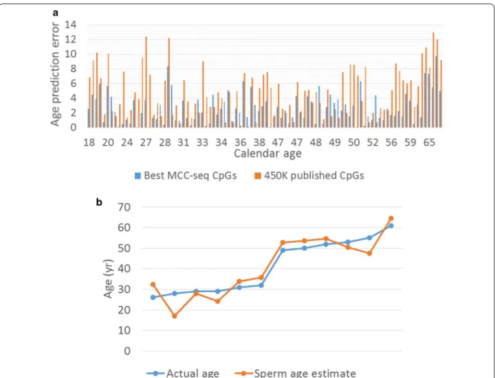
CpGs) detected in the LM age model. In all these cases, the CpGs measured by all samples were eventually used, i.e. CpGs with missing data were removed for training. Finally, as an independent test (replication), we tested our novel sperm age predictor on a recently published sperm cohort [20] where the sperm age predictor was retrained by considering the CpGs measured in all training set (94 samples) and testing set (12 samples). As shown in Fig. 3, the predictor based on novel sites was most accurate with an average error of 2.68 years (years) (Fig. 3a), and, even with limited training set, the replication shows age prediction accuracy with 4.43 year error in the small set of independent samples (n = 12) (Fig. 3b). These results underscore the widespread age-related changes in sperm methylome and suggests that age predictors based on sperm methylome alterations can be further refined.
Characteristics of sperm aging CpGs
We first examined the overall genomic distribution of the genome-wide significant age-associated CpGs based on relative hypo and hypermethylation as a function of age. Hypomethylation was observed to be less common (~ 38% of significant sites, n = 8409 CpGs) with distribu-tion of CpGs biased strongly towards proximal regulatory elements as compared to more prevalent (~ 62% of sig-nificant sites, n = 13,562 CpGs) hypermethylation upon aging (Fig. 4a, b). This new observation of predominance of age-associated hypermethylation is solely driven by Fig. 2 Sperm age associations from MCC-seq are specific. CpG association results for sperm aging intervals reported earlier are shown in green
with earlier blood associated CpGs highlighted in red. Only two blood methylation age predicting CpGs reach qv < 0.01, whereas > 100 CpGs in 50 regions are showing significance for sperm methylation age
sperm age-associated regions not detected by Illumina microarrays [21] (Table 2).
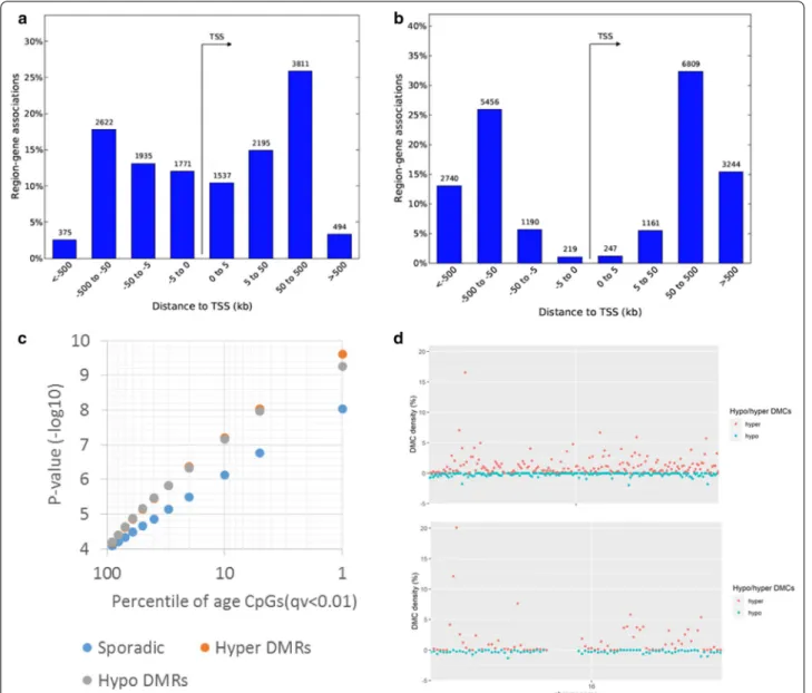
Furthermore, we observe that 26% of age-associated CpGs were clustered in DMRs where five or more CpGs in a 500 bp window showed significant association with either hypomethylation (n = 315 HypoDMRs) or with hypermethylation (n = 483 HyperDMRs) among the total 798 DMRs. The DMRs were characterized by show-ing stronger associations (Fig. 4c) and particularly hyper DMRs with localized clusters in chromosomes 4 and 16 leading to high density of age CpGs in these loci (Fig. 4d). Of note, chr4 DMR cluster overlaps with the peroxisome proliferator-activated receptor (PPAR)-gamma coacti-vator (PGC)-1α locus, a protein involved in metabolic aging [31] and chr16 is overlapping RNA binding fox-1 homolog 1 (RBFOX1) gene, implicated in neurodevelop-mental disease [32].
The distribution of age‑related hypo‑ or hyper‑methylated CpGs in sperm is not random
Using the GREAT [33], we then explored the distri-bution of age-associated hyper- and hypomethylated CpGs in relation to gene region and to in-chromosome location. We annotated each CpG to its closest tran-scription start site (TSS), and found that the age-asso-ciated hypomethylation is proximal to genes, whereas age-associated hypermethylation occupies gene dis-tal regions (Fig. 4a, b). Furthermore, density of hypo-methylation is relatively evenly distributed across the genome. However, overall sex chromosomes are rela-tively depleted of age-associated CpGs (Additional file 5: Figure S3). Taken together, our results indicate that sperm age-related hypo- or hypermethylated CpGs are not randomly distributed.
Fig. 3 Sperm age prediction analysis. a Comparison of age predictor error between model using previously published CpGs detected on Illumina
450 K Human Methylation array (“450 K published CpGs”, orange bars) and the model with top 5000 CpGs (> 99% novel) from our genomewide aging EWAS (“Best MCC-seq CpGs”, blue bars), which provided higher prediction accuracy. b Replication of the age predictor in 12 independent samples from our earlier sperm capture methylome study
Sperm aging CpGs in relations to chromatin states
The divergence of age-associated hypo- vs. hypermeth-ylated CpGs is also clear from relatively lower average methylation across all samples at age hypomethylated sites 39% ± 20% vs. 60% ± 16% (average ± SD). Conse-quently, nearly 50% of age-associated hypomethylation localized to open chromatin (DNaseI Hypersensitive Site Master List (125 cell types) from ENCODE/Analysis [34], whereas only 20% age-related hypermethylated sites over-lapped open chromatin in multiple human cell types. In
line with this observation, the chromatin states enriched strongly for relatively hypermethylated CpGs are strongly biased towards heterochromatin for all ENCODE cell lines; representative distribution of hypo/hypermethyl-ated regions in sperm within Human ES cells (H1/ESC) mapped to chromatin states is shown in Additional file 1: Table S3. Similarly, we observed an enrichment (32% vs. 9%) of CpGs in hypo-DMRs in human sperm H3K27me3 histone modification regions [35] where developmental promoter-like regions were reported in human sperm. Fig. 4 Distribution of age-associated hypo- and hypermethylated CpGs in relation to gene region and in chromosome. a Age-associated
hypomethylation is proximal to genes, whereas b hypermethylation occupies gene distal regions. We used GREAT (PMID 20436461) analyses to annotate each CpG to its closeset transcription start site (TSS). c A subset of age-associated CpGs are clustered in differentially methylated regions (DMRs) showing higher significances than sporadic age-associated CpGs. d Local density of sperm age-associated hypermethylation (red dots) shows two outlier regions in the genome reaching > 10% density in chr4 and chr16. Density of hypomethylation (green dots) is relatively evenly distributed across the genome. Overall, sex chromosomes are relatively depleted of age-associated CpGs
Analysis of the evolutionarily constrained hyper‑ and hypomethylated region
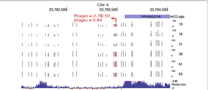
Genomic element rate profiling (GERP) analysis shows that evolutionarily constraint is similar among hyper- and hypomethylated CpGs (Additional file 5: Figure S4). Particularly, approximately 15% of CpGs lie in areas showing similar constraint as known functional elements (GERP ++ score > 1.7) [36]. However, we also observed regions with high conservation. For example, a HyperDMR spanning over 20 CpGs was observed at the evolutionarily constrained region 3′ of PGC1α locus in chromosome 4 (shown in Fig. 5). Specifically, this high evolutionarily constraint region is evident by Phy-lop constraint scores > 4.88 at 3′ end of the transcript and distal intergenic region, spanned by age-related hypermethylation where the top CpG within this region shows large effect size of 0.84 (p = 2.7e−10).
We further studied the DMRs showing significant evo-lutionary (GERP ++ score > 1.7) constraint (Additional file 2: Table S4) observing that disease (OMIM) relevant gene loci were slightly less likely to be found among constrained HyperDMRs (61/211, or 29%) as compared to constrained HypoDMRs (39/114 or 34%). However, among HyperDMRs at disease loci we observed pri-mary neuropsychiatric/neurodevelopmental/neurologi-cal traits for 31% (19/61), whereas only 10% (4/39) of HypoDMRs at disease loci had primary neurological/ neurodevelopmental picture. An example of sperm aging HyperDMR (chr2:166900333–166900544) is overlapping
exon 11 of SCN1A, most common genetic cause of epi-lepsy, intriguingly most de novo mutations arise in the paternal chromosome [37], but paternal age has not yet been associated with occurrence of mutations.
Gene networks implicated by age‑associated methylation
The CpG enrichments analyses in GREAT were done to look for genes linked to relative hypo- or hypermethyla-tion with higher density of CpG associahypermethyla-tions in ± 1 Mb for genes than expected by chance (Additional file 3: Table S5). We performed gene ontology (GO) analyses and found an enrichment of 420 genes in age-depend-ent hypoDMRs, and 606 genes in age dependage-depend-ent hyper-DMRs. The top 20 terms were listed for hypoDMRs (Fig. 6a) and hyperDMRs (Fig. 6b); the lists of enriched genes in hyper- and hypomethylated regions are shown in Additional file 4: Table S6. Overall, GO analyses revealed developmental pathways in both hypo- and hypermeth-ylated regions and markedly strong association signals for pathways associated with the central nervous system (CNS) including neuron projection, differentiation and recognition, and behaviour-related terms in hypermeth-ylated regions with age (Fig. 6). It is interesting to note that pathways associated with spermatogenesis or sperm function are not identified in this analysis.
Fig. 5 Evolutionarily constrained region 3′ of PGC1α locus in chromosome 4 with cluster of age-induced hypermethylation. Top associated CpG
is highlighted in red. Raw methylation values (0–100%) are shown (tracks with black tick marks) for six samples representing extremes of the age distribution in our sample set (ages shown on the right). Bottom track shows 100 vertebrates Basewise Conservation by PhyloP (phyloP100wayAll): high evolutionary constraints are evident by Phylop constraint scores > 4.88 at 3′ end of the transcript and distal intergenic region, spanned by age-related hypermethylation
Weighted gene co‑expression network analysis (WGCNA) in sperm MCC‑seq
WCGNA is an unsupervised method used to iden-tify clusters (modules) of highly correlated features and favours a scale-free network clustering pattern [38]. We have previously utilized this in identifying correlated CpGs in diseases [39]. We reasoned that the approach could identify important sets of CpGs, which may not reach marginal significance in site-wise analyses. We then performed trait correlation against modules identified in WGCNA (Additional file 5: Figure S5). We observed two highly significant module-trait correlations (|Aver-ageCorr|> 0.6) with each linked to age and composed of hundreds of CpGs and involving > 100 gene loci (Addi-tional file 4: Table S6). These large modules (MEblue corr = 0.62, p = 2.00E−11 and MEbrown corr = −0.62, p = 3.00E−11) show GO enrichments to neuronal as well as developmental processes (Additional file 5: Figure S6) with moderate overlap (> 15%) to marginally significant age-associated CpGs. Several other age trait correlated
(|Corr|> 0.3, p < 0.005) modules are driven by fewer CpGs and protein coding gene loci most of which were not cap-tured by marginal age associations and may represent important additional loci for aging. In addition, one mod-erately correlated module (MEtan corr = 0.33, p = 0.001) was observed with infertility trait and not detected in pointwise analyses for infertility associations (Additional file 5: Figure S5, Additional file 4: Table S6). Remarkably, the infertility associated CpG module is driven strongly by single chromosome 8 locus (87 of 97 module CpGs), which is hypermethylated in sperm from infertile men (Fig. 7a,b). These CpGs are all in the area of 9 kb region map proximal to testis-selective long-noncoding RNA (LINC01606) (Fig. 7c).
Discussion
To our knowledge, this is the first study that uses a high throughput, customized MCC-seq approach to enrich bisulfite sequencing coverage to regions of variable and/ or dynamic DNA methylation in human spermatozoa Fig. 6 Gene networks implicated by age-associated methylation. a Gene ontology (GO) analyses of age genes (n = 420) enriched in age dependent
designed to identify the effects of both increasing pater-nal age and fertility. In comparison with previously commonly used methods, the merit of higher resolu-tion is that it allows for the identificaresolu-tion of thousands of age-related and age-specific epigenetic alterations. In addition, we found that there are more hypermethyl-ated (62%) than hypomethylhypermethyl-ated (38%) CpG sites during aging. Interestingly, the hypermethylated CpG sites were frequently located in the distal region to genes, whereas hypomethylated sites were often near to gene region. Furthermore, developmental pathways were enriched for both age hypo- and hypermethylated subsets; neu-ron function and behaviour-related genes were markedly strong and enriched for age hypermethylated regions in spermatozoa. However, these dynamic DNA methylation changes during aging remain to be supported by further experiments using in vivo cell models to detect biologi-cal effect on developmental transcription and signaling; transgenerational persistence of these changes could be identified in a longitudinal cohort or in multi-genera-tional families.
We demonstrated that age prediction from our data improved by increasing the number of age-associated regions from earlier reported loci to thousands of novel regions. This suggests that by specific targeting of the full complement of age-associated CpGs and generating deep sequencing data (improving pointwise accuracy of
methylation data) across this subset of sperm dynamic methylome could allow for the development of a fine-tuned sperm epigenome age tool.
Our results clearly show that the identified DNA meth-ylation alterations are age-related and specific to germ cells. Sperm DNA methylation patterns were evaluated in 17 fertile donors in a longitudinal study by compar-ing the sperm methylome of 2 samples collected from each individual 9–19 years apart; in that study, using the Illumina 450 K microarray, 139 age-related hypomethyl-ated regions and 8 age-relhypomethyl-ated hypermethylhypomethyl-ated regions were identified [21]. In our study (Fig. 2), we found more than 100 age-associated CpGs in previously reported 50 age DMR regions are reaching significance for sperm [22], whereas only two age-related CpGs are significant in blood [23]. This strong replication of previous sperm aging data along with no signal enrichment for blood aging provides clear evidence of sensitivity and specificity of our technology and dataset. Overall, the results sug-gest that there is a pervasive influence of aging in shaping the sperm methylome patterns.
Other than examining the effects of aging, we also investigated whether the fertility status of these subjects as well as other covariates affected the sperm methyl-ome. Surprisingly, fertility status did not result in signifi-cant changes in the DNA methylation pattern. Previous studies indicated that sperm DNA methylation patterns Fig. 7 WGCNA revealed a hypermethylated CpG module associated with infertility trait in Chromosome 8 locus. a Sperm DNA methylation track
pattern was shown in representative fertile and infertile subjects (their ID and age on the left), the hypermethylated CpG module maps proximal to a long noncoding RNA, LINC01606. b Mean CpG methylation levels of sperm from fertile (green) and infertile (red) men on chr8, p-values were shown in blue (fertile vs. infertile). c Gene expression for LINC01606 in different tissues indicated its testis-selective
differed significantly with fertility status; > 8500 DMCs were identified and were proposed to be predictive of embryo quality after in vitro fertilization (IVF) [26]. The reasons for the discrepant results between the two stud-ies are not clear. Male infertility is a multi-factorial disor-der; infertility of unknown origin accounts for 37–58% of all cases [40]. Although the fertility status of subjects is well defined in this study, and the average sperm concen-tration was reduced significantly in infertile versus fertile group (Table 1), we did not have severe oligozoospermic men in this study. It has been reported that moderate or severe oligozoospermia is often associated with aber-rant DNA methylation of imprinted genes, especially in patient with less than 10 million spermatozoa per ml [41,
42], but the global DNA methylation was normal [14, 43,
44]; this could, in part, explain why we did not find fertil-ity associated DNA methylation alterations in our study cohort.
Although we do not detect fertility associated DNA methylation in pointwise analyses in human sperm, we do observe a moderately correlated CpGs cluster (mod-ule) with infertility trait in WGCNA analysis. Specifically, the infertility associated CpG module is hypermethyl-ated on chromosome 8 locus in sperm from infertile men, and is located nearby the upstream of LINC01606, a testis-selective long-noncoding RNA gene. LINC01606 has been shown to be elevated aberrantly and correlated with metastasis and invasion of gastric cancer, and its expression level is associated with Wnt/β-catenin sign-aling pathway activation through the regulation of miR-423-5P [45]. There is no evidence yet that LINC01606 plays a role in spermatogenesis or male fertility, but whether the hypermethylated CpG module is correlated to this long non-coding RNA gene in infertile men, and which miRNA species are subsequently regulated by LINC01606 in human sperm is worth considering for further study.
The distribution of age-related either hypo- or hyper-methylated CpGs in sperm is not random, as age-associ-ated CpGs in DMRs regions are highly significant (Fig. 4). We identified 798 age-associated DMRs (483 Hyper-DMRs and 315 HypoHyper-DMRs) that are relatively evenly distributed in comparison with only 147 DMRs (8 Hyper-DMRs and 139 HypoHyper-DMRs) with strong bias toward hypomethylation regionally that were identified by Jen-kins and colleagues [21]. This suggests that the 450 K array, which has inherent biases with the use of higher density of probes at promoter or gene dense regions, pro-vides a selective perspective on the impact of aging on the methylome.
We identified a high density of age-associated CpGs changes in chromosomes 4 and 16, particularly HyperD-MRs with localized clusters. It is interesting to note that
the chr4 DMR cluster overlaps PGC1α locus, a protein involved in metabolic aging [31] and that chr16 DMR cluster overlaps RBFOX1 locus, a gene implicated in neu-rodevelopmental disease [32]. The latter was also identi-fied within a MEblue module by WGCNA, in which GO enrichments to neuronal and developmental processes were shown. PGC1α is a transcription coactivator that is involved in a wide variety of biological responses includ-ing adaptive thermogenesis, mitochondrial biogenesis, glucose/fatty acid metabolism and heart development. PGC-1α is a powerful regulator of energy metabolism in both health and disease [31]; a close relationship exists among PGC-1α function, insulin sensitivity, and Type 2 diabetes. With aging, a potentially important defect has been found in the mitochondrial fatty acid oxidation pathway associated with insulin resistance [46]. RBFOX1 gene has been recently shown to serve as a ‘hub’ in autism spectrum disorder (ASD) transcriptome networks and in neurodevelopmental and psychiatric disorder including intellectual disability (ID), attention deficit hyperactivity disorder (ADHD) and schizophrenia [32]. While there is an indication that sperm DNA methylation patterns do not persist trans-generationally [47], it has been shown that paternal age effects on sperm FOX1 and KCNA7 methylation can be transmitted to the next generation [48]. We speculate that age-induced hypermethylation might be related to PGC-1α dysfunction, at least in part, resulting in insulin resistance development in children of aged father, and that RBFOX1 dysfunction is implicated in the higher risk of mental disorders of children of older fathers.
Epigenetic clocks, including DNA methylation clocks, are a novel class of biomarkers of aging; they also appear to be a robust measure of chronological age in humans, and reflect the biological age of individual [49]. The best established epigenetic clocks pioneered by Hovarth [23,
50] combined blood DNA methylation values in selected sets of CpG sites in order to predict an individual’s age. Deviation of epigenetic age from chronological age (epi-genetic age acceleration) has been shown to be predic-tive of deleterious health outcomes [51, 52]. The DNA methylation clock may not only be a measure of age, but also a regulator of age. In this study, we show a dissocia-tion between methylome changes during aging between somatic and male germ cells. It remains to be determined whether the methylation changes found in spermatozoa during aging will be able to serve as predictors of the pat-tern of health and disease of men’s progeny.
Conclusions
Using high throughput and customized MCC-seq tech-nique, we identified thousands of novel age-related epigenomic alterations in human spermatozoa. Our
results constitute important new insight on human sperm DNA methylation dynamics during the process of aging. These findings provide important new leads to determine how advanced paternal age results in DNA methylation that may have consequences on the health of their progeny.
Methods
Subject recruitment and study design
Both fertile and infertile subjects were recruited from the Men’s Health Clinic at the Royal Victoria Hos-pital, Montreal, Quebec over the period of January 2016 to December 2018. Research ethics protocol was approved by McGill University Health Centre (REB#: Human Subjects Research 15-068-MUH). All the sub-jects were evaluated by physicians, informed consent was obtained from all participants, with the assistance of the research nurse coordinators. Semen samples were collected by masturbation with at least 3 days abstinence. Semen parameters including semen vol-ume, sperm count, concentration, and motility were assessed, according to the WHO guidelines [53]. Sam-ples were aliquoted, frozen and stored at −80 °C until further analysis. Plasma hormone levels and metabolic factors were evaluated.
Inclusion criteria for fertile subjects were: (1) over the age of 18 years; (2) history of natural fecundity; (3) able to ejaculate by masturbation to provide semen samples. Exclusion criteria for fertile subjects are: (1) history of infertility (defined as inability to achieve natural concep-tion with a reproductively health female partner after one year of unprotected sexual intercourse) at any point of his life; (2) inability to provide informed consent; (3) inadequate sperm per ejaculate (total sperm count below 5 × 10⁶ /ml).
Inclusion criteria for infertile subjects were: (1) over the age of 18 years; (2) history of idiopathic male-factor infertility (defined above), with or without anomalies in semen parameters, in the absence of identifiable causes; (3) ability to ejaculate by masturbation to provide semen samples.
For both fertile and infertile groups, the following con-founding clinical conditions, known to affect sperma-tozoa, were used as additional exclusion criteria: recent (< 6 months) infection/inflammation of the genitourinary tract; history of hypogonadism and metabolic syndrome; varicoceles; and history of reconstructive surgery of the male reproductive tract.
In this study, 94 participants were recruited, 48 fer-tile controls and 46 inferfer-tile subjects. Within ferfer-tile and infertile groups, subjects were categorized in young (18– 38 years) and old (46–71 years) groups.
Extraction of genomic DNA from spermatozoa
Sperm genomic DNA (gDNA) was extracted from 5 to 10 million spermatozoa using QIAamp DNA Mini Kit (Qiagen #51306, Qiagen Mississauga, ON) accord-ing to the manufacturer’s protocols. Prior to proceed-ing with DNA extraction, sperm were washed usproceed-ing cold 0.45% NaCl for 3 times at 1500 g, 5 min at 4 °C to burst somatic cells. Sperm were then pelleted, and prior to proceeding, a visual inspection of each sample was done to ensure the absence of potentially contaminat-ing cells. Sperm lysis was done by pipettcontaminat-ing sperm pel-lets with sperm lysis buffer containing 150 mM EDTA, 10 mM Tris and 40 mM DTT, with the addition of 10% proteinase K (Sigma P4850) and 3.25% of Sarkosyl (30% N-Lauroylsarcosine sodium salt solution, Sigma 61747), and incubated at 56 °C for 2 h. Sperm DNA concentra-tions were quantified using a NanoDrop 2000 spectro-photometer (Thermo Fisher Scientific, Montreal, QC).
Purity of sperm preparation assessment by pyrosequencing of imprinted gene loci
Determination of the purity of the sperm preparation, i.e., absence of contaminating somatic cells, was veri-fied by determining DNA methylation levels of CpG dinucleotides located on the germline DMRs of mater-nally and patermater-nally imprinted genes using bisulfite pyrosequencing as previously described [19] with minor modifications. Briefly, 100–500 ng of sperm DNA were subjected to bisulfite conversion treat-ment using the EpiTect Plus DNA Bisulfite Kit (Qiagen #59124). Pyrosequencing was done and bisulfite con-verted DNA as template was subject to PCR reaction using HotStartTAq Master Mix Kit (Qiagen #203443) and in a GeneAmp PCR System 2400 (Perkin Elmer, Montreal, QC). Primers were designed using Qiagen PyroMark Assay Design SW 2.0 for assessment of two paternally methylated imprinted gene loci H19, DLK/ GLT2 IG-DMR, and two maternally methylated gene loci MEST, KCNQ10T1. The sequences of PCR primers and sequencing primers, and the PCR conditions are provided in Additional file 1: Table S7. Amplicons were sequenced using the PyroMark Q24 Reagents Kit (Qia-gen #970802), the PyroMark Q24 Vacuum Prep Work-station and Instrument (Qiagen, Valencia, CA, USA) following the manufacturer’s protocol. All samples used in this study had methylation profiles confirming a lack of somatic cell contamination. Sperm gDNA samples (1 µg) were sent to McGill University Genome Centre and sequencing (subject for Quality Control again prior to library preparation for MethylC-capture MCC-seq).
MethylC‑capture sequencing (MCC‑seq)
MCC-seq was done using a dedicated system to enrich dynamic CpG sites in human sperm as previously described [20]. WGBS and targeted bisulfite sequenc-ing were done as previously described [29, 54]. The WGBS libraries were constructed using the KAPA High Throughput Library Preparation Kit (Roche/KAPA Bio-systems) from 1 µg of the sperm DNA spiked with 0.1% (w/w) unmethylated lambda and pUC19 DNA (Pro-mega). DNA was sonicated (Covaris) and fragments sizes of 300-400 bp were controlled on a Bioanalyzer DNA 1000 Chip (Agilent). Following fragmentation, DNA end repair of double stranded DNA breaks, 3′-end adenylation, adaptor ligation and clean-up steps were conducted according to KAPA Biosystems’ pro-tocols. The sample was then bisulfite converted using the Epitect Fast DNA bisulfite kit (Qiagen) following manufacturer’s protocol. The resulting bisulfite DNA was quantified with OliGreen (Life Technology) and amplified with 9–12 PCR cycles using the KAPA HiFi HotStart Uracil + DNA Polymerase (Roche/KAPA Bio-systems) according to suggested protocols. The final WGBS libraries were purified using Ampure Beads, validated on Bioanalyzer High Sensitivity DNA Chips (Agilent) and quantified by PicoGreen (ThermoFisher). For WGBS libraries the SeqCap Epi Enrichment Sys-tem protocol (Roche NimbleGen) was used to capture the regions of interest. Equal amounts of multiplexed libraries (84 ng of each; 12 samples per capture) were combined to obtain 1 µg of total input library and was hybridized to the capture panel at 47 °C for 72 h. Washing, recovery, PCR amplification of the captured libraries as well as final purification were conducted as recommended by the manufacturer. Bioanalyzer High Sensitivity DNA Chips (Agilent) were used to deter-mine quality, concentration and size distribution of the final captured libraries. The capture libraries were sequenced with a 200-cycle S2 kit (100-bp paired end) on the NovaSeq 6000 following the NovaSeq XP work-flow, at a depth of 0.5 lanes per library.
Sequencing data processing
Targeted sperm panel MCC-Seq HiSeq reads were aligned using the Epigenome Pipeline available from the DRAGEN Bio-IT platform (Edico Genomics/Illumina). Specifically, the MCC-Seq paired-end raw reads were first demultiplexed into FASTQ files using Illumina’s bcl2Fastq2-2.19.1 software. Reads were then trimmed for quality (phred33 ≥ 20) and Illumina adapters using trimgalore v.0.4.2, a wrapper tool around Cutadapt [55] and FastQC. Then the trimmed reads were aligned, to the bisulfite-converted GRCh37 reference genome using DRAGEN EP v2.6.3 or later in paired end mode using
the directional/Lister methylation protocol presets; alignments were calculated for both Watson and Crick strands and the highest quality unique alignment was retained. A genome-wide cytosine methylation report was generated by DRAGEN to record counts of methyl-ated and unmethylmethyl-ated cytosines at each cytosine posi-tion in the genome. Methylaposi-tion counts are provided for the CpG, CHG and CHH cytosine contexts. DNA meth-ylation level of each CpG was calculated by the number of methylated reads over the total number of sequenced reads. CpGs that were found to be overlapping with SNPs (dbSNP 137), the DAC Blacklisted Regions or Duke Excluded Regions (generated by the ENCODE project) were removed. CpG sites with less than 20× read cover-age were also discarded.
Age prediction analysis
Sperm age predictor for discovery dataset was built using the R package glmnet [56]. Starting with the top 5000 significant DMCs, CpGs which were covered by all sam-ples in the discovery cohort were selected to build the predictor. The DNA methylation level was ranged from 0 to 1. Similar to Horvath’s strategy, the alpha parameter of glmnet was chosen to 0.5 (elastic net regression) and the lambda value was chosen using leave-one-out cross-validation (LOOCV) on the training data [23].
Genomic enrichment analyses
We utilized Genomic Regions Enrichment of Annota-tions Tool (GREAT) [33] to explore general characteris-tics of CpGs associated with sperm age in a random set of 88,000 CpGs targeted for custom capture and sequenc-ing [20] as background. To characterize gene networks potentially linked to age-associated CpGs, we fetched ENSEMBL gene annotations for significantly GREAT enriched “foreground” regions (as compared to all tar-geted CpGs). The analysis of enriched gene ontologies among GREAT significant “foreground” gene regions was then pursued by selecting genes with significantly increased density (± 1 Mb) of multiple testing (qv < 0.01) adjusted hypo- or hypermethylated CpGs as compared to random (targeted) CpG background. To further identify the hypo- and hyper-DMC relative density, we calculated the ratio of DMCs over the total number of CpGs within 1 Mb sliding windows over the genome.
We also performed GO analyses using Metascape [57] to enrich genes in age dependent hypo- and hyper-methylated regions. We investigated the distribution of chromatin states (ChromHMM from ENCODE data) of sperm age-related hypo- and hypermethylated CpGs in human hESC (H1) [34].
Evolutionarily constraint among hypo- and hypermeth-ylated age-CpGs was done using genomic element rate
profiling (GERP) analysis, and evolutionary constrained hypo- and hypermethylated DMRs were localized to dis-ease (OMIM) relevant gene loci [58].
Weighted gene‑co‑expression network analysis (WGCNA)
Weighted gene co-expression network analysis (WGCNA), aimed at detecting highly correlated mod-ule of CpGs, was done using 20,000 variable CpGs. The input CpGs were chosen based on uniform (no missing values) coverage (i.e. ≥20X) and population variance (i.e. top 20,000 most variable CpGs based on their variance, and roughly top 5% of variance) among our samples. The WGCNA was ran using the WGCNA package in R [38] with the default parameters. A soft-thresholding power of 8, which is the lowest power for which the scale-free topology fit index curve flattens out upon reaching a high value of signed R-square of 0.9, was chosen. A maximum block size of 20,000 and unsigned network were used (to construct a topological overlap matrix) and minimum number of CpGs per module = 30 and cut height = 0.4 were used to detect methylation modules. The module eigengene (ME) value was calculated for each module and further the Pearson correlation between MEs and traits was computed to quantify module-trait associations.
The CpGs from significant module trait correlations (p < 0.005, absolute correction |Corr|> 0.3) were used as input to GREAT protein coding gene enrichment using top 20 K variable CpGs as above. For the top 2 most com-plex modules involving 100 genes, we further performed GO enrichment analyses.
Statistical analyses
Statistical analyses for the characterization of subjects were done using GraphPad Prism 6 Software (San Diego, CA). Comparisons of semen parameters, biochemical and metabolic factors between fertile and infertile sub-jects were done using Student’s t-test. Pearson correla-tions were used for age and other semen parameters and metabolic factors. p < 0.05 was considered statistically significant.
Linear regression models (LMs) were built to inves-tigate the association between the DNA methylation level (methylation proportion defined above) and age. The age LM model was adjusted by correcting infertil-ity status, sperm concentration, smoking status, and other clinic indices such as total testosterone, bioavail-able testosterone concentrations, plasma FSH level, high density lipoprotein (HDL), and other metabolic parameters including BMI, plasma triglycerides and the risk ratio of total cholesterol to HDL. We used the R function lm() to fit the models, and calculated p-values for variables of interest. QQ-plot and Manhattan plot were reported for each LM. We further evaluated the
obtained p-values by generating false discovery pro-portion q-values using the R package q-values [59]. We reported qv < 0.01 as the genome-wide significant DMCs. The detailed number of CpGs used in each model is listed in Additional file 1: Table S2.
Supplementary information
Supplementary information accompanies this paper at https ://doi. org/10.1186/s1314 8-020-00988 -1.
Additional file 1: Table S1. Pearson correlation analysis of the age of subjects and clinical indices in the fertile and infertile subjects. Table S2. Association results for age models with different corrections. Table S3. Chromatin states distribution (ChromHMM from ENCODE data) of sperm age hypo –and hypermethylated regions in human hESC (H1). Table S7. Bisulfite pyrosequencing primers and PCR conditions for human sperm germline differentially methylated regions (DMRs)
Additional file 2: Table S4. Age related hypermethylated DMRs and clos-est genes with OMIM genes annotated.
Additional file 3: Table S5. Enriched gene lists by GREAT in hyper- or Hypomethylated regions.
Additional file 4: Table S6. WGCNA module enriched genes and CpGs. Additional file 5: Figure S1. DNA methylation profiles of imprinted gene loci approved sperm DNA sample purity. Two paternal methylated gene loci a H19 and b DLK1/GTL2 IG-DMR, two maternal methylated gene loci c MEST and d KCNQ10T1. Blue area in each genes indicated the CpG sites examined in pyrosequencing analysis. Sperm DNA methylation of each individual by MCC-seq further validated the pyrosequencing results. Figure S2. QQ-plots for associations tests. Unadjusted association tests for a age and b fertility show excess of age associations (black dots) above null expectation (red line), whereas for fertility no associations survive multiple testing correction. Figure S3. Genome-wide density of hyper/ hypomethylated regions upon sperm aging. Density of hypomethyla-tion is relatively even across the genome. Overall, sex chromosomes are relatively depleted of age associated CpGs. Figure S4. Genomic element rate profiling (GERP) analysis shows evolutional constrain is similar among hyper-and hypomethylated CpGs. X-axis shows data deciles and Y-axis GERP++ scores for hypermethylated (blue) and hypomethylated CpGs as function of age. Approximately 15% of CpGs lie in areas showing similar constraint as known functional elements (GERP++ > 1.7) (PMID: 21152010). Figure S5. Heatmap of module-trait relationships revealed by WGCNA analysis. Forty-three module eigengenes (MEs) listed in different colors on the left, were correlated to age, infertility, smoking and BMI traits shown on the bottom. The average module-trait correlation values and p-values were indicated individually, correlation value scale was shown on the right. Note that two highly significant modules each linked to age, and one moderate but highly significant module was associated with infertil-ity trait. Figure S6. GO enrichments for genes loci near age-linked CpG modules identified by WGCNA. Both a MEblue (corr = 0.62, P = 2.00E-11) and b ME brown (corr = −0.62, P = 3.00E-11) modules show GO enrich-ment to neuronal as well as developenrich-mental processes with significant age association.
Abbreviations
ADHD: Attention deficit hyperactivity disorder; ASD: Autism spectrum disor-der; BMI: Body mass index; CNS: Central nervous system; DMCs: Differentially methylated CpG sites; DMRs: Differentially methylated regions; FSH: Follicle stimulating hormone; GERP: Genomic element rate profiling; GO: Gene Ontol-ogy; GREAT: Genomic regions enrichment of annotations tool; HDL: High density lipoprotein; ID: Intellectual disability; IVF: In vitro fertilization; LDL: Low density lipoprotein; LH: Luteinizing hormone; LOOCV: Leave-one-out cross-validation; MCC-seq: MethylC-capture sequencing; ME: Module eigengene; PGC-1α: Peroxisome proliferator-activated receptor (PPAR)-gamma coactivator 1-alpha; RRBS: Reduced representation bisulfite sequencing; RBFOX1: RNA binding fox-1 homolog 1; TSH: Thyroid stimulating hormone; TSS: Transcription
start site; WGBS: Whole genome bisulfite sequencing; WGCNA: Weighted gene co-expression network analysis.
Acknowledgements
We are grateful to all the subjects who participated in this study for their con-tributions. We thank all the members of research team, in particular project coordinator Elise Boivin-Ford at McGill University, Dr. Marie-France Lusignan and Lorraine Lavigne, RN in MUHC-Research Institute for subjects recruitment and semen preparation. XS is supported by the NRC Artificial Intelligence for Design program. We thank Marie-Michell Simon at the McGill University Genome Centre for sequencing library preparation.
Authors’ contributions
M.C. contributed to performing the experiment in wet lab, data analyses and interpretation, manuscript draft and revision; X.S. participated in manuscript draft and revision, performed the data analysis including the EWAS analysis, WGCNA analysis and built the ageing model; P.C. recruited patients, and participated in obtaining funding and manuscript preparation; W.C. and T.K. assisted with data analysis; T.P. oversaw data analysis and manuscript prepara-tion. B.R. conceived the project, obtained funding, and participated in data analysis and manuscript preparation. All authors read and approved the final manusript.
Funding
This research has been funded by the CIHR Institute for Gender and Health Team Grant TE1-138298. BR is a James McGill Professor.
Availability of data and materials
The raw reads data generated by MCC-seq during the current study have been submitted to the European Genome-phenome Archive under the acces-sion number EGAS00001004168.
Ethics approval and consent to participate
Participants were recruited from the Men’s Health Clinic at the Royal Victoria Hospital, Montreal, Quebec over the period of January 2016 to December 2018. Research ethics protocol was approved by McGill University Health Centre (REB#: Human Subjects Research 15-068-MUH).
Consent for publications Not applicable.
Competing interests
The authors declare that they have no competing interests. Author details
1 Department of Pharmacology and Therapeutics, McGill University, 3655 Promenade Sir William Osler, Montreal, QC H3G 1Y6, Canada. 2 Department of Human Genetics, McGill University, 740 Docteur-Penfield Avenue, Montreal, QC H3A 0G1, Canada. 3 McGill University Genome Quebec Innovation Centre, 740 Docteur-Penfield Avenue, Montreal, QC H3A 0G1, Canada. 4 Digital Technologies Research Centre, National Research Council Canada, 1200 Montreal Road, Ottawa, ON K1A 0R6, Canada. 5 Department of Urology, McGill University Health Centre, 1001 Boulevard Decarie, Montreal, QC H4A 3J1, Canada. 6 Center for Pediatric Genomic Medicine, Children’s Mercy Kansas City, 2401 Gilham Road, Kansas City, MO 64108, USA. 7 Department of Obstetric and Gynecology, McGill University, 1001 Boulevard Decarie, Montreal, QC H4A 3J1, Canada.
Received: 13 September 2020 Accepted: 25 November 2020
References
1. Paul C, Robaire B. Ageing of the male germ line. Nat Rev Urol. 2013;10(4):227–34.
2. Selvaratnam J, Fice H, Noblanc A, Robaire B. Effects of aging on sperm chromatin. In: Leung PCK, Qiao J, editors. Human reproductive and prenatal genetics. London: Elsevier Academic Press; 2019. p. 85–103.
3. Deenadayal Mettler A, Govindarajan M, Srinivas S, Mithraprabhu S, Even-son D, Mahendran T. Male age is associated with sperm DNA/chromatin integrity. Aging Male. 2019:1–8.
4. Crow JF. The origins, patterns and implications of human spontaneous mutation. Nat Rev Genet. 2000;1(1):40–7.
5. Kong A, Frigge ML, Masson G, Besenbacher S, Sulem P, Magnusson G, et al. Rate of de novo mutations and the importance of father’s age to disease risk. Nature. 2012;488(7412):471–5.
6. Cioppi F, Casamonti E, Krausz C. Age-dependent de novo mutations during spermatogenesis and their consequences. Adv Exp Med Biol. 2019;1166:29–46.
7. Risch N, Reich EW, Wishnick MM, McCarthy JG. Spontaneous mutation and parental age in humans. Am J Hum Genet. 1987;41(2):218–48. 8. Ziller MJ, Gu H, Muller F, Donaghey J, Tsai LT, Kohlbacher O, et al. Charting
a dynamic DNA methylation landscape of the human genome. Nature. 2013;500(7463):477–81.
9. Xie K, Ryan DP, Pearson BL, Henzel KS, Neff F, Vidal RO, et al. Epigenetic alterations in longevity regulators, reduced life span, and exacerbated aging-related pathology in old father offspring mice. Proc Natl Acad Sci USA. 2018;115(10):E2348–57.
10. Marttila S, Kananen L, Hayrynen S, Jylhava J, Nevalainen T, Hervonen A, et al. Ageing-associated changes in the human DNA methylome: genomic locations and effects on gene expression. BMC Genomics. 2015;16:179.
11. Trasler JM. Epigenetics in spermatogenesis. Mol Cell Endocrinol. 2009;306(1–2):33–6.
12. Oakes CC, La Salle S, Smiraglia DJ, Robaire B, Trasler JM. A unique configu-ration of genome-wide DNA methylation patterns in the testis. Proc Natl Acad Sci USA. 2007;104(1):228–33.
13. Eckhardt F, Lewin J, Cortese R, Rakyan VK, Attwood J, Burger M, et al. DNA methylation profiling of human chromosomes 6, 20 and 22. Nat Genet. 2006;38(12):1378–85.
14. Kobayashi H, Sato A, Otsu E, Hiura H, Tomatsu C, Utsunomiya T, et al. Aberrant DNA methylation of imprinted loci in sperm from oligospermic patients. Hum Mol Genet. 2007;16(21):2542–51.
15. Marques CJ, Costa P, Vaz B, Carvalho F, Fernandes S, Barros A, et al. Abnor-mal methylation of imprinted genes in human sperm is associated with oligozoospermia. Mol Hum Reprod. 2008;14(2):67–74.
16. Hammoud SS, Purwar J, Pflueger C, Cairns BR, Carrell DT. Alterations in sperm DNA methylation patterns at imprinted loci in two classes of infertility. Fertil Steril. 2010;94(5):1728–33.
17. Hammoud SS, Cairns BR, Carrell DT. Analysis of gene-specific and genome-wide sperm DNA methylation. Methods Mol Biol. 2013;927:451–8.
18. Denham J, O’Brien BJ, Harvey JT, Charchar FJ. Genome-wide sperm DNA methylation changes after 3 months of exercise training in humans. Epigenomics. 2015;7(5):717–31.
19. Aarabi M, San Gabriel MC, Chan D, Behan NA, Caron M, Pastinen T, et al. High-dose folic acid supplementation alters the human sperm methy-lome and is influenced by the MTHFR C677T polymorphism. Hum Mol Genet. 2015;24(22):6301–13.
20. Chan D, Shao X, Dumargne MC, Aarabi M, Simon MM, Kwan T, et al. Cus-tomized MethylC-capture sequencing to evaluate variation in the human sperm DNA methylome representative of altered folate metabolism. Environ Health Perspect. 2019;127(8):87002.
21. Jenkins TG, Aston KI, Pflueger C, Cairns BR, Carrell DT. Age-associated sperm DNA methylation alterations: possible implications in offspring disease susceptibility. PLoS Genet. 2014;10(7):e1004458.
22. Jenkins TG, Aston KI, Cairns B, Smith A, Carrell DT. Paternal germ line aging: DNA methylation age prediction from human sperm. BMC Genomics. 2018;19(1):763.
23. Horvath S. DNA methylation age of human tissues and cell types. Genome Biol. 2013;14(10):R115.
24. Denomme MM, Haywood ME, Parks JC, Schoolcraft WB, Katz-Jaffe MG. The inherited methylome landscape is directly altered with paternal aging and associated with offspring neurodevelopmental disorders. Aging Cell. 2020:e13178.
25. Molaro A, Hodges E, Fang F, Song Q, McCombie WR, Hannon GJ, et al. Sperm methylation profiles reveal features of epigenetic inheritance and evolution in primates. Cell. 2011;146(6):1029–41.
•fast, convenient online submission
•
thorough peer review by experienced researchers in your field
• rapid publication on acceptance
• support for research data, including large and complex data types
•
gold Open Access which fosters wider collaboration and increased citations maximum visibility for your research: over 100M website views per year
•
At BMC, research is always in progress. Learn more biomedcentral.com/submissions
Ready to submit your research
Ready to submit your research ? Choose BMC and benefit from: ? Choose BMC and benefit from:
26. Aston KI, Uren PJ, Jenkins TG, Horsager A, Cairns BR, Smith AD, et al. Aber-rant sperm DNA methylation predicts male fertility status and embryo quality. Fertil Steril. 2015;104(6):1388–97.
27. Jenkins TG, Aston KI, Meyer TD, Hotaling JM, Shamsi MB, Johnstone EB, et al. Decreased fecundity and sperm DNA methylation patterns. Fertil Steril. 2016;105(1):51–7.
28. Moran S, Arribas C, Esteller M. Validation of a DNA methylation microar-ray for 850,000 CpG sites of the human genome enriched in enhancer sequences. Epigenomics. 2016;8(3):389–99.
29. Allum F, Shao X, Guenard F, Simon MM, Busche S, Caron M, et al. Characterization of functional methylomes by next-generation capture sequencing identifies novel disease-associated variants. Nat Commun. 2015;6:7211.
30. Turner SD. qqman: an R package for visualizing GWAS results using Q–Q and manhattan plots. bioRxiv. 2014.
31. Liang H, Ward WF. PGC-1alpha: a key regulator of energy metabolism. Adv Physiol Educ. 2006;30(4):145–51.
32. Hamada N, Ito H, Nishijo T, Iwamoto I, Morishita R, Tabata H, et al. Essential role of the nuclear isoform of RBFOX1, a candidate gene for autism spec-trum disorders, in the brain development. Sci Rep. 2016;6:30805. 33. McLean CY, Bristor D, Hiller M, Clarke SL, Schaar BT, Lowe CB, et al. GREAT
improves functional interpretation of cis-regulatory regions. Nat Biotech-nol. 2010;28(5):495–501.
34. Thurman RE, Rynes E, Humbert R, Vierstra J, Maurano MT, Haugen E, et al. The accessible chromatin landscape of the human genome. Nature. 2012;489(7414):75–82.
35. Hammoud SS, Nix DA, Zhang H, Purwar J, Carrell DT, Cairns BR. Distinctive chromatin in human sperm packages genes for embryo development. Nature. 2009;460(7254):473–8.
36. Davydov EV, Goode DL, Sirota M, Cooper GM, Sidow A, Batzoglou S. Identifying a high fraction of the human genome to be under selective constraint using GERP++. PLoS Comput Biol. 2010;6(12):e1001025. 37. Heron SE, Scheffer IE, Iona X, Zuberi SM, Birch R, McMahon JM, et al.
De novo SCN1A mutations in Dravet syndrome and related epilep-tic encephalopathies are largely of paternal origin. J Med Genet. 2010;47(2):137–41.
38. Langfelder P, Horvath S. WGCNA: an R package for weighted correlation network analysis. BMC Bioinform. 2008;9:559.
39. Hudson M, Baron M, Colmegna I, Bernatsky S, Klein Oros K, Pastinen T, et al. Novel approaches to discovery of biomarkers in rheumatoid arthritis: comment on the article by Oswald et al. Arthritis Rheumatol. 2015;67(8):2276–2277.
40. Hamada A, Esteves SC, Agarwal A. Unexplained male infertility: potential causes and management. Hum Androl. 2011;1(1):2–16.
41. Dada R, Kumar M, Jesudasan R, Fernandez JL, Gosalvez J, Agarwal A. Epigenetics and its role in male infertility. J Assist Reprod Genet. 2012;29(3):213–23.
42. Pacheco SE, Houseman EA, Christensen BC, Marsit CJ, Kelsey KT, Sigman M, et al. Integrative DNA methylation and gene expression analyses identify DNA packaging and epigenetic regulatory genes associated with low motility sperm. PLoS ONE. 2011;6(6):e20280.
43. Kobayashi N, Miyauchi N, Tatsuta N, Kitamura A, Okae H, Hiura H, et al. Factors associated with aberrant imprint methylation and oligozoo-spermia. Sci Rep. 2017;7:42336.
44. Tang Q, Pan F, Yang J, Fu Z, Lu Y, Wu X, et al. Idiopathic male infertility is strongly associated with aberrant DNA methylation of imprinted loci in sperm: a case-control study. Clin Epigenetics. 2018;10(1):134. 45. Luo Y, Tan W, Jia W, Liu Z, Ye P, Fu Z, et al. The long non-coding RNA
LINC01606 contributes to the metastasis and invasion of human gastric cancer and is associated with Wnt/beta-catenin signaling. Int J Biochem Cell Biol. 2018;103:125–34.
46. Petersen KF, Befroy D, Dufour S, Dziura J, Ariyan C, Rothman DL, et al. Mitochondrial dysfunction in the elderly: possible role in insulin resist-ance. Science. 2003;300(5622):1140–2.
47. Jenkins TG, James ER, Aston KI, Salas-Huetos A, Pastuszak AW, Smith KR, et al. Age-associated sperm DNA methylation patterns do not directly persist trans-generationally. Epigenetics Chromatin. 2019;12(1):74. 48. Atsem S, Reichenbach J, Potabattula R, Dittrich M, Nava C, Depienne
C, et al. Paternal age effects on sperm FOXK1 and KCNA7 meth-ylation and transmission into the next generation. Hum Mol Genet. 2016;25(22):4996–5005.
49. Gensous N, Franceschi C, Santoro A, Milazzo M, Garagnani P, Bacalini MG. The Impact of Caloric Restriction on the Epigenetic Signatures of Aging. Int J Mol Sci. 2019;20(8).
50. Hannum G, Guinney J, Zhao L, Zhang L, Hughes G, Sadda S, et al. Genome-wide methylation profiles reveal quantitative views of human aging rates. Mol Cell. 2013;49(2):359–67.
51. Marioni RE, Shah S, McRae AF, Chen BH, Colicino E, Harris SE, et al. DNA methylation age of blood predicts all-cause mortality in later life. Genome Biol. 2015;16:25.
52. Chen BH, Marioni RE, Colicino E, Peters MJ, Ward-Caviness CK, Tsai PC, et al. DNA methylation-based measures of biological age: meta-analysis predicting time to death. Aging (Albany NY). 2016;8(9):1844–65. 53. WHO. World Health Organization laboratory manual for the examination
and processing of human semen, 5th ed. 2010.
54. Cheung WA, Shao X, Morin A, Siroux V, Kwan T, Ge B, et al. Functional variation in allelic methylomes underscores a strong genetic contribu-tion and reveals novel epigenetic alteracontribu-tions in the human epigenome. Genome Biol. 2017;18(1):50.
55. Martin M. Cutadapt removes adapter sequences from high-throughput sequencing reads. EMBnetjournal. 2011;17(1):10–2.
56. Friedman J, Hastie T, Tibshirani R. Regularization paths for generalized linear models via coordinate descent. J Stat Softw. 2010;33(1):1–22. 57. Zhou Y, Zhou B, Pache L, Chang M, Khodabakhshi AH, Tanaseichuk O,
et al. Metascape provides a biologist-oriented resource for the analysis of systems-level datasets. Nat Commun. 2019;10(1):1523.
58. Lindblad-Toh K, Garber M, Zuk O, Lin MF, Parker BJ, Washietl S, et al. A high-resolution map of human evolutionary constraint using 29 mam-mals. Nature. 2011;478(7370):476–82.
59. Storey JD, Bass AJ, Dabney A, Robinson D. qvalue: Q-value estimation for false discovery rate control. R package. version 2.0. 0. ed. Available at github. com/jdstorey/qvalue: github; 2017. Accessed 14 April 2017
Publisher’s Note
Springer Nature remains neutral with regard to jurisdictional claims in pub-lished maps and institutional affiliations.