Publisher’s version / Version de l'éditeur:
Pulmonary Research and Respiratory Medicine, 2, 1, pp. 52-62, 2015-02-18
READ THESE TERMS AND CONDITIONS CAREFULLY BEFORE USING THIS WEBSITE.
https://nrc-publications.canada.ca/eng/copyright
Vous avez des questions? Nous pouvons vous aider. Pour communiquer directement avec un auteur, consultez la
première page de la revue dans laquelle son article a été publié afin de trouver ses coordonnées. Si vous n’arrivez pas à les repérer, communiquez avec nous à PublicationsArchive-ArchivesPublications@nrc-cnrc.gc.ca.
Questions? Contact the NRC Publications Archive team at
PublicationsArchive-ArchivesPublications@nrc-cnrc.gc.ca. If you wish to email the authors directly, please see the first page of the publication for their contact information.
This publication could be one of several versions: author’s original, accepted manuscript or the publisher’s version. / La version de cette publication peut être l’une des suivantes : la version prépublication de l’auteur, la version acceptée du manuscrit ou la version de l’éditeur.
For the publisher’s version, please access the DOI link below./ Pour consulter la version de l’éditeur, utilisez le lien DOI ci-dessous.
https://doi.org/10.17140/PRRMOJ-2-108
Access and use of this website and the material on it are subject to the Terms and Conditions set forth at
Role of microRNAs in progression and recurrence of early-stage lung
adenocarcinoma
Singh, Rama K.; Bethune, Drew C.; Xu, Zhaolin; Douglas, Susan E.
https://publications-cnrc.canada.ca/fra/droits
L’accès à ce site Web et l’utilisation de son contenu sont assujettis aux conditions présentées dans le site LISEZ CES CONDITIONS ATTENTIVEMENT AVANT D’UTILISER CE SITE WEB.
NRC Publications Record / Notice d'Archives des publications de CNRC:
https://nrc-publications.canada.ca/eng/view/object/?id=36412f77-cc69-4873-9900-248c489b858d https://publications-cnrc.canada.ca/fra/voir/objet/?id=36412f77-cc69-4873-9900-248c489b858d
Role of MicroRNAs in Progression and
Recurrence of Early-Stage Lung
Adenocarcinoma
Rama K. Singh1#, Drew C. Bethune2#, Zhaolin Xu3# and Susan E. Douglas1#*
#These authors contributed equally to this work
1National Research Council Halifax, 1411 Oxford Street, Halifax, NS, B3H 3Z1, Canada 2Department of Surgery, Queen Elizabeth II Health Sciences Centre and Dalhousie University,
1278 Tower Rd., Halifax, NS, B3H 2Y9, Canada
3Department of Pathology, Queen Elizabeth II Health Sciences Centre and Dalhousie
Univer-sity, 5788 University Ave., Halifax, NS, B3H 1V8, Canada
*Corresponding author:
Susan E. Douglas, PhD National Research Council Halifax, 1411 Oxford Street Halifax NS, B3H 3Z1 Canada E-mail: susan.douglas@nrc-cnrc.gc.ca Article History: Received: January 27th, 2015 Accepted: February 16th, 2015 Published: February 18th, 2015 Citation:
Singh RK, Bethune DC, Xu Z, Doug-las SE. Role of microRNAs in pro-gression and recurrence of early-stage lung adenocarcinoma. Pulm
Res Respir Med Open J. 2015; 2(1):
52-62.
Copyright:
© 2015 Douglas SE. This is an open access article distributed under the Creative Commons Attribution Li-cense, which permits unrestricted use, distribution, and reproduction in any medium, provided the origi-nal work is properly cited. Volume 2 : Issue 1
Article Ref. #: 1000PRRMOJ2108
Research
ABSTRACT
Lung cancer is the leading cause of cancer-related death worldwide, and the majority of cases (77%) are not diagnosed until the disease has spread to regional lymph nodes or distant sites. Even among Non-Small Cell Lung Cancer (NSCLC) adenocarcinoma patients who have been diagnosed early and where there has been no spread to lymph nodes, recurrence after surgical intervention is high. Improved understanding of the molecular alterations involved in aggressivity and recurrence of these tumors may provide better biomarkers for the identiica-tion of patients who would beneit from adjuvant chemotherapy. By comparing the expression of microRNAs in advanced Stage II/III tumors with those expressed in earlier Stage I tumors, we aimed to identify differentially expressed molecular biomarkers that could underly progres-sion and recurrence of Stage I tumors. This pilot study utilized TaqMan qPCR assays to assess the expression of microRNAs in tumor tissue, matched normal tissue and plasma samples from Stage I and Stage II/III lung adenocarcinoma patients. Seven microRNAs were identiied from plasma that could distinguish between patients with Stage I and Stage II/III adenocarcinoma. The up-regulation of miR-29a in plasma of patients with later-stage adenocarcinoma would result in enhanced expression of several molecules involved in integrin signaling, migration and proliferation. Analysis of differential expression of microRNAs in later-stage compared to early-stage lung adenocarcinoma implicates focal adhesion and ECM-receptor pathways in progression and recurrence. Plasma miR-29a is a promising biomarker that can be assessed non-invasively and whose clinical utility should be pursued.
KEYWORDS: Adenocarcinoma; Biomarker; microRNAs; Non-Small Cell Lung Cancer;
Recur-rence; Transcriptome
ABBREVIATIONS:ECM: Extracellular Matrix; KEGG: Kyoto Encyclopedia of Genes and
Ge-nomes; MAPK: Mitogen-Activated Protein Kinase; NSCLC: Non-Small Cell Lung Cancer; qPCR: quantitative Polymerase Chain Reaction; RNA: Ribonucleic Acid
Note: All gene names are abbreviated according to theHuman Genome Nomenclature Com-mittee.
INTRODUCTION
Lung cancer remains the leading cause of cancer-relat-ed death worldwide, accounting for 1.59 million of the 8.2
mil-lion total cancer deaths each year.1NSCLC accounts for ~80%
of all lung cancers, and adenocarcinoma is the main histologi-cal subtype. Smoking is responsible for 87% of lung cancers in the United States, and the majority of NSCLC adenocarcino-mas. While historically, cigarette smoking was associated with NSCLC squamous cell lung cancer, since the transition to iltered cigarettes there has been a rise in the number of adeocarcinomas among smokers. Most NSCLC patients are diagnosed at an ad-vanced stage that is associated with high recurrence and less than 15% survival over 5 years. Even for patients with Stage I disease who have had a complete surgical resection, the 5-year survival
is only 52%,2 mostly because of recurrence. This suggests that
Stage I tumors can be misclassiied, especially when lymph node metastases are small and escape detection. Furthermore, intra-tumor heterogeneity may not be detected by standard
single-site biopsy, leading to inaccurate classiication and prognosis.3,4
Classiication and staging by Tumor, Node, Metastasis (TNM)5
have only modest prognostic utility and patients with resected Stage IA tumors of identical histology, differentiation, vascular invasion, and margins may differ widely in their survival time and response to therapy. The use of alternative sources of biopsy material that relect tumor heterogeneity, such as readily avail-able blood or plasma, may provide valuavail-able information.
Cells and cellular nucleic acids in sputum, bronchial bi-opsies, brushing specimens, bronchial lavage luid, blood, pleu-ral effusions and solid tumor biopsies provide material for
clas-siication, diagnosis and prognosis of NSCLC.6Several cellular
tumor markers have been shown to be useful in prognosis of lung cancer including positive immunohistochemical staining of
mTOR in early stage NSCLC,7overexpression of Apolipoprotein
E in lung adenocarcinomas with malignant pleural effusion,8and
overexpression of SOX2 in Stage I adenocarcinomas, especially
those with pleural invasion.9Recently, a 14-gene expression
as-say has been developed to examine recurrence risk in early-stage
NSCLC.10
Small non-coding RNAs such as snoRNAs11and
mi-croRNAs (“oncomirs”)12 are a promising class of cancer
bio-markers. The expression proiles of these highly stable mole-cules in tumor tissue and/or plasma have been associated with occurrence of NSCLC, tumor tissue type, stage of
differentia-tion, and prognosis (for review see13). microRNAs are generally
down-regulated in tumor tissue compared to matched normal tissue. However, tumor-derived microRNAs are found within
exosomes or in free circulation in the plasma of cancer patients14
and have been shown to be generally up-regulated compared to
plasma from normal donors.15 A panel of four plasma
microR-NAs (mir-486, -30d, -1 and -499) was recently shown to dis-criminate between NSCLC patients with good and poor
prog-nosis.15Serum levels of miR-142-3p and miR-29b were elevated
in lung adenocarcinoma patients suffering recurrence within 24
months.16
There is an urgent need for better understanding of the molecular alterations that occur during progression of early-stage lung adenocarcinoma, and more reliable prognostic and predictive biomarkers. Such biomarkers could improve survival by identifying patients at high risk for recurrence who may ben-eit from adjuvant chemotherapy. Here we report a pilot study that determines microRNA proiles in tumors and plasma from patients with Stage I and Stage II/III adenocarcinoma, the most common form of NSCLC, and relate these indings to the bio-logical processes that may be involved in early recurrence.
MATERIALS AND METHODS Study population
Patient biopsy samples (frozen tumor and plasma) from Stage I (with no lymph node involvement) and Stage II/III (with lymph node involvement) were selected from the Lung Cancer Tumor Bank at Capital District Health Authority (CDHA; Hal-ifax, NS, Canada). In order to limit the clinical variability as much as possible, a uniform study population was used based on the following criteria: adenocarcinoma, age >45 years; all but one were current or past smokers (Table 1).
Most cases of adenocarcinoma are associated with smoking, and these were highly represented in the Tumor Bank. Recurrence is the time in months from surgery until detection of recurrence. Control plasma (Precision Biologics, Dartmouth, NS, Canada) contained pooled plasma from 20 healthy subjects. This study was approved by the Capital Health Research Ethics Board (CDHA-RS/2004-336), and all participating individuals signed informed consent.
ID Stage Age Gender Smoking Status Recurrence (months) L202 IB 76 Male Current L218 IA 65 Male Past L247 IB 77 Male Past L252 IA 81 Male Past L272 IA 79 Female Past L278 IA 69 Male Past
L194 IIA 53 Male Past 12.8
L212 IIA 71 Male Current
L229 IIA 66 Male Past 8.9
L240 IIA 74 Male Past
L258 IIIA 45 Male Never
L262 IIA 76 Female Past
L300a IIB 54 Female Past
aremoved from analyses due to high hemolysis in plasma samples. Table 1: Patient characteristics and samples used for microRNA analysis.
RNA extraction
Approximately 50 mg of frozen lung tissue was pulver-ized in a MultiSample BioPulverizer (BioSpec, Bartlesville, OK,
USA) and homogenized in 1 mL TRIZOL® (Life Technologies,
Burlington, ON, Canada) using a FastPrep®-24 (MP
Biomedi-cals, Santa Ana, CA, USA). Total RNA was extracted according to the manufacturer’s protocol and treated with TURBO DNase (Applied Biosystems, Carlsbad, CA, USA) prior to further pu-riication with the Total RNA Pupu-riication Kit (Norgen Biotech Corp., Thorold, ON, Canada). RNA was puriied from 300 μL of plasma using the Total RNA Puriication Kit and both prepara-tions were enriched for small RNAs using the PureLink miRNA Isolation Kit (Invitrogen, Burlington, ON, Canada). The RNA was quantiied on the NanoDrop-1000 (NanoDrop Products, Wilmington, DE, USA) and RNA quality was determined on the Bioanalyzer-2100 (Agilent, Wilmington, DE, USA).
MicroRNA assay of tissue and plasma
The miRNA PCR Array for Cancer (MAH-102F; SA Biosciences, Burlington, ON, Canada) containing 88 cancer-relevant microRNA-probes and 8 controls in a 96-well plate was employed to assess expression of microRNAs in pooled plasma samples of ive Stage I (L202, 218, 247, 252 and 278) and ive Stage II (194, 212, 229, 240 and 262) lung adenocarcinoma pa-tients compared to pooled normal plasma (Precision Biologics). RNA was puriied from 300 μL of plasma (60 μL each of ive in-dividual samples from each stage). 200 ng enriched small RNA was reverse-transcribed using the RT² First Strand Kit (SA Bio-sciences, Burlington, ON, Canada) and qPCR was performed on a Light Cycler-480 (Roche Applied Science, Laval, QC, Canada) using conditions speciied by the manufacturer. Fold
regulation was calculated from 2nd derivative Cp values using
the SA Biosciences web-based qPCR analysis software (http://
pcrdataanalysis.sabiosciences.com/pcr/arrayanalysis.php) and
SNORD48 was used as a reference.
Based on the results of the PCR array (Supp Table 1)
and literature survey, a multiplex TaqMan® microRNA Assay
(Applied Biosystems) was designed for 31 microRNAs that were at least two-fold up-regulated in cancer relative to control (Supp Table 2). RNU48 was used as a reference, allowing all 32 reac-tions to be performed in triplicate in 96-well plates. Expression was evaluated in an expanded group of six Stage I and seven Stage II/III tissue and plasma samples. Briely, 100 ng of
to-tal RNA was reverse-transcribed using the TaqMan® microRNA
Reverse Transcription Kit (Applied Biosystems) and qPCR re-actions were cycled on a LightCycler-480 once at 95 °C for 10 min; 45 times at 95 °C for 15 sec, 60 °C for 60 sec; and once at
40 °C for 30 sec. Individual samples contained 1 μL cDNA, 5
μL of 2X qPCR mix and 1 μL of microRNA-speciic TaqMan Probe/Primer mix in a 10 μL total reaction volume. For tumor samples, matched normal tissue samples in triplicate were used as controls and for plasma, pooled normal samples were used. In the latter case, control samples were assayed twice
indepen-dently in triplicate reactions and the average was used.
Normalized data was analyzed by Student’s t-test and
p-values were based on replicate 2nd derivative Cp values for
each microRNA in the control group and patient group. Those microRNAs that were associated with high p-values (>0.05) were assigned as missing values. The Student’s t-test and SAM
modules of MeV17were then used to identify microRNAs that
were signiicantly differentially expressed between the Stage I and Stage II/III samples. MicroRNA expression was analyzed
from 2nd derivative Cp values using SA Bioscience web-based
PCR data analysis software (Qiagen, Mississauga, ON, Canada) and the stably expressed RNU48 and miR-210 to normalize for tissue and plasma samples, respectively. Targets for microRNAs
were identiied by searching miRDB(www.mirDB.org). The list
of microRNA targets with a score greater than 80 was used to identify KEGG pathways potentially involved in progression
using gene enrichment analysis in WebGestalt (http://bioinfo.
vanderbilt.edu/webgestalt).
RESULTS
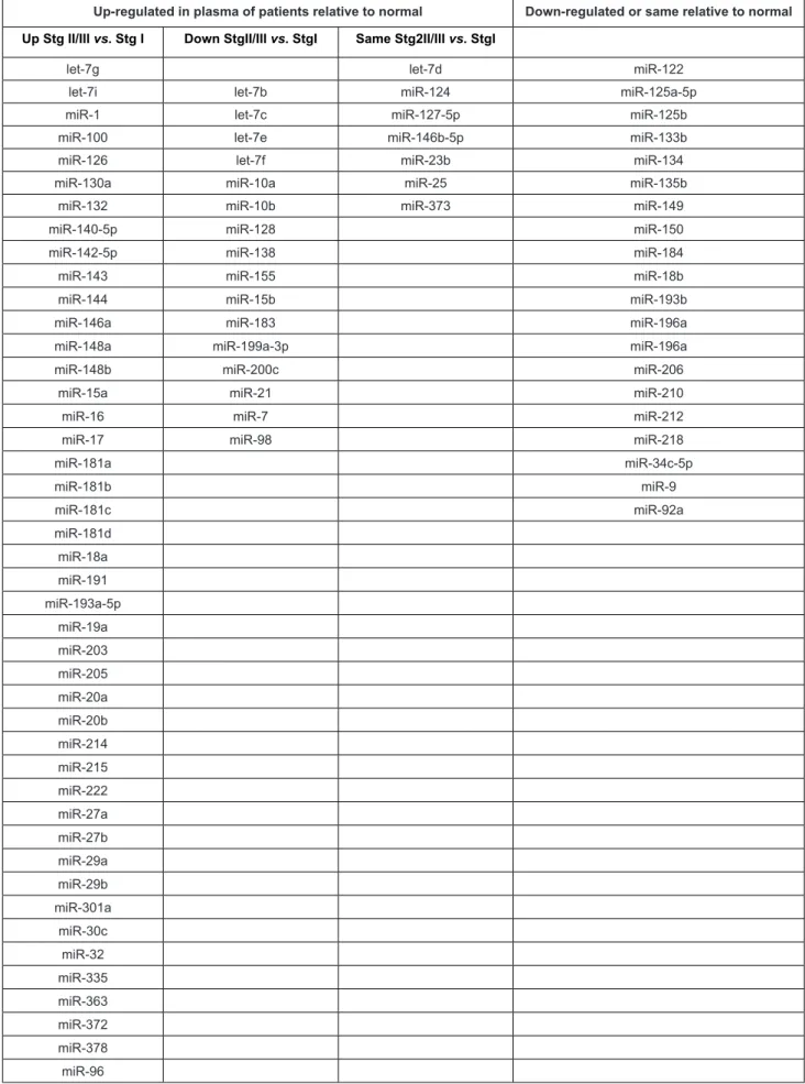
In order to identify clinically relevant microRNA bio-markers, we screened 88 cancer-relevant microRNAs by array. We selected those that were up-regulated at least two-fold in plasma of lung adenocarcinoma patients relative to plasma from pooled normal subjects as these would be most easily detectable in a clinical assay. Sixty-eight microRNAs were up-regulated in plasma of NSCLC patients relative to normal subjects. Of these 44 microRNAs showed a higher level in Stage II/III vs. Stage I, 17 showed a lower level in Stage II/III vs. Stage I, and 7 did not differ between Stage II/III and Stage I (Table 2).
Of these, 20 for which there was corroborating evidence in the literature were chosen for TaqMan assays along with 11 other well-supported microRNAs from the literature (miR-17-3p, 34a, 92a, 106a, 141, 182, 201, 218, 221, 451 and 486-5p; Supp Table 2).
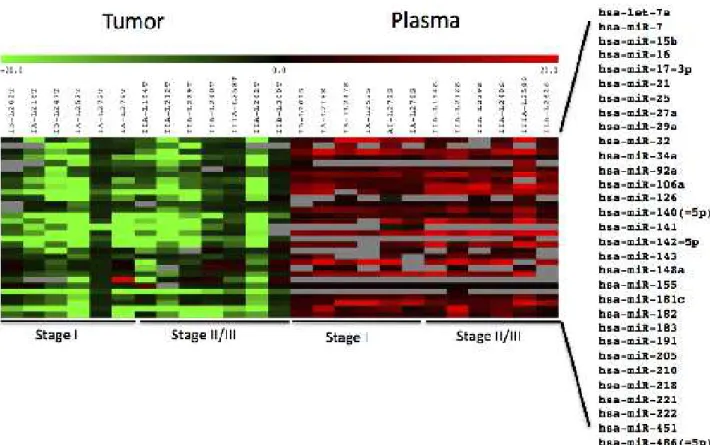
Expression data for these 31 microRNAs were obtained using TaqMan assays for all tissue/matched normal samples and all but one plasma sample (L300 plasma was unusable due to a high level of lysed erythrocytes and was excluded from analy-sis). Several microRNAs (miR-17-3p, 141, 143, 205 and 218)
exhibited inconsistent, often low, signal intensities in most
plas-ma samples (grey boxes, Figure 1).
microRNAs were mostly down-regulated in tumor samples relative to normal adjacent tissue but up-regulated in plasma relative to control pooled plasma from normal subjects (Figure 1, Supp Table 3) . The single Stage IIIA sample (L258) exhibited generally higher, but not statistically signiicant, up-regulation of plasma microRNAs than the other samples.
There were no statistically signiicant differences in tumor tissue microRNA expression between different stages.
Up-regulated in plasma of patients relative to normal Down-regulated or same relative to normal Up Stg II/III vs. Stg I Down StgII/III vs. StgI Same Stg2II/III vs. StgI
let-7g let-7d miR-122
let-7i let-7b miR-124 miR-125a-5p
miR-1 let-7c miR-127-5p miR-125b
miR-100 let-7e miR-146b-5p miR-133b
miR-126 let-7f miR-23b miR-134
miR-130a miR-10a miR-25 miR-135b
miR-132 miR-10b miR-373 miR-149
miR-140-5p miR-128 miR-150
miR-142-5p miR-138 miR-184
miR-143 miR-155 miR-18b
miR-144 miR-15b miR-193b
miR-146a miR-183 miR-196a
miR-148a miR-199a-3p miR-196a
miR-148b miR-200c miR-206
miR-15a miR-21 miR-210
miR-16 miR-7 miR-212
miR-17 miR-98 miR-218
miR-181a miR-34c-5p miR-181b miR-9 miR-181c miR-92a miR-181d miR-18a miR-191 miR-193a-5p miR-19a miR-203 miR-205 miR-20a miR-20b miR-214 miR-215 miR-222 miR-27a miR-27b miR-29a miR-29b miR-301a miR-30c miR-32 miR-335 miR-363 miR-372 miR-378 miR-96
Table 2: Expression of 88 cancer-relevant miRNAs in plasma of NSCLC Stage I and Stage II/III patients relative to normal plasma, determined by PCR Array. MicroRNAs that are shaded were included in the TaqMan assay.
Expression of 31 miRNAs in tumor relative to matched normal tissue and patient plasma relative to pooled control plasma from six patients each with Stage I and Stage II/III NSCLC adenocarcinoma. Red, up-regulated; green, down-regulated; grey, high p-values (>0.05) were assigned as missing data. Log2 ratios are presented.
Figure 1: Heat map of miRNA expression in tumor tissue.
Signiicantly differentially expressed plasma miRNAs identiied by SAM analysis. Those marked with a single asterisk or double asterisk were also identiied as signiicant by t-test (p<0.1 and p<0.01, respectively). Red, up-regulated; green, down-regulated; grey, high p-values (>0.05) were assigned as missing data. Log2 ratios are presented.
Figure 2: Heat map of miRNA expression in plasma.
However, SAM analysis identiied seven plasma microRNAs that could distinguish between patients with Stage I and Stage II/III lung adenocarcinoma (Figure 2).
Four of these (miR-25, 32, 142-5p, and 148a) ap-proached signiicance (p<0.1) and miR-29a was identiied at p<0.01 by t-test (Table 3).
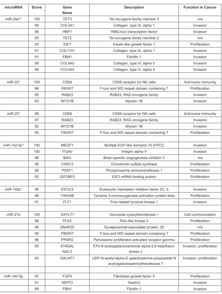
Searching miRDB for targets of these seven microRNAs that were up-regulated in Stage II/III compared to Stage I pa-tients resulted in 416 unique targets with a score greater than 80 in the human genome (Supp Table 4). Enrichment analysis of these targets identiied ive major KEGG pathways: cancer, focal adhesion, ECM-receptor, p53 signalling, and MAPK signaling pathway (Supp Table 5). The highest-scoring cancer-relevant targets are shown in Table 3.
microRNA Score Gene Name
Description Function in Cancer
miR-29a** 100 TET3 Tet oncogene family member 3 n/a
99 COL3A1 Collagen, type III, alpha 1 Invasion
98 HBP1 HMG-box transcription factor Invasion
95 TET2 Tet oncogene family member 2 n/a
93 IGF1 Insulin-like growth factor 1 Proliferation
91 COL11A1 Collagen, type XI, alpha 1 Invasion
91 FBN1 Fibrillin 1 Invasion
89 COL4A5 Collagen, type IV, alpha 5 Invasion
88 COL4A4 Collagen, type IV, alpha 4 Invasion
miR-32* 100 CD69 CD69 receptor for NK cells Anti-tumor immunity
96 FBXW7 F-box and WD repeat domain containing 7 Proliferation
95 RAB23 RAB23, RAS oncogene family Invasion
93 MYO1B Myosin 1B Invasion
miR-25* 98 CD69 CD69 receptor for NK cells Anti-tumor immunity
97 RAB23 RAB23, RAS oncogene family Invasion
92 MYO1B Myosin 1B Invasion
90 FBXW7 F-box and WD repeat domain containing 7 Proliferation
miR-142-5p* 100 MEGF1 Multiple EGF-like domains 10 (FAT2) Invasion
100 ITGAV Integrin alpha V Invasion
98 BAI3 Brain-speciic angiogenesis inhibitor 3 n/a
96 CHSY3 Chondroitin sulfate synthase Proliferation
96 PSAT1 Phosphoserine aminotransferase 1 Proliferation
93 IGF2BP3 IGF2 mRNA binding protein Proliferation
miR-148a* 96 EIF2C4 Eukaryotic translation initiation factor 2C, 4 Invasion 96 YWHAB Tyrosine 3-monooxygenase activation protein beta Proliferation
91 FLT1 Fms-related tyrosine kinase 1 Invasion
miR-27a 100 GXYLT1 Glucoside xylosyltransferase 1 Cell communication
99 PLK2 Polo-like kinase 2 Proliferation
96 SNAP25 Synaptosomal-associated protein, 25 n/a
96 FBXW7 F-box and WD repeat domain containing 7 Proliferation 96 PPARG Peroxisome proliferator-activated receptor gamma Proliferation
95 ST6GAL
NAC3
ST6 N-acetylgalactosaminide alpha-2,6-sialyltrans-ferase 3
Invasion, proliferation
93 GALNT7 UDP-N-acetyl-alpha-D galactosamine:polypeptide N acetylgalactosaminyltransferase 7
Invasion, proliferation
miR-140-5p 97 FGF9 Fibroblast growth factor 9 Proliferation
91 SEPT2 Septin2 Invasion
89 FBN1 Fibrillin 1 Invasion
Table 3: Cancer-relevant targets of plasma microRNAs that distinguish between Stage I and Stage II/III tumors. microRNAs were identiied using SAM and those marked with a single or double asterisk were signiicant by t-Test (p<0.1 and p<0.01, respectively). Targets were identiied by searching miRDB. n/a, no direct role in NSCLC.
microRNA Gene target Function Tumor type Reference
miR-29a nd Invasion Gastric 21
MMP2 Invasion, apoptosis Oral squamous 22
nd Prognosis Colorectal 23
nd Early detection Colorectal 24
COL11A1 Progression Gastric 28
COL11A1 Cancer-associated stromal cells Colorectal 29
miR-142-5p MEGF (FAT2) Invasion Squamous carcinoma 35
PSAT Proliferation Colorectal 36
IGF2BP3 Prognosis Colorectal 38
IGF2BP3 Prognosis Pancreatic ductal 40
CHSY3 Invasion Colorectal 39
miR-148a MMP7 Invasion Gastric 21
EIF2C4 Metastasis Colorectal 42
miR-27a GALNT7* Invasion, proliferation Cervical 44
miR-140-5p SEPT2 Proliferation Liver 47
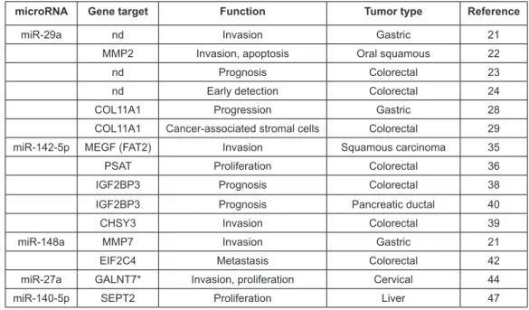
Table 4: microRNAs dysregulated in this study that have been reported to be prognostic or have targets that function in other cancer types. Only in the case of miR-29a and MMP2 (shaded) have the microRNA and target gene been demonstrated to be co-regulated in the other tumor types. *GALNT7 regulation in cervical cancer is associated with miR-214 expression. nd, not determined.
DISCUSSION
In this study, we found a consistent down-regulation of microRNAs in tumor compared to adjacent normal tissue and up-regulation of plasma microRNAs from lung cancer patients relative to normal controls. Of the 31 microRNAs included in the TaqMan assay, most were consistently detectable; however ive microRNAs had highly variable, often low, values between triplicates, giving a low p-value. Two of these, miR-17-3p and 205, have been reported to have very weak intensities in plasma
and serum.18There was a slightly lower overall expression of
tumor microRNAs and corresponding higher overall expression of plasma microRNAs for the youngest patient (L258), but this was not statistically signiicant. This patient was also the only non-smoker and had the most advanced stage (IIIA). Although biological features of the patient can impact microRNA levels, it would be dificult to draw any valid conclusions concerning mi-croRNA dysregulation based on these features from the results of a single patient.
We were unable to detect any statistically signiicant changes in tumor tissue microRNAs between Stage I and Stage II/III patients. However, patient plasma did exhibit several mi-croRNAs (miR-25, 27, 29a, 32, 140-5p, 142-5p, and 148a) that could distinguish between patients with Stage I and Stage II/III lung adenocarcinoma, with miR-29a being the most statistically signiicant (p<0.01). Since plasma is a more easily accessible biopsy material, plasma proiles rather than tissue proiles were used to investigate alterations in cancer pathways.
There is limited information on the expression of these
microRNAs and their target genes in other solid tumor types. There have, however, been some studies describing these mi-croRNAs as prognostic biomarkers and others describing the same genes targeted by the dysregulated microRNAs we have identiied. These studies mainly include tumors associated with the digestive system (particularly colorectal) and lend support to our indings (Table 4).
Similar to lung adenocarcinoma, expression of miR-29a
is also down-regulated in gastric tumors19and oral squamous
carcinoma,20resulting in increased expression of Matrix
Met-alloproteinase2(MMP2) in the latter case. MMP2 degrades
ex-tracellular matrix and promotes tumor invasiveness. In Stage II colorectal cancer, down-regulated tissue miR-29a is associated
with high recurrence21and elevated plasma levels are associated
with advanced stage.22Consistent with our results, the miR-29
family has been reported to be down-regulated in NSCLC
tu-mors relative to normal tissue23,24and up-regulated in serum of
early stage lung adenocarcinoma patients who recurred within
24 months.16We also found miR-142-5p to be up-regulated in
plasma of later-stage patients. This microRNA is derived from the same precursor as miR-142-3p, which is signiicantly up-regulated in serum of early stage lung adenocarcinoma patients who recurred within 24 months16 and in plasma of patients with
aggressive NSCLC.25
Of the gene targets of differentially regulated microR-NAs listed in Table 3, several do not have a known direct role in NSCLC and are not discussed. These include Brain-speciic An-giogenesis Inhibitor (BAI3), SNAP25 (involved in neurotrans-mitter release) and TET2 and TET3 oncogenes (epigenetic
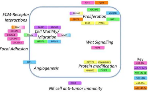
regu-Gene targets are represented by colored boxes with colors indicating microRNA involved. If the gene is targeted by more than one microRNA a gradient of the appropriate color is applied. The tumor cell membrane is represented by a blue line and targets are shown as extracellular, intracellular or membrane-bound. Names of pathways are shown in bold italic font.
Figure 3: Cancer pathways affected by microRNAs differentially regulated in NSCLC progression.
lators in malignant hematopoiesis).26
The remaining gene targets have roles in ECM-receptor interactions, focal adhesion, cell motility and migration, angio-genesis, protein modiication, invasion, proliferation, cell sig-nalling and NK-cell anti-tumor immunity (Figure 3).
This is in agreement with a recent microarray study we
performed on the same tumor biopsies27in which ECM-receptor
interactions and components of the focal adhesion pathway were the main elements differentially regulated between early and lat-er-stage lung adenocarcinomas.
miR-29a targets collagens COL3A1, COL11A1, CO-L4A5, and COL4A4, and Fibrillin1 (FBN1), which participate in integrin signaling, focal adhesion and migration. In gastric cancer, transcripts for several collagens, including COLA11A1, are also up-regulated in malignant compared to pre-malignant
tumors28and COLA11A1 is also up-regulated in
cancer-associ-ated ibroblasts of colon adenocarcinomas.29 Insulin-like Growth
Factor 1 (IGF1) is a key player in NSCLC tumorigenesis and
metastasis,30 initiating cell signaling, promoting mitosis,
pro-liferation and differentiation, and inhibiting apoptosis. HBP1 is a transcriptional repressor that plays a role in Wnt signaling
and induces cellular transformation.31 Reduction in tumor tissue
miR-29a would result in enhanced IGF-1 and Wnt signalling. Interestingly, we found two microRNAs (miR-25 and miR-32) that both targeted the same four genes (CD69, FBXW7, RAB23 and MYO1B). As with any biomarker discovery, there is high potential for false positives due to the large number of variables relative to the number of patient samples. Therefore,
this overlap in targets suggests that these microRNAs and their target transcripts warrant further investigation as biomarkers in early lung adenocarcinoma. Their higher expression in plasma and concomitant low expression in tumor tissue would promote translation of the hedgehog-signalling factor, RAB23 and the cytoskeletal protein MYO1B, which are necessary for cell
mo-tility and metastasis.32 FBXW7 is part of the E3 ubiquitin ligase
complex and targets cyclin E for degradation, thus impacting cell proliferation.
microRNAs in the plasma can affect immune cell function. CD69 is a marker of NK-cell activation and a
posi-tive regulator of anti-tumor activity.33It has been shown that
administration of several synthetic microRNAs or microRNAs in exosomes from healthy donors to NK-cells and mice bear-ing tumors resulted in TLR1 activation and increased NF-kB signaling. This resulted in increased NK-cell activation via
up-regulation of CD69 and increased anti-tumor immunity.34
How-ever, high plasma levels of miR-32 and -25, which repress CD69 expression, would result in reduced NK-cell anti-tumor activity leading to tumor progression.
miR-142-5p targets MEGF1 (FAT2), which mediates
migration of human cutaneous squamous carcinoma cells,35and
PSAT1, which has been shown to stimulate proliferation in
col-orectal cancer.36miR-142-5p also targets ITGAV and its
reduc-tion in tumor tissue would result in an increase in ITGAV.
Inter-estingly, miR-32 and miR-25 also target ITGAV37and miR-32
is reported to be down-regulated in another lung cancer study.23
CHSY3 and IGF2BP3 are two other relevant targets of miR-142 -5p and both are over-expressed in colorectal cancer patients
of glycosaminoglycans on the cell surface and shows elevated
expression in colorectal cancer;39IGF2BP3 has been suggested
as a biomarker of poor prognosis in pancreatic ductal
adenocar-cinoma.40
miR-148a, like miR-29a, is down-regulated in gastric cancer and has a major impact on the expression of MMP7 and
invasiveness.19miR-148a also targets FLT1, the receptor for
Vas-cular Endothelial Growth Factor (VEGFR1) and thus could play an important role in tumor angiogenesis. Decreased levels of tu-mor miR-148a would result in higher levels of FLT1 available for angiogenic signaling. NSCLC tumors expressing FLT1 are
more malignant and associated with poorer outcomes.41
Simi-larly, higher levels of YWHAB, a 14-3-3 protein involved in cell cycle control and inhibition of apoptosis, would result in a more malignant phenotype. EIF2C4 is an argonaute protein in the RNA-induced silencing complex. Argonaute proteins are up-regulated in colon cancer and higher levels are associated with
distant metastasis.42
Two other microRNAs that showed statistically signii-cant differential expression by SAM (but not t-test) target key genes of importance in cancer progression. miR-27a regulates expression of three enzymes that participate in post-second-ary modiication of important proteins (GXYLT1, GALNT7, ST6GALNAC3). GXYLT1 glycosylates NOTCH proteins and
reduces cancer signalling43whereas GALNT7 and
ST6GAL-NAC3 glycosylate mucins, thus impacting proliferation, migra-tion, and invasion. Oncogenesis in cervical cancer cells is
ef-fected through miR-214 regulation of GALNT7.44PPAR-γ is a
nuclear hormone receptor that is involved in cell proliferation and is over-expressed in many human cancers. It has recently been shown to prevent metastasis in lung cancer cells by
inhibit-ing TGF-β induced epithelial-to-mesenchymal transition.45PLK2
is a serine-threonine protein kinase that is essential for cell
divi-sion.46miR-140-5p, like miR-29a, targets FBN1. SEPT2, which
plays an important role regulation of cell proliferation through its effect on actin ilament formation and has been suggested as
a target for liver cancer therapy,47 is also a target of miR-140-5p.
The growth factor FGF9 is also involved in cell proliferation and can synergize with the EGFR oncogenic pathway in lung
adenocarcinoma48 leading to recurrence.49
The use of microRNAs as clinical biomarkers is
increas-ingly being described in the literature.50 They are highly stable
in circulating blood plasma and can be readily isolated. They are tissue-speciic and their expression can be dysregulated in response to physiological changes such as disease. Furthermore, they play a key role in differentiation, proliferation and apop-tosis making them ideal diagnostic, prognostic and predictive biomarkers in cancer. Although there are some published stud-ies describing microRNA signatures associated with recurrence in lung cancer, they tend to include lung cancer patients with
multiple NSCLC tumor types and from multiple stages.15,51,52It
is well known that different types of NSCLC (such as squamous
and adenocarcinoma) express different miRNAs. In our small-scale study, we have restricted our comparison to only adenocar-cinomas of Stage I and Stage II/III in order to more accurately identify plasma microRNAs likely to be involved in recurrence in early-stage patients. Of the six Stage I and six Stage II/III pa-tients studied, recurrences only occurred in two papa-tients both of whom were Stage II.
CONCLUSIONS
In conclusion, our study indicates that several micro-RNAs that distinguish between Stage I and Stage II/III patients are involved in the regulation of important pathways in cancer biology including focal adhesion, ECM-receptor interactions, p53 signalling, and MAPK signaling pathway. Because of the small number of samples in this study, their role in lung adeno-carcinoma should be veriied in a larger patient cohort. Their presence in plasma provides a readily accessible bioluid for clinical testing and the signiicant up-regulation of miR-29a in-dicates it could be a promising biomarker.
ACKNOWLEDGEMENT
We gratefully acknowledge the technical assistance of Jeffrey Gallant and Evelyn Teh, and bioinformatic analysis by Susanne Penny, National Research Council (NRC). We thank John Fris, database manager of the Lung Tumor Bank at Capital District Health Authority, for collating clinical annotations. Funding was provided by NRC and Dalhousie University Departments of Pa-thology and Surgery.
REFERENCES
1. WHO. Cancer Fact Sheet 297, 2012.
http://www.who.int/me-diacentre/factsheets/fs297/en/ Accessed 2015.
2. Reed MF, Molloy M, Dalton EL, Howington JA. Survival af-ter resection for lung cancer is the outcome that mataf-ters. Am
J Surg. 2004; 188: 598-602. doi: http://dx.doi.org/10.1016/j.
amjsurg.2004.07.037
3. Zhang J, Fujimoto J, Wedge DC, et al. Intratumor heteroge-neity in localized lung adenocarcinomas delineated by
multi-region sequencing. Science. 2014; 346: 256-259. doi: 10.1126/
science.1256930
4. Seoane J, De Mattos-Arruda L. The challenge of intratumour heterogeneity in precision medicine. J Intern Med. 2014; 276:
41-51. doi: 10.1111/joim.12240
5. Tsim S, O’Dowd CA, Milroy R, Davidson S. Staging of non-small cell lung cancer (NSCLC): A review. Respir. Med. 2010;
104: 1767-1774. doi: 10.1016/j.rmed.2010.08.005
the management of non-small cell lung cancer. Cancer Biomark-ers. 2009; 6: 197-210. doi: 10.3233/CBM-2009-0130
7. Dhillon T, Mauri FA, Bellezza G, et al. Overexpression of the mammalian target of rapamycin: a novel biomarker for poor sur-vival in resected early stage non-small cell lung cancer. J Thorac Oncol. 2010; 5: 314-319. doi: 10.1097/JTO.0b013e3181ce6604 8. Su WP, Chen YT, Lai WW, Lin CC, Yan JJ, Su WC. Apo-lipoprotein E expression promotes lung adenocarcinoma pro-liferation and migration and as a potential survival marker in
lung cancer. Lung Cancer. 2011; 71: 28-33. doi: 10.1016/j.
lungcan.2010.04.009
9. Sholl LM, Barletta JA, Yeap BY, Chirieac LR, Hornick JL. Sox2 protein expression is an independent poor prognostic indi-cator in stage I lung adenocarcinoma. Am J Surg Pathol. 2010;
34: 1193-1198. doi: 10.1097/PAS.0b013e3181e5e024
10. Woodard GA, Gubens MA, Jahan TM, et al. Prognostic mo-lecular assay might improve identiication of patients at risk for recurrence in early-stage non-small-cell lung cancer. Clin Lung Cancer. 2014; 15: 426-432. doi: 10.1016/j.cllc.2014.07.004 11. Liao J, Yu L, Mei Y, et al. Small nucleolar RNA signatures as biomarkers for non-small-cell lung cancer. Mol. Cancer. 2010;
9: 198-208. doi: 10.1186/1476-4598-9-198
12. Esquela-Kerscher A, Slack FJ. Oncomirs - microRNAs with a role in cancer. Nat. Rev. Cancer. 2006; 6: 259-269. doi: 10.1038/nrc1840
13. Shen J, Jiang F. Applications of microRNAs in the diagnosis and prognosis of lung cancer. Expert Opin. Med. Diagn. 2012; 6: 197-207.
14. Ohshima K, Inoue K, Fujiwara A, et al. Let-7 microRNA family is selectively secreted into the extracellular environment
via exosomes in a metastatic gastric cancer cell line. PLoS One.
2010; 5: e13247. doi: 10.1371/journal.pone.0013247
15. Hu Z, Chen X, Zhao Y, et al. Serum microRNA signatures identiied in a genome-wide serum microRNA expression proil-ing predict survival of non-small-cell lung cancer. J. Clin. On-col. 2010; 28: 1721-1726. doi: 10.1200/JCO.2009.24.9342 16. Kaduthanam S, Gade S, Meister M, et al. Serum miR-142-3p is associated with early relapse in operable lung
adenocarci-noma patients. Lung Cancer. 2013; 80: 223-227. doi: 10.1016/j.
lungcan.2013.01.013
17. Saeed AI, Sharov V, White J, et al. TM4: a free, open-source system for microarray data management and analysis. BioTech-niques. 2003; 34: 374-378.
18. Heegaard NH, Schetter AJ, Welsh JA, Yoneda M, Bowman
ED, Harris CC. Circulating micro-RNA expression proiles in early stage nonsmall cell lung cancer. Int J Cancer. 2012; 130:
1378-1386. doi: 10.1002/ijc.26153
19. Sakamoto N, Naito Y, Oue N, et al. MicroRNA-148a is downregulated in gastric cancer, targets MMP7, and indicates tumor invasiveness and poor prognosis. Cancer Sci. 2013. doi: 10.1111/cas.12330
20. Lu L, Xue X, Lan J, Gao Y, et al. MicroRNA-29a upregulates MMP2 in oral squamous cell carcinoma to promote cancer inva-sion and anti-apoptosis. Biomed Pharmacother. 2013; in press. 21. Weissmann-Brenner A, Kushnir M, Lithwick Yanai G, et al. Tumor microRNA-29a expression and the risk of recurrence in stage II colon cancer. Int J Oncol. 2012; 40: 2097-2103. doi: 10.3892/ijo.2012.1403
22. Huang Z, Huang D, Ni S, Peng Z, Sheng W, Du X. Plasma microRNAs are promising novel biomarkers for early detec-tion of colorectal cancer. Int J Cancer. 2010; 127: 118-126. doi: 10.1002/ijc.25007
23. Yanaihara N, Caplen N, Bowman E, et al. Unique microR-NA molecular proiles in lung cancer diagnosis and prognosis. Cancer Cell. 2006; 9: 189-198. doi: http://dx.doi.org/10.1016/j. ccr.2006.01.025
24. Barh D, Jain N, Tiwari S, et al. A novel in silico reverse-transcriptomics-based identiication and blood-based validation of a panel of sub-type speciic biomarkers in lung cancer. BMC Genomics. 2013; 14: S5. doi: 10.1186/1471-2164-14-S6-S5 25. Boeri M, Verri C, Conte D, et al. MicroRNA signatures in tissues and plasma predict development and prognosis of com-puted tomography detected lung cancer. Proc Natl Acad Sci USA. 2011; 108: 3713-3718. doi: 10.1073/pnas.1100048108 26. Cheng J, Guo S, Chen S, et al. An extensive network of TET2-targeting MicroRNAs regulates malignant
hematopoie-sis. Cell Rep. 2013; 5: 471-481. doi: http://dx.doi.org/10.1016/j.
celrep.2013.08.050
27. Douglas S, Bethune D, Xu Z. Microarray analysis implicates focal adhesion and ECM-receptor pathways in progression of early stage lung adenocarcinoma. Pulm Res Respir Med. 2015; 1: 21-31.
28.Zhao Y, Zhou T, Li A, Yao H, He F, Wang L, et al. A potential role of collagens expression in distinguishing between prema-lignant and maprema-lignant lesions in stomach. Anat Rec (Hoboken).
2009; 292: 692-700. doi: 10.1002/ar.20874
29. Galvan JA, Garcia-Martinez J, Vazquez-Villa F, et al. Valida-tion of COL11A1/procollagen 11A1 expression in TGF-beta1-activated immortalised human mesenchymal cells and in stromal
cells of human colon adenocarcinoma. BMC Cancer. 2014; 14:
867. doi: 10.1186/1471-2407-14-867
30. Wang Z, Liang Z, Liu J, et al. Expression and clinical signii-cance of IGF-1, IGFBP-3, and IGFBP-7 in serum and lung can-cer tissues from patients with non-small cell lung cancan-cer. Onco Targets Ther. 2013; 6: 1437-1444. doi: 10.2147/OTT.S51997 31. Lavender P, Vandel L, Bannister AJ, Kouzarides T. The HMG-box transcription factor HBP1 is targeted by the pocket proteins and E1A. Oncogene. 1997; 14: 2721-2728.
32.Gonzalez L, Eiro N, Gonzalez-Reyes S, et al. Clinical signii-cance of myosin in colorectal signii-cancer. Ann Diagn Pathol. 2012;
16: 260-266. doi: 10.1016/j.anndiagpath.2011.11.004
33. Esplugues E, Vega-Ramos J, Cartoixa D, et al. Induction of tumor NK-cell immunity by anti-CD69 antibody therapy. Blood. 2005; 105: 4399-4406.
34. He S, Chu J, Wu LC, et al. MicroRNAs activate natural kill-er cells through Toll-like receptor signaling. Blood. 2013; 121:
4663-4671. doi: 10.1182/blood-2012-07-441360
35. Matsui S, Utani A, Takahashi K, Mukoyama Y, Miyachi Y, Matsuyoshi N. Knockdown of Fat2 by siRNA inhibits the migra-tion of human squamous carcinoma cells. J Dermatol Sci. 2008;
51: 207-210. doi: 10.1016/j.jdermsci.2008.04.006
36. Vie N, Copois V, Bascoul-Mollevi C, et al. Overexpres-sion of phosphoserine aminotransferase PSAT1 stimulates cell growth and increases chemoresistance of colon cancer cells. Mol Cancer. 2008; 7: 14. doi: 10.1186/1476-4598-7-14
37. Dalmay T, Edwards DR. MicroRNAs and the hallmarks
of cancer. Oncogene. 2006; 25: 6170-6175. doi: 10.1038/
sj.onc.1209911
38. Lochhead P, Imamura Y, Morikawa T, et al. Insulin-like growth factor 2 messenger RNA binding protein 3 (IGF2BP3) is a marker of unfavourable prognosis in colorectal cancer. Eur J Cancer. 2012; 48: 3405-3413. doi: 10.1016/j.ejca.2012.06.021 39. Kalathas D, Theocharis DA, Bounias D, et al. Chondroitin synthases I, II, III and chondroitin sulfate glucuronyltransferase expression in colorectal cancer. Mol Med Rep. 2011; 4: 363-368.
doi: 10.3892/mmr.2011.431
40. Schaeffer DF, Owen DR, Lim HJ, et al. Insulin-like growth factor 2 mRNA binding protein 3 (IGF2BP3) overexpression in pancreatic ductal adenocarcinoma correlates with poor survival. BMC Cancer. 2010; 10: 59. doi: 10.1186/1471-2407-10-59 41. Fischer C, Mazzone M, Jonckx B, Carmeliet P. FLT1 and its ligands VEGFB and PlGF: drug targets for anti-angiogenic
ther-apy? Nat Rev Cancer. 2008; 8: 942-956. doi: 10.1038/nrc2524
42. Li L, Yu C, Gao H, Li Y. Argonaute proteins: potential bio-markers for human colon cancer. BMC Cancer. 2010; 10: 38.
doi: 10.1186/1471-2407-10-38
43. Lee TV, Sethi MK, Leonardi J, et al. Negative regulation of notch signaling by xylose. PLoS Genet. 2013; 9: e1003547. doi: 10.1371/journal.pgen.1003547
44. Peng RQ, Wan HY, Li HF, Liu M, Li X, Tang H. MicroRNA-214 suppresses growth and invasiveness of cervical cancer cells by targeting UDP-N-acetyl-alpha-D-galactosamine:polypeptide N-acetylgalactosaminyltransferase 7. J Biol Chem. 2012; 287:
14301-14309. doi: 10.1074/jbc.M111.337642
45. Reka AK, Kurapati H, Narala VR, et al. Peroxisome pro-liferator-activated receptor-gamma activation inhibits tumor metastasis by antagonizing Smad3-mediated epithelial-mesen-chymal transition. Mol Cancer Ther. 2010; 9: 3221-3232. doi: 10.1158/1535-7163.MCT-10-0570
46. Strebhardt K. Multifaceted polo-like kinases: drug targets and antitargets for cancer therapy. Nat Rev Drug Discov. 2010;
9: 643-660. doi: 10.1038/nrd3184
47. Yu W, Ding X, Chen F, et al. The phosphorylation of SEPT2 on Ser218 by casein kinase 2 is important to hepatoma carci-noma cell proliferation. Mol Cell Biochem. 2009; 325: 61-67.
doi: 10.1007/s11010-008-0020-2
48. Yin Y, Betsuyaku T, Garbow JR, Miao J, Govindan R, Or-nitz DM. Rapid induction of lung adenocarcinoma by ibroblast growth factor 9 signaling through FGF receptor 3. Cancer Res.
2013; 73: 5730-5741. doi: 10.1158/0008-5472.CAN-13-0495
49. Ohgino K, Soejima K, Yasuda H, et al. Expression of ibro-blast growth factor 9 is associated with poor prognosis in pa-tients with resected non-small cell lung cancer. Lung Cancer.
2014; 83: 90-96. doi: 10.1016/j.lungcan.2013.10.016
50. Schwarzenbach H. Circulating nucleic acids as biomarkers in breast cancer. Breast Cancer Res. 2013; 15: 211.
51. Liu XG, Zhu WY, Huang YY, et al. High expression of serum miR-21 and tumor miR-200c associated with poor prognosis in patients with lung cancer. Med. Oncol. 2012; 29: 618-626. doi: 10.1007/s12032-011-9923-y
52. Patnaik SK, Kannisto E, Knudsen S, Yendamuri S. Evalua-tion of microRNA expression proiles that may predict recurrence of localized stage I non-small cell lung cancer after surgical
re-section. Cancer Res. 2010; 70: 36-45. doi: 10.1158/0008-5472.