Divambal APPAVOO-GUPTA
Chimiste diplômée de l’Université de Johannesburg, Johannesburg (Afrique du Sud)
Exploring New Avenues for Arene-Ruthenium Complexes:
Coordination to [60]Fullerene, Hydrogen Bonding
Assemblies and Liquid-Crystalline Materials
Thèse présentée à la Faculté des Sciences,
Pour l’obtention du grade de Docteur ès Sciences
Membres du jury:
Prof. Robert Deschenaux Université de Neuchâtel, Directeur de thèse Prof. Bruno Therrien Université de Neuchâtel, Rapporteur interne Prof. Gilles Gasser Université de Zurich, Rapporteur externe
Institut de Chimie Université de Neuchâtel Soutenue le 20 July 2015
Rue Emile-Argand 11 2000 Neuchâtel - Suisse Tél: + 41 (0)32 718 2100 E-mail: [email protected]
Imprimatur pour thèse de doctorat www.unine.ch/sciences
IMPRIMATUR POUR THESE DE DOCTORAT
La Faculté des sciences de l'Université de Neuchâtel
autorise l'impression de la présente thèse soutenue par
Madame Divambal APPAVOO-GUPTA
Titre:
“Exploring New Avenues for Arene-Ruthenium
Complexes: Coordination to Fullerene,
Hydrogen Bonding Assemblies
and Liquid-Crystalline Materials”
sur le rapport des membres du jury composé comme suit:
- Prof. Robert Deschenaux, directeur de thèse, Université de Neuchâtel - Prof. ass. Bruno Therrien, Université de Neuchâtel
- Prof. Gilles Gasser, Université de Zürich
Professor Robert Deschenaux, thank you for having welcomed me in your group. You have been of constant support during my entire Ph.D. and I am grateful for that.
Professor Bruno Therrien, thank you for having given me the opportunity to do research in Switzerland.
Professor Gilles Gasser, thank you for accepting to be in my jury.
Dr Diego Carnevale, thank you for the NMR analysis. Your contribution has been crucial to my work.
Dr Armelle Vallat, thank you for MS spectrometry analysis and for your advice.
I am very grateful to my previous and current colleagues, without whom I would probably not be where I am today. Thank you Luyen, Anaïs, Nguyet, Sébastiano, Sylvain, Virginie, Tung, Ester, LeAnh and Steeve. Marion and Aurélie, thank you for your huge support and for keeping the “Hakuna Matata” and joyful ambiance in the lab.
I want to thank Damien and Christian for their useful advice as well as Thomas, David and Bing, who helped me whenever I needed an inorganic expert’s advice.
A big thank you to all my friends here in Neuchâtel who made my stay a bit more fun and comfortable, Jon, Manu, Ana, Christian, Simla, Theju, Bindhu and Justin.
I want to give an enormous thank you to my parents, mom, dad and Sel and my entire family for always caring and supporting me. Finally, thank you Mayank, for standing by my side, for trusting and believing in me more than I believe in myself.
The thesis aims at using arene-ruthenium complexes as building blocks for the synthesis of diverse compounds to obtain potential mesomorphic and/or biological properties. The thesis consists of three main projects.
The first project deals with supramolecular assemblies. New supramolecular di- and tetranuclear ruthenium arrangements, the latter bearing a cavity, were designed. H-bonding was the key interaction involved in the synthesis of the spacer ligands, which exist as dimers. Different spacer ligands and different ruthenium clips were used in synthesizing a whole family of the corresponding ruthenium assemblies/cages. Several analytical methods were employed to characterise the compounds and to study their arrangements.
The second project involves more classical ruthenium cages, including hexanuclear prisms and octanuclear cubes. Anthracene- and pyrene-core dendrimers were developed as potential guests, using key reactions such as click, Suzuki and Sonogashira coupling. Owing to the poor stability of the anthracene derivatives, their proper characterization and encapsulation could not be carried out. Pyrene derivatives showed better stability and were therefore studied further. The pyrene dendrimers, bearing cyanobiphenyl dendrons, displayed liquid-crystalline properties. Two pyrene dendrimers could be successfully encapsulated into hexanuclear arene-ruthenium prisms by the carceplex method. However, no mesomorphic properties were observed for the encapsulated systems.
The third project had as objective to study the biological properties of a system comprising of an arene-ruthenium moiety and [60]fullerene. Two such compounds were successfully synthesised. However, these compounds could not be tested for their potential biological activities because of their poor solubility in aqueous media.
1 Introduction ... 1
1.1 Ruthenium Complexes ... 3
1.1.1 Catalytic Applications ... 3
1.1.2 Biological Applications ... 4
1.1.3 Arene-Ruthenium Complexes ... 7
1.1.4 Mononuclear Arene-Ruthenium Complexes as Anticancer Drugs ... 8
1.1.5 Multinuclear Arene-Ruthenium Complexes for Anticancer Treatment ... 9
1.2 Host-Guest Chemistry ... 12
1.2.1 Carceplex Systems ... 13
1.2.2 Trojan Horse Concept ... 14
1.2.3 Encapsulation of Pyrene-Core Dendrimers and their Biological Properties ... 15
1.3 Aim of the Present Thesis ... 16
2 Hydrogen Bonded Metallo-Assemblies ... 19
2.1 Hydrogen Bonding ... 21
2.2 Aim ... 29
2.3 Synthesis ... 29
2.3.1 Synthesis of the Spacer Ligands ... 29
2.3.2 Macromolecular Assemblies ... 31
2.4 Characterization ... 33
2.4.1 Proton NMR Spectroscopy ... 33
2.4.2 DOSY ... 36
2.4.3 NOE/ROE and DFT Calculations ... 38
2.4.4 UV-Vis Spectroscopy ... 42
2.4.5 Electrospray Ionization Mass Spectrometry ... 45
2.5 Conclusion ... 46
2.6 Experimental Part ... 46
3 Encapsulation of Anthracene-Core Dendrimers into Arene-Ru Metallo-Cycles ... 69
3.1 Introduction ... 71
3.1.1 Anthracene ... 71
3.1.2 Suzuki Coupling Reaction ... 71
3.1.3 Click Chemistry ... 73
3.1.4 Sonogashira Coupling Reaction ... 75
3.2 Aim ... 77
3.3 Synthesis ... 77
3.3.1 Synthesis of Anthracene Compounds ... 77
3.3.2 Carceplex Assemblies ... 81
3.4 Characterization ... 83
4 Encapsulation of Pyrene-Core Dendrimers into Arene-Ru Metallo-Cycles ... 93
4.1 Introduction ... 95
4.2 Aim ... 96
4.3 Synthesis ... 96
4.3.1 Synthesis of Pyrene Dendrimers ... 96
4.3.2 Carceplex Assemblies ... 98 4.4 Characterization ... 104 4.4.1 Proton NMR Spectroscopy ... 104 4.4.2 DOSY ... 105 4.4.3 X-Ray Crystallography ... 107 4.4.4 Mesomorphic Properties ... 108 4.5 Conclusion ... 113 4.6 Experimental Part ... 114 5 [60]Fullerene-Ruthenium Complexes ... 131 5.1 The [60]Fullerene ... 133 5.2 Aim ... 137 5.3 Synthesis ... 137 5.4 Characterization ... 140 5.4.1 Proton NMR Spectroscopy ... 140 5.4.2 UV-Vis Spectroscopy ... 141 5.5 Conclusion ... 142 5.6 Experimental Part ... 142
6 Conclusion and Perspectives ... 149
6.1 Conclusion ... 151 6.2 Perspectives ... 152 7 References ... 155 Abbreviations ... 167 Annex 1 ... 171 Annex 2 ... 177 Annex 3 ... 179
1
3
1.1 Ruthenium Complexes 1.1.1 Catalytic Applications
Ruthenium, the d-block element that belongs to the “platinum group metals”, is one of the rarest metals on Earth. It is obtained commercially from the wastes of nickel refining. Ru complexes are mostly used as catalysts for several, mechanically diverse, processes. Their ability to assume a vast range of oxidation states (-II to +VIII) and coordination geometries, and the major influence of ligands on the complexes have made Ru complexes very versatile catalysts.[1] Before the 1980s, a limited number of reactions, such as oxidation with RuO4 and
hydrogenation with ruthenium hydroxide were known.[2] Since then, the chemistry of Ru has progressed and is better understood, leading to higher scope for Ru catalysts. Thus, their ability of high electron transfer, high Lewis acidity, low redox potentials and their stability have given rise to the development of a large number of new reactions catalysed by Ru complexes, some of which are listed below:[3]
Hydrogenation: homogeneous or heterogeneous catalysis of the hydrogenation of substrates, like olefins, with either molecular hydrogen or by hydrogen transfer reactions.
Oxidation: oxidative transformations of alcohols, amines, amides and hydrocarbons catalysed by low-valent Ru complexes.
Isomerization: isomerization, like Claisen rearrangement of β,γ-unsaturated oxygen and nitrogen containing compounds catalysed by low-valent Ru complexes.
Nucleophilic addition: nucleophilic attack on alkynes and nitriles activated by Ru catalysts.
C-C bond formation: selective C-C bond formation reactions catalysed by low-valent Ru complexes.
Among all these Ru-catalysed reactions, the C-C bond formation, more specifically olefin metathesis, is a powerful method used extensively in organic synthesis. The work on olefin metathesis has been pioneered by Yves Chauvin, Robert H. Grubbs and Richard R. Schrock who were awarded the Nobel Prize in 2005. Ruthenium alkylidenes have been successful catalysts for metathesis transformations, including ring-closing metathesis, cross-metathesis, enyne metathesis and ring-opening metathesis polymerisation (Figure 1.1). Most of these reactions are catalysed by Grubbs 1st and 2nd generation Ru catalysts or Hoveyda-Grubbs Ru catalysts (Figure 1.2).[4]
4
Figure 1.1 Schematic representation of common Ru catalysed metathesis reactions
Figure 1.2 The 1st and 2nd generation of Grubbs and Hoveyda-Grubbs catalysts
Besides the widespread applications of Ru as catalysts, the unique properties of this precious metal have made its complexes to find interesting applications in other fields. For instance, many Ru complexes are being explored for their potential biological activities.[5]
1.1.2 Biological Applications
Belonging to the same group, ruthenium has mostly been inspired by the well-established chemistry and applications of platinum and its compounds. Just as in the case of catalysis, Ru has followed in the steps of Pt into biological applications. The area of metal-based anticancer drugs has been dominated by the Pt drug, cisplatin and closely related Pt antitumor agents, such as carboplatin and oxaliplatin (Figure 1.3). Since it got FDA (US Food and Drug Administration) approval in 1978, cisplatin has been used extensively in the treatment of several cancers including ovarian and testicular cancers.[6] Despite its resounding success in treating some cancers, cisplatin has several limitations, among which kidney toxicity, nausea, and bone marrow suppression are a few.[6c] Hence, the search for a better anticancer drug continues.
5
Figure 1.3 Structures of cisplatin, carboplatin and oxaliplatin
The application of platinum compounds in the treatment of cancer questions the ability of other metal complexes to do the same. In fact, metals such as Fe,[7] Ti,[8] Ga,[9] and Ru[10] have compounds that show interesting medicinal properties. Among these metals, Ru has attracted the attention of several research groups worldwide as complexes of this d7 metal display ligand exchange kinetics similar to those of platinum complexes.[11] Ruthenium is being considered as a promising alternative to platinum because it shows many advantages compared to platinum. Ru has more coordination sites, can undergo more changes in oxidation state, is less toxic than Pt and is able to mimic Fe in binding to biomolecules. All these properties have led many researchers to believe that the future of metal-based anticancer drug might be a Ru drug that will show anticancer activity similar to or even better than cisplatin and that can treat a broader range of tumours and with less side effects.[12]
The idea of using Ru complexes as potential anticancer agent first came to Clarke in 1980 after Durig et al. reported, in 1976, the ability of fac-Ru(NH3)3Cl3 to induce filamentous
growth of E. Coli cells in a manner comparable to cisplatin.[13] Following this discovery, Clarke et al. tested the anticancer properties of fac-Ru(NH3)3Cl3 and related cis-Ru(NH3)4Cl2
(Figure 1.4). Despite their good antitumor activity, the poor solubility of both compounds made them unsuitable for pharmaceutical use.[14]
Figure 1.4 Structures of fac-Ru(NH3)3Cl3 and cis-Ru(NH3)4Cl2
This work of Clarke instigated the study of the cytotoxic properties of Ru complexes, eventually resulting in a large number of these compounds that can be categorised according to the ligand type; amine and imine: {mer-Ru(tpy)Cl3, where tpy = 2,2′:6′,2′′-terpyridine[15]
and (HIm)[trans-(Im)2Cl4Ru],[16] where Im = imidazole}, polyaminopolycarboxylate:
6
where pdta = 1,2-propylendiaminetetraacetate}, DMSO: cis- and trans-Cl2(MeSO)4Ru[19]
(Figure 1.5).
Figure 1.5 Structures of octahedral cytotoxic Ru complexes
From all the biologically active ruthenium complexes, only two entered clinical trials, imidazolium-trans-dimethylsulfoxide-imidazole-tetrachlororuthenate (NAMI-A) and imidazolium-trans-bis(1H-indazole)-tetrachlororuthenate (KP1019) (Figure 1.6).[6c, 14, 20] Even though their structures resemble, these two Ru(III) complexes have different modes of action as anticancer drugs. While NAMI-A shows little cytotoxicity towards many cancer cell lines, KP1019 has high antitumoral activity and NAMI-A has pronounced antimetastatic effects (ability to prevent the release of cancer cells from the tumour into other parts of the body) on several cancer models whereas none is shown by KP1019.[21]
Figure 1.6 Structures of NAMI-A and KP1019
Both Ru(III) complexes have successfully completed Phase I of clinical trial and it has been observed that the reduction of the Ru(III) of NAMI-A to Ru(II), with ascorbic acid prior to administration, gives rise to increased antimetastatic activity. This is in agreement with the hypothesis made earlier by Clarke about “activation by reduction”, whereby the Ru(III) is believed to be reduced to Ru(II) species, the transformation being favoured by the chemically reducing environment created by the low molecular oxygen concentration and lower pH in tumour cells than in healthy ones.[11b, 14, 22] Ru(II) species, in general, show better binding
7
ability and hence better reactivity than Ru(III). In view of this, the area of Ru(II) arene complexes is rapidly growing, with the synthesis and evaluation of a series of these compounds for their potential biological activities.[5, 6c, 23]
1.1.3 Arene-Ruthenium Complexes
Arene is a ligand that commonly binds to a metal through more than two atoms. η6
-Arene, whereby the ligand gives three electron pairs to the metal, tends to maintain its planar structure after coordination to the metal. For these ligands, a mixture of π-bonding (co-axial bonding from the p orbitals of the ligand to the vacant d orbitals of the metal) and back π-bonding (lateral π-bonding from filled d orbitals of the metal to the π* antiπ-bonding p orbitals of the ligand) helps to generate stable complexes. This and its strong π-acceptor behaviour has made arene one of the mostly used ligands in Ru(II) chemistry.[6c, 12, 23-24]
Hence, since the discovery of the first dinuclear chloride-bridged arene complexes [(η6 -arene)Ru(µ2-Cl)Cl]2 by Winkhaus and Singer in 1967, this area of Ru chemistry has
undergone exponential expansion. These dimers, which can be easily synthesised from RuCl3·xH2O starting material, show very good stability under atmospheric conditions and
hence do not require special storage. They also show good solubility in common organic solvents as well as in water. This very important amphiphilic property of the arene-Ru unit comes from the hydrophobic nature of the arene ligand combined with the hydrophilicity of the metal centre. Moreover, the chloride ligands on the dimers undergo facile replacement with other ligands, while the arene remains intact, due to the robust Ru-arene bond (Scheme 1.1). All these properties make them popular building blocks for metallo-assemblies.[1] The arene also plays the crucial role of protecting the metal against oxidation to Ru(III). Among the many different assemblies, arene-ruthenium metallo-cages are under intensive investigation. Their adaptable cavity and their host-guest properties make these arene-Ru metallo-cages versatile drug-delivery systems.[23c, 24c] In our group, a wide range of η6-arene ligands have been used to coordinate to Ru(II), such as benzene, hexamethylbenzene, p-cymene and other functionalised arenes.[6c, 23]
8
Scheme 1.1 The synthesis of arene-Ru building blocks
1.1.4 Mononuclear Arene-Ruthenium Complexes as Anticancer Drugs
A large number of mononuclear Ru complexes have been synthesised and evaluated for their antitumor activity after the entrance of NAMI-A and KP1019 into clinical trial. Furthermore, with the discovery of the higher activity of Ru(II) compared to Ru(III), research has been growing in the Ru(II) chemistry. As seen from the previous section, arene is found to be a crucial ligand for many reasons, one of which being the stability of the Ru(II) species. Also, the facile cleavage of the chloride bridges allows for the synthesis of a series of half-sandwich arene-Ru complexes, which in turn allows multiple anchoring possibilities to introduce biologically active groups to the three coordination sites.
Recently, two classes of ruthenium anticancer agents have been developed, centred on arene-Ru compounds (Figure 1.7). The first class, developed by Sadler, consists of an arene-arene-Ru with a chelating ethylenediamine ligand (en) and a chloride occupying the three remaining coordination sites, [(η6
-arene)Ru(en)Cl]+ (Figure 1.7(a)).[24g] It is water soluble and shows activity against A2780 human ovarian cancer cells in vitro and in vivo. Its cytotoxicity against primary tumours was found to be similar to anticancer agent carboplatin and it shows better activity than cisplatin.[12]
9
Figure 1.7 The two half-sandwich mononuclear arene-Ru complexes, (a) [(η6 -biphenyl)Ru(en)Cl]+ PF6- and (b) RAPTA-C
The second class comprises of the ruthenium arene pta (pta = 1,3,5-triaza-7-phosphaadamantane), RAPTA agents developed by Dyson. The RAPTA complexes are a family of Ru(II) compounds with monodentate pta ligand and a η6
-arene ligand. The synthesis is very simple, leading to mononuclear arene-Ru complexes that are stable under physiological environment.[25] RAPTA-C, the most popular member of the RAPTA family, displays antimetastatic activity in vivo and is able to selectively target cancer cell lines (Figure 1.7(b)). It has a relatively lower activity than cisplatin, but it exhibits selective cytotoxicity towards TS/A (a metastasizing mouse cell line) cancer cell lines in vivo, with the pta ligand playing a crucial role in determining the selectivity.[26]
1.1.5 Multinuclear Arene-Ruthenium Complexes for Anticancer Treatment
Inspired by the higher cytotoxicity shown by the trinuclear platinum complex, [trans, trans,
trans-(NH3)2NH2Pt(Cl)(CH2)6NH2Pt(NH3)2NH2(CH2)6NH2Pt(NH3)2(Cl)][NO3]4,[27]
researchers started exploring the possibility of having better anticancer activities from multinuclear complexes.[28] Mendoza-Ferri et al. reported the structure-activity relationships of dinuclear pyridinone-derived arene-Ru complexes (Figure 1.8(a)). In vitro studies show the antiproliferative activity, determined against the human cancer cell lines SW480 (colon) and A2780 (ovary), to be similar to oxaliplatin in SW480 and better cytotoxicity than cisplatin in A2780.[29] Inspired by these results, other groups started research in this area, one of which is the diruthenium complex, [(Ru(tpy)Cl)2(µ-bbn)]2+ (tpy = 2,2′:6′,2′′-terpyridine and bbn =
bis[4(4′-methyl-2,2′-bipyridyl)]-1 (Figure 1.8(b)). For the complex n = 12, higher activity compared to cisplatin was observed against two breast cancer cell lines.[30]
10
Figure 1.8 Dinuclear Ru complexes, (a) pyridinone-derived arene-Ru complex and (b) [(Ru(tpy)Cl)2(µ-bbn)]2+
The chemistry of multinuclear complexes does not stop at dinuclear compounds. Numerous molecular assemblies, tetra-, hexa-, octanuclear Ru complexes have been produced by combining two different strategies developed by Stang and Fujita who work on “molecular clip”[31]
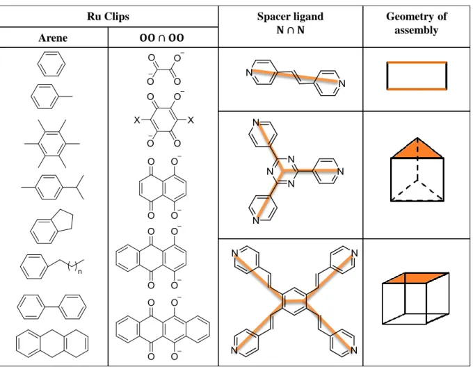
and “molecular panelling”[32] respectively. These two strategies constitute a very important part in modern supramolecular chemistry. With controlled architecture of the shape and size of the complex, a variety of coordination-driven self-assembled 2D and 3D supramolecular ensembles have been built. The desired frameworks can be obtained by the choice of appropriate building blocks. Hence, if the precursor units of rigid structures with predefined bite angles are combined in the appropriate stoichiometric ratio, the desired arrangement is formed. A panorama of spatially and electronically tunable supramolecular arrangements now exists and some have found applications in the biomedical field. For instance, a cationic hexanuclear metallo-prism is formed from arene-Ru building blocks with 𝑂𝑂 ∩ 𝑂𝑂 bridges and triazine trigonal panels (or spacer ligands); these complexes show anticancer properties (Figure 1.9).[33]
11
Among the many research groups whose work was inspired by this supramolecular chemistry, our group has reported the synthesis of a wide range of multinuclear arene-ruthenium complexes in the form of metallo-cages.[23b] These cages are supramolecular arrangements bearing a cavity formed during the assembly of the different subunits. Changing the Ru clips and the spacer ligands changes the shape and size of the cavity. Generally, the spacer ligand is the one that determines the geometry of the metallo-assembly, giving rise to the formation of cages in the shape of rectangles, prisms and cubes (Table 1.1). There are some cases where the clip has an as important role to play in the architecture of the assembly as the spacer ligand. For instance, two complexes of platinum and palladium have been reported recently, consisting of π-extended tetrathiafulvalene ligand with four pyridyl units that bind to the metal centres, each metal coordinated to a (diphenylphosphino)ferrocene unit.[34] These octacationic self-assembled complexes possess an ovoid internal cavity, which is different from the common cubic or cuboid formed by tetradentate spacer ligands (Figure 1.10).
Table 1.1 The different clips and spacer ligands and examples of the geometries they give
Ru Clips Spacer ligand Geometry of
assembly Arene
12
Figure 1.10 The X-ray crystal structures of the Pd complex (a) lateral view and (b) top view[34]
1.2 Host-Guest Chemistry
The term “Host-Guest Chemistry” was coined by Cram in 1974, to describe the “complexation and decomplexation” happening during an enzymatic reaction during which molecular recognition is of utmost importance for the substrate-enzyme systems.[35] In 1967, Pedersen reported the binding of metal ions with crown ethers to give highly structured complexes.[36] Two years later, Lehn, Sauvage and Dietrich published their work on cryptands that involved the study of the binding properties of these molecules.[37] The basic principle behind this chemistry is the size compatibility between the host and the guest. Also, the complementary binding forces, which are weak non-covalent interactions like van der Waals, eventually leading to the assembly of the host and the guest are crucial.[35a] In 1987, Donald J. Cram, Jean-Marie Lehn and Charles J. Pedersen were awarded the Noble Prize for their work on this concept, which has found widespread applications. The term host-guest chemistry is well defined in the following citation by Cram:[35a]
“… a host-guest relationship involves a complementary stereoelectronic arrangement of binding sites in host and guest… the host component is defined as an organic molecule or ion whose binding sites converge in the complex… the guest component is defined as any molecule or ion whose binding sites diverge in the complex…”
Thus, the host possesses convergent binding sites while the guest has divergent ones. The interactions existing in the host-guest system are important in determining the stability of the
13
assembly. These interactions can be hydrophobic, H-bonding, π-π aromatic interactions and ion-ion and dipole interactions. Apart from the selective binding affinity between the host and the guest, the two components need to have complementary geometric size/shape according to the lock and key principle.
In order to get a better understanding of how the host-guest strategy works, thermodynamic experiments, like isothermal titration calorimetry, were performed. Results from the experiments between guest molecules and the cavity confirmed that depending on the type of interaction happening inside the cavity, the encapsulation will be either entropy- or enthalpy-driven. Since entropy changes occur mostly when there is desolvation or a change in conformation, the host-guest systems, in general, are most likely to be enthalpy-driven. Thus, the π-π and C-H-π interactions between the guest molecule and the inner surface of the cavity give rise to enthalpic combination. Therefore, encapsulation is mainly enthalpy-driven, with probably some favourable entropic changes in terms of loss of solvent molecules surrounding the guest during encapsulation.[38]
Chemists have designed a large array of host-guest systems ranging from crown ethers to cyclodextrins to calixarenes, capable of hosting guest molecules or ions based on complementary/attractive forces between the two species. Host-guest chemistry is now applied in many areas including catalysis, sensors and pharmaceuticals. Host-guest systems have become a very popular method of drug-delivery in recent years and are believed to give rise to improvement in efficiency and biocompatibility.[23b] Researchers have even encapsulated cisplatin in order to reduce the severe side effects of the drug.[39] Thus, the arene-ruthenium arrangements allow the encapsulation of organic molecules of sizes that can fit in the cavity. The guest, usually a planar aromatic compound, is held by π-stacking to the spacer ligand of the host. A series of these complexes have been tested for their medicinal properties and interesting results have been obtained, comparable to cisplatin.[40]
1.2.1 Carceplex Systems
For small aromatic molecules that need to be encapsulated, the concept of host-guest has worked very well, allowing the guest molecule to enter and exit the cavity without disruption of the host molecule (Figure 1.11(a)). But in the case of highly branched aromatic molecules that can fit in the cavity, the movement in and out of the cage is restricted as the passage is not large enough.[41] In such instances, carceplex systems are used, whereby the guest molecule
14
gets trapped permanently as the host is assembled around it (Figure 1.11(b)). The driving force for the incarceration is the interaction between the guest molecule and the interior of the forming shell.[42] With the portal size being too small, the guest molecule can only be liberated via rupture of the cage. It is also possible for several molecules to be imprisoned inside the cavity that is big enough to accommodate the guests.[43] Besides its growing use in drug-delivery, carceplex systems are being applied in other areas, for instance, it is being employed as stabilizers for highly reactive species.[43b]
Figure 1.11 Schematic representation of encapsulation of a guest molecule by the (a) host-guest and (b) carceplex methods
1.2.2 Trojan Horse Concept
Drug-delivery strategies are engineered technologies that allow controlled release of drugs into targeted cells. This system has helped in avoiding many side effects associated with unwanted interactions of the drug with parts of the body that are not targeted. Trojan-Horse is a common way of referring to drug-delivery based on the strategy of transporting potential drugs, which are not water-soluble, inside the hydrophobic cavity of a cage with hydrophilic exterior and releasing the drug in the target cells. Drug-delivery employing Trojan-Horse concept can be advantageous compared to other modes used. Some of the advantages can be the higher local drug concentration and improved therapeutic efficacy.[44] From the various types of Trojan-Horse published, the horse was found to adopt different forms such as liposomes, engineered viruses,[45] nanotubes[46] and supramolecular assemblies.[23d, 47]
Host Guest
+
3 + 3 +
a
15
Introduced by our group, the supramolecular assemblies have been used to encapsulate drugs into the hydrophobic cavity of the metallo-assembly, with two main objectives: (1) transportation of mostly hydrophobic drug and (2) release of the treatment inside the targeted cancer cells.[47a] The first supramolecular Trojan Horse system involved the encapsulation of non-water soluble M(acac)2 complexes into water-soluble hexanuclear arene-ruthenium prism
via carceplex assembly until its release in the cancer cells (Figure 1.12). This
“complex-in-a-complex” assembly was confirmed by X-ray crystallography, which demonstrates the bending of the plane of the triazine ligands due to strong interactions with the guest complex.
Figure 1.12 Encapsulation of M(acac)2 into supramolecular Trojan Horse
1.2.3 Encapsulation of Pyrene-Core Dendrimers and their Biological Properties
In our group, a series of successful arene-Ru assemblies showing interesting anticancer properties have been developed. One such example is a family of pyrene-core dendrimers encapsulated in tetra- and hexanuclear arene-Ru complexes with the pyrene-core inside the hydrophobic cavity of the cage and the dendritic arm left dangling out (Figures 1.13, 1.14). This system was found to exhibit enhanced cytotoxicity compared to either the host or the guest alone. The IC50 values (the half maximal inhibitory concentration that indicates how
much of a particular drug is needed to inhibit a given biological process by half) of these systems were comparable to those of cisplatin when evaluated against human ovarian A2780 (cisplatin sensitive) and A2780cisR (acquired resistance to cisplatin) cancer cell lines.[48]
16
Figure 1.13 Encapsulation in a hexanuclear metallo-cage of dendrimers of second generation: (a) poly(arylester), (b) poly(benzylether) and (c) polyester
Figure 1.14 HyperChem simulation of the structure of the encapsulation of the poly(arylester) dendrimer of second generation into a hexanuclear Ru prism
1.3 Aim of the Present Thesis
The work done by Anaïs Pitto-Barry during her thesis has shown that dendrimers displaying liquid-crystalline properties could be successfully encapsulated into arene-Ru metallo-cages
Ru Ru Ru Ru Ru Ru R
17
by host-guest chemistry.[48] Moreover, these macromolecular assemblies show biological activities against human ovarian cancer cells. This thesis work is a continuation of the project initiated by Anaïs Pitto-Barry, consisting of the synthesis and the study of liquid-crystalline dendrimers in combination with arene-Ru metallo-cages as potential anticancer drugs and as hybrid liquid-crystalline materials.
So far, only monosubstituted pyrene-core dendrimers have been tested for their biological activities, alone and encapsulated. Therefore, the goal of this work was to synthesise new aromatic-core dendrimers possessing two dendritic chains that have liquid-crystalline properties and to encapsulate them into arene-Ru metallo-cages in order to determine any possible biological activities they might display inside cancer cells. Moreover, studying the liquid-crystalline property of the host-guest systems was also envisaged.
Furthermore, there were two other objectives for this work. One was to use a different approach in assembling the supramolecular cages other than the coordination-driven method; hence, self-assembly of arene-Ru cages by H-bonding was tested. The description of new and larger spacer ligands and the syntheses of the corresponding half-sandwich complexes and metallo-rectangles have been studied. The properties of these new metallo-assemblies as host-guest as well as carceplex systems have been evaluated. The other aim involved the association of arene-Ru units with [60]fullerene. Half-sandwich mononuclear [60]fullerene-based arene-Ru complexes were thus synthesised for their potential biological activities. The introduction for these two topics, hydrogen bonding self-assembly and [60]fullerene, will be done in the corresponding chapters.
19
21
2.1 Hydrogen Bonding
When hydrogen is bonded to electronegative atoms, such as N, O and F, the covalently bonded electron pair is pulled strongly to the nucleus of the electronegative atom, leaving a partial positive charge on the hydrogen atom and a partial negative charge on the electronegative atom. The partial positive charge allows the hydrogen to attract negatively charged species, an anion or a lone pair of electrons. This dipole-dipole attraction between the H atom, covalently bonded to an electronegative atom, and another electronegative atom is called hydrogen bonding. Hence, the IUPAC definition of hydrogen bonding is the “association between an electronegative atom and a hydrogen atom attached to a second, relatively electronegative atom”. The terms “donor” and “acceptor” are commonly used when dealing with hydrogen bonding: the donor (D) is the electronegative atom (X) to which the H atom is covalently bonded and the acceptor (A) is the neighbouring electronegative atom (Y) that possesses the negative charge or lone pair of electrons (Figure 2.1).[49]
Figure 2.1 Schematic representation of a H-bonded system
The strength of H-bond is intermediate between that of dipole forces between molecules and covalent bonds within a molecule. The H-bond is about 1/10 as strong as covalent bonds. Nevertheless, multiple H-bonds between two molecules result in a union that displays sufficient strength and good stability.
There have not been any Nobel Prize winners for the discovery of H-bonding because since the realization of its significance in the early 1900s, many important researchers have been working on and contributed to the understanding of this complex concept, among whom were Werner, Huggins, Rodebush and Pauling.[50] Hydrogen bond is still today a growing area of research due to its enormous importance. One example of the role of H-bonding is water (Figure 2.2). Each water molecule can potentially form four intermolecular hydrogen bonds with neighbouring water molecules. Compared to other binary compounds formed between hydrogen and elements of group 6, like H2S, where H-bonding is absent, H2O has the highest
boiling point, despite its smaller molar mass. Also, compared to other larger molar mass
X H Yδ- δ+ : donor acceptor
H-bonding covalent bond
22
molecules where H-bonding is present, like HF, the boiling point of H2O is still higher as it
partakes in more hydrogen bonds per molecule. Hence, H-bonding can account for the unique properties of water (Figure 2.2).[51]
Figure 2.2 Hydrogen bonding in water
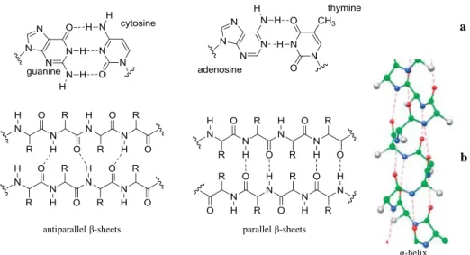
H-bonding has a very important role to play in the structure and function of several biological systems, like DNA. The double strands in DNA are held together by hydrogen bonds between the H atoms attached to N on one strand and lone pairs on another N or an O on the other strand. The H-bonding between the complementary nucleotide Watson-Crick base pairs and π-stacking interactions give rise to the stable double helical structure of DNA. Two H-bonds occur between the adenosine and the thymine base pairs and triple hydrogen bonds exist between the cytosine and the guanine (Figure 2.3(a)). Furthermore, the construction of polypeptides uses extensively H-bonds between N-H and C=O groups. Intra- and inter-chain H-bonding interactions in α-helices and β-sheets, respectively, establish the secondary structure of proteins (Figure 2.3(b)).[49]
Figure 2.3 Hydrogen bonding between base pairs (a) and peptides (b) δ+ δ+ δ+ δ -δ -δ -δ+
antiparallel β-sheets parallel β-sheets
α-helix
a
23
Supramolecular chemistry, as defined by Nobel Prize winners Lehn, Cram and Pedersen, is the “chemistry beyond the molecule”, and two fundamental features of this discipline are molecular recognition and self-assembly. Non-covalent interactions are used for the synthesis of supramolecules, enabling preparation of these complex assemblies from simple fragments, which may sometimes be difficult by covalent interactions. Hydrogen bonding is one of the preferred types of non-covalent interactions in supramolecular chemistry because H-bonded mediated self-assembly has been shown to be a good strategy for creating well-defined supramolecular architectures. Its directionality, stability, reversibility and specificity constitute some of the features that make H-bonding so versatile compared to other non-covalent interactions.[52] Appropriate choice of donor/acceptor pairs makes it possible for H-bonding to be directional and to have tunable strengths. Hence, H-bonds can be classified as follows:
Strong H-bonds: formed between charged species and have bond strengths in the range 15-40 kcal·mol-1 and have the shortest bond lengths.
Moderate H-bonds: are mostly electrostatic interactions found in biomolecules with bond strengths in the order 4-15 kcal·mol-1 with moderate bond lengths.
Weak H-bonds: formed between an electronegative species and a significantly less electronegative complement of bond strengths less than 4 kcal·mol-1 and have the longest bond lengths.
Based on the several biological examples where H-bonding exists, researchers have been inspired to build synthetic systems with H-bonding to benefit from its unique properties. The careful selection of acceptor/donor pairs is able to give the desired strength and stability of each hydrogen bond as well as the number and arrangement of the hydrogen bonds in the system. Similar to the base pairing in DNA, the well-defined through-space directionality and hence high selectivity of H-bonds have been used by many researchers. Thus, many examples of supramolecular assemblies involving H-bonding interactions have been reported in different fields of chemistry, especially in organic and polymer chemistry.[52a, 53]
Studies have shown that H-bonds could be made stronger and/or more selective by introducing arrays of donors (D) and acceptors (A) sites. These H-bonded systems are built by the use of parallel arrays of H-bonds and they feature two or more contiguous H-bonds on a heterocyclic scaffold. Units consisting of up to six consecutive H-bonds have been reported, but those of the triple and quadruple units are the most studied derivatives in molecular
24
recognition and self-assembly processes. It was found that the arrangement of the D and A sites, as well as the solvent in which the complexes are formed are determining factors for the strength of the H-bonds, called the association constant (Ka) or dimerization constant (Kdim) in
case of homodimers.[52c]
For triple H-bonded complexes, the general trend in their stability is as follows:
ADA-DAD < ADD-DAA < AAA-DDD
The DDD-AAA arrangement was found to have the highest association constant (Ka = 105
-106 M-1) and ADA-DAD pair the lowest (Ka = 102-103 M-1).[54] With the same primary
H-bonding interactions between the D/A pairs, the difference in Ka can only be the result of
another type of interaction. Hence, Jorgensen rationalised these differences as the impact of the secondary electrostatic interactions on the stability of H-bonding arrays.[55] Since the D/A partners are in close proximity, due to the H-bonding between them, this gives rise to substantial electrostatic interactions. The partial positive charges on the protons and the partial negative charges on the electronegative atoms result in attractive forces between opposite charges and repulsive forces between like charges. The net of the secondary interactions can therefore account for the stronger/weaker binding, depending on the arrangement of the D/A sites in the molecules. Hence, parallel H-bond arrays, having all the H-bond donor groups in one partner and the acceptor sites in the other in an AAA-DDD arrangement seems to result in the strongest association with all secondary electrostatic interactions between neighbouring H-bond pairs being attractive.[52c]
Among the many triple H-bonding modes, the H-bonded melamine-cyanuric acid motif with a ADA-DAD array has been one of the most widely used in the formation of the so-called “rosette” supramolecular structures. In this system, a single rosette constitutes of a cyclic aggregate of three melamine and three cyanuric acid units. The lattice formed in organic solution shows very good stability arising from nine hydrogen bonds surrounding each melamine or cyanuric acid molecule (Figure 2.4).[56] Based on this arrangement, a series of macromolecules have been developed using melamine and cyanuric acid derivatives. Two strategies were adopted in designing these rosette structures: preorganization and peripheral crowding, favouring the formation of 1:1 cyclic aggregates.[56-57]
25
Figure 2.4 Melamine and cyanuric acid rosette structure
Four H-bonding systems have also attracted the attention of many research groups, as a result of which a vast series of quadruple H-bond complexes have been synthesised. Out of the six possible H-bond permutations, only three have been extensively studied: ADAD-DADA, AADD-DDAA, ADDA-DAAD (Figure 2.5).[52c, 58] The AAAA-DDDD array is not much explored and even though these complexes with four consecutive H-bond donors and acceptors show the highest stability (Ka ˃ 1012 M-1), similar to triple systems, their application
is not so popular. One example of an AAAA-DDDD quadruple H-bond array is the highly stable cationic complex formed by the equimolar assembly between guanidinium dibenzimidazole and diisoquinoline-[1,8]-naphthyridine (Figure 2.5).[58c]
Figure 2.5 Examples of quadruple H-bond arrays and their Ka values
Many different scaffolds have been used in designing and building up quadruple H-bonding self-assembled systems, and the most popular one is probably the 2-ureido-4-[1H]-pyrimidinone (UPy). Upy dimers, with a DDAA H-bond array, have high dimerization constants (Kdim ˃ 107 M-1) and they can be readily synthesised.[61f] The high dimerization
26
secondary interactions within the motif, as well as the intramolecular H-bond that creates order within the molecule by prearrangement of the motif.[59] UPy unit can also associate with other units to form heterocomplexes that are stable with high association constants. Thus, Meijer and co-workers have developed a series of UPy derivatives.[60] UPy consisting of a linear four H-bond DDAA-AADD array has been used as building block for the construction of various assemblies. Inspired by this unit, numerous derivatives of UPy have been used as DDAA modules, with varying dimerization constants (Figure 2.6).[52b, 52d, 53c, 61]
Figure 2.6 Homodimer structures of DDAA-AADD array modules, UPy, DeAP, UN and UC
Developed by Zimmerman and co-workers in the late 1990s, ureidodeazapterin (DeAP) shows very high stability (Figure 2.6). Three major DDAA-AADD dimers are present in the DeAP systems: two homodimers, X.X and Y.Y (arising from the two possible tautomers) and one heterodimer X.Y. Some undesired DADA-ADAD homodimers, Z.Z, are also detected in small amount (Figure 2.7).[53c]
27
Figure 2.7 The different protomeric forms of the AADD H-bonding array of DeAP
A few years later, the same group developed a ureidodeazapterin-based module, linking two DeAP units with a semi-rigid spacer bearing a Fréchet-type dendron (of first, second and third generation) attached to it. This molecule exists as two tautomers, with a DDAADDAA H-bond array in both forms. The two different conformations, symmetrical and non-symmetrical, give rise to different aggregates, polymeric and cyclic, respectively (Figure 2.8).[52a]
Figure 2.8 Different conformations of spacer unit DeAP and the corresponding assemblies with third generation dendron
The dimerization constant of ureidonaphthyridine (UN) is the lowest in this series because intramolecular H-bonding occurs to stabilise the folded conformer rather than the unfolded
28
one (Figure 2.9).[62] This problem is less in the ureidocytosine (UC) units, developed by Hailes and co-workers, where the unfolded dimer is the most stable conformer.[63] Similar to UC, ureidoimidazo[1,2-a]pyrimidine (UImp) has highly stable unfolded dimer.[52b]
Figure 2.9 The folded and unfolded conformers of UC dimer
Among the different macrostructures that have been successfully designed and constructed by H-bonds, there are several networks bearing cavities that have been formed as a result of non-covalent, complementary interactions. The formation of these cyclic aggregates, or cages, by H-bonding requires a high binding/association constant and a highly preorganised monomeric subunit.[61a] Another type of interaction that has been found to produce macrocycles and cages is metal coordination. Fujita and Stang have worked, independently, on the design of cyclic frameworks by coordinating different ligands, like 4,4′-dipyridine, to the metal centres, like Pt(II).[32c, 64] Mendoza et al. generated molecular squares and triangles by combining H-bonding and metal-templated self-assembly. Upon mixing the difunctional UPy dimers, which can easily be preorganised by functionalisation of the phenylene ring or the amine, and a Pd(II) species in a 1:1 molar ratio, different cyclic aggregates were formed (Figure 2.10). These results led to the start of the application of H-bonding and metal complexation in designing molecular architectures of higher complexity.[61f]
29
Figure 2.10 Schematic representation of cyclic aggregates (a) propeller-shaped triangle, (b) propeller-shaped square and (c) tubular-shaped tetramer.
2.2 Aim
Supramolecular chemistry combining H-bonding and metal complexation has flourished since Mendoza and co-workers published their findings on this method. Thus, the objective of this project was to move from directional-bonding coordination-driven self-assembly of Ru cages to H-bonding and metal coordination self-assembly, in order to take advantage of the pool of information associated with each technique. Hence, a series of new assemblies was developed consisting of arene-Ru clips and ureidopyrimidone derivatives with a DDAA H-bond array as the spacer ligand. Five new dinuclear Ru panels and a family of new arene-Ru rectangles were generated from this work.
2.3 Synthesis
2.3.1 Synthesis of Spacer Ligands
Four different ligands (4-6, 10) were synthesised by a four step synthetic route (Scheme 2.1). The first step involves an alkylation reaction between diethyl carbonate and a ketone in the presence of NaH in refluxing THF. The product obtained after purification was found to be a mixture of tautomers: the enol form and the desired ketone form as the major product (1 and 7). Isolation of the ketone was not carried out, and the product was used for further reactions without separation.
= =
30
Scheme 2.1 Synthesis of H-bonded systems (4-6, 10). Reagents and conditions: i. NaH, THF, 66°C, 16h, 1: 76%, 7: 88%, ii. EtOH, 90°C, 16h, 2: 75%, 8: 43%, iii. THF, rt, 8h, 3: quantitative yield, 9: 64%, iv. DMF/NEt3, 70°C, 16h, 4: 37%, 5: 72%, 6: 50%, 10: 73%
The second step is the ring-closure of the β-ketoester (1 and 7) with guanidinium carbonate in refluxing ethanol. The isocytosine derivative (2 and 8) reacts with 1,1′-carbonyldiimidazole (CDI) in THF, due to limited solubility of the isocytosines, to give the blocked isocytosine isocyanate derivate (3 and 9). An excess (two equivalents) of CDI was used to ensure complete conversion of the isocytosine. The imidazolide was then reacted with different amines in DMF/NEt3 solvent mixture in a sealed tube to produce head-to-tail
ureidopyrimidinone dimer with a DDAA H-bond array (4, 6 and 10). The different ligands were synthesised to test the effect of: (1) adding a methyl group to the methylene spacer, (2) elongating the spacer chain, hence increasing flexibility and (3) reducing the length of the aliphatic chain from C11 to C4.
31
2.3.2 Macromolecular Assemblies
[(η6-p-cym)Ru(µ2-Cl)Cl]2, its hexamethylbenzene derivative, [(η6-hmb)Ru(µ2-Cl)Cl]2, and
four other dinuclear arene-Ru clips synthesised from [(η6-p-cym)Ru(µ2-Cl)Cl]2 were used for
molecular assembly. As in the case of the ligands, different clips were used for complexation to test the effect of the bridge and the arene on the ability of the resulting product to self-assemble and to form monocrystals.
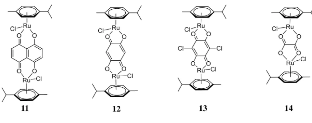
Clips 11-14 (Figure 2.11) were synthesised, following literature procedures.[43a, 47a] In these complexes, each ruthenium is coordinated to a chloride ligand that allows substitution by other ligands.
Figure 2.11 Structures of Ru clips 11-14
These Ru clips were then reacted with the ureido dimers under different conditions to give either the dinuclear panels (15, 20, 25, 30, 31) or the tetranuclear rectangular assemblies (16-19, 21-24, 26-29) (Schemes 2.2 and 2.3). The dinuclear panels are obtained by simply stirring the ruthenium dimer with the ligand in CHCl3 at room temperature for 24h. In these panels,
the dichloro-bridge is broken, leaving each Ru with two chloride ligands and one vacant site that is taken up by the ureido ligand with the coordination of the pyridyl nitrogen to the Ru(II). With the ligand behaving as one single component with two available coordinating sites, two ruthenium dichloride species get attached to the two pyridyl nitrogen atoms of every bidentate ligand (Scheme 2.2).
i i ii iii
32
Scheme 2.2 Synthesis of dinuclear Ru assemblies (15, 20, 25, 30, 31). Reagents and conditions: i. CHCl3, rt, 24h, 15: 98%, 20: 96%, 25: 93%, 30: 95%, 31: 90%
In the case of the cages, a vacant site is created on each Ru centre by loss of the chloride ligand in the form of AgCl, resulting in the in-situ preparation of the activated intermediate species in the syn symmetry exclusively. When the ligand is added to the mixture, two bidentate dimers link together two Ru clips to form a cationic tetrameric cyclic arrangement adopting a rectangular shape, with four triflate counter anion (Scheme 2.3).
33
Scheme 2.3 Synthesis of tetranuclear Ru assemblies (16-19, 21-24, 26-29). Reagents and conditions: i. AgCF3SO3, CH2Cl2, rt, 24h or MeOH, rt, 16h, 16: 23%, 17: 72%, 18: 72%, 19:
38%, 21: 46%, 22: 65%, 23: 53%, 24: 23%, 26: 21%, 27: 61%, 28: 57%, 29: 21%
2.4 Characterization
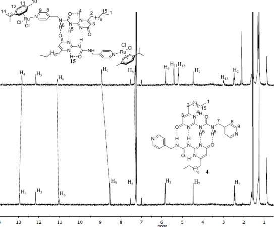
2.4.1 Proton NMR Spectroscopy
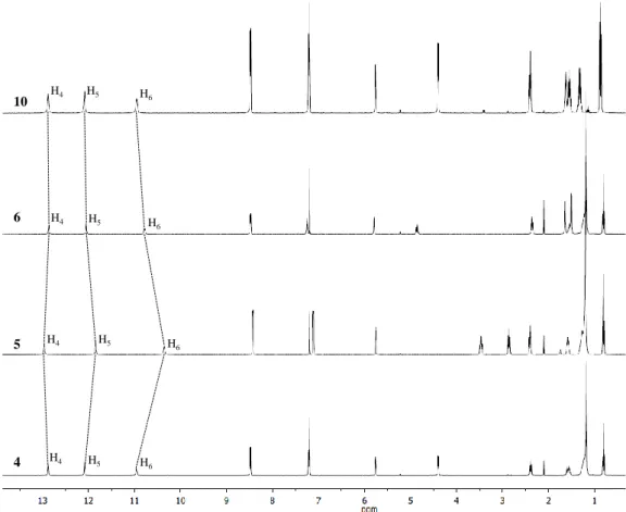
For all the ligands (4-6, 10), sharp 1H NMR spectra in CDCl3 were observed (Figure 2.12).
Well-defined typical signature for UPy’s NH was observed downfield, in the region expected for H-bonded dimers. It was also observed that changes brought to the molecule gave rise to shifting of some proton signals (Figure 2.12). For instance, adding a methyl group to the spacer results in an upfield shift for H5 and H6 protons from 12.16 and 11.03 ppm to 12.10
34
upfield shift of H5 and H6 protons is observed from 12.16 and 11.03 ppm to 11.91 and 10.42
ppm. On the other hand, shortening the aliphatic side chain from C11 to C4 causes no change
in the chemical shifts of H5 and H6 protons (Figure 2.12). The H4 protons are the most
downfield in 5, when the spacer is more flexible and shortening the aliphatic side chain results in an upfield shift of the H4 proton from 13.02 ppm in 5 to 12.95 ppm in 4 (Figure 2.12).
Hence, alterations brought to the dimer have an effect on the H-bonds holding the dimer together and inter- and intramolecular H-bondings are affected in different ways.
Figure 2.12 1H NMR spectra (400 MHz, CDCl3) of ligands 4-6 and 10
The effect of concentration on the chemical shifts of the NH protons (H4, H5 and H6) in dimer
4 was tested. There was no apparent change in the chemical shift across a broad range of concentration (1 mM to 50 µM). From this observation, only a lower limit to the dimerization constant, Kdim > 107 M-1 could be estimated, which is among the largest stability constants
reported to date for neutral, H-bonded species.
For the two sets of Ru complexes, the dinuclear and the tetranuclear, the resolution of the proton signals was found to differ. The spectra of the dinuclear species show peaks that are
H4 H5 H6 10 6 5 4 H4 H5 H6 H4 H5 H6 H4 H5 H6
35
sharp and well-defined, like in the case of the uncoordinated dimers (Figure 2.13). Besides a slight upfield shift of the H4 proton of the ureido derivatives after coordination of the dimer to
the Ru, the other two NH signals (H5 and H6) did not undergo any major shifting. The H9
peaks, however, underwent a downfield shift due to their close proximity to the Ru centre. Only a slight upfield shift was observed for the H8 peaks, being further away and therefore
less affected by the Ru(II) centre. An example of the comparison between uncoordinated ligand and the corresponding dinuclear Ru complex is shown in Figure 2.13, whereby the 1H NMR spectra of compound 4 and its dinuclear Ru complex 15 are compared. The shifts observed between the two spectra are indicated by dotted lines, namely the shifting of the H4,
H6, H8 and H9 protons.
In the case of the rectangles, for most of the complexes, broad and ill-defined peaks were obtained, with some signals not visible on the spectra. For instance, the NH signals (H4, H5,
H6)are broadened completely into the baseline for most of the rectangular systems (see Figure
2.20 for 1H NMR spectrum of 17).
Figure 2.13 1H NMR spectra (400 MHz, CDCl3) of 4 and 15
15 4 H4 H4 H5 H5 H6 H6 H9 H9 H8 H8 H3 H7 H2 H2 H7 H13 H3H11H12
36
2.4.2 DOSY
Diffusion-ordered NMR spectroscopy (DOSY) is a 2D NMR experiment developed in 1992 to measure diffusion coefficients and in case of spherical molecules, the hydrodynamic radii of such molecules in solution using the Stokes-Einstein equation can be determined.[65] The 2D spectrum consists of one axis representing typical chemical shift information and diffusion coefficient (D) on another. This technique allows the different species contained in a mixture to be distinguished, as D is related to the size of the particle. The diffusion coefficient is dependent on the size and shape of the molecule in a particular solvent at a particular temperature.[66] This NMR tool has found widespread applications in biology and chemistry where DOSY is being used to determine the dimensions of polydispersed supramolecular aggregates.[67]
In order to confirm the composition of the metal coordination/H-bonding self-assembly of the Ru/ureido systems, DOSY experiments were run on the complexes. For all the dinuclear complexes (15, 20, 25, 30 and 31) DOSY experiments were run in CDCl3 using similar
concentrations and at room temperature. Figure 2.14 shows the 2D spectra of compounds 15, 20 and 25, which display single diffusion coefficient for each compound, indicating the formation of single species. It was also observed that the diffusion coefficients differ only very slightly for the five complexes (-9.30 > log D > -9.41) and hence, the standard errors associated with the differences between these values are too significant to be ignored. Comparing the DOSY spectrum of the free ligand to its corresponding dinuclear Ru complex, a decrease in log D value was observed. For example, compound 4 has log D value of -9.25 while compound 15 has a log D value around -9.32. It can be concluded that for the dinuclear system, the complexes have very similar diffusion coefficients and that the minor differences among the UPy ligands do not have any apparent significance on the diffusion of the corresponding complexes, which have similar hydrodynamic volumes in solution.
37
Figure 2.14 DOSY spectra (400 MHz, CDCl3) of compounds 15, 20 and 25
For the tetranuclear complexes, the DOSY experiments were run mostly in CD3CN, and it
was observed that the complexes have diffusion coefficients in the range of -9.12 to -9.30 x 10¹⁰. Similar to the dinuclear system, the differences are too small to account for any structure/diffusion relationship among these complexes. Thus, even with clips of different lengths, for example 11 and 14, their corresponding complexes show very close D values and hence have almost the same hydrodynamic volumes in solution. The difference in D values is however slightly more important when comparing a tetranuclear assembly to its corresponding dinuclear complex bearing the same UPy ligand. Thus, complexes 15 and 17 have diffusion coefficients of -9.38 x 10¹⁰ and -9.54 x 10¹⁰ respectively in CDCl3. This
difference can be associated to the larger hydrodynamic volume of the rectangle than the dinuclear panel. An example of the comparison of free ligand 4 and its corresponding tetranuclear Ru complex 17 is shown in Figure 2.15.
Figure 2.15 DOSY spectra (400 MHz, CDCl3) of 4 and 17
15 20 25 Log D ppm Log D 4 17 ppm
38
2.4.3 NOE/ROE and DFT Calculations
A very powerful tool in NMR spectroscopy for structural studies of chemical systems is the Nuclear Overhauser Effect (NOE). This technique relies on relaxation phenomena between dipolar coupled spins and manifests itself in changes in signal intensities of nuclear sites when other specific nuclear sites are perturbed by means of a radio-frequency (rf) pulse.[68] Therefore, the experimental observation of NOE supplies information about, and implies, spatial proximities between given nuclear sites and can prove extremely useful in structural studies of the supramolecular systems considered in this thesis (See Annex 2).[69] A widespread variation of the NOE experiment is the Rotating-frame Overhauser Effect (ROE), where an rf pulse is applied so as to spin-lock the magnetization in the transverse plane. This technique is utilised in cases where NOE enhancements are close to zero. Also this latter technique may therefore be utilised for structural studies aiming to determine internuclear distances between sites experiencing non-negligible dipolar couplings.
In order to corroborate the experimental findings, a computational study has also been undertaken (See Annex 2).[70] The structures thus obtained were used to calculate the magnetic shielding tensors of both 1H and 13C nuclei with the GIAO method at the same level of theory of the DFT optimizations.[71] The geometry optimizations supplied internuclear distances to be compared to those determined experimentally by means of NMR/NOE measurements whereas the calculations of the magnetic shieldings allowed comparison with the experimental 1H and 13C chemical shifts observed for structure 4 in simple 1D solution-phase NMR spectra.
Structure 4 is expected to form in solution a dimer via formation of four H-bonds, resulting in the head-to-tail configuration as shown in Figure 2.16.
Figure 2.16 Dimeric structure of 4 in a head-to-tail configuration a
39
Figure 2.17 1D ROE spectra (400 MHz, CDCl3) of 4 with protons H9 (a), H7 (b) and H3 (c)
irradiated
Figure 2.17(a), (b) and (c) shows 1D ROE spectra obtained when irradiating protons H3, H7
and H9, respectively. Figure 2.18 shows, as histogram, the differences between internuclear
distances measured from the NOE enhancements of the spectra in (a), (b) and (c), and those obtained from the structures optimised with DFT methods. The smaller those differences, the better the agreement between experiments and calculations are. In this histogram, the differences calculated, assuming a monomeric non-H-bonded structure 4, are shown in blue whereas those calculated, assuming the head-to-tail dimer, are shown in red. This histogram clearly shows a much better agreement of the experimental data sets when the dimeric structure is considered, with differences generally smaller than 1 Å in both cases (Figure 2.18). In particular, proton sites that are on opposite sides of structure 4, such as, H3 and H9,
necessarily require intermolecular pathways that may only exist in the dimeric form in order to interpret the experimentally-observed NOE enhancements.
H9 H3 H7 H4 H5 H6 H9 H8 H3 H7 H2 a c b 4
40
Figure 2.18 Difference in Δr between DFT-calculated and ROE measured distances
The experimental data based on ROE measurements strongly suggests the existence of 4 as a H-bonded dimer in solution. An independent further assessment may be also carried out by considering the chemical shifts of the 13C and 1H resonances as measured in conventional 1D spectra and as calculated with DFT methods, as described above. Figure 2.19(a) shows the correlation between experimental 13C chemical shifts and those calculated for the monomeric structure. The quality of the correlation may be indicated by a coefficient R2 = 0.996. Figure 2.19(b) shows the good correlation when the dimeric structure was used to calculate the chemical shifts. In this latter case R2 = 0.999, with all carbon sites being very accurately described by DFT calculations. Figure 2.19(c) shows the correlation between experimental 1H chemical shifts and those calculated for the monomeric structure. In this case, the NH protons lay completely out of the correlation line where all other proton sites may be found. These protons are involved in the H-bonding network that assembles the dimeric structure. If solvent effects are included in the calculations, no substantial improvement is obtained. Figure
H3 H9 H3 H9 H9 Monomer Dimer H7 H7 H2 H5 H3 H4 H6 H7 H8 H5 H4 H3 H5 H9 H6 H7 H9 H9 H7 H6 : dimer : monomer Δr (Å)
41
2.19(d) shows the analogous correlation when the dimeric structure was considered in the calculations. In this case the chemical shifts of the NH protons correlate very well with the calculated ones, with a coefficient R2 = 0.998. These results indicate that, in order to interpret the chemical shifts observed experimentally for 13C and 1H in structure 4, a dimeric arrangement needs to be considered. This is particularly true for the protons of the NH groups involved in the H-bonding system.
Figure 2.19 Experimental v/s DFT-calculated 13C and 1H chemical shifts for the (a and c) monomeric and (b and d) dimeric forms
All the NMR data experimentally observed as ROE 1H enhancements and 1H and 13C chemical shifts strongly support the hypothesis of a structure 4 existing as a head-to-tail dimer in solution. In the case of the tetranuclear species, qualitative ROE/NMR analysis of compound 17 confirms the predicted rectangular self-assembled arrangement. Figure 2.20 shows the NOE spectrum of 17: with H9 being irradiated, the peak corresponding to the
benzoquinone Ru bridge of the clip, H19 was observed on the spectrum (See Annex 2).
0 2 4 6 8 10 12 14 0 2 4 6 8 10 12 H6 H5 H4 Experimental (ppm) Calculated (ppm)
Monomer: 1H chemical shifts
y = 0.9828x + 0.5029 R² = 0.998 0 2 4 6 8 10 12 14 0 2 4 6 8 10 12 14
Dimer: 1H chemical shifts
Calculated (ppm) Experimental (ppm) H6 H5 H4 y = 0.9876x + 1.7495 R² = 0.9955 0 20 40 60 80 100 120 140 160 180 200 0 50 100 150 200
Monomer: 13C chemical shifts
Experimental (ppm) Calculated (ppm) y = 0.9613x + 3.4619 R² = 0.9989 0 20 40 60 80 100 120 140 160 180 200 0 50 100 150 200 Experimental (ppm) Calculated (ppm)
Dimer: 13C chemical shifts
a)
b)
c)
42
Figure 2.20 1D ROE spectra (400 MHz, CDCl3) of 17 with protons H9 (a) and H19 (b)
irradiated
2.4.4 UV-Vis Spectroscopy
The binding of the various metallo-assemblies were studied by UV-visible spectroscopy; the electronic absorption spectra were acquired in CH2Cl2 at concentrations around 10-5 M in the
range 250-700 nm (Table 2.1). The di- and tetranuclear complexes were found to have different spectra. The dinuclear complexes have similar spectra, with a high energy shoulder around 288 nm, which may be attributed to ligand 𝜋 → 𝜋 ∗ transitions and a broad and low energy band at around 410 nm.[72] The latter may be assigned to mixed metal-ligand charge transfer (MLCT), intra-ligand charge transfer (ILCT) from spacer ligand to 𝑂𝑂 ∩ 𝑂𝑂 bridging ligand and ligand 𝜋 → 𝜋 ∗ transitions.[73] Figure 2.21 shows the stacking of the UV-Vis spectra of compounds 16, 21 and 26 measured in CH2Cl2 and the similarity between these
three naphthoquinone ruthenium derivatives. The spectra show that the differences between the three compounds bearing three different spacer ligands are negligible.
H9 H8 H11, H12 H3 H19 H7 17 H9 H11, H12 H19
43
Table 2.1 λmax values of the different series of metallo-cycles
Clips Metallo-cycles λmax (nm) ε (M-1·cm-1) x 105
11 16 306 2.38 433 1.66 639 0.55 693 0.62 21 310 2.56 434 4.32 644 0.55 696 0.58 26 310 2.57 434 1.57 641 0.51 693 0.51 12 17 299 3.64 498 3.39 22 300 3.72 497 2.23 27 298 3.42 496 3.07 13 18 324 2.27 504 2.27 23 326 2.20 509 2.28 28 325 2.31 503 2.40 14 19 383 0.28 24 384 0.26 29 387 0.28