Distinct Roles of the ATR Kinase and the Mre11-Rad50-Nbs1
Complex in the Maintenance of Chromosomal Stability
in Arabidopsis
Simon Amiard,
1Cyril Charbonnel,
1Elisabeth Allain, Annie Depeiges, Charles I. White, and Maria Eugenia Gallego
2 Ge´ne´tique, Reproduction et De´veloppement, Centre National de la Recherche Scientifique, Unite´ Mixte de Recherche6247-Clermont Universite´-Institut National de la Sante´ et de la Recherche Me´dicale U931, F-63177 Aubie`re, France
Signaling of chromosomal DNA breaks is of primary importance for initiation of repair and, thus, for global genomic stability. Although the Mre11-Rad50-Nbs1 (MRN) complex is the first sensor of double-strand breaks, its role in double-strand break
(DSB) signaling is not fully understood. We report the absence of g-ray–induced, ATM/ATR-dependent histone H2AX
phosphorylation in Arabidopsis thaliana rad50 and mre11 mutants, confirming that the MRN complex is required for H2AX phosphorylation by the ATM and ATR kinases in response to irradiation-induced DSB in Arabidopsis. rad50 and mre11
mutants spontaneously activate a DNA damage response, as shown by the presence ofg-H2AX foci and activation of cell
cycle arrest in nonirradiated plants. This response is ATR dependent as shown both by the absence of these spontaneous foci and by the wild-type mitotic indices of double rad50 atr and mre11 atr plants. EdU S-phase labeling and fluorescence in situ hybridization analysis using specific subtelomeric probes point to a replicative S-phase origin of this chromosome damage in the double mutants and not to telomere destabilization. Thus, the data presented here show the exclusive involvement of ATR in DNA damage signaling in MRN mutants and provide evidence for a role for ATR in the avoidance of S-phase DNA damage.
INTRODUCTION
Maintenance of genome stability is of critical importance for all cellular organisms. Cellular DNA is subject to many types of damage resulting from cellular metabolism (such as reactive radicals or stalled replication forks) or through the action of exogenous agents, such as radiation and chemical mutagens. Double-strand DNA breaks are extremely toxic lesions, poten-tially causing chromosomal translocations and rearrangements and ultimately cell death, senescence, or tumorigenesis (Rich et al., 2000). In response to DNA damage, cells activate complex signaling pathways that activate DNA repair, cell cycle arrest, and eventually apoptosis. These processes are based on signal transduction initiated by sensor proteins that recognize the damage and activate the transducers, which send the signal to the effector proteins (Shiloh, 2006; Cimprich and Cortez, 2008). The major regulators of the DNA damage response are the protein kinases Ataxia telangiectasia mutated (ATM) and ATM and Rad3 related (ATR), which belong to the phosphatidyl inositol 3-kinase family (PIKKs). Each is activated in response to a different type of damage. ATR responds primarily to stalled replication forks where the generation of replication protein A–coated single-stranded DNA activates its kinase activity, while the kinase activity of ATM is enhanced in response to the
presence of double-strand breaks (DSBs). DSBs are first bound by the Mre11-Rad50-Nbs1 (MRN) complex, which then recruits and activates ATM (Harper and Elledge, 2007). ATR activation in response to replicative stress, however, does not require the MRN complex (Cimprich and Cortez, 2008). Once activated, PIKKs are necessary to maintain genomic integrity by initiating multiple events, including cell cycle arrest, chromatin remodel-ing, repair, and eventually cell death. Phosphorylation by PIKKs of the histone variant H2AX, forming g-H2AX, plays a key role in the recruitment and accumulation of DNA repair proteins at sites of DSB damage (Paull et al., 2000; Fernandez-Capetillo et al., 2003; Fillingham et al., 2006), and detection of this phosphory-lation event using antibodies to g-H2AX has emerged as a highly specific and sensitive molecular marker for monitoring DNA DSB damage and its repair (Kinner et al., 2008).
The MRN complex is a highly conserved complex composed of three proteins, Meiotic recombination 11 (MRE11), Radiation sensitive 50 (RAD50), and Nijmegen Breakage Syndrome 1 (NBS1; x-ray sensitive 2 [XRS2] in Saccharomyces cerevisiae), and has been shown to be involved in DNA damage repair, DNA replication, meiosis, and telomere maintenance (Czornak et al., 2008; Mimitou and Symington, 2009; Lamarche et al., 2010). The MRN complex recognizes DSBs by its ability to bind the ends of DNA, and, once bound, it unwinds (Paull and Gellert, 1999) and initiates processing of DNA ends (review in Mimitou and Symington, 2009). The interaction with NBS1 activates ATM kinase activity, resulting in phosphorylation of target proteins H2AX, Mre11, Nbs1/Xrs2, and Sae2/CtIP (for Sporulation in the absence of Spo11-2 and C-terminal binding protein-interacting protein, respectively). Interaction between Sae2/CtIP and the MRN complex activates the nuclease activity of Mre11,
1These authors contributed equally to this work.
2Address correspondence to megalleg@univ-bpclermont.fr.
The author responsible for distribution of materials integral to the findings presented in this article in accordance with the policy described in the Instructions for Authors (www.plantcell.org) is: Maria Eugenia Gallego (megalleg@univ-bpclermont.fr).
www.plantcell.org/cgi/doi/10.1105/tpc.110.078527
preparing the DNA ends and initiating resection of the 59-ended
strand for the generation of 39 single-stranded DNA (ssDNA),
which is required for the initiation of homologous recombination and subsequent activation of the ATR kinase in response to DSBs (reviewed in Mimitou and Symington, 2009; Lamarche et al., 2010).
Yeast cells mutated for any member of the MRN complex are sensitive to replication stress (D’Amours and Jackson, 2001; Shor et al., 2002), and in mammals the MRN complex has been shown to localize with proliferating cell nuclear antigene and to interact with replication protein A (RPA) during hydroxyurea (HU) treatment (Mirzoeva and Petrini, 2003; Robison et al., 2004; Olson et al., 2007). All these data suggest a key role for the MRN complex in stabilizing replication forks. Thus, ATM, ATR, and the MRN complex are all required for chromosomal integrity. How-ever, defining their functional relationship in the maintenance of chromosomal stability has been hampered by the early lethality associated with null mutations in these genes in vertebrate cells. By contrast, Arabidopsis thaliana plants mutated for ATR, ATM, or the MRN complex proteins are viable. atr and atm mutants are both phenotypically wild type, except for a partial sterility in the atm mutant (Garcia et al., 2003; Culligan et al., 2004). The atm mutant is sensitive to DSB-inducing agents (e.g., ionizing radiation and methyl methane sulphonate), and the atr mutant is sensitive to replication stress inducing agents (e.g., Aphidicolin or Hydroxyurea), indicating conservation of the roles of these proteins in plants. rad50 and mre11 mutants are fully sterile, indicating an essential role for RAD50 and MRE11 in meiosis (Gallego et al., 2001; Bundock and Hooykaas, 2002; Bleuyard et al., 2004). That these plants are also genetically unstable is shown by the presence of anaphase bridges in 10% of the anaphases in mitotic cells (Gallego and White, 2001; Puizina et al., 2004; Vannier et al., 2006). Fluorescent in situ hybridization (FISH) analysis of rad50 mutant cells using specific subtelomeric probes has shown that half of these chromosome fusions involve the end of a chromosome as a result of loss of telomeric repeats in the mutant plants. Concerning DNA damage signaling, the only information comes from experiments that reveal that ionizing radiation induction of g-H2AX foci is com-pletely dependent on ATM and ATR with a preponderant role of ATM (Friesner et al., 2005); however, no role has been described for the MRN complex.
The aim of the work presented here is to define the role of the MRN complex in the activation of the DNA damage response in plant cells. We show that the activation of both ATM and ATR kinase activities in response to g-irradiation–induced DSBs is dependent upon the MRN complex. The essential role of the MRN complex in activation of the ATM and ATR kinases in response to DSB induction is thus conserved in plants. In the absence of RAD50 or MRE11, Arabidopsis cells activate a DNA damage response as shown by the presence of g-H2AX foci. In these cells, phosphorylation of the histone H2AX is dependent on ATR kinase, and not ATM activity. Such spontaneous activation of g-H2AX foci formation in the absence of MRN has not to our knowledge been previously tested in any organism. This ATR-dependent phosphorylation is strongly enriched in S-phase nuclei, and only a subset of these g-H2AX foci are found to be associated with telomeres. Finally, plants defective in both ATR
and the MRN complex show severe growth defects related to increased genomic instability. Thus, we show the exclusive in-volvement of ATR in DNA damage signaling in m r n mutants and provide evidence of a role for ATR in avoidance of S-phase DNA damage and assuring proper replication.
RESULTS
The MRN Complex Is Essential for Activation of the ATM and ATR Kinases in Response to Irradiation
Activation of ATM in response to g-irradiation in vertebrate cells is dependent upon the presence of the MRN complex at the sites of DSBs. To establish whether the MRN complex is needed in plant cells for the activation of ATM and/or ATR kinases after irradiation, we performed in situ immunostaining experiments in rad50 and mre11 Arabidopsis root tip nuclei using g-H2AX antibodies.
As expected, no foci were detected when in situ immunostain-ing was performed in root tip mitotic nuclei of nonirradiated wild-type or heterozygous plants (designated as wt for both in all the figures). However, in rad50 (and in mre11) mutants, 68% (and 64%) of the mitotic nuclei present g-H2AX foci with a mean of 1.4 (or 1.3) foci per nucleus, revealing the presence of spontaneous DSBs in mutants defective in the MRN complex (Figure 1). When plants were irradiated at 25 Gy and fixed 10 min after irradiation, 100% of the nuclei from wild-type plants show g-H2AX foci with a mean of 14 foci per nucleus, as expected from previous results (Friesner et al., 2005; Charbonnel et al., 2010). However, in irradiated rad50 mutant plants, the proportion of nuclei showing g-H2AX foci (64%) and the number of foci per nucleus (1.2) were similar to those observed in the nonirradiated plants (1.4). Similar results were obtained after 25 Gy irradiation of mre11 mutant plants (Figure 1B). These results show that the MRN complex is required for H2AX phosphorylation in response to irradiation-induced DSBs. Given that g-irradiation–irradiation-induced g-H2AX focus formation in Arabidopsis depends on the ATM and ATR kinases (Friesner et al., 2005), these results lead us to conclude that interaction of the complex with the irradiation-induced lesion causes activation of both ATM and ATR. Thus, as is the case in mammalian cells (Lamarche et al., 2010), the MRN complex is required for ATM- and ATR-dependent H2AX phosphorylation in response to ionizing radiation in Arabidopsis plants.
H2AX Phosphorylation in rad50 and mre11 Mutant Plants Is Increased in Replicated Nuclei
We and others have previously shown that rad50 and mre11 Arabidopsis mutant plants exhibit high levels of anaphase bridges as a consequence of the repair of DSBs generated in the absence of MRN as a result of replicative stress or dysfunc-tional telomeres (Puizina et al., 2004; Vannier et al., 2006). As described above (Figure 1), g-H2AX foci are detected in mitotic nuclei of unirradiated rad50 or mre11 plants. Thus, Arabidopsis plants lacking the MRN complex accumulate chromosomal instability associated with DSBs that can be visualized as g-H2AX foci.
It is possible that the spontaneous DSBs in these mutants result from replicative stress related to the absence of the MRN complex and, if so, we should expect an enrichment of g-H2AX foci in the S-phase or early G2-phase cells of these mutants. To test for the presence of g-H2AX foci in replicating nuclei, root tips were incubated with the thymidine analog EdU. EdU is incorpo-rated into chromosomal DNA during replication; thus, S-phase nuclei can be identified. Preliminary experiments to establish the length of the G2-phase showed that the first EdU-positive M-phase nuclei were detected 360 min after the addition of
EdU to plantlets growing in liquid medium (S. Amiard and C. White, unpublished data; Charbonnel et al., 2010). Thus, by fixing plantlets after 60 min of incubation with EdU, we are sure that only S- and early G2-phase nuclei will be labeled. EdU detection and immunostaining of phosphorylated H2AX were then per-formed on these plantlets. The results in Figures 2A and 2B show that 90% (or 89% in the case of mre11) of S- or early G2-phase nuclei of rad50 mutant plants present at least one g-H2AX focus (compared with 68% [or 64%] in M-phase figures). Furthermore, 38% of S- or early G2-phase nuclei of rad50 (and 39% of mre11) Figure 1. The MRN Complex Is Essential for Activation of the ATM and ATR Kinases in Response to Irradiation.
(A) Detection of g-H2AX immunofluorescence in mitotic root tip nuclei. In nonirradiated plants, no foci were detected in wild-type (left panel) plants, and a small number are seen in rad50 mutants (right panel). 25 Gy g-irradiation induces g-H2AX foci in wild-type plants, but there is no change in the number of foci in rad50 mutants. DNA is stained with DAPI (blue), g-H2AX foci are colored in green, and merged images overlay g-H2AX foci onto chromosomes. A 2-mm scale bar is shown at the bottom left.
(B) Graphic representation of the number of foci detected in nonirradiated and irradiated wild-type, rad50, and mre11 mutant plants. The numbers of foci per nucleus corresponding to the differently colored blocks are given to the right, except for the irradiated wild-type bar, for which these are given in the bar. The table underneath gives the percentage of nuclei containing at least one focus (% of nuclei +) and the mean number of foci per mitotic nucleus (n = 25; numbers in brackets are standard deviations).
present four or more foci (compared with 12% [or 8%] in the M-phase figures; Figure 1B). These results demonstrate an enrichment of g-H2AX foci in S- or early G2-phase nuclei of cells lacking the MRN complex, suggesting an important role for the MRN complex during replication in plant cells.
In addition to problems in replication of the genome, the loss of telomeric repeats observed in the absence of RAD50 should induce a DNA damage response and H2AX phosphorylation at the unprotected telomeres. FISH of root tip nuclei with nine
subtelomere-specific probes, corresponding to nine of the 10 ends of the five Arabidopsis chromosomes, was performed to verify if this is so in Arabidopsis. Approximately 14% (25/179) of the g-H2AX foci in these nuclei colocalize with subtelomeric FISH signals, showing that unprotected chromosome ends lacking most of the telomeric repeats do activate a DNA damage response in Arabidopsis rad50 and mre11 mutants (Figure 2C). Plant Cells Mutated in the MRN Complex Activate the ATR Kinase
The presence of g-H2AX foci in root tip nuclei of rad50 and mre11 mutant plants suggests activation of a kinase that does not require the MRN complex. In mammalian cells, the ATR kinase is activated by RPA-associated ssDNA, which accumulates as the result of replicative stress. This type of activation of the ATR kinase by replicative stress does not require the MRN complex. To determine whether the g-H2AX foci in rad50 mutant cells are ATR dependent, we analyzed the formation of g-H2AX foci in double mutant rad50 atr and mre11 atr plants. Root tip nuclei from wild-type, atr, rad50, mre11, rad50 atr, and mre11 atr mutant plants were immunostained using the g-H2AX anti-bodies, and the number of g-H2AX foci were counted in mitotic cells. No foci were detected in the rad50 atr or mre11 atr nuclei (Figure 3). Thus, lesions resulting from the absence of the MRN complex activate ATR-dependent damage signaling in plant cells, and this activation does therefore not require the MRN complex.
To confirm these data, we tested the effect of the inhibitor Ku55933 on formation of g-H2AX foci in rad50 mutant plants. Ku55933 (IATM) has been shown to inhibit specifically the ATM kinase in mammalian cells. (Hickson et al., 2004) We tested the ability of IATM to inhibit ATM kinase in Arabidopsis. Seeds from wild-type plants were grown in the presence of the inhibitor and irradiated at 25 Gy of g-rays. Immunostaining of root tip nuclei showed 0.5 g-H2AX foci/cell in plants grown in the presence of the inhibitor compared with 11 foci/cell in the control plants (Figure 4A). As expected, low levels of ATR-dependent g-H2AX foci were still observed in 30% of nuclei. Thus, as has been shown in mammalian cells, Ku55933 specifically inhibits ATM kinase in plant cells. We next tested the effect of the inhibitor on the formation of g-H2AX foci in rad50 mutant plants. As expected from the absence of g-ray–induced g-H2AX foci in the rad50 atr mutant, no effect on the number of foci was observed in these plants in the presence of the inhibitor (Figure 4B). These data thus confirm the specificity of the inhibitor for the ATM kinase in plants and that DNA lesions in the rad50 mutant cells activate the ATR kinase.
Severe Growth Defects and Increased Cell Mortality in rad50 atr and mre11 atr Mutants
Root growth was retarded to a greater extent in the rad50 atr and mre11 atr double mutant plants than in the single atr, rad50, or mre11 mutant plants. The double mutant plants also exhibited reduced leaf size and disrupted phylotaxis compared with single mutant plants, with the appearance of short floral stems with distorted floral phylotaxy being retarded more than 4 weeks in Figure 2. H2AX Phosphorylation in rad50 and mre11 Mutant Plants Is
Increased in S- and Early G2-Phase Nuclei.
(A) EdU labeling shows enrichment of g-H2AX foci in S- and early G2-phase nuclei of rad50 and mre11 mutants. DNA is stained with DAPI (blue), EdU incorporation is shown in red, and g-H2AX foci are colored in green. Bar = 2 mm.
(B) Graphic representation of numbers of g-H2AX foci in EdU+ nuclei. The numbers of foci per nucleus corresponding to the differently colored blocks are given to the right. The percentages of nuclei without foci (blue) or with more than four foci (purple) are indicated. In each case, 120 EdU-positive nuclei were counted.
(C) Immunostaining and subtelomeric FISH labeling of root tip nuclei of
rad50 mutants reveal that g-H2AX signal is partially located at telomeres.
Nuclei were stained with DAPI (blue), g-H2AX foci are colored in green, and FISH signals are red. Images are a single focal plane from a de-convolved three-dimensional image. Colocalized foci are indicated with white arrows. A 2-mm scale bar is shown at the bottom left.
the double mutants (Figure 5). To determine whether this phe-notype results from reduced cell division associated with cell cycle arrest, we quantified the number of mitotic figures in the root meristems of these plants. atr and wild-type mutant plants show similar numbers of mitotic figures per root tip; however, the number of mitoses per root tip is reduced in rad50 and mre11 mutant plants (Figure 6A). The absence of RAD50 or MRE11 thus causes retardation or arrest of the cell cycle, in accordance with the observation of g-H2AX foci in these cells. Given that we found that ATM activation is dependent upon the presence of the MRN complex (Figure 1), it is likely that the observed cell cycle arrest in
MRN-deficient plants is elicited by ATR activation. This conclu-sion is confirmed by the observation that there are a similar number of mitotic cells per root meristem in rad50 atr and mre11 atr mutant plants as there are in wild-type plants. Thus, in the absence of the MRN complex, plant cells accumulate DNA lesions that activate cell cycle arrest through activation of the ATR kinase signaling pathway. The growth phenotype observed in the rad50 atr and mre11 atr double mutants is thus not due to cell cycle arrest.
Another possible explanation for the growth defects observed in the m r n atr double mutant plants is that cell death increases as Figure 3. Plant Cells Mutated in the MRN Complex Activate the ATR Kinase.
(A) Immunofluorescence of root tip mitotic nuclei of rad50 single (left panel) and rad50 atr double (right panel) mutants shows the absence of g-H2AX foci in the rad50 atr mutant. DNA is stained with DAPI (blue), g-H2AX foci are colored in green, and merged images overlay g-H2AX foci onto chromosomes. Bar = 2 mm.
(B) Graphic representation of the number of foci detected in wild-type, atr, rad50, mre11, rad50 atr, and mre11 atr mutant plants. The numbers of foci per nucleus corresponding to the differently colored blocks are given in the bars. The table underneath gives the percentage of nuclei containing at least one focus (% of nuclei +) and the mean number of foci per mitotic nucleus (n = 25; numbers in brackets are standard deviations).
a consequence of the accumulation of unrepaired or misrepaired DNA damage. We thus stained living plantlets with propidium iodide as a marker of cell death. Propidium iodide is excluded from living cells by the cytoplasmic membrane, and the loss of cytoplasmic membrane integrity in dead cells permits uptake of propidium iodide and nuclear staining (Curtis and Hays, 2007). rad50 and mre11 mutant plants show cell death mainly in the region around the quiescent center. By contrast, rad50 atr and mre11 atr double mutant plants have a higher number of dead cells spread throughout the meristem (Figure 6B). It has been shown that radiation-induced DSBs in root plant cells activate ATM- and ATR-dependent cell death (Friesner et al., 2005; Fulcher and Sablowski, 2009). We thus tested whether the cell death observed in rad50 atr mutant plants was ATM dependent by germinating seeds in the presence of the ATM inhibitor. No effect on cell death was observed after inhibition of the ATM kinase activity, which is in agreement with the requirement for the MRN complex in ATM signaling. These results thus show that accumulation of DNA lesions in the rad50 atr mutants causes cell death and that this, not cell cycle arrest (which is ATR depen-dent), is at the origin of the growth defects of these plants. Increased DNA Fragmentation in rad50 Mutant Plants in the Absence of ATR
The MRN complex has been shown to participate in stabilizing replication forks through tethering of sister chromatids (see review in Lamarche et al., 2010). In mammalian cells, the MRN
complex colocalizes with sites of BrdU incorporation during S-phase (Olson et al., 2007), and depletion of Mre11 in Xenopus laevis extracts has been shown to induce accumulation of DSBs in the absence of damage (Costanzo et al., 2001). We previously showed that rad50 mutant plants present frequent end-to-end chromosome fusions and that half of these anaphase bridges involve chromosome ends that have lost telomeric repeats (Vannier et al., 2006). We thus asked whether ATR is needed to prevent chromosome instability in plants lacking the MRN com-plex. Mitotic figures were analyzed from pistil cells of rad50 or rad50 atr mutant plants. As previously shown by Vannier et al. (2006), chromosome bridges were detected in 10% of mitotic anaphase cells in the rad50 mutant plants. Careful observation of the micrographs showed that approximately half of these bridges are thin and complete (classical bridges), whereas half are thick and fragmented (fragmented bridges) (Figures 7A to 7C).
In the rad50 atr double mutant, the number of anaphase bridges increases from 10.2% of anaphases (63/617) in rad50 plants to 33.1% (155/468) in the rad50 atr plants. Strikingly, 83.33% (125/155) of the bridges in the rad50 atr anaphase cells are fragmented bridges (44% in rad50 plants). ATR thus plays an important role in the maintenance of genomic stability in cells lacking the MRN complex.
That the observed increase in chromosome fragmentation in rad50 atr is related to a role of ATR in signaling replication fork-associated lesions generated in the absence of the MRN com-plex is supported by the relative enrichment of g-H2AX foci in Figure 4. ATM Independence of Spontaneous g-H2AX Foci in rad50.
Immunofluorescence of mitotic root tip nuclei indicates that g-H2AX foci formation 20 min after irradiation is almost abolished in the presence of IATM (ku55933) (A) and that foci detected in rad50 mutants are not ATM dependent (B). DNA was stained with DAPI (blue), g-H2AX foci are colored in green, and merged images overlay g-H2AX foci onto chromosomes. The percentage of nuclei containing at least one focus (% of nuclei +) and the mean number of foci per nucleus from counting 25 nuclei are indicated. Bars = 2 mm.
S-and early G2-phase nuclei of rad50 and mre11 mutants (above and Figure 2). Nevertheless, a role for ATR in preserving func-tional telomeres cannot be excluded. In fact, accelerated telo-mere shortening has been reported in atr tert mutant plants compared with tert plants (Vespa et al., 2005). If we accept this hypothesis that ATR stabilizes telomeres in cells lacking RAD50, we would expect an increase in the number of chromosome
fusions involving a chromosome end in rad50 atr cells relative to rad50 cells. We performed FISH analyses in mitotic cells from rad50 and rad50 atr plants using a mixture of nine chromosome-end specific subtelomeric BAC probes (Figures 7B and 7C). Fifty-eight of 155 anaphase bridges in rad50 atr hybridize to these subtelomeric probes. Thus, 37.4% of the observed chromosome fusions include a chromosome end, less than the 55.6% de-tected in single rad50 mutant plants. The increase in chromo-some breakage observed in rad50 atr double mutant plants is thus not due to an increase in telomere uncapping. Rather than stabilizing telomeres, ATR contributes to the signaling of stalled replication forks, in agreement with the enrichment of g-H2AX foci in replicating rad50 and mre11 mutant cells. Interestingly, activation of DNA damage signaling is not essential for the generation of chromosome fusions.
DISCUSSION
Conservation of the Requirement for the MRN Complex in
Activating ATM and ATR in Response tog-Radiation
One of the earliest responses to radiation-induced DSBs is the phosphorylation of the histone variant H2AX. This extends from the break site into surrounding chromatin and can be detected as cytological foci by the use of the appropriate antibody. In Arab-idopsis plants, it has been shown that formation of g-H2AX foci in response to ionizing radiation damage is dependent on the kinase activity of ATM and ATR (Friesner et al., 2005). In turn, activation of ATM and ATR has been shown in mammalian cells to be dependent on the MRN complex (Carson et al., 2003; Uziel et al., 2003; Horejsi et al., 2004; Lee and Paull, 2004; Adams et al., 2006; Jazayeri et al., 2006; Myers and Cortez, 2006). We show here a complete absence of induction of g-H2AX foci in rad50 or mre11 mutant plants by g-irradiation, confirming that the MRN complex is required for H2AX phosphorylation by the ATM and ATR kinases in response to irradiation-induced DSBs in Arabi-dopsis. Thus, the essential role of the MRN complex in activation of the ATM and ATR kinases in response to DSB induction is conserved in plants.
Formation of Spontaneousg-H2AX Foci in MRN
Mutant Plants
Arabidopsis rad50 and mre11 mutants present spontaneous chromosomal instability, as gauged by the presence of dicentric chromosomes and mitotic anaphase bridges (Puizina et al., 2004; Vannier et al., 2006). In this study, we show that this instability is associated with the presence of g-H2AX foci in mitotic figures and also that these g-H2AX foci are enriched in nuclei of S- and early G2-phase cells. This latter observation points to an important role for the Arabidopsis MRN complex in replication and agrees with reports concerning other organisms. In mammals, the MRN complex colocalizes with replication forks (Zhao et al., 2002; Mirzoeva and Petrini, 2003) and has been shown to interact with replication protein A at the sites of stalled replication forks (Robison et al., 2004; Olson et al., 2007). In X. laevis, MRE11 prevents DSB accumulation during replication
Figure 5. rad50 atr or mre11 atr Mutants Present Severe Morphological Defects.
Phenotypic appearance of the wild type, rad50, mre11, atr, rad50 atr, and
mre11 atr mutants 5 d (top photograph; bar = 5 mm) or 6 weeks after
germination. Phenotypes of double rad50 atr and mre11 atr mutants are also presented 10 weeks after germination.
stress by promoting replication fork restart (Costanzo et al., 2001; Trenz et al., 2006). A recent report shows that the S. cerevisiae MRX complex stabilizes the association of replisome components with stalled replication forks independently of the S-phase checkpoint, and disruption of the complex provokes failure to complete DNA replication in the presence of replication stress (Tittel-Elmer et al., 2009). Telomere dysfunction provides a possible alternative explanation for the origin of spontaneous g-H2AX foci in rad50 and mre11 mutants. We have previously shown that absence of RAD50 leads to rapid shortening of a subpopulation of telomeres (Gallego and White, 2001; Vannier et al., 2006), and we hypothesized that these uncapped telo-meres could elicit a DNA damage response and H2AX phosphor-ylation. We confirm this here with FISH analyses using specific subtelomeric probes, showing that a proportion of the
sponta-neous g-H2AX foci in rad50 plants colocalize with subtelomeric DNA. These data thus indicate that both cellular DNA replication and chromosome uncapping activate the DNA damage re-sponse in rad50 and mre11 Arabidopsis mutants.
ATR Dependence of Spontaneousg-H2AX Foci in MRN
Mutant Plants
In rad50 and mre11 mutants, we thus observe both enrichment of spontaneous g-H2AX foci in replicating cells and also the pres-ence of these foci at telomeres. Given the MRN dependpres-ence of ATM and ATR activation and formation of g-H2AX foci in irradi-ated cells, we next asked whether these spontaneous foci arising in the absence of MRN depend upon signaling of DNA damage via the ATM and/or ATR kinases (Arabidopsis has no identifiable Figure 6. Cell Cycle Regulation and Cell Death Profile in Root Tips of rad50 atr and mre11 atr Mutants.
(A) The reduction in the number of mitotic nuclei per root tip observed in rad50 and mre11 mutants depends on ATR. Numbers of mitotic nuclei per root tip are shown for 5-d-old wild-type and rad50, mre11, atr, atr rad50, and atr mre11 seedlings. Error bars indicate standard errors (n = 5) and an asterisk significant differences (t test; P < 0.05) between the wild type and rad50 or mre11 mutants.
(B) Abundant cell death in double rad50 atr or mre11 atr mutants. Representative images of root tips stained with propidium iodide, which stains dead cells (images are representative of 10 root tips). No cell death is observed in wild-type plants, limited cell death is observed in the region around the quiescent center in rad50 or mre11 mutants, and abundant cell death is observed all along the root tip in rad50 atr and mre11 atr. Cell death in the atr
rad50 and atr mre11 mutants is not affected by inhibition of ATM kinase with IATM. Bar = 50 mm.
DNA-PKcs). To answer this question, we first used the specific ATM inhibitor (ku55933), which is frequently used in mammalian studies (Hickson et al., 2004). As this drug has not to our knowl-edge been used previously in plants, we first tested its efficacy in Arabidopsis plantlets. Growth in the presence of ku55933 com-pletely abolishes irradiation-induced, ATM-dependent g-H2AX foci in wild-type plants, thus validating the specificity of the inhibitor for ATM. The presence of this drug does not, however, affect the appearance of spontaneous g-H2AX foci in rad50 mutant plants, suggesting that the DNA damage signaling in these mutants is ATR dependent and does not require ATM. We confirmed this by constructing rad50 atr and mre11 atr double mutants and showing that no g-H2AX foci are detected in these plants. Thus, in contrast with the ATM-dependent H2AX phosphorylation seen after DSB induction by irradiation, ATR-mediated phosphorylation of H2AX observed in cells under replicative stress due to absence of the MRN complex is not dependent on ATM. Consistent with this observation, MRN-independent H2AX phosphorylation can occur efficiently in mammalian cells after HU or UV treatment, as demonstrated
by Stiff et al. (2005) and Zhong et al. (2005). These two studies show that, following HU-induced replication arrest, MRN or ATR-deficient cells exhibit impaired G2/M cell cycle arrest, which is correlated with a defect in Chk1 phosphorylation. A third study, however, showed that the MRN complex is not indispensable for ATR-dependent phosphorylation of Chk1 in response to UV and agents that induce replication stress (Myers and Cortez, 2006). The reduction of mitotic index we observed in rad50 and mre11 root tips suggests activation of G2 arrest; however, atr rad50, atr mre11, and wild-type root tips have similar mitotic indices, show-ing that activation of G2-M cell cycle arrest in MRN-deficient plants is ATR dependent. Arabidopsis plants can thus activate ATR-dependent H2AX phosphorylation and cell cycle arrest in the absence of the MRN complex.
In human cells, ATR plays a role in the response to DSBs, following the action of ATM and MRN to generate RPA-coated ssDNA (Jazayeri et al., 2006; Myers and Cortez, 2006). Our detection of ATR-dependent g-H2AX foci in mitotic cells of rad50 and mre11 mutants shows that DSBs in Arabidopsis can be processed in the absence of the MRN complex. This conclusion Figure 7. Increased Cytological Abnormalities and Chromosome Fragmentation in the rad50 atr Mutant.
(A) Cytogenetic analysis of flower bud nuclei of rad50 atr mutant plants. Mitotic anaphase bridges are classified in two distinct categories: classical (thin and complete bridges) or fragmented (thick and fragmented bridges). DNA stained with DAPI is white.
(B) FISH analysis using a pool of nine subtelomeric (subtel) BAC fluorescent probes (red). DAPI (blue), FISH signal (red), and merged images are shown for two anaphase nuclei: one with subtelomeric signals in the bridge (right) and a second without subtelomeric signals (left). Bar = 2 mm.
(C) Numeric results recapitulating the percentage of anaphases with chromosomal bridges and the percentage of subtelomeric signal in bridges in flower buds of rad50 and rad50 atr mutants (nb, number of nuclei).
agrees with the recent report of a low level of ATM/MRN-independent activation of ATR in mammalian cells (Tomimatsu et al., 2009).
ATR and MRN Contributions to Genome Stability
While ATM is not essential in mice and humans, RAD50, MRE11, and ATR are essential for viability of cultured human and mouse cells and of mouse embryos (Yamaguchi-Iwai et al., 1999; Brown and Baltimore, 2000; de Klein et al., 2000). Functional analyses of the roles of these proteins in the maintenance of chromosome stability have been hampered by the lethality of null mutations. Drosophila mre11, rad50, and atm mutants die at late larval stages and show both telomeric fusions and chromosome break-age (Bi et al., 2005). By contrast, null alleles of MEI-41 (ATR ortholog) are not zygotic lethal in Drosophila melanogaster; thus, ATR appears to be nonessential (Laurencon et al., 2003). Arabi-dopsis plants mutated for ATR are phenotypically wild type but hypersensitive to agents that induce replication stress (Culligan et al., 2004), and we show here that double atr rad50 and atr
mre11 mutant plants show severe developmental defects but are viable. Cytogenetic analyses show that rad50 atr and mre11 atr plants displayed higher chromosomal instability than either sin-gle mutant, which very probably underlies the observed devel-opmental defects in these double mutants. This would accord with observations in Drosophila mei41 nbs1 and mei41 rad50 double mutant cells, which show higher frequencies of end-to-end chromosome fusions and chromosome breaks than the single mutants (Bi et al., 2005; Ciapponi et al., 2006). Our mitotic FISH analysis shows similar proportions of chromosome fusions involving a chromosome end in rad50 atr and rad50 mutant plants. Thus, in Arabidopsis plants, ATR does not contribute to stabilizing the telomeres but prevents chromosome breakage.
Absence of ATR thus strongly increases the number of DSBs and genomic instability in rad50 and mre11 mutant plants. These results are in agreement with data that show that ATR responds principally to problems arising at replication forks (Culligan et al., 2004; Cimprich and Cortez, 2008; Friedel et al., 2009). ATR could contribute to the activation of recombination at stalled and/or collapsed replication forks. In support of this hypothesis, ATR
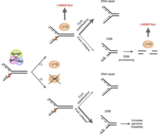
Figure 8. Model Explaining the Role for MRN and ATR in Maintaining Proper Replication.
Absence of functional MRN complex induces stalled replication forks (represented by a red cross). In presence of ATR (1), the kinase is activated through the presence of ssDNA generated at the stalled fork. ATR kinase will then phosphorylate H2AX (visible in S-phase), stabilize blocked replication forks, and facilitate completion of S-phase synthesis (major pathway symbolized by a thick arrow). Failure to rescue replication forks results in fork collapse and chromatid breakage, giving rise to activation of ATR and appearance of g-H2AX foci either in S-phase or in mitosis (minor pathway symbolized by a thin arrow). In absence of ATR in rad50 atr or mre11 atr double mutants (2), fork stabilization is less efficient, leading to increased DSB and genomic instability.
has been shown in human cells to phosphorylate proteins in-volved in the regulation of homologous recombination (Tibbetts et al., 2000). An alternative hypothesis is that ATR could promote fork stability and/or recovery of stalled forks in rad50 mutant plants, and, in support of this, a number of replisome compo-nents are substrates of the ATR kinase (Yoo et al., 2004; Liu et al., 2006; Trenz et al., 2008). A growing body of evidence suggests that human ATR and the Werner’s Syndrome helicase work together to suppress fragile replication site instability (Pichierri et al., 2003; Pirzio et al., 2008). Furthermore, it has been shown that in response to replicative stress (i.e., in the presence of HU or Aphidicolin), ATR activates the Bloom’s Syndrome helicase to restore correct fork restart and hence prevent subsequent ge-nomic instability (Davies et al., 2004, 2007). Moreover, in various organisms, H2AX phosphorylation by ATR could contribute through recruitment of cohesin and repair factors to repair of DSBs generated in the absence of MRN (Unal et al., 2004; Lowndes and Toh, 2005; Fillingham et al., 2006; Stucki and Jackson, 2006; Kinner et al., 2008).
The accumulation of unrepaired or misrepaired DNA in the single (rad50 and mre11) and double (rad50 atr and mre11 atr) mutant plants is expected to lead to cell death and thus to developmental defects in the growing plants. Using propidium iodide staining as a marker of cell death, we found that cell death in rad50 and mre11 mutant plant roots is restricted to the meristem, while cell death in rad50 atr and mre11 atr double mutant plants spreads throughout the root. It has recently been shown that irradiation of plants with UV or g-rays preferentially kills stem cells, and this cell death involves signaling of DNA damage through activation of both ATM and ATR kinases (Fulcher and Sablowski, 2009; Furukawa et al., 2010). While g-irradiation–induced programmed cell death (PCD) in Arabi-dopsis is dependent upon ATM signaling, UV-B–induced PCD is elevated in the absence of ATM or ATR. In contrast with the ATM-and ATR-dependent PCD induced by replication stress, we describe here the death of root initials in rad50 or mre11 mutants, independently of both ATR and ATM. Thus, in the absence of RAD50, intrinsic replication stress presumably causes fork col-lapse and accumulation of DSB, leading to aneuploidy and/or mitotic catastrophe and, thus, the observed cell death.
In conclusion, our data show an absence of induction of g-H2AX foci in rad50 and mre11 mutant plants by g-irradiation, confirming that the MRN complex is required for H2AX phos-phorylation by the ATM and ATR kinases in response to irradi-ation-induced DSBs in Arabidopsis. The essential role of the MRN complex in activation of the ATM and ATR kinases in response to DSB induction is thus conserved in plants. Sponta-neous activation of g-H2AX foci formation in the absence of MRN has not to our knowledge been tested in any organism. We show that this phosphorylation is ATR dependent and strongly en-riched in S-phase nuclei, and only a subset of these g-H2AX foci are found to be associated with telomeres. Furthermore, our detection of ATR-dependent g-H2AX foci in mitotic cells of rad50 and mre11 mutants shows that DSB in Arabidopsis can be processed in the absence of the MRN complex. Taken together, these data suggest a key role for MRN and ATR in maintaining proper replication. In the absence of RAD50 or MRE11, cells activate ATR, presumably through ssDNA generated at stalled
replication forks. Activation of the ATR kinase will then not only activate cell cycle arrest, but stabilize blocked replication forks and facilitate completion of S-phase synthesis. Failure to rescue replication forks in rad50 atr mutant plants will result in fork collapse and chromatid breakage and give rise to the appear-ance of dicentric chromosomes. The high degree of chromo-somal instability in these plants and the resulting cell death explain their severe developmental defects (Figure 8). The em-bryonic lethality of rad50, mre11, and atr in mammals could well result from the critical roles of the MRN complex and ATR in maintaining proper replication as observed in Arabidopsis. The striking tolerance to these mutations, both individually and in combination, makes Arabidopsis a particularly interesting model for studies of the fundamental cellular metabolic pro-cesses that assure the replication and stability of the eukaryotic genome.
METHODS
Plant Material and Growth Conditions
The T-DNA insertional Arabidopsis thaliana mutants of rad50, mre11-3, and atr-2 have been described previously (Gallego et al., 2001; Culligan et al., 2004; Puizina et al., 2004). Seeds of the mre11-3 Arabidopsis line SALK_054418 and the atr-2 Arabidopsis line SALK_032841 were ob-tained from the Nottingham Arabidopsis Stock Centre (Scholl et al., 2000). Homozygous mutant and heterozygous plants for the RAD50 or MRE11 alleles were distinguished by phenotypic and PCR analyses, as described previously (Gallego et al., 2001; Puizina et al., 2004). The double rad50 atr or mre11 atr mutants were produced by crossing rad50 and mre11 heterozygotes with an atr homozygote using standard tech-niques.
Seeds were surface sterilized in 7% calcium hypochlorite for 15 min and rinsed four times with sterile water. Seeds were sown on Petri dishes containing 13 Murashige and Skoog (MS) with vitamins, MES buffer (M0255; Duchefa Biochimie) and1% sucrose (Duchefa), and solidified with 0.8% agar (Bacto-Agar; DIFCO Laboratories). Petri dishes were placed at 48C overnight (15 h) and transferred to a growth chamber (16 h light, 8 h dark cycle) at 238C.
Plant Treatments
To inhibit ATM kinase, Arabidopsis plants were grown in Petri dishes on solid MS medium containing 10 mM ku55933 (ATM inhibitor or IATM) (Hickson et al., 2004).
EdU detection (Vanstraelen et al., 2009) was performed as indicated by the manufacturer (Click-It EdU Alexa Fluor 594 imaging kit; Invitrogen) with modifications. For EdU (5-ethynyl-2’-deoxyuridine) incorporation, 5-d-old seedlings growing in liquid 0.53 MS medium were transferred to the same medium containing 10 mM EdU and incubated for 1 h. All the following steps were performed at room temperature. Seedlings were fixed for 15 min in 3.7% (v/v) formaldehyde in PBS, pH 7.4, rinsed twice with PBS/3% BSA, and placed in 0.5% (v/v) Triton X-100/PBS. After 20 min at room temperature, the Triton X-100/PBS was replaced with Click-It reaction solution and incubation was continued for 30 min with gentle agitation. The Click-It solution was removed, and the seedlings were rinsed in PBS/BSA and transferred to PBS.
g-Irradiation of 5-d-old seedlings was performed with a 137Cs source (C.I.S. Bio International) at an absorbed dose of 25 Gy (1 Gy/7.2 s), and the seedlings were then fixed with 4% (v/v) paraformaldehyde, 10 or 20 min after the start of irradiation.
Cell Death Assay
Five days after germination, seedlings were placed in propidium iodide solution (5 mg/mL in water) for 1 min and rinsed three times with water. Root tips were then transferred to slides in a drop of water and covered with a cover slip for observation under the fluorescence microscope with a Zeiss filter set #43HE (adapted from Curtis and Hays, 2007). Determination of the Mitotic Index
Roots were fixed in a solution of 4% paraformaldehyde in PBS for 45 min, washed twice in PBS/Tween 1%, incubated for 30 min in Hoechst 33258 (3 mg/mL), rinsed in PBS/Tween, and mounted under cover slips in 40% glycerol. The roots were analyzed for mitotic stages (metaphase and anaphase/telophase) using fluorescence microscopy with Zeiss filter set #49.
g-H2AX Antibodies
Antiserum was raised and purified by Eurogentec against a phospho-specific Arabidopsis H2AX peptide following the method published by the Britt group (Friesner et al., 2005). Using a sample of antiserum kindly sent by Anne Britt, we verified that this new antiserum gives identical results in our hands to that published by Friesner et al. (Friesner et al., 2005; Charbonnel et al., 2010). We verified the number of foci at different doses and found identical results to those published by Friesner et al., with a dose of 25 Gy from a 137Cs source inducing a mean of 15 g-H2AX foci per 4C M-phase root nucleus in wild-type plants. No or very few foci were observed in nonirradiated wild-type plants.
Five days after germination, root tips were prepared as previously described (Charbonnel et al., 2010). Briefly, root tips were fixed for 45 min in 4% paraformaldehyde in PME (50 mM PIPES, pH 6.9, 5 mM MgSO4, and 1 mM EGTA) and then washed 33 5 min in PME. Tips were digested for 30 min in a 1% (w/v) cellulase, 0.5% (w/v) cytohelicase, and 1% (w/v) pectolyase (from Sigma-Aldrich; Refs. C1794, C8274, and P5936) solu-tion prepared in PME and then washed 33 5 min in PME. These roots were gently squashed onto slides as described (Liu et al., 1993), air dried, and stored at2808C. Each slide was incubated overnight at 48C with 50 to 100 mL rabbit, anti-plant g-H2AX antiserum diluted 1:600 in fresh blocking buffer (3% BSA, 0.05% Tween 20 [Sigma–Aldrich] in 13 PBS). Slides were washed 33 5min in 13 PBS solution and then incubated for 2 to 3 h at room temperature in 50 to 100 mL blocking buffer consisting of Alexa 488–conjugated goat anti-rabbit (Molecular Probes, 1:1000) secondary antibodies. Finally, slides were washed 33 5 min in 13 PBS and mounted in Vectashield mounting medium with 4’,6-diamidino-2-phenylindole (DAPI) (2 mg/mL) (Vector Laboratories).
DAPI Staining of Mitosis
As described (Caryl et al., 2000), whole inflorescences were collected and fixed, and the mitotic chromosomes of fixed flower pistils were squashed on a slide. Slides were mounted using Vectashield (Vector Laboratories) mounting medium with 1.5 mg/mL DAPI and observed by fluorescence microscopy. Images were further processed and enhanced using Adobe Photoshop software.
FISH
FISH was performed according to Mokros et al. (2006) with the modifi-cations described by Vannier et al. (2009), using BACs from subtelomeric regions of Arabidopsis chromosomes (F6F3, F23A5, F17A22, F4P13, T20O10, F6N15, T19P19, F7J8, and K9I9), labeled either with Cy5-dUTP or Cy3-dUTP (Amersham) by standard nick translation reactions (Roche). FISH after immunostaining required a postfixation step of 30 min in 4% formaldehyde.
Microscopy andg-H2AX Foci Analysis
Images were acquired on a Zeiss epifluorescence microscope using Axiovision software. Measurements were performed using the same software, and images were treated using Adobe Photoshop software to enhance contrast. Images stacks were captured in three dimensions (x, y, and z), and were deconvolved with the deconvolution module (theoretical PSF, iterative algorithm) of Axiovision 4.6.2 (Zeiss) software to affine g-H2AX foci, which were counted manually. The images presented are collapsed Z-stack projections obtained using the Extended-focus mod-ule (projection method) of the Axiovision 4.6.2 software, except for immunostaining and subtelomeric FISH labeling images (Figure 2C) that correspond to the single focal plane from a deconvolved three-dimensional image.
Accession Numbers
Sequence data from this article can be found in the Arabidopsis Genome Initiative or GenBank/EMBL databases under the following accession numbers: RAD50 (locus AT2G31970; GenBank NM_128757); Mre11 (locus AT5G5426; GenBank NM_124806); ATR (locus AT5G40820; GenBank NM_123447); and ATM (locus AT3G48190; GenBank NM_ 114689.6).
ACKNOWLEDGMENTS
We thank the members of the recombination group for their help and discussions. Spencer Brown and Marlene Van Straelen (Centre National de la Recherche Scientifique) are thanked for sending us the Arabidopsis EdU labeling protocol prior to publication. We thank the Etablissement Franc¸ais du Sang and Dr. P. Fabrigly for access to the 137Cs irradiator. This work was partly financed by a European Union research grant (LSHG-CT-2005-018785), a French Government ANR grant (ANR-07-BLAN-0068), the Centre National de la Recherche Scientifique, the Universite´ Blaise Pascal, the Universite´ d’Auvergne, and the Institut National de la Sante´ et la Recherche Medicale. C.C. was supported by a doctoral fellowship from the Ministe`re de l’Education Nationale, de la Recherche et de la Technologie, and S.A. is an ANR postdoctoral fellow (ANR-07-BLAN-0068).
Received August 3, 2010; revised September 4, 2010; accepted Sep-tember 13, 2010; published SepSep-tember 28, 2010.
REFERENCES
Adams, K.E., Medhurst, A.L., Dart, D.A., and Lakin, N.D. (2006). Recruitment of ATR to sites of ionising radiation-induced DNA dam-age requires ATM and components of the MRN protein complex. Oncogene 25: 3894–3904.
Bi, X., Srikanta, D., Fanti, L., Pimpinelli, S., Badugu, R., Kellum, R., and Rong, Y.S. (2005). Drosophila ATM and ATR checkpoint kinases control partially redundant pathways for telomere maintenance. Proc. Natl. Acad. Sci. USA 102: 15167–15172.
Bleuyard, J.-Y., Gallego, M.E., and White, C.I. (2004). Meiotic defects in the Arabidopsis rad50 mutant point to conservation of the MRX complex function in early stages of meiotic recombination. Chromo-soma 113: 197–203.
Brown, E.J., and Baltimore, D. (2000). ATR disruption leads to chro-mosomal fragmentation and early embryonic lethality. Genes Dev. 14: 397–402.
Bundock, P., and Hooykaas, P. (2002). Severe developmental defects,
hypersensitivity to DNA-damaging agents, and lengthened telomeres in Arabidopsis MRE11 mutants. Plant Cell 14: 2451–2462.
Carson, C.T., Schwartz, R.A., Stracker, T.H., Lilley, C.E., Lee, D.V., and Weitzman, M.D. (2003). The Mre11 complex is required for ATM activation and the G2/M checkpoint. EMBO J. 22: 6610–6620. Caryl, A.P., Armstrong, S.J., Jones, G.H., and Franklin, F.C. (2000). A
homologue of the yeast HOP1 gene is inactivated in the Arabidopsis meiotic mutant asy1. Chromosoma 109: 62–71.
Charbonnel, C., Gallego, M.E., and White, C.I. (2010). XRCC1-dependent and KU-XRCC1-dependent DNA double-strand break repair knetics in Arabidopsis plants. Plant J. http://dx.doi.org/10.1111/ j.1365-313X.2010.04331.x.
Ciapponi, L., Cenci, G., and Gatti, M. (2006). The Drosophila Nbs protein functions in multiple pathways for the maintenance of genome stability. Genetics 173: 1447–1454.
Cimprich, K.A., and Cortez, D. (2008). ATR: An essential regulator of genome integrity. Nat. Rev. Mol. Cell Biol. 9: 616–627.
Costanzo, V., Robertson, K., Bibikova, M., Kim, E., Grieco, D., Gottesman, M., Carroll, D., and Gautier, J. (2001). Mre11 protein complex prevents double-strand break accumulation during chromo-somal DNA replication. Mol. Cell 8: 137–147.
Culligan, K., Tissier, A., and Britt, A. (2004). ATR regulates a G2-phase cell-cycle checkpoint in Arabidopsis thaliana. Plant Cell 16: 1091– 1104.
Curtis, M.J., and Hays, J.B. (2007). Tolerance of dividing cells to replication stress in UVB-irradiated Arabidopsis roots: requirements for DNA translesion polymerases eta and zeta. DNA Repair (Amst.) 6: 1341–1358.
Czornak, K., Chughtai, S., and Chrzanowska, K.H. (2008). Mystery of DNA repair: The role of the MRN complex and ATM kinase in DNA damage repair. J. Appl. Genet. 49: 383–396.
D’Amours, D., and Jackson, S.P. (2001). The yeast Xrs2 complex functions in S phase checkpoint regulation. Genes Dev. 15: 2238– 2249.
Davies, S.L., North, P.S., Dart, A., Lakin, N.D., and Hickson, I.D. (2004). Phosphorylation of the Bloom’s syndrome helicase and its role in recovery from S-phase arrest. Mol. Cell. Biol. 24: 1279–1291. Davies, S.L., North, P.S., and Hickson, I.D. (2007). Role for BLM in
replication-fork restart and suppression of origin firing after replicative stress. Nat. Struct. Mol. Biol. 14: 677–679.
de Klein, A., Muijtjens, M., van Os, R., Verhoeven, Y., Smit, B., Carr, A.M., Lehmann, A.R., and Hoeijmakers, J.H. (2000). Targeted dis-ruption of the cell-cycle checkpoint gene ATR leads to early embry-onic lethality in mice. Curr. Biol. 10: 479–482.
Fernandez-Capetillo, O., Celeste, A., and Nussenzweig, A. (2003). Focusing on foci: H2AX and the recruitment of DNA-damage re-sponse factors. Cell Cycle 2: 426–427.
Fillingham, J., Keogh, M.-C., and Krogan, N.J. (2006). GammaH2AX and its role in DNA double-strand break repair. Biochem. Cell Biol. 84: 568–577.
Friedel, A.M., Pike, B.L., and Gasser, S.M. (2009). ATR/Mec1: Coor-dinating fork stability and repair. Curr. Opin. Cell Biol. 21: 237–244. Friesner, J.D., Liu, B., Culligan, K., and Britt, A.B. (2005). Ionizing
radiation-dependent gamma-H2AX focus formation requires ataxia telangiectasia mutated and ataxia telangiectasia mutated and Rad3-related. Mol. Biol. Cell 16: 2566–2576.
Fulcher, N., and Sablowski, R. (2009). Hypersensitivity to DNA damage in plant stem cell niches. Proc. Natl. Acad. Sci. USA 106: 20984– 20988.
Furukawa, T., Curtis, M.J., Tominey, C.M., Duong, Y.H., Wilcox, B. W., Aggoune, D., Hays, J.B., and Britt, A.B. (2010). A shared DNA-damage-response pathway for induction of stem-cell death by UVB and by gamma irradiation. DNA Repair (Amst.) 9: 940–948.
Gallego, M.E., Jeanneau, M., Granier, F., Bouchez, D., Bechtold, N., and White, C.I. (2001). Disruption of the Arabidopsis RAD50 gene leads to plant sterility and MMS sensitivity. Plant J. 25: 31–41. Gallego, M.E., and White, C.I. (2001). RAD50 function is essential for
telomere maintenance in Arabidopsis. Proc. Natl. Acad. Sci. USA 98: 1711–1716.
Garcia, V., Bruchet, H., Camescasse, D., Granier, F., Bouchez, D., and Tissier, A. (2003). AtATM is essential for meiosis and the somatic response to DNA damage in plants. Plant Cell 15: 119–132. Harper, J.W., and Elledge, S.J. (2007). The DNA damage response:
Ten years after. Mol. Cell 28: 739–745.
Hickson, I., Zhao, Y., Richardson, C.J., Green, S.J., Martin, N.M.B., Orr, A.I., Reaper, P.M., Jackson, S.P., Curtin, N.J., and Smith, G.C.M. (2004). Identification and characterization of a novel and specific inhibitor of the ataxia-telangiectasia mutated kinase ATM. Cancer Res. 64: 9152–9159.
Horejsı´, Z., Falck, J., Bakkenist, C.J., Kastan, M.B., Lukas, J., and Bartek, J. (2004). Distinct functional domains of Nbs1 modulate the timing and magnitude of ATM activation after low doses of ionizing radiation. Oncogene 23: 3122–3127.
Jazayeri, A., Falck, J., Lukas, C., Bartek, J., Smith, G.C., Lukas, J., and Jackson, S.P. (2006). ATM- and cell cycle-dependent regulation of ATR in response to DNA double-strand breaks. Nat. Cell Biol. 8: 37–45.
Kinner, A., Wu, W., Staudt, C., and Iliakis, G. (2008). Gamma-H2AX in recognition and signaling of DNA double-strand breaks in the context of chromatin. Nucleic Acids Res. 36: 5678–5694.
Lamarche, B.J., Orazio, N.I., and Weitzman, M.D. (2010). The MRN complex in double-strand break repair and telomere maintenance. FEBS Lett. 584: 3682–3695.
Laurenc¸on, A., Purdy, A., Sekelsky, J., Hawley, R.S., and Su, T.T. (2003). Phenotypic analysis of separation-of-function alleles of MEI-41, Drosophila ATM/ATR. Genetics 164: 589–601.
Lee, J.H., and Paull, T.T. (2004). Direct activation of the ATM protein kinase by the Mre11/Rad50/Nbs1 complex. Science 304: 93–96. Liu, B., Marc, J., Joshi, H.C., and Palevitz, B.A. (1993). A
gamma-tubulin-related protein associated with the microtubule arrays of higher plants in a cell cycle-dependent manner. J. Cell Sci. 104: 1217–1228.
Liu, J.S., Kuo, S.R., and Melendy, T. (2006). Phosphorylation of replication protein A by S-phase checkpoint kinases. DNA Repair (Amst.) 5: 369–380.
Lowndes, N.F., and Toh, G.W. (2005). DNA repair: The importance of phosphorylating histone H2AX. Curr. Biol. 15: R99–R102.
Mimitou, E.P., and Symington, L.S. (2009). DNA end resection: Many nucleases make light work. DNA Repair (Amst.) 8: 983–995. Mirzoeva, O.K., and Petrini, J.H.J. (2003). DNA replication-dependent
nuclear dynamics of the Mre11 complex. Mol. Cancer Res. 1: 207–218. Mokros, P., Vrbsky, J., and Siroky, J.(2006). Identification of chro-mosomal fusion sites in Arabidopsis mutants using sequential bi-colour BAC-FISH. Genome 49: 1036–1042.
Myers, J.S., and Cortez, D. (2006). Rapid activation of ATR by ionizing radiation requires ATM and Mre11. J. Biol. Chem. 281: 9346–9350. Olson, E., Nievera, C.J., Lee, A.Y., Chen, L., and Wu, X. (2007). The
Mre11-Rad50-Nbs1 complex acts both upstream and downstream of ataxia telangiectasia mutated and Rad3-related protein (ATR) to regulate the S-phase checkpoint following UV treatment. J. Biol. Chem. 282: 22939–22952.
Paull, T.T., and Gellert, M. (1999). Nbs1 potentiates ATP-driven DNA unwinding and endonuclease cleavage by the Mre11/Rad50 complex. Genes Dev. 13: 1276–1288.
Paull, T.T., Rogakou, E.P., Yamazaki, V., Kirchgessner, C.U., Gellert, M., and Bonner, W.M. (2000). A critical role for histone H2AX in
recruitment of repair factors to nuclear foci after DNA damage. Curr. Biol. 10: 886–895.
Pichierri, P., Rosselli, F., and Franchitto, A. (2003). Werner’s syn-drome protein is phosphorylated in an ATR/ATM-dependent manner following replication arrest and DNA damage induced during the S phase of the cell cycle. Oncogene 22: 1491–1500.
Pirzio, L.M., Pichierri, P., Bignami, M., and Franchitto, A. (2008). Werner syndrome helicase activity is essential in maintaining fragile site stability. J. Cell Biol. 180: 305–314.
Puizina, J., Siroky, J., Mokros, P., Schweizer, D., and Riha, K. (2004). Mre11 deficiency in Arabidopsis is associated with chromosomal instability in somatic cells and Spo11-dependent genome fragmen-tation during meiosis. Plant Cell 16: 1968–1978.
Rich, T., Allen, R.L., and Wyllie, A.H. (2000). Defying death after DNA damage. Nature 407: 777–783.
Robison, J.G., Elliott, J., Dixon, K., and Oakley, G.G. (2004). Repli-cation protein A and the Mre11.Rad50.Nbs1 complex co-localize and interact at sites of stalled replication forks. J. Biol. Chem. 279: 34802– 34810.
Scholl, R.L., May, S.T., and Ware, D.H. (2000). Seed and molecular resources for Arabidopsis. Plant Physiol. 124: 1477–1480.
Shiloh, Y. (2006). The ATM-mediated DNA-damage response: Taking shape. Trends Biochem. Sci. 31: 402–410.
Shor, E., Gangloff, S., Wagner, M., Weinstein, J., Price, G., and Rothstein, R. (2002). Mutations in homologous recombination genes rescue top3 slow growth in Saccharomyces cerevisiae. Genetics 162: 647–662.
Stiff, T., Reis, C., Alderton, G.K., Woodbine, L., O’Driscoll, M., and Jeggo, P.A. (2005). Nbs1 is required for ATR-dependent phosphor-ylation events. EMBO J. 24: 199–208.
Stucki, M., and Jackson, S.P. (2006). gammaH2AX and MDC1: An-choring the DNA-damage-response machinery to broken chromo-somes. DNA Repair (Amst.) 5: 534–543.
Tibbetts, R.S., Cortez, D., Brumbaugh, K.M., Scully, R., Livingston, D., Elledge, S.J., and Abraham, R.T. (2000). Functional interactions between BRCA1 and the checkpoint kinase ATR during genotoxic stress. Genes Dev. 14: 2989–3002.
Tittel-Elmer, M., Alabert, C., Pasero, P., and Cobb, J.A. (2009). The MRX complex stabilizes the replisome independently of the S phase checkpoint during replication stress. EMBO J. 28: 1142–1156. Tomimatsu, N., Mukherjee, B., and Burma, S. (2009). Distinct roles of
ATR and DNA-PKcs in triggering DNA damage responses in ATM-deficient cells. EMBO Rep. 10: 629–635.
Trenz, K., Errico, A., and Costanzo, V. (2008). Plx1 is required for chromosomal DNA replication under stressful conditions. EMBO J. 27: 876–885.
Trenz, K., Smith, E., Smith, S., and Costanzo, V. (2006). ATM and ATR promote Mre11 dependent restart of collapsed replication forks and prevent accumulation of DNA breaks. EMBO J. 25: 1764–1774. Unal, E., Arbel-Eden, A., Sattler, U., Shroff, R., Lichten, M., Haber,
J.E., and Koshland, D. (2004). DNA damage response pathway uses histone modification to assemble a double-strand break-specific cohesin domain. Mol. Cell 16: 991–1002.
Uziel, T., Lerenthal, Y., Moyal, L., Andegeko, Y., Mittelman, L., and Shiloh, Y. (2003). Requirement of the MRN complex for ATM activa-tion by DNA damage. EMBO J. 22: 5612–5621.
Vannier, J.-B., Depeiges, A., White, C., and Gallego, M.E. (2006). Two roles for Rad50 in telomere maintenance. EMBO J. 25: 4577– 4585.
Vannier, J.-B., Depeiges, A., White, C., and Gallego, M.E. (2009). ERCC1/XPF protects short telomeres from homologous recombina-tion in Arabidopsis thaliana. PLoS Genet. 5: e1000380.
Vanstraelen, M., Baloban, M., Da Ines, O., Cultrone, A., Lammens, T., Boudolf, V., Brown, S.C., De Veylder, L., Mergaert, P., and Kondorosi, E. (2009). APC/C-CCS52A complexes control meristem maintenance in the Arabidopsis root. Proc. Natl. Acad. Sci. USA 106: 11806–11811.
Vespa, L., Couvillion, M., Spangler, E., and Shippen, D.E. (2005). ATM and ATR make distinct contributions to chromosome end protection and the maintenance of telomeric DNA in Arabidopsis. Genes Dev. 19: 2111–2115.
Yamaguchi-Iwai, Y., Sonoda, E., Sasaki, M.S., Morrison, C., Haraguchi, T., Hiraoka, Y., Yamashita, Y.M., Yagi, T., Takata, M., Price, C., Kakazu, N., and Takeda, S. (1999). Mre11 is essential for the maintenance of chromosomal DNA in vertebrate cells. EMBO J. 18: 6619–6629.
Yoo, H.Y., Shevchenko, A., Shevchenko, A., and Dunphy, W.G. (2004). Mcm2 is a direct substrate of ATM and ATR during DNA damage and DNA replication checkpoint responses. J. Biol. Chem. 279: 53353–53364.
Zhao, S., Renthal, W., and Lee, E.Y. (2002). Functional analysis of FHA and BRCT domains of NBS1 in chromatin association and DNA damage responses. Nucleic Acids Res. 30: 4815–4822.
Zhong, H., Bryson, A., Eckersdorff, M., and Ferguson, D.O. (2005). Rad50 depletion impacts upon ATR-dependent DNA damage re-sponses. Hum. Mol. Genet. 14: 2685–2693.