ARTICLE
The coactivator PGC-1
α regulates skeletal muscle oxidative
metabolism independently of the nuclear receptor PPAR
β/δ
in sedentary mice fed a regular chow diet
Joaquín Pérez-Schindler&Kristoffer Svensson&Elyzabeth Vargas-Fernández&Gesa Santos&
Walter Wahli&Christoph Handschin
Received: 18 December 2013 / Accepted: 22 July 2014 / Published online: 13 August 2014 # Springer-Verlag Berlin Heidelberg 2014
Abstract
Aims/hypothesis Physical activity improves oxidative capaci-ty and exerts therapeutic beneficial effects, particularly in the context of metabolic diseases. The peroxisome proliferator-activated receptor (PPAR)γ coactivator-1α (PGC-1α) and the nuclear receptor PPARβ/δ have both been independently discovered to play a pivotal role in the regulation of oxidative metabolism in skeletal muscle, though their interdependence remains unclear. Hence, our aim was to determine the func-tional interaction between these two factors in mouse skeletal muscle in vivo.
Methods Adult male control mice, PGC-1α muscle-specific transgenic (mTg) mice, PPARβ/δ muscle-specific knockout (mKO) mice and the combination PPARβ/δ mKO + PGC-1α mTg mice were studied under basal conditions and following PPARβ/δ agonist administration and acute exercise.
Whole-body metabolism was assessed by indirect calorimetry and blood analysis, while magnetic resonance was used to mea-sure body composition. Quantitative PCR and western blot were used to determine gene expression and intracellular signalling. The proportion of oxidative muscle fibre was de-termined by NADH staining.
Results Agonist-induced PPARβ/δ activation was only disrupted by PPARβ/δ knockout. We also found that the disruption of the PGC-1α–PPARβ/δ axis did not affect whole-body metabolism under basal conditions. As expected, PGC-1α mTg mice exhibited higher exercise performance, peak oxygen consumption and lower blood lactate levels following exercise, though PPARβ/δ mKO + PGC-1α mTg mice showed a similar phenotype. Similarly, we found that PPARβ/δ was dispensable for PGC-1α-mediated enhance-ment of an oxidative phenotype in skeletal muscle.
Conclusions/interpretation Collectively, these results indicate that PPARβ/δ is not an essential partner of PGC-1α in the control of skeletal muscle energy metabolism.
Keywords Coregulators . Exercise . Nuclear receptors . Skeletal muscle metabolism
Abbreviations
AMPK AMP-activated protein kinase CON Control mice
GTT Glucose tolerance test
IMTG Intramyocellular triacylglycerol ITT Insulin tolerance test
mKO Muscle-specific knockout mTg Muscle-specific transgenic PGC PPARγ coactivator
PPAR Peroxisome proliferator-activated receptor qPCR Quantitative PCR
RER Respiratory exchange ratio
Electronic supplementary material The online version of this article (doi:10.1007/s00125-014-3352-3) contains peer-reviewed but unedited supplementary material, which is available to authorised users. J. Pérez-Schindler
:
K. Svensson:
E. Vargas-Fernández:
G. Santos
:
C. Handschin (*)Biozentrum, University of Basel, Klingelbergstrasse 50/70, 4056 Basel, Switzerland
e-mail: christoph.handschin@unibas.ch W. Wahli
Center for Integrative Genomics, National Research Center Frontiers in Genetics, University of Lausanne, Le Génopode, Lausanne, Switzerland
W. Wahli
Lee Kong Chian School of Medicine, Nanyang Technological University, Singapore, Republic of Singapore
Present address: J. Pérez-Schindler
School of Sport, Exercise and Rehabilitation Sciences, University of Birmingham, Edgbaston, Birmingham, UK
TBP TATA binding protein UCP3 Uncoupling protein 3
V⋅O2peak Peak oxygen consumption
Introduction
The regulation of energy metabolism in skeletal muscle is highly controlled by the peroxisome proliferator-activated receptor (PPAR) γ coactivator-1α (PGC-1α) [1]. PGC-1α
drives the expression of genes involved in catabolic processes leading to aerobic ATP synthesis [1] while concomitantly promoting anabolic processes, including de novo lipogenesis [2]. Once activated, PGC-1α boosts the activity of different
transcription factors to control gene programmes resembling an endurance-trained phenotype in skeletal muscle [1, 3]. These adaptations are associated with an enhanced oxidative capacity, which contributes to an increased skeletal muscle fatigue resistance ex vivo and exercise performance in vivo [4–6]. Importantly, exercise is in fact one of the most efficient stimuli to induce PGC-1α in skeletal muscle [3].
Among the transcription factors regulated by PGC-1α, the nuclear receptor PPARβ/δ has been proposed to be a key partner of PGC-1α in the regulation of skeletal muscle me-tabolism and function, though mainly based on cell culture and pharmacological studies [7]. PGC-1α acts as a coactivator
of PPARβ/δ [8–10], while PPARβ/δ can directly regulate PGC-1α expression [11,12], indicating that this nuclear re-ceptor acts both upstream and downstream of PGC-1α. Fur-thermore, transgenic mouse models for PPARβ/δ exhibit a similar phenotype to their counterparts for PGC-1α [4,5,13,
14]. Nevertheless, although the PGC-1α–PPARβ/δ axis
ap-pears to play a key role in the regulation of energy metabo-lism, the epistatic interaction between these proteins is cur-rently unclear. We therefore aimed at directly assessing the functional interplay between PGC-1α and PPARβ/δ in the regulation of skeletal muscle oxidative metabolism in vivo.
Methods
Animals Mice were housed in a conventional facility with a 12 h night/day cycle and had free access to food/water. Ex-periments were performed on adult male mice with approval of the Swiss authorities. PGC-1α muscle-specific transgenic (mTg) mice have been described previously [5]. PPARβ/δ muscle-specific knockout (mKO) mice were generated by crossing PPARβ/δloxP/loxP
mice with HSA-Cre transgenic mice [11]. Finally, PGC-1α mTg and HSA-Cre positive PPARβ/ δloxP/loxPmKO mice were crossed to generate PPARβ/δ mKO + PGC-1α mTg mice. PPARβ/δloxP/loxP
mice without Cre and Pgc-1α (also known as Ppargc1a) transgene expression were
used as control (CON) mice. All mice had mixed sv129 and C57BL/6 background. Genotypes were confirmed through PCR procedures (data not shown) and quantitative PCR analysis in kidney and skeletal muscle (Fig.1a, b).
PPARβ/δ agonist administration CON mice were subjected to an intraperitoneal injection of either 0.9% NaCl (control) or 1 mg/kg of body weight of the PPARβ/δ agonist GW0742 (Tocris No. 2229; Tocris, Bristol, UK), as previously de-scribed [15]. Muscles were collected 8 h following drug administration.
Body composition analysis Lean and fat mass were measured via magnetic resonance imaging (EchoMRI, Houston, TX, USA).
Blood and plasma analysis Blood samples were collected under basal conditions or immediately after maximal exercise from fed and/or overnight-fasted mice, as previously described [9].
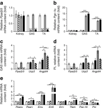
Fig. 1 PGC-1α and PPARβ/δ mouse models. (a, b) Pparβ/δ and Pgc-1α mRNA levels in kidney, gastrocnemius (GAS) and tibialis anterior (TA) (n=6 per group). (c, d) Pparβ/δ, Ucp3 and Angptl4 mRNA levels in GAS and plantaris (PL) 8 h after the injection of 0.9% NaCl (as control) or 1 mg/kg of body weight of the PPARβ/δ agonist GW0742 (n=6 per group). (e) mRNA level of different transcriptional regulators in GAS (n= 6 per group). In (a), (b) and (e): white bars, CON; hatched bars, PPARβ/δ mKO; grey bars, PGC-1α mTg; black bars, PPARβ/δ mKO + PGC-1α mTg. In (c) and (d): white bars, CON + NaCl; black bars, PPARβ/δ mKO + NaCl; grey bars, CON + GW0742; hatched bars, PPARβ/δ mKO + GW0742. Values are mean ± SEM. *p<0.05, **p<0.01 and ***p<0.001 for the indicated comparisons. In (a), (c) and (d) ***p<0.001 vs CON and/or PGC-1α mTg for same tissue/treatment. When shown, fold changes are reported vs CON group
Glucose and insulin tolerance test Intraperitoneal glucose tolerance tests (GTTs) were carried out by injecting 2 g/kg of body weight of glucose after mice had been fasted for 16 h. Insulin tolerance tests (ITTs) were performed by injecting 0.8 U/kg of body weight of insulin (Novo Nordisk, Bagsvaerd, Denmark) after mice had been fasted for 6 h. Indirect calorimetry Mice were individually housed in a Comprehensive Lab Animal Monitoring System (Columbus Instruments, Columbus, OH, USA) for an acclimatisation period of 48 h during which they were allowed free access to food and water. Subsequently, indirect calorimetry was performed for 48 h and data was analysed with the Oxymax software (Columbus Instruments).
Maximal exercise test Exercise tests were performed as previ-ously described [9]. Briefly, 2 days after acclimatisation, mice performed a maximal exercise test in a closed treadmill (Colum-bus Instruments), allowing the measurement of peak oxygen consumption (V⋅O2peak) and respiratory exchange ratio (RER; CO2produced divided by consumed O2[V
⋅
CO2/V⋅O2]). Histology NADH staining was performed on 10μm cross sections from tibialis anterior by exposing the sections to 1 mg/ml NADH (Sigma, St Louis, MO, USA) in the presence of 1 mg/ml nitro blue tetrazolium (Sigma).
NEFA measurement Plasma NEFA were measured using a commercial kit (HR Series NEFA-HR(2); Wako Diagnostics, Richmond, VA, USA), according to the manufacturer’s instruc-tions. Blood samples were collected under basal conditions and following 1 h of treadmill running at 13 m/min with 5° slope. Intramyocellular triacylglycerol extraction Quadriceps intramyocellular triacylglycerols (IMTGs) were extracted by standard procedures using a solid-phase extraction column (UPTI-CLEAN NH2-S 100 mg/1 mL SPE Columns; Interchim, Montluçon, France) and quantified with a commercial kit (Tri-glyceride enzymatique PAP 150; Biomérieux, Marcy-l’Etoile, France), according to the manufacturer’s instructions.
RNA isolation and quantitative PCR Total RNA isolation from fed (ad libitum) mice and quantitative PCR (qPCR) analysis was performed by standard procedures [9]. Se-quences of qPCR primers are depicted in electronic supple-mentary material (ESM) Table1. Analysis was performed by the ΔΔCt method using TATA binding protein (TBP) as endogenous control. TBP transcript levels were not different between genotypes or between experimental conditions. Protein isolation and western blot Protein isolation and west-ern blot was conducted as previously described [9]. Proteins were detected with primary antibodies to Akt (Cell Signaling
No.9272; Cell Signaling, Danvers, MA, USA), p-AktT308 (Cell Signaling No. 4056), AMP-activated protein kinase (AMPK)α (Cell Signaling No. 2603), p-AMPKαT172(Cell Signaling No. 2535), total OXPHOS (ab110413; Abcam, USA) and eEF2 (Cell Signaling No. 2332).
Statistical analysis Values are expressed as mean ± SEM. Statistical significance was determined with unpaired two-tailed t tests or one-way ANOVA with Tukey’s post hoc test. Significance was considered with a p<0.05.
Results
PGC-1α overexpression and PPARβ/δ deletion in mouse skel-etal muscle To elucidate the functional requirement for PPARβ/δ in the metabolic adaptations induced by PGC-1α, we crossed PPARβ/δ mKO mice with PGC-1α mTg mice, referred to as PPARβ/δ mKO + PGC-1α mTg mice. As expected, both PPARβ/δ mKO and PPARβ/δ mKO + PGC-1α mTg mice showed a reduction in Pparβ/δ (Ppard) mRNA specifically in skeletal muscle, while Pgc-1α mRNA was upregulated by∼12-fold in skeletal muscle of PGC-1α mTg and PPARβ/δ mKO + PGC-1α mTg mice compared with control (CON) mice (Fig.1a, b). To validate the functional consequence of Pparβ/δ deletion in skeletal muscle, we assessed the effects of the PPARβ/δ agonist GW0742 on the expression levels of PPARβ/δ target genes [7, 16]. Acute treatment with GW0742 did not affect Pparβ/δ mRNA in gastrocnemius and plantaris muscles whereas Ucp3 mRNA levels were induced in CON, but not in PPARβ/δ mKO mice (Fig.1c, d). Moreover, as previously reported [16], Angptl4 was upregulated by GW0742 in a way that was partially dependent on PPARβ/δ (Fig.1c, d). Importantly, Pparβ/δ
deletion did not affect the transcript levels of Pparα (Ppara) and Pparγ (Pparg) (Fig.1e). We subsequently measured the expression levels of other transcription factors and coactivators regulating metabolism, including the oestrogen-related receptors, mitochondrial transcription factor A, Pgc-1β and PGC-1-related coactivator. The expression levels of genes encoding these factors/coactivators (Errα [Esrra], Errβ [Esrrb], Tfam, Pgc-1β [Ppargc1b], Prc [Pprc1]) were altered in skeletal muscle of PGC-1α mTg and PPARβ/δ mKO + PGC-1α mTg mice, thus independent of PPARβ/δ ablation (Fig.1e).
Effects of skeletal muscle disruption of the PGC-1α–PPARβ/δ axis on whole-body metabolism Body composition assess-ment revealed equal body weight, fat mass and lean mass in PPARβ/δ mKO, PGC-1α mTg, PPARβ/δ mKO + PGC-1α mTg and CON mice (Fig.2a). Analysis of plasma triacylglyc-erol, cholesttriacylglyc-erol, LDL-cholesterol and HDL-cholesterol
during the fed and fasted state exhibited no differences except for a significant decrease in fed cholesterol in the PPARβ/δ mKO + PGC-1α mTg mice (Fig.2b, c). Moreover, indirect calorimetry during 48 h revealed no differences in V⋅O2 or RER between any of the genotypes (Fig. 2d, e and ESM Fig.1a, b).
Pharmacological activation of PPARβ/δ attenuates the det-rimental effects of obesity and type 2 diabetes on systemic glucose homeostasis [13,17,18]. Compared with CON mice, neither GTTs nor ITTs were affected by PGC-1α overexpres-sion and/or Pparβ/δ deletion in skeletal muscle in mice fed a regular chow diet (Fig.2f, gand ESM Fig.1c, d). Moreover, we did not find any differences in blood glucose levels in fed mice between the four different genotypes (Fig.2h). These findings were corroborated by unchanged expression of genes involved in glucose transport and catabolism, such as Glut4 (Slc2a4), Tbc1d1, Pfk and Hk2 (encoding glucose transporter 4, TBC domain family member 1, phosphofructokinase and hexokinase 2, respectively), in skeletal muscle of PPARβ/δ mKO, PGC-1α mTg and PPARβ/δ mKO + PGC-1α mTg mice (Fig.2i). In contrast, Tbc1d4 (As160, and which encodes
Akt substrate of 160 kDa) was significantly upregulated in PGC-1α mTg mice (Fig.2i). Finally, we observed an increase in total Akt protein levels following PGC-1α overexpression, with no substantial effect of Pparβ/δ deletion (Fig.2jand ESM Fig. 1e). Consistently, PGC-1α overexpression slightly
de-creased relative AktT308phosphorylation levels, although this effect was not statistically significant (ESM Fig. 1f). These data hence suggest that the PGC-1α–PPARβ/δ axis is not essential for the modulation of whole-body metabolism and glucose homeostasis under basal conditions in chow-fed mice. Modulation of skeletal muscle metabolism by the PGC-1α– PPARβ/δ axis Skeletal muscle PGC-1α and PPARβ/δ have been proposed to be key regulators of exercise performance and lactate metabolism [19, 20]. Consequently, we next assessed exercise performance in treadmill-based tests, which revealed a higher exercise performance in PGC-1α mTg mice as expected (Fig.3a–c). Interestingly, Pparβ/δ muscle knock-out did not reduce this difference when PPARβ/δ mKO + PGC-1α mTg mice were compared with CON mice (Fig.3a–c). Moreover, V⋅O2 was significantly enhanced in PGC-1α mTg
Fig. 2 Body composition, systemic variables and glucose handling. (a) Assessment of body composition (n=10–12 mice per group). (b, c) Plasma concentration of TG, cholesterol (Chol), LDL-cholesterol and HDL-cholesterol under fed and fasted conditions (n=10–12 mice per group). (ofV⋅O2(d) and RER (e) over a period of 48 h (n = 10–14
mice per group). (f, g) Blood glucose levels during GTTs (f) and ITTs (g). (h) Blood glucose levels in fed mice (n = 10–12 per group). (i) Gastrocnemius mRNA levels of genes involved in glucose metabolism (n=6 mice per group). (j) Western blot assessment of Akt
phosphorylation status in gastrocnemius muscle (n=6 mice per group). In (a–c), (h) and (i): white bars, CON; hatched bars, PPARβ/δ mKO; grey bars, PGC-1α mTg; black bars, PPARβ/δ mKO + PGC-1α mTg. *p<0.05 and **p<0.01 for the indicated comparisons. In (d–g): solid black line, CON; dashed black line, PPARβ/δ mKO; solid grey line, PGC-1α mTg; dashed grey line, PPARβ/δ mKO + PGC-1α mTg.
†p< 0.05, PPARβ/δ mKO vs PGC-1α mTg;‡‡‡p< 0.001 PPARβ/δ
mKO vs PPARβ/δ mKO + PGC-1α mTg. In graphs, values are mean ± SEM. When shown, fold changes are reported vs CON group
and PPARβ/δ mKO + PGC-1α mTg mice during maximal exercise (Fig.3d), thus altered by PGC-1α independent of
PPARβ/δ. In contrast, the decrease in the RER in PGC-1α mTg mice was attenuated by concomitant Pparβ/δ deletion (Fig. 3e). Blood lactate concentration increased following maximal exercise in CON mice (Fig.3f). This effect was attenuated in PPARβ/δ mKO mice and virtually abolished in both PGC-1α mTg and PPARβ/δ mKO + PGC-1α mTg mice (Fig. 3f). Similarly, pre-exercise blood lactate levels were reduced only in the mouse models with elevated skeletal muscle PGC-1α (Fig. 3f). Consistently, mRNA levels of genes encoding lactate dehydrogenase A (Ldha) and mono-carboxylic acid transporter 4 (Mct4 [Slc16a3]) were reduced only by PGC-1α overexpression in skeletal muscle, while in the same mice, Ldhb and Mct1 (Slc16a1) genes were upreg-ulated (Fig.3g), reflecting an attenuated lactate production as well as higher catabolism. To assess substrate availability, we measured IMTG content and, consistent with the function of PGC-1α in de novo lipogenesis [2], both PGC-1α mTg and
PPARβ/δ mKO + PGC-1α mTg mice showed elevated IMTG levels, though Pparβ/δ knockout had no effect (Fig. 3h).
Finally, we measured plasma levels of NEFA before and after exercise. Exercise significantly increased plasma NEFA levels in PPARβ/δ mKO mice, while CON and PPARβ/δ mKO + PGC-1α mTg mice showed a trend toward an increase (Fig.3i). These data show that, in response to maximal exer-cise, skeletal muscle PGC-1α is a pivotal regulator of whole-body metabolism, mainly in a PPARβ/δ-independent manner. Next, we investigated the relevance of PGC-1α and PPARβ/δ interaction in the regulation of skeletal muscle metabolism. We therefore determined the mRNA levels of genes regulating skeletal muscle oxidative metabolism, sev-eral of which have been suggested to be both PGC-1α and PPARβ/δ targets. Interestingly, we observed that Pparβ/δ deletion in skeletal muscle did not change the transcript abun-dance of genes involved in the tricarboxylic acid cycle, β-oxidation and electron transport chain (Fig.4a, b). In con-trast, most of these genes were strongly upregulated by PGC-1α overexpression in a PPARβ/δ-independent manner (Fig. 4a, b). Assessment of the protein content of different components of mitochondrial complexes supported the mRNA data, although the overall effects were milder
Fig. 3 Skeletal muscle PGC-1α modulates whole-body metabolism in mice during maximal exercise. (a–c) Maximal speed, time and work achieved during exercise tests to exhaustion (n=10–12 mice per group). (d, e) Measurement ofV⋅O2 and RER during the maximal exercise test (n=10–12 mice per group). (f) Blood lactate levels before (Pre) and after (Post) maximal exercise (n=10–12 mice per group). (g) mRNA levels of key genes of lactate metabolism in gastrocnemius (n=6 per group). (h) Quadriceps IMTG content (n=5 per group). (i) Plasma levels of NEFA before (Pre) and after (Post) exercise (n=4–6 mice per group). Values are mean ± SEM. In (a–c, f–i): white bars, CON; hatched bars, PPARβ/δ
mKO; grey bars, PGC-1α mTg; black bars, PPARβ/δ mKO + PGC-1α mTg. In (d, e): black continuous line, CON; black discontinuous line, PPARβ/δ mKO; grey continuous line, PGC-1α mTg; grey discontinuous line, PPARβ/δ mKO + PGC-1α mTg. *p<0.05, **p<0.01 and ***p < 0.001 for the indicated comparisons. In (d, e): †p < 0.05,
††p<0.01 and†††p<0.001, CON vs PGC-1α mTg;‡‡p<0.01, PPARβ/δ
mKO vs PPARβ/δ mKO + PGC-1α mTg;§p<0.05 and§§§p<0.001,
PPARβ/δ mKO vs PGC-1α mTg; ¶p<0.05 and¶¶p<0.01, CON vs
PPARβ/δ mKO + PGC-1α mTg. In (i)¥p=0.067;¤p=0.065. When shown, fold changes are reported vs CON group
(Fig.4cand ESM Fig.2a). We then assessed the metabolic muscle phenotype by determining the proportion of oxidative fibre using NADH staining. This revealed a higher oxidative activity and proportion of oxidative fibres in PGC-1α mTg and PPARβ/δ mKO + PGC-1α mTg mice independent of a functional Pparβ/δ gene (Fig.4d). The total protein content and phosphorylation levels of the key metabolic regulator AMPK did not differ between PPARβ/δ mKO, PGC-1α mTg and PPARβ/δ mKO + PGC-1α mTg mice, suggesting that there was no alteration in energy status in any of these models (Fig.4eand ESM Fig.2b, c). We finally explored the relevance of the PGC-1α–PPARβ/δ axis in the context of the PPARβ/δ agonist GW0742-induced gene expression. As shown in ESM Fig.2d, GW0742 enhanced the expression of the PPARβ/δ target genes Angptl4 and Ucp3, whereas it did not affect the mRNA levels of the key regulators of oxidative metabolism. Moreover, the effects of PGC-1α overexpression on gene expression were not affected by GW0742 (ESM Fig.2d). Finally, as expected, Pparβ/δ gene ablation likewise
abrogated any effect of the synthetic ligand (Fig.1c, dand ESM Fig.2d).
Discussion
The oxidative phenotype of skeletal muscle is strongly linked to physical activity levels and it has been associated with
beneficial health effects in metabolic diseases and other pa-thologies. Even though the molecular mechanisms controlling exercise-induced adaptation in skeletal muscle have not been fully elucidated, PGC-1α is thought to promote mitochondrial function, myofibrillar gene expression, vascularisation and other gene programmes that are characteristic of oxidative muscle fibres [1]. Interestingly, PPARβ/δ is able to
recapitu-late several of these effects [7], although the functional inter-action between PGC-1α and PPARβ/δ has not been elucidat-ed in this tissue so far. We now provide strong evidence indicating the almost complete PPARβ/δ independence of PGC-1α overexpression in its effects on the metabolic phe-notype of skeletal muscle.
Importantly, supporting our hypothesis, contrary to the effects observed in PGC-1α muscle-specific transgenic mice, the enhancement of skeletal muscle oxidative metabolism is weaker in a bona fide muscle-specific PPARβ/δ gain-of-function mouse model [14]. Moreover, ligand-based activa-tion of PPARβ/δ only increases exercise performance in trained mice and not in sedentary animals [10]. Interestingly, oxidative metabolism and exercise performance can be boosted by fusing the PPARβ/δ protein to the heterologous VP16 activation domain, which strongly increases its tran-scriptional activity in the absence of ligand or coactivator recruitment [13]. These data demonstrate that the reported functions of PPARβ/δ upstream and downstream of PGC-1α thereby are dispensable for PGC-1α function in an overexpression context. These observations are consistent
Fig. 4 Oxidative metabolism of gastrocnemius is enhanced by PGC-1α even in the absence of PPARβ/δ. (a, b) mRNA levels of genes regulating oxidative and fatty acid metabolism (n=6 per group). (c) Western blot analysis of key proteins regulating the electron transport chain (n=6 per group). (d) Assessment of oxidative muscle fibres (dark blue) via NADH staining (n = 3 per group). (e) Western blot analysis of AMPK
phosphorylation status (n=6 per group). In (a, b): white bars, CON; hatched bars, PPARβ/δ mKO; grey bars, PGC-1α mTg; black bars, PPARβ/δ mKO + PGC-1α mTg. Values are mean ± SEM. *p<0.05, **p<0.01 and ***p<0.001 for the indicated comparisons. When shown, fold changes are reported vs CON group
with cell culture-based experiments showing that PGC-1α strongly increases oxidative metabolism in the absence of PPARβ/δ in skeletal muscle cells [21]. It thus appears that PGC-1α regulates skeletal muscle oxidative metabolism by increasing the transcriptional activity of alternative transcrip-tion factors, some of which might even compensate for the loss of PPARβ/δ. In fact, Pparα, Errα and Errγ were signif-icantly upregulated in the skeletal muscle of both PGC-1α mTg and PPARβ/δ mKO + PGC-1α mTg mice, suggesting that these transcription factors might have a more relevant function in this context. Importantly, our results indicate that Pparβ/δ deletion by itself does not result in a compensatory activation of such related transcription factors. In fact, PPARβ/δ mKO mice do not exhibit an upregulation of PPARs, oestrogen-related receptors or mitochondrial transcription factor A in skeletal muscle. In addition, several target genes of these tran-scription factors were unaltered in PPARβ/δ mKO mice.
The contribution of PPARβ/δ to the regulation of skeletal muscle metabolism seems to be more relevant in the context of ligand-induced activation. Accordingly, PPARβ/δ activation with synthetic ligands is an efficient treatment for metabolic disorders [13,17,18,22], though it remains unclear whether this effect is mediated by skeletal muscle PPARβ/δ. Converse-ly, overexpression of PGC-1α in skeletal muscle is insufficient to evoke similar therapeutic benefits in young mice and even accelerates the development of insulin resistance when such mice are fed a high-fat diet [23], unless the mice are concom-itantly exercised [24]. In elderly animals, however, overexpres-sion of PGC-1α in muscle prevents age-induced insulin resis-tance [25]. These findings indicate that in some pathological settings, PPARβ/δ activation might be more relevant than PGC-1α, particularly in the absence of physical activity.
Surprisingly, in our study, PPARβ/δ mKO mice had a similar phenotype to CON mice, with minimal or no changes in body composition, blood variables and gene expression. In contrast, Schuler et al have reported higher body weight and fat, in addition to increased serum levels of glucose, insulin and TG, in the same mouse model [11]. Intriguingly, similar dis-crepancies have been reported in global PPARβ/δ KO mouse models in regard to whole-body metabolism assessed under basal conditions [18,26–29]. These differences in the pheno-type of PPARβ/δ KO mouse models in the chow-fed sedentary condition might stem from different environmental factors (e.g. diet and temperature), which could lead to a partial PPARβ/δ activation in CON mice and thus lead to more pronounced phenotypic differences in metabolic variables when compared with knockout mice. Importantly, most of the effects of skeletal muscle Pparβ/δ deletion reported by Schuler et al on energy metabolism are observed following high-fat diet feeding and/or in elderly mice [11]. Moreover, in the same study, the pheno-type of adult PPARβ/δ mKO mice fed chow diet is rather mild and not substantially different from our results, reflected by the magnitude and variability of the data [11].
During exercise, skeletal muscle exerts a greater impact on whole-body metabolism. Accordingly, PGC-1α mTg mice exhibit a higher V⋅O2and lower RER during treadmill run-ning, reflecting an enhanced oxidative capacity and increased fatty acid oxidation [4]. Interestingly, while the
PGC-1α-mediated improvement in V⋅O2 during exercise was main-tained in the absence of a functional PPARβ/δ gene, knockout of Pparβ/δ attenuated the decrease in the RER in PPARβ/δ mKO + PGC-1α mTg mice. In line with our observations, it has been shown that PPARβ/δ overexpression in skeletal muscle does not affect V⋅O2and RER during treadmill run-ning [20]. Moreover, PPARβ/δ has been proposed to
specif-ically regulate fatty acid metabolism and, only to a smaller extent, other oxidative metabolic genes in cultured muscle cells [21]. Surprisingly, the effect of PPARβ/δ knockout on
RER during maximal exercise appears to be unrelated to mRNA level of genes controlling fatty acid transport and oxidation. Interestingly, Pparβ/δ deletion attenuated the up-regulation of Pdk4 induced by PGC-1α overexpression. Im-portantly, skeletal muscle pyruvate dehydrogenase kinase 4 has been extensively shown to be a key regulator of fatty acid oxidation during exercise [30], suggesting a possible mecha-nism by which PPARβ/δ modulates RER and thus energy substrate use during maximal exercise. It should be noted that Pparβ/δ knockout induced the upregulation of Pdk4, an effect that supports the idea that this nuclear receptor can actively repress target genes in the absence of ligand [16, 31]. Together, these data suggest that the effects of skeletal muscle PGC-1α on V⋅O2are not dependent upon PPARβ/δ,
even though this nuclear receptor appears to be partially involved in the PGC-1α-mediated increase in β-oxidation during exercise. In addition, our findings support previous data suggesting that PGC-1α-controlled lactate metabolism is predominantly regulated by oestrogen-related receptor α and not by PPARβ/δ [19].
In summary, our results reveal important insights into the regulatory networks that control skeletal muscle plasticity. Here, we show that in normal/physiological conditions, PPARβ/δ is dispensable for the effect of PGC-1α on skeletal muscle remodelling. Importantly, the different therapeutic effects of PPARβ/δ and PGC-1α in the context of metabolic diseases during sedentary vs exercise/ageing state, strongly suggest that the relative importance of these molecules in controlling the metabolic phenotype of skeletal muscle varies significantly depending on the physiological and pathological context. Therefore, we hope that these findings will allow a more targeted dissection and modulation of skeletal muscle plasticity in health and disease in the future.
Funding This project was funded by the Swiss National Science Foun-dation, the Muscular Dystrophy Association USA (MDA), the SwissLife ‘Jubiläumsstiftung für Volksgesundheit und medizinische Forschung’,
the Swiss Society for Research on Muscle Diseases (SSEM), the Swiss Diabetes Association, the Roche Research Foundation, the United Mito-chondrial Disease Foundation (UMDF), the Association Française contre les Myopathies (AFM), the Neuromuscular Research Association Basel (NeRAB), the Gebert-Rüf Foundation‘Rare Diseases’ Program, the University of Basel and the Biozentrum.
Duality of interest The authors declare that there is no duality of interest associated with this manuscript.
Contribution statement JPS, WW and CH contributed to the study conception and design, being responsible for the integrity of the work as a whole. All the authors contributed to the acquisition of data or analysis and interpretation of data, in addition to drafting the article, and approved its final version.
References
1. Handschin C (2010) Regulation of skeletal muscle cell plasticity by the peroxisome proliferator-activated receptor gamma coactivator 1alpha. J Recept Signal Transduct Res 30:376–384
2. Summermatter S, Baum O, Santos G, Hoppeler H, Handschin C (2010) Peroxisome proliferator-activated receptorγ coactivator 1α (PGC-1α) promotes skeletal muscle lipid refueling in vivo by activating de novo lipogenesis and the pentose phosphate pathway. J Biol Chem 285:32793–32800
3. Pérez-Schindler J, Handschin C (2013) New insights in the regulation of skeletal muscle PGC-1α by exercise and metabolic diseases. Drug Discov Today Dis Model 10:e79–e85
4. Calvo JA, Daniels TG, Wang X et al (2008) Muscle-specific expres-sion of PPARγ coactivator-1α improves exercise performance and increases peak oxygen uptake. J Appl Physiol 104:1304–1312 5. Lin J, Wu H, Tarr PT et al (2002) Transcriptional co-activator
PGC-1α drives the formation of slow-twitch muscle fibres. Nature 418:797–801
6. Summermatter S, Thurnheer R, Santos G et al (2012) Remodeling of calcium handling in skeletal muscle through PGC-1α: impact on force, fatigability, and fiber type. Am J Physiol Cell Physiol 302:C88–C99
7. Ehrenborg E, Krook A (2009) Regulation of skeletal muscle physi-ology and metabolism by peroxisome proliferator-activated receptor delta. Pharmacol Rev 61:373–393
8. Dressel U, Allen TL, Pippal JB, Rohde PR, Lau P, Muscat GE (2003) The peroxisome proliferator-activated receptor β/δ agonist, GW501516, regulates the expression of genes involved in lipid catabolism and energy uncoupling in skeletal muscle cells. Mol Endocrinol 17:2477–2493
9. Perez-Schindler J, Summermatter S, Salatino S et al (2012) The corepressor NCoR1 antagonizes PGC-1α and estrogen-related recep-torα in the regulation of skeletal muscle function and oxidative metabolism. Mol Cell Biol 32:4913–4924
10. Narkar VA, Downes M, Yu RT et al (2008) AMPK and PPARδ agonists are exercise mimetics. Cell 134:405–415
11. Schuler M, Ali F, Chambon C et al (2006) PGC1α expression is controlled in skeletal muscles by PPARβ, whose ablation results in fiber-type switching, obesity, and type 2 diabetes. Cell Metab 4:407–414 12. Hondares E, Pineda-Torra I, Iglesias R, Staels B, Villarroya F, Giralt M (2007) PPARδ, but not PPARα, activates PGC-1α gene transcription in muscle. Biochem Biophys Res Commun 354:1021–1027 13. Wang YX, Zhang CL, Yu RT et al (2004) Regulation of muscle fiber
type and running endurance by PPARδ. PLoS Biol 2:e294
14. Luquet S, Lopez-Soriano J, Holst D et al (2003) Peroxisome proliferator-activated receptorδ controls muscle development and oxidative capability. FASEB J 17:2299–2301
15. Gaudel C, Schwartz C, Giordano C, Abumrad NA, Grimaldi PA (2008) Pharmacological activation of PPARβ promotes rapid and calcineurin-dependent fiber remodeling and angiogenesis in mouse skeletal muscle. Am J Physiol Endocrinol Metab 295:E297–E304 16. Adhikary T, Kaddatz K, Finkernagel F et al (2011) Genomewide
analyses define different modes of transcriptional regulation by peroxisome proliferator-activated receptor-β/δ (PPARβ/δ). PLoS One 6:e16344
17. Tanaka T, Yamamoto J, Iwasaki S et al (2003) Activation of perox-isome proliferator-activated receptor δ induces fatty acid beta-oxidation in skeletal muscle and attenuates metabolic syndrome. Proc Natl Acad Sci U S A 100:15924–15929
18. Lee CH, Olson P, Hevener A et al (2006) PPARδ regulates glucose metabolism and insulin sensitivity. Proc Natl Acad Sci U S A 103:3444–3449
19. Summermatter S, Santos G, Perez-Schindler J, Handschin C (2013) Skeletal muscle PGC-1α controls whole-body lactate homeostasis through estrogen-related receptorα-dependent activation of LDH B and repression of LDH A. Proc Natl Acad Sci U S A 110:8738–8743 20. Gan Z, Burkart-Hartman EM, Han DH et al (2011) The nuclear receptor PPARβ/δ programs muscle glucose metabolism in cooper-ation with AMPK and MEF2. Genes Dev 25:2619–2630
21. Kleiner S, Nguyen-Tran V, Bare O, Huang X, Spiegelman B, Wu Z (2009) PPARδ agonism activates fatty acid oxidation via PGC-1α but does not increase mitochondrial gene expression and function. J Biol Chem 284:18624–18633
22. Salvado L, Serrano-Marco L, Barroso E, Palomer X, Vazquez-Carrera M (2012) Targeting PPARβ/δ for the treatment of type 2 diabetes mellitus. Expert Opin Ther Targets 16:209–223 23. Choi CS, Befroy DE, Codella R et al (2008) Paradoxical effects
of increased expression of PGC-1α on muscle mitochondrial function and insulin-stimulated muscle glucose metabolism. Proc Natl Acad Sci U S A 105:19926–19931
24. Summermatter S, Shui G, Maag D, Santos G, Wenk MR, Handschin C (2013) PGC-1α improves glucose homeostasis in skeletal muscle in an activity-dependent manner. Diabetes 62:85–95
25. Wenz T, Rossi SG, Rotundo RL, Spiegelman BM, Moraes CT (2009) Increased muscle PGC-1α expression protects from sarcopenia and metabolic disease during aging. Proc Natl Acad Sci U S A 106:20405–20410
26. Feng X, Luo Z, Ma L et al (2011) Angiotensin II receptor blocker telmisartan enhances running endurance of skeletal muscle through activation of the PPAR-δ/AMPK pathway. J Cell Mol Med 15:1572–1581
27. He H, Yang D, Ma L et al (2010) Telmisartan prevents weight gain and obesity through activation of peroxisome proliferator-activated receptor-δ-dependent pathways. Hypertension 55:869–879 28. Akiyama TE, Lambert G, Nicol CJ et al (2004) Peroxisome
proliferator-activated receptorβ/δ regulates very low density lipo-protein production and catabolism in mice on a Western diet. J Biol Chem 279:20874–20881
29. Peters JM, Lee SS, Li W et al (2000) Growth, adipose, brain, and skin alterations resulting from targeted disruption of the mouse peroxisome proliferator-activated receptor β(δ). Mol Cell Biol 20:5119–5128
30. Peters SJ (2003) Regulation of PDH activity and isoform expression: diet and exercise. Biochem Soc Trans 31:1274–1280
31. Lee CH, Kang K, Mehl IR et al (2006) Peroxisome proliferator-activated receptor delta promotes very low-density lipoprotein-derived fatty acid catabolism in the macrophage. Proc Natl Acad Sci U S A 103:2434–2439