Copper detection with bio-inspired
mems-based electrochemical sensor
The MIT Faculty has made this article openly available.
Please share
how this access benefits you. Your story matters.
Citation
Wang, N., E. Kanhere, M.S. Triantafyllou and J. M. Miao. "Copper
detection with bio-inspired mems-based electrochemical sensor."
19th International Conference on Miniaturized Systems for
Chemistry and Life Sciences (MicroTAS 2015) (25-29 October 2015,
Gyeongju, Korea).
As Published
http://www.proceedings.com/29077.html
Publisher
Chemical and Biological Microsystems Society (CBMS)
Version
Author's final manuscript
Citable link
http://hdl.handle.net/1721.1/110679
Terms of Use
Creative Commons Attribution-Noncommercial-Share Alike
Detailed Terms
http://creativecommons.org/licenses/by-nc-sa/4.0/
COPPER DETECTION WITH BIO-INSPIRED MEMS-BASED
ELECTROCHEMICAL SENSOR
N. Wang
1, E. Kanhere
1, M.S. Triantafyllou
2and J.M. Miao
1*1
School of Mechanical and Aerospace Engineering, Nanyang Technological University,
SINGAPORE and
2
Department of Mechanical Engineering, Massachusetts Institute of Technology,
Cambridge, MA, USA
ABSTRACT
A bio-inspired, miniaturized, compact and disposable electrochemical sensor fabricated by microelectromechanical systems (MEMS) technology for copper detection is proposed. The biomimetic design circumvents the usage of mechanical stirring, rendering the MEMS-based sensor to exhibit high sensitivity of 32 nA/ppb and low detection limit of 0.4 ppb with short deposition time of 40 s.
KEYWORDS: Bio-inspired working electrode, MEMS electrochemical sensor, Copper detection INTRODUCTION
Recently, due to the possibility of mass production, commercial screen-printed electrodes (SPEs) have been considered as a potential alternative to conventional macro-sized electrodes for field meas-urement of heavy metal pollution. The applicability of SPEs for copper detection is studied in different mediums, such as atmospheric sample [1], fuel bioethanol [2] and water [3]. However, the ability of SPEs to detect low concentration metal ions highly relies on the employment of mechanical intervention, especially magnetic stirring to induce sufficient mass transport towards working electrode. Sensitivity will be significantly reduced if the mechanical intervention is not involved. Besides, boundary layer gen-erated by moving flow may cover the sensing surface of SPEs, which further lowers the mass transport efficiency of target ions. As an attempt to address these challenges, we propose a MEMS electrochemical sensor with bio-inspired configuration of three-dimensional (3D) arch-shaped working electrode array. The idea of designing 3D electrode array comes from the biomimicry of shark’s olfactory receptor cells, considering the fact that shark has developed ultrasensitive odorant perception among aquatic animals. As shown in Figure 1, shark’s olfactory sensing organ is filled with abundant layers of olfactory lamellae, on which large quantities of ciliated receptor cells are scattered [4]. The standing structure of these re-ceptor cells not only enlarges the total area that is in contact with odorant molecules, but also extends from the boundary layer, enabling shark to quickly and accurately pinpoint the location of prey.
Figure 1: (A) Cross-sectional SEM image (permission obtained from ref. [4]) of shark’s olfactory organ to show abundant layers of olfactory lamellae (ol), where ‘ic’, inlet chamber; ‘r’, raphe. Inset depicts enlarged view of one piece of lamella, which is enclosed in red rectangular box. (B) SEM image (permission obtained from ref. [5]) to show standing receptor cells on the sensory epithelium (S), where ‘NS’, nonsensory epithelium.
EXPERIMENTAL
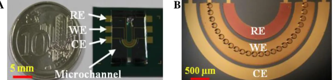
Fabrication of proposed bio-inspired electrochemical sensor was performed by means of MEMS technology. As shown in Figure 2(A), the sensor consisted of a polydimethylsiloxane (PDMS) microchannel and a silicon substrate on which three miniaturized base electrodes, i.e. reference electrode,
working electrode and counter electrode, were photolithographically patterned. A silicon dioxide layer was deposited on the silicon substrate in order to insulate the base electrodes. Figure 2(B) shows the close-up of arch-shaped electrode array with 24 micropillars functioning as 3D working electrodes. The height and diameter of each micropillar are 120 µm and 100 µm, respectively. The material of reference electrode is silver/silver chloride, while the working and counter electrodes are all made of gold.
Figure 2: (A) Photograph of bio-inspired MEMS-based electrochemical sensor after fabrication. (B) A close-up of 3D arch-shaped working electrode array under optical microscope, where ‘RE’, reference electrode; ‘WE’, working electrode; ‘CE’, counter electrode.
As for the electrochemical experiments, all the chemicals used were of analytical grade. Atomic absorption standard copper (Cu(II)) solution (1000 mg/L) and acetate buffer (pH 4.6) were purchased from Sigma-Aldrich (Singapore). Diluted solution was prepared with deionized water (18.2 MΩ·cm) collected from a Milli-Q system (Millipore, Singapore). All measurements with square wave anodic stripping voltammetry (SWASV) were carried out by using a CHI 600C electrochemical workstation (CH Instruments, USA).
RESULTS AND DISCUSSION
To optimize the electrochemical performance of proposed sensor, the influences of frequency, ampli-tude and step potential regarding SWASV were studied in the range of 20 to 70 Hz, 5 to 55 mV and 1 to 11 mV, respectively. As shown in Figure 3, the stripping peak for the solution containing 30 ppb Cu(II) gradually decreases from 20 to 50 Hz and remains stable when the frequency is higher than 50 Hz. For the amplitude, the peak current significantly increases from 5 to 45 mV and then reaches a plateau value. However, in terms of step potential, the magnitude of Cu(II) stripping peak initially rises up from 1 to 5 mV and then quickly reduces once the step potential crosses 5 mV.
Figure 3: Influences of (A) frequency, (B) amplitude and (C) step potential on the stripping peak of 30 ppb Cu(II). Measurement parameters: deposition potential of -0.2 V and deposition time of 30 s.
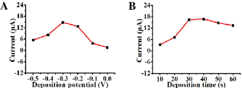
The effects of deposition potential as well as deposition time were also investigated to select appro-priate parameters. As shown in Figure 4, a considerable increase of Cu(II) stripping peak is observed when the deposition potential varies from 0 to -0.3 V, after which peak current is distinctly dropped. For the deposition time, the peak current ascends up to 40 s, whereas prolonged deposition time does not con-tribute to the enhancement of stripping peak. Under the optimal conditions, a series of SWASV were recorded by increasing the concentration of Cu(II) from 10 ppb to 110 ppb, as shown in Figure 5(A). The corresponding calibration curve is depicted in Figure 5(B). High sensitivity of 32 nA/ppb and low detec-tion limit of 0.4 ppb were achieved under short deposidetec-tion time of 40 s, demonstrating favorable capabil-ity of proposed bio-inspired electrochemical sensor for on-site/in-situ copper detection.
A
B
Figure 4: Influences of (A) deposition potential and (B) deposition time on the stripping peak of 30 ppb Cu(II). Measurement parameters: deposition time of 30 s for (A), deposition potential of -0.3 V for (B), frequency of 20 Hz, amplitude of 45 mV and step potential of 5 mV.
Figure 5: (A) Square wave stripping voltammograms for increasing concentration of Cu(II) (from 10 to 110 ppb) under optimized conditions and (B) corresponding calibration curve. Measurement parameters: deposition potential of -0.3 V, deposition time of 40 s, frequency of 20 Hz, amplitude of 45 mV and step potential of 5 mV.
CONCLUSION
A bio-inspired MEMS-based electrochemical sensor is presented. The performance of proposed sen-sor regarding copper detection has been comprehensively investigated by using SWASV. Under the con-dition of optimum experimental parameters, well-defined and legible stripping peaks with good linearity are obtained. High sensitivity of 32 nA/ppb and low detection limit of 0.4 ppb are achieved.
ACKNOWLEDGEMENTS
This research was supported by the Singapore Ministry of Education and the Singapore National Re-search Foundation through the Singapore MIT Alliance for ReRe-search and Technology's Center for Envi-ronmental Sensing and Modeling interdisciplinary research program.
REFERENCES
[1] F. Rueda-Holgado, E. Bernalte, M.R. Palomo-Marin, L. Calvo-Blazquez, F. Cereceda-Balic, and E. Pinilla-Gil, “Miniaturized Voltammetric Stripping on Screen Printed Gold Electrodes for Field De-termination of Copper in Atmospheric Deposition,” Talanta, 101, 435-439, 2012.
[2] E.S. Almeida, E.M. Richter, and R.A.A. Munoz, “On-Site Fuel Electroanalysis: Determination of Lead, Copper and Mercury in Fuel Bioethanol by Anodic Stripping Voltammetry Using Screen-Printed Gold Electrodes,” Anal. Chim. Acta, 837, 38-43, 2014.
[3] H. Wan, Q.Y. Sun, H.B. Li, F. Sun, N. Hu, and P. Wang, “Screen-Printed Gold Electrode with Gold Nanoparticles Modification for Simultaneous Electrochemical Determination of Lead and Copper,”
Sens. Actuators B, 209, 336-342, 2015.
[4] E. Zeiske, B. Theisen, and S.H. Gruber, “Functional Morphology of the Olfactory Organ of Two Carcharhinid Shark Species,” Can. J. Zool., 65, 2406-2412, 1987.
[5] V. Schluessel, M.B. Bennett, H. Bleckmann, and S.P. Collin, “The Role of Olfaction Throughout Juvenile Development: Functional Adaptations in Elasmobranchs,” J. Morphol., 271, 451-461, 2010.
CONTACT
* J.M. Miao; phone: +65-6790-6038; [email protected]
![Figure 1: (A) Cross-sectional SEM image (permission obtained from ref. [4]) of shark’s olfactory organ to show abundant layers of olfactory lamellae (ol), where ‘ic’, inlet chamber; ‘r’, raphe](https://thumb-eu.123doks.com/thumbv2/123doknet/14493668.526387/2.918.249.687.770.910/figure-sectional-permission-obtained-olfactory-abundant-olfactory-lamellae.webp)