HAL Id: inserm-00624805
https://www.hal.inserm.fr/inserm-00624805
Submitted on 19 Sep 2011
HAL is a multi-disciplinary open access
archive for the deposit and dissemination of
sci-entific research documents, whether they are
pub-lished or not. The documents may come from
teaching and research institutions in France or
abroad, or from public or private research centers.
L’archive ouverte pluridisciplinaire HAL, est
destinée au dépôt et à la diffusion de documents
scientifiques de niveau recherche, publiés ou non,
émanant des établissements d’enseignement et de
recherche français ou étrangers, des laboratoires
publics ou privés.
ribonucleoprotein L as a novel component of
SLM/Sam68 Nuclear Bodies.
Prabhakar Rajan, Caroline Dalgliesh, Cyril F Bourgeois, Monika Heiner,
Kaveh Emami, Emma Clark, Albrecht Bindereif, James Stevenin, Craig
Robson, Hing Leung, et al.
To cite this version:
Prabhakar Rajan, Caroline Dalgliesh, Cyril F Bourgeois, Monika Heiner, Kaveh Emami, et al..
Proteomic identification of heterogeneous nuclear ribonucleoprotein L as a novel component of
SLM/Sam68 Nuclear Bodies.. BMC Cell Biology, BioMed Central, 2009, 10 (1), pp.82.
�10.1186/1471-2121-10-82�. �inserm-00624805�
Open Access
Research article
Proteomic identification of heterogeneous nuclear
ribonucleoprotein L as a novel component of SLM/Sam68 Nuclear
Bodies
Prabhakar Rajan
†1,2, Caroline Dalgliesh
†1, Cyril F Bourgeois
3,4,5,6,
Monika Heiner
7, Kaveh Emami
8, Emma L Clark
9, Albrecht Bindereif
7,
James Stevenin
3,4,5,6, Craig N Robson
9, Hing Y Leung*
2and David J Elliott*
1 Address: 1Institute of Human Genetics, Newcastle University, Central Parkway, Newcastle-upon-Tyne, NE1 3BZ, UK, 2Beatson Institute for CancerResearch, Garscube Estate, Switchback Road, Glasgow, G61 1BD, UK, 3IGBMC Department of Functional Genomics, Illkirch, F-67400, France, 4INSERM U596, Illkirch, F-67400, France, 5CNRS UMR 7104, Illkirch F-67400, France, 6University of Strasbourg, Strasbourg, F-67000, France, 7Institute of Biochemistry, Department of Biology and Chemistry, Justus-Liebig-University of Giessen, Heinrich-Buff-Ring 58, D-35392 Giessen,
Germany, 8North East Proteome Analysis Facility, Devonshire Building, Newcastle University, Devonshire Terrace, Newcastle-upon-Tyne, NE1
7RU, UK and 9Northern Institute for Cancer Research, Newcastle University, Framlington Place, Newcastle-upon-Tyne, NE2 4HH, UK
Email: Prabhakar Rajan - p.rajan@beatson.gla.ac.uk; Caroline Dalgliesh - caroline.dalgliesh@ncl.ac.uk; Cyril F Bourgeois - cyril@igbmc.fr; Monika Heiner - monika.heiner@chemie.bio.uni-giessen.de; Kaveh Emami - kaveh.emami@ncl.ac.uk; Emma L Clark - e.clark@ncl.ac.uk; Albrecht Bindereif - albrecht.bindereif@chemie.bio.uni-giessen.de; James Stevenin - stevenin@igbmc.fr;
Craig N Robson - c.n.robson@ncl.ac.uk; Hing Y Leung* - h.leung@beatson.gla.ac.uk; David J Elliott* - David.Elliott@ncl.ac.uk * Corresponding authors †Equal contributors
Abstract
Background: Active pre-mRNA splicing occurs co-transcriptionally, and takes place throughout
the nucleoplasm of eukaryotic cells. Splicing decisions are controlled by networks of nuclear RNA-binding proteins and their target sequences, sometimes in response to signalling pathways. Sam68 (Src-associated in mitosis 68 kDa) is the prototypic member of the STAR (Signal Transduction and Activation of RNA) family of RNA-binding proteins, which regulate splicing in response to signalling cascades. Nuclear Sam68 protein is concentrated within subnuclear organelles called SLM/Sam68 Nuclear Bodies (SNBs), which also contain some other splicing regulators, signalling components and nucleic acids.
Results: We used proteomics to search for the major interacting protein partners of nuclear
Sam68. In addition to Sam68 itself and known Sam68-associated proteins (heterogeneous nuclear ribonucleoproteins hnRNP A1, A2/B1 and G), we identified hnRNP L as a novel Sam68-interacting protein partner. hnRNP L protein was predominantly present within small nuclear protein complexes approximating to the expected size of monomers and dimers, and was quantitatively associated with nucleic acids. hnRNP L spatially co-localised with Sam68 as a novel component of SNBs and was also observed within the general nucleoplasm. Localisation within SNBs was highly specific to hnRNP L and was not shared by the closely-related hnRNP LL protein, nor any of the other Sam68-interacting proteins we identified by proteomics. The interaction between Sam68 and hnRNP L proteins was observed in a cell line which exhibits low frequency of SNBs suggesting that this association also takes place outside SNBs. Although ectopic expression of hnRNP L and Sam68
Published: 13 November 2009
BMC Cell Biology 2009, 10:82 doi:10.1186/1471-2121-10-82
Received: 9 July 2009 Accepted: 13 November 2009 This article is available from: http://www.biomedcentral.com/1471-2121/10/82
© 2009 Rajan et al; licensee BioMed Central Ltd.
This is an Open Access article distributed under the terms of the Creative Commons Attribution License (http://creativecommons.org/licenses/by/2.0), which permits unrestricted use, distribution, and reproduction in any medium, provided the original work is properly cited.
proteins independently affected splicing of CD44 variable exon v5 and TJP1 exon 20 minigenes, these proteins did not, however, co-operate with each other in splicing regulation of these target exons.
Conclusion: Here we identify hnRNP L as a novel SNB component. We show that, compared with
other identified Sam68-associated hnRNP proteins and hnRNP LL, this co-localisation within SNBs is specific to hnRNP L. Our data suggest that the novel Sam68-hnRNP L protein interaction may have a distinct role within SNBs.
Background
Alternative splicing is regulated in part by a network of sig-nalling pathways which respond to extracellular stimuli [1]. One molecule with a key role linking signalling and splicing is Sam68 (Src-associated in mitosis 68 kDa). Sam68 is the prototypic member of the STAR (Signal Transduction and Activation of RNA) family of RNA-binding proteins [2]. Sam68-dependent splicing events impact upon important cellular decisions such as choices between cell survival and cell death [3]. Sam68 has also been recently shown to play an important role in neuro-genesis through splicing regulation of specific pre-mRNAs important for neural development [4]. Homozygous null Sam68 mice have pleiotropic defects in bone morphogen-esis, spermatogenesis and motor coordination suggesting widespread anatomical functions for the encoded protein [5,6].
Sam68 has been reported to be associated with a number of different proteins involved in RNA processing, tran-scription, and cell signalling. Sam68 induces ERK (extra-cellular signal-regulated kinase)-mediated inclusion of
CD44 variable exon v5 in response to Ras activation [7].
Activation of CD44 exon v5 splicing by Sam68 involves interactions with U2AF65 to facilitate v5 exon definition [8]. Sam68 also interacts with the splicing repressor hnRNP A1 [3] and nuclear transcriptional regulators [9,10]. The amino acid sequence of Sam68 protein con-tains several consensus motifs that mediate protein-pro-tein and proprotein-pro-tein-RNA interactions in response to different stimuli [2]. In most somatic cells, Sam68 protein is exclu-sively nuclear, but Sam68 can also interact with cytoplas-mic signalling molecules [2]. Sam68 protein has also been observed in the cytoplasm of secondary spermatocytes, where it is associated with polysomes and is involved in translational regulation [11,12].
In cancer cells, Sam68 protein exhibits a general nucleo-plasmic distribution but is also concentrated within sub-nuclear structures called SLM/Sam68 Nuclear Bodies (SNBs) [13]. Although the exact function of SNBs is unknown, they have been shown to contain some other splicing regulators, signalling components and nucleic acids. Although Sam68 has a number of reported interact-ing protein partners, its major associated proteins are not
yet known. In this study, we searched by proteomics for the major interacting protein partners of nuclear Sam68. Our data reveal hnRNP (heterogeneous nuclear ribonu-cleoprotein) L as a novel Sam68-associated nuclear pro-tein and a novel component of SNBs.
Results
hnRNP L is a novel Sam68-interacting protein
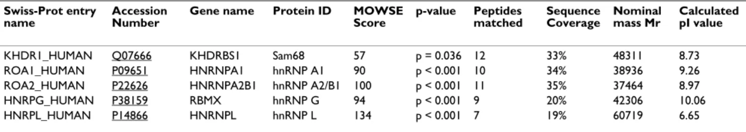
Sam68 protein is expressed at high levels in the nuclei of prostate cancer cells [10,14]. We immunoprecipitated endogenous Sam68 protein in nuclear extracts prepared from LNCaP cells. Western analysis confirmed that Sam68 protein was efficiently immunoprecipitated by rabbit antisera specific for Sam68, but not by normal rabbit IgG (Additional File 1). Further analysis of the immunopre-cipitates using SDS-PAGE followed by Coomassie staining revealed a number of immunoprecipitated proteins, including a protein of ~68 kDa and a protein co-migrating with IgG heavy chain (Figure 1A; compare lanes 1 and 4), as well as a number of other distinct proteins. Peptide mass fingerprinting by MALDI-TOF (Matrix-Assisted Laser Desorption/Ionization Time Of Flight) mass spectrometry was carried out on each of the immunoprecipitated pro-teins. The Mascot search engine was used to match pep-tides with statistically significant MOWSE (Molecular Weight Search) scores mapping across full-length protein sequences obtained from the Swiss-Prot database (Figure 1B to 1E and Table 1). We identified the ~68 kDa band as Sam68 (Figure 1B), and other co-immunoprecipitated proteins as the known Sam68-interacting proteins hnRNP A1 [3] (Figure 1C), hnRNP G [15] (Figure 1D), and hnRNP A2/B1 [16] (Figure 1C).
In addition to the three previously known Sam68-associ-ated proteins, we also identified hnRNP L as a novel potential Sam68-interacting protein partner (Figure 1E). By Coomassie staining, the levels of co-precipitated hnRNP A1 and hnRNP G were similar to the level of immunoprecipitated Sam68, but there was considerably less co-immunoprecipitated hnRNP L (Figure 1A, lane 1). Hence, to independently confirm the novel Sam68-hnRNP L interaction, both Sam68 and Sam68-hnRNP L proteins were separately immunoprecipitated in LNCaP cell nuclear extracts using their cognate antibodies prior to Western analysis (Figure 2A, left and right panels). hnRNP
Proteomic analysis identifies hnRNP L as a major nuclear Sam68-interacting protein
Figure 1
Proteomic analysis identifies hnRNP L as a major nuclear Sam68-interacting protein. (A) LNCaP cell nuclear
extracts were subjected to immunoprecipitation (IP) using anti-Sam68 rabbit antisera. IP was carried out in the presence or absence (+/-) of antisera to Sam68, with or without (+/-) Dynabeads Protein A, and either with or without (+/-) prior cross-linking of the antisera to the Dynabeads Protein A. Immunoprecipitates were resolved by SDS-PAGE, and six bands were excised from the Coomassie-stained gel for identification by peptide mass fingerprinting, of which 4 were positively identified. (B to E) MASCOT search results for Sam68-interacting proteins: Swiss-Prot entry names KHDR1_HUMAN (B: Sam68), ROA1_HUMAN (C: hnRNP A1), ROA2_HUMAN (C: hnRNP A2/B1), HNRPG_HUMAN (D: hnRNP G), and
HNRPL_HUMAN (E: hnRNP L). Histograms show the MOWSE score distributions for identified peptides, with matched pep-tide sequences shown in bold red. The band corresponding to histogram (C) contained a mixture of peppep-tides matching to Swiss-Prot entry names ROA1_HUMAN and ROA2_HUMAN.
1 2 3 4 130kDa 100kDa 70kDa 35kDa unidentifiable Sam68
IgG heavy chain hnRNPL hnRNPG hnRNPA2/B1 hnRNPA1 40kDa 50kDa (A) Sam68 Ab + + - + Dynabeads + + + -Cross-linked + - - -IP: Sam68 unidentifiable 1 MQRRDDPAAR MSRSSGRSGS MDPSGAHPSV RQTPSRQPPL PHRSRGGGGG
51 SRGGARASPA TQPPPLLPPS ATGPDATVGG PAPTPLLPPS ATASVKMEPE 101 NKYLPELMAE KDSLDPSFTH AMQLLTAEIE KIQKGDSKKD DEENYLDLFS
151 HKNMKLKERV LIPVKQYPKF NFVGKILGPQ GNTIKRLQEE TGAKISVLGK
201 GSMRDKAKEE ELRKGGDPKY AHLNMDLHVF IEVFGPPCEA YALMAHAMEE
251 VKKFLVPDMM DDICQEQFLE LSYLNGVPEP SRGRGVPVRG RGAAPPPPPV 301 PRGRGVGPPR GALVRGTPVR GAITRGATVT RGVPPPPTVR GAPAPRARTA 351 GIQRIPLPPP PAPETYEEYG YDDTYAEQSY EGYEGYYSQS QGDSEYYDYG 401 HGEVQDSYEA YGQDDWNGTR PSLKAPPARP VKGAYREHPY GRY
1 MSKSESPKEP EQLRKLFIGG LSFETTDESL RSHFEQWGTL TDCVVMRDPN
51 TKRSRGFGFV TYATVEEVDA AMNARPHKVD GRVVEPKRAV SREDSQRPGA
101 HLTVKKIFVG GIKEDTEEHH LRDYFEQYGK IEVIEIMTDR GSGKKRGFAF
151 VTFDDHDSVD KIVIQKYHTV NGHNCEVRKA LSKQEMASAS SSQRGRSGSG
201 NFGGGRGGGF GGNDNFGRGG NFSGRGGFGG SRGGGGYGGS GDGYNGFGND 251 GGYGGGGPGY SGGSRGYGSG GQGYGNQGSG YGGSGSYDSY NNGGGRGFGG 301 GSGSNFGGGG SYNDFGNYNN QSSNFGPMKG GNFGGRSSGP YGGGGQYFAK
351 PRNQGGYGGS SSSSSYGSGR RF
1 MEKTLETVPL ERKKREKEQF RKLFIGGLSF ETTEESLRNY YEQWGKLTDC
51 VVMRDPASKR SRGFGFVTFS SMAEVDAAMA ARPHSIDGRV VEPKRAVARE
101 ESGKPGAHVT VKKLFVGGIKEDTEEHHLRD YFEEYGKIDT IEIITDRQSG
151 KKRGFGFVTF DDHDPVDKIV LQKYHTINGH NAEVRKALSR QEMQEVQSSR
201 SGRGGNFGFG DSRGGGGNFG PGPGSNFRGG SDGYGSGRGF GDGYNGYGGG
251 PGGGNFGGSP GYGGGRGGYG GGGPGYGNQG GGYGGGYDNY GGGNYGSGNY 301 NDFGNYNQQP SNYGPMKSGN FGGSRNMGGP YGGGNYGPGG SGGSGGYGGR
351 SRY
(B)
KHDR1_HUMAN ROA1_HUMAN
ROA2_HUMAN
1 MVEADRPGKL FIGGLNTETN EKALEAVFGK YGRIVEVLLM KDRETNKSRG
51 FAFVTFESPA DAKDAARDMN GKSLDGKAIK VEQATKPSFE SGRRGPPPPP
101 RSRGPPRGLR GGRGGSGGTR GPPSRGGHMD DGGYSMNFNM SSSRGPLPVK 151 RGPPPRSGGP PPKRSAPSGP VRSSSGMGGR APVSRGRDSY GGPPRREPLP
201 SRRDVYLSPR DDGYSTKDSY SSRDYPSSRD TRDYAPPPRD YTYRDYGHSS
251 SRDDYPSRGY SDRDGYGRDR DYSDHPSGGS YRDSYESYGN SRSAPPTRGP
301 PPSYGGSSRY DDYSSSRDGY GGSRDSYSSS RSDLYSSGRD RVGRQERGLP
351 PSMERGYPPP RDSYSSSSRG APRGGGRGGS RSDRGGGRSR Y
HNRPG_HUMAN
(E)
1 MVKMAAAGGG GGGGRYYGGG SEGGRAPKRL KTDNAGDQHG GGGGGGGGAG
51 AAGGGGGGEN YDDPHKTPAS PVVHIRGLID GVVEADLVEA LQEFGPISYV 101 VVMPKKRQAL VEFEDVLGAC NAVNYAADNQ IYIAGHPAFV NYSTSQKISR 151 PGDSDDSRSV NSVLLFTILN PIYSITTDVL YTICNPCGPV QRIVIFRKNG 201 VQAMVEFDSV QSAQRAKASL NGADIYSGCC TLKIEYAKPT RLNVFKNDQD
251 TWDYTNPNLS GQGDPGSNPN KRQRQPPLLG DHPAEYGGPH GGYHSHYHDE 301 GYGPPPPHYE GRRMGPPVGG HRRGPSRYGP QYGHPPPPPP PPEYGPHADS
351 PVLMVYGLDQ SKMNGDRVFN VFCLYGNVEK VKFMKSKPGA AMVEMADGYA
401 VDRAITHLNN NFMFGQKLNV CVSKQPAIMP GQSYGLEDGS CSYKDFSESR
451 NNRFSTPEQA AKNRIQHPSN VLHFFNAPLE VTEENFFEIC DELGVKRPSS 501 VKVFSGKSER SSSGLLEWES KSDALETLGF LNHYQMKNPN GPYPYTLKLC
551 FSTAQHAS
HNRPL_HUMAN
(D)
L protein was efficiently immunoprecipitated by a mono-clonal antibody specific for hnRNP L, and also co-immu-noprecipitated by the antisera to Sam68 (Figure 2A, left panel). Likewise, Sam68 protein was efficiently immuno-precipitated by its cognate antisera, and also co-immu-noreprecipitated by the anti-hnRNP L antibody (Figure 2A, right panel). Similar to previous reports [17], we observed hnRNP L to migrate as a doublet, and both of these isoforms interacted with Sam68 (Figure 2, left panel). No or very weak immunoprecipitation was observed in the absence of either antibodies (Figure 2A, both panels, lane 4).
To further verify the novel Sam68-hnRNP L detected inter-action, HEK293 cells were transiently transfected with expression constructs encoding FLAG-tagged Sam68 and GST-tagged hnRNP L proteins. Anti-FLAG M2 agarose was used to immunoprecipitate FLAG-tagged proteins from HEK293 cells, and co-immunoprecipitated GST fusion proteins were detected using antisera specific to GST. Con-sistent with the observed interactions of endogenous pro-teins, hnRNP L-GST was efficiently co-immunoprecipitated with Sam68-FLAG (Figure 2B, com-pare lanes 1 and 3), but could not be co-immunoprecipi-tated in cells transfected with an empty FLAG vector (Figure 2B, compare lanes 2 and 4).
Note that all immunoprecipitations of both endogenous (Figure 2A) and FLAG-tagged proteins (Figure 2B) were carried out in the presence of Benzonase nuclease to ensure that the detected associations were mediated by protein-protein interactions and not bridged by nucleic acids.
Sam68 and hnRNP L are both present in small protein complexes which are quantitatively associated with nucleic acids
Sam68 is quantitatively associated with small nuclear pro-tein complexes of a size compatible with either mono-mers or dimono-mers [18]. In order to investigate and compare the size ranges of any endogenous nuclear complexes con-taining hnRNP L protein, we analysed velocity gradient fractions of HEK293 cell nuclear extracts prepared either
without pretreatment or after prior micrococcal nuclease (MNase) treatment to eliminate any nucleic acid depend-ent complexes (Figures 2C and [18]). Similarly to previ-ously published data for Sam68, following MNase treatment, hnRNP L (monomeric molecular weight ~70 kDa) protein was mainly present in small stable protein complexes (of less than 158 kDa) corresponding to the expected size of monomeric or dimeric molecular com-plexes (Figure 2C, upper panel). Also like Sam68, hnRNP L protein was quantitatively associated with nucleic acids since it migrated with much larger complexes without prior MNase digestion (Figure 2C, lower panel).
hnRNP L uniquely co-localises with Sam68 as a novel component of SNBs
Within the nuclei of cancer cells, Sam68 protein exhibits a general nucleoplasmic distribution but is also concen-trated within distinct peri-nucleolar structures called SNBs [13]. We compared the localization of Sam68 and hnRNP L proteins in cancer cell lines using indirect immunofluo-rescence (representative images are shown in Figure 3A and 3B, and Additional File 2). Confocal microscopy demonstrated that the endogenous Sam68 and hnRNP L proteins directly overlapped within SNBs in 100% of all cells examined. In addition to SNBs, further large pools of hnRNP L and Sam68 proteins were seen in the general nucleoplasm, and in the case of hnRNP L, towards the nuclear periphery in Saos-2 cells (the position of repre-sentative SNBs are indicated by arrows in Figures 3A, B and Additional File 2: note also a large population of both Sam68 and hnRNP L proteins outside SNBs). In parallel experiments, using the same combination of secondary antisera, no co-localisation was observed between Sam68 protein (identified using specific rabbit antisera) and SC35-containing splicing speckles (identified using a spe-cific mouse monoclonal antibody) (Figure 3C). The distri-bution of Sam68 and hnRNP L proteins was examined in three different cell lines (LNCaP, Saos-2 and HeLa) and found to be very similar suggesting that the hnRNP L asso-ciation with SNBs may be ubiquitous (Figure 3A, B, and Additional File 2). Furthermore, the other identified Sam68-associated proteins (hnRNP A1, A2/B1, and G) identified in our proteomic screen exhibited diffuse
sub-Table 1: Identities of Sam68-interacting proteins. Swiss-Prot entry
name
Accession Number
Gene name Protein ID MOWSE
Score p-value Peptides matched Sequence Coverage Nominal mass Mr Calculated pI value KHDR1_HUMAN Q07666 KHDRBS1 Sam68 57 p = 0.036 12 33% 48311 8.73 ROA1_HUMAN P09651 HNRNPA1 hnRNP A1 90 p < 0.001 10 34% 38936 9.26
ROA2_HUMAN P22626 HNRNPA2B1 hnRNP A2/B1 100 p < 0.001 11 35% 37464 8.97
HNRPG_HUMAN P38159 RBMX hnRNP G 94 p < 0.001 9 20% 42306 10.06
HNRPL_HUMAN P14866 HNRNPL hnRNP L 134 p < 0.001 7 19% 60719 6.65
Swiss-Prot names and primary accession numbers for proteins identified by Mascot searches. p-values were calculated using the MOWSE algorithm. Nominal mass (Mr) and isoelectric point (pI) were calculated using the integer mass of the most abundant isotope of each constituent element and the individual ionizing groups in each peptide, respectively.
Sam68 and hnRNP L proteins associate via protein-protein interactions and are not bridged by nucleic acids
Figure 2
Sam68 and hnRNP L proteins associate via protein-protein interactions and are not bridged by nucleic acids.
(A) LNCaP cell nuclear extracts were subjected to IP using either Sam68 antisera or an hnRNP L monoclonal anti-body. Recovered material was subjected to Western analysis with antibodies specific to hnRNP L (left panel) or Sam68 (right panel). (B) HEK293 cells were transfected with expression vectors for FLAG-Sam68 or FLAG alone, and hnRNP L-GST, and subjected to IP using anti-FLAG M2 agarose. Recovered material was subjected to Western analysis with antisera to GST. (C) HEK293 cell nuclear extracts were fractionated on sucrose gradients before (upper panel) or after (lower panel) treatment with MNase to digest nucleic acids. Fraction 20 (containing low molecular weight material) was taken from the top of the gra-dient, and fraction 1 (containing high molecular weight material) was taken from the bottom. Pelleted material is indicated as P. The migration of individual proteins in each fraction was monitored by SDS-PAGE and Western analysis. The mobility of size markers on the gradients is shown.
Extract + + + + FLAG-Sam68 + - + -Empty FLAG - + - + hnRNP L-GST + + + + FLAG M2 agarose - - + + Extract + + + + -Sam68 Ab - + - - -hnRNP L Ab - - + - + Dynabeads - + + + + Extract + + + + -hnRNP L Ab - + - - -Sam68 Ab - - + - + Dynabeads - + + + + hnRNPL
(A)
Sam68 83 kDa 64 kDa hnRNPL 1 2 3 4 5 1 2 3 4 5 GST-hnRNPL 95 kDa IP: Sam68/hnRNP L IB: hnRNP L 6 % Input IP: hnRNP L/Sam68 IB: Sam68 6 % Input IP: FLAG IB: GST 100 % Input(B)
1 2 3 4(C)
670,000 440,000 158,000 44,000 670,000 440,000 158,000 44,000 hnRNP L hnRNP L P 1 2 3 4 5 6 7 8 9 10 11 12 13 14 15 16 17 18 19 20 P 1 2 3 4 5 6 7 8 9 10 11 12 13 14 15 16 17 18 19 20nuclear distributions and did not localise within SNBs (Additional File 2 and [19]).
Only a small number of molecules have been identified as SNB components, and each of these share a role in cell sig-nalling and/or RNA processing. hnRNP L has a closely-related homolog within the cell called hnRNP LL (hnRNP L-like), which also regulates signal-dependent splicing. We carried out further experiments to test if hnRNP LL protein would be also found within SNBs. Surprisingly, these experiments indicated that hnRNP LL protein exhib-its a nucleoplasmic distribution outside SNBs (Figure 3D and Additional File 2). The observed hnRNP LL distribu-tion was punctate and somewhat reminiscent to that of splicing speckles. We compared the localisation of hnRNP LL protein with SC35, but found that the speckled distri-bution of hnRNP LL is in fact distinct from the SC35-con-taining splicing speckles (Figure 3E).
To verify these localization findings, HeLa cells were tran-siently transfected with expression vectors encoding hnRNP L-GST, hnRNP LL-GST, or GST alone. We then detected the localisation of the ectopically expressed pro-teins using antisera to detect the GST tag, and compared this distribution with that of endogenous Sam68 protein (Figure 4). Consistent with the data described above, hnRNP L-GST but not hnRNP LL-GST, was found within SNBs (Figure 4A and 4B). The GST moiety had no effect on protein localization, since GST alone was detected both within the nuclei and cytoplasm of transfected cells, and not within SNBs (Figure 4C).
Sam68 and hnRNP L proteins associate in a cell line which exhibits a low frequency of SNBs
The frequency of cells in a population containing SNBs is cell line-dependent. To test whether the association of Sam68 and hnRNP L was dependent on the presence of SNBs, we performed side-by-side immunoprecipitations in whole cell extracts obtained from LNCaP and NIH3T3 cells (only ~5.5% of NIH3T3 cells have SNBs) [13]. Sam68 protein was efficiently immunoprecipitated by its cognate antisera from both these cell lines (Figure 5A and 5B, left panels). Consistent with the interaction of Sam68 and hnRNP L also occurring outside of SNBs, virtually equal levels of hnRNP L protein was co-immunoprecipi-tated by the antisera to Sam68 from NIH3T3 cells as LNCaP cells (Figure 5A and 5B, right panels). Notice that neither Sam68 nor hnRNP L proteins were immunopre-cipitated by normal rabbit IgG or beads alone. All immu-noprecipitations were carried out in the presence of Benzonase nuclease to confirm that the detected associa-tions were mediated by protein-protein interacassocia-tions and not bridged by nucleic acids.
The Sam68-hnRNP L protein interaction does not significantly impact on splicing regulation of two known target exons
The above experiments indicate that Sam68 and hnRNP L proteins interact and are both present within SNBs. Although SNBs contain some splicing regulators and RNA, they do not detectably contain key spliceosome components [13]. Hence SNBs are not thought to be sites for active pre-mRNA splicing, which takes place in the general nucleoplasm [20]. Both Sam68 and hnRNP L pro-teins are independently implicated in the regulation of distinct alternative splicing events during which exon inclusion can be either activated or repressed depending on the target transcript [4,21]. Although active splicing of Sam68 and hnRNP L target pre-mRNAs is unlikely to take place within SNBs, we also detected significant nucleo-plasmic populations of both Sam68 and hnRNP L pro-teins outside SNBs, and protein interactions between Sam68 and hnRNP L occurred equally well in a cell line which exhibits a low frequency of SNBs. These observa-tions raise the question as to whether the Sam68-hnRNP L interaction might have a co-operative effect on pre-mRNA splicing in the general nucleoplasm.
Since ectopically expressed Sam68 protein stimulates splicing inclusion of CD44 variable exon v5 [7], and TJP1 exon 20 inclusion is repressed by hnRNP L [21], we exam-ined the effect of both Sam68 and hnRNP L protein expression on these known target exons (Figure 6). We firstly monitored splicing in HEK293 cells transfected with a minigene containing CD44 variable exon v5 together with expression constructs encoding Sam68, hnRNP L or both (Figure 6A). Ectopic expression of hnRNP L protein stimulated exon v5 inclusion albeit with a lower potency than Sam68 (Figure 6B, compare lane 1 with lanes 2 and 3). We carried out similar experiments with a minigene containing TJP1 exon 20 (Figure 6C). Due to the low levels of expression of TJP1 exon 20, it was not easy to draw clear conclusions from this experiment, however ectopic expression of either hnRNP L or Sam68 proteins alone appeared to repress exon 20 inclusion (Fig-ure 6D, compare lane 1 with lanes 2 and 3). Together, these experiments indicate that both Sam68 and hnRNP L proteins can regulate the same pre-mRNA splicing events in the same direction, with hnRNP L activating CD44 exon v5 inclusion like Sam68, and Sam68 repressing TJP1 exon 20 inclusion like hnRNP L. Importantly in either case, however, co-expression of hnRNP L and Sam68 pro-teins did not have a significant additive effect on either
TJP1 exon 20 skipping or CD44 exon v5 inclusion (Figures
6B and 6D, compare lanes 2, 3 and 4). Hence it seems unlikely that the general nucleoplasmic populations of hnRNP L and Sam68 proteins co-operate in splicing con-trol of these shared target exons, despite their observed
Endogenous hnRNP L protein co-localises with Sam68 within SNBs
Figure 3
Endogenous hnRNP L protein co-localises with Sam68 within SNBs. Representative images of LNCaP and Saos-2
cells captured by confocal laser scanning microscopy using indirect immunofluorescence and antibodies specific to Sam68, hnRNP L, hnRNP LL and SC35. (A and B) Sam68 co-localises with hnRNP L to SNBs (arrowed), but not with SC35 to splicing speckles (C). (D) hnRNP LL exhibits a different subnuclear localisation to hnRNP L, which is again distinct from SC35-contain-ing splicSC35-contain-ing speckles (E), and does not co-localise to SNBs (arrowed). (Bar = 10 μm).
Sam68 hnRNP L Merge Sam68 hnRNP L Merge hnRNP LL SC35 Merge LNC aP Saos-2
(A)
(B)
Saos-2 hnRNP LL hnRNP L Merge Saos-2(D)
Sam68 SC35 Merge LNC aP(C)
(E)
interaction, localisations, and ability to regulate these tar-get exons by themselves.
Discussion
Although Sam68 has been associated with a number of proteins, our proteomics screen identified hnRNP A1,
hnRNP A2/B1, hnRNP G and hnRNP L as its four major nuclear interacting protein partners in LNCaP cells. The stoichiometry of the Sam68-associated proteins recovered by immunoprecipitation, their spatial localisations, and analysis of endogenous complexes using velocity gradient centrifugation analysis [18] are most consistent with a
An ectopically-expressed hnRNP L fusion protein localises within SNBs
Figure 4
An ectopically-expressed hnRNP L fusion protein localises within SNBs. Representative indirect
immunofluores-cence images of HeLa cells transfected with vectors encoding hnRNP L-GST (A), hnRNP LL-GST (B), or GST alone (C), and captured by confocal laser scanning microscopy using indirect immunofluorescence and antibodies to Sam68 and GST. (A) Direct co-localisation within SNBs of endogenous Sam68 and ectopically-expressed hnRNP L-GST fusion protein (arrowed). (B) No co-localisation within SNBs was observed between endogenous Sam68 and the ectopically-expressed hnRNP LL-GST fusion protein. (C) GST protein was generally distributed within the nucleus and cytoplasm and showed no subnuclear localisa-tion within SNBs, (Bar = 10 μm).
hnRNP L-GST Sam68 Merge GST Sam68 Merge
HeLa
HeLa
hnRNP LL-GST Sam68 MergeHeLa
(A)
(B)
(C)
number of distinct small Sam68-containing protein com-plexes (predominantly monomeric and dimeric-sized), rather than a single large complex containing each of the identified Sam68-interacting proteins. Like Sam68 and the Sam68-interactor hnRNP G, but not hnRNP A1, hnRNP L protein was quantitatively associated with nucleic acids since it migrated with much larger com-plexes without prior MNase digestion before velocity gra-dient centrifugation.
hnRNP L protein has previously been described in discrete perinucleolar structures [17], which we here now identify as SNBs. Although the exact functions of SNBs are not fully understood, they are known to contain other STAR proteins (SLM-1 and SLM-2) [13], the splicing factor YT521B [22], Scaffold Attachment Factors SAFB1 and SAFB2 [18] and the protein kinase BRK/Sik (Breast Tumour Kinase/Src-related Intestinal Kinase) [23]. SNBs also contain nucleic acids including RNA, and on heat-shock additionally recruit hnRNP A1 and other splicing factors including arginine-serine-rich (SR) proteins [24]. Like Sam68, hnRNP L is implicated in coupling signaling
and splicing: hnRNP L regulates signal-dependent splicing of CD45 exon v4 [25] and STREX (stress axis-regulated exon) [26]. Despite the spatial and physical associations of Sam68 and hnRNP L detected in our study, Sam68 and hnRNP L proteins did not either co-operate or antagonize each others' splicing activity on target exons. In contrast, the generally nucleoplasmic Sam68-associated protein hnRNP G potently inhibited Sam68-mediated splicing of
CD44 exon v5 ([27] and Additional File 3). Our protein
interaction analyses indicated that the interaction between Sam68 and hnRNP L is independent of nucleic acids. Using a directed yeast two-hybrid system, a direct protein-protein interaction has been demonstrated between the STAR-family protein rSLM-2 and hnRNP L [27], suggesting, by analogy, that the Sam68-hnRNP L interaction may also be direct. However, we were unable to detect such an interaction by directed yeast two hybrid analysis (unpublished observations).
Amongst all the Sam68-associated proteins we investi-gated in this study, the subnuclear distribution of hnRNP L protein is specifically within SNBs. Consistent with the
Sam68 and hnRNP L proteins associate in a cell line which exhibits a low frequency of SNBs
Figure 5
Sam68 and hnRNP L proteins associate in a cell line which exhibits a low frequency of SNBs. (A) LNCaP and (B)
NIH3T3 whole cell extracts were subjected to IP using either anti-Sam68 antisera or normal rabbit IgG. Recovered material was subjected to Western analysis with antibodies specific to Sam68 (left hand panels) or hnRNP L (right hand panels).
(A)
Sam68 72 kDa 1 2 3 4 1 2 3 4 72 kDaIgG heavy chain
hnRNP L Extract + + + + Sam68 Ab - + - - Rabbit IgG - - + - Dynabeads - + + IP: Sam68 IB: Sam68 3 % Input hnRNP L
(B)
1 2 3 4 72 kDa Extract + + + + Sam68 Ab - + - - Rabbit IgG - - + - Dynabeads - + + + IP: Sam68 IB: hnRNP L 3 % Input 72 kDa IgG heavy chain 1 2 3 4 Sam68 Extract + + + + Sam68 Ab - + - - Rabbit IgG - - + - Dynabeads - + + + IP: Sam68 IB: Sam68 3 % Input Extract + + + + Sam68 Ab - + - - Rabbit IgG - - + - Dynabeads - + + + IP: Sam68 IB: hnRNP L 3 % Input +The Sam68-hnRNP L protein interaction does not significantly impact on splicing regulation of two known target exons
Figure 6
The Sam68-hnRNP L protein interaction does not significantly impact on splicing regulation of two known tar-get exons. (A) Minigene pETv5 contains the CD44 variable exon v5 cloned downstream of a constitutively-active RSV
pro-moter. (C) Minigene pcDNA3-TJP1-WT contains TJP1 exon 20 downstream of a constitutively-active CMV propro-moter. (A and C) Splicing events are shown by broken lines, and arrows show the location of primers for RT-PCR. (B) HEK293 cells were transfected with the pETv5 minigene (150 ng), and expression vectors for hnRNP L-GST or GFP-Sam68 (500 ng). (D) HEK293 cells were transfected with pcDNA3-TJP1-WT (150 ng), and expression vectors for hnRNP L-GST or GFP-Sam68 (500 ng). (B and D) Gel images shown are representative of at least three independent experiments, from which densitometric assessment of RT-PCR product (lower panels) was performed to obtain means +/- standard error which are plotted in the bar chart. The lane marked M shows migration of the 1 Kb plus DNA ladder (Invitrogen).
(B)
100bp 200bp exon inclusion exon skipping Empty vector + - - -hnRNP L - + - + Sam68 - + + pETv5 1 2 3 4 M 0 0.2 0.4 0.6 0.8 1 1.2 1.4 1.6 Empty vector hnRNP L Sam68 hnRNP L + Sam68 R a ti o e xon in cl us ion /s k ip p in g(A)
RSVexon v5 insulin exon insulin exon CD44 0 0.02 0.04 0.06 0.08 0.1 0.12 0.14 0.16 0.18 0.2
Empty vector hnRNP L Sam68 hnRNP L + Sam68 R a tio ex on inclusion/sk ipping
(D)
200bp 500bp exon inclusion exon skipping Empty vector + - - -hnRNP L - + - + Sam68 - + + TJP1 1 2 3 4 M 300bp(C)
CMV exon 20 exon 21 exon 19 TJP1known nuclear functions of hnRNP L, SNBs have been suggested as potential nuclear sites for the regulation of RNA processing by signalling pathways. However, we found that the homologous hnRNP LL protein which is also implicated in signal-dependent CD45 splicing [28-30], did not localize to SNBs. Hence SNBs are not a default location for all nuclear RNA-binding proteins involved in signaling and RNA processing. hnRNP LL pro-tein lacks some proline- and glycine-rich regions present within the hnRNP L amino acid sequence, and by analogy with Sam68, it is possible that these motifs are required for interactions with signaling molecules and SNB locali-zation. The distinct nuclear localization we observe for hnRNP LL may suggest somewhat diverged functions and so provide some explanation for the evolutionary mainte-nance of both hnRNP L and LL proteins within cells.
Conclusion
We have identified hnRNP L as a novel Sam68-interacting protein partner and component of SNBs. Despite hnRNP L and Sam68 proteins also localizing to the general nucle-oplasm, where pre-mRNA splicing occurs, we did not observe a role for the novel Sam68-hnRNP L protein inter-action on splicing of known target exons for these RNA-binding proteins. The spatial localization of hnRNP L pro-tein within SNBs further implicates a role for these organelles in coupling signaling to RNA processing.
Methods
Cell Culture and DNA transfection
LNCaP cells were cultured in RPMI-1640 (PAA Laborato-ries) supplemented with 10% foetal calf serum (FCS) (PAA Laboratories). HEK293, HeLa, NIH3T3, and Saos-2, and cells were cultured in Dulbecco's Modified Eagle Media (D-MEM) with glutamax-1 (PAA Laboratories) with 10% FCS. Plasmid transfections were performed as previously described [18] using Genejammer (Stratagene) according to manufacturer's instructions.
Antibodies and plasmids
The following antibodies were used: normal rabbit IgG sc-2027 and anti-Sam68 sc-333 rabbit antisera (Santa Cruz Biotechnology); anti-hnRNP A1 9H10, anti-hnRNP L 4D11, anti-SC35 SC-35, and anti-β-actin AC15 mouse monoclonal antibodies (Sigma); anti-hnRNP A2/B1 DP3B2 mouse monoclonal antibody (Abcam); hnRNP LL rabbit antisera (Aviva Systems Biology); anti-GST (glutathione S-transferase) goat antisera (GE Health-care).
The following plasmids have been described previously: FLAG-Sam68 [10]; pGFP3-Sam68 [31]; pETv5 [32] (from Stefan Stamm, University of Kentucky, USA). The
follow-ing plasmids were made usfollow-ing standard clonfollow-ing proce-dures, the details of which are available on request: pcDNA-GST, pcDNA-hnRNP L-GST, and pcDNA-hnRNP LL-GST encode full-length GST, hnRNP L [33], and hnRNP LL with a C-terminal GST tag, respectively. pcDNA-hnRNP G encodes full-length hnRNP G. pcDNA3-TJP1-wWT contains exon 19, 20 and 21 of TJP1 with intervening intronic sequence.
Peptide mass fingerprinting and mass spectrometry (MS) Immunoprecipitated proteins were resolved on NuPAGE Novex 10% Bis-Tris gels (Invitrogen) and stained with Coomassie blue (GE Healthcare). Excised bands were digested with trypsin, and identified by peptide mass fin-gerprinting. Mass spectra were obtained using the Ultraflex II MALDI-TOF (matrix-assisted laser desorption/ ionization time-of-flight) mass spectrometer (Bruker Dal-tonics) in standard MALDI-TOF mass spectrometry (MS) mode. The peak lists were searched against the non-redun-dant Swiss-Prot protein sequence database using the Mas-cot [34] search engine version 2.2 (Matrix Science). Probability-based MOWSE (Molecular Weight Search) [35] scores greater than 55 were considered as significant (p < 0.05).
Immunoprecipitation
LNCaP cell nuclear extracts were obtained using the Cel-Lytic NuCLEAR Extraction Kit (Sigma) according to man-ufacturer's instructions. Benzonase nuclease (Sigma) was used at 100 units/ml where indicated. Immunoprecipita-tions were performed using Dynabeads Protein A (Invitro-gen) or anti-FLAG M2 agarose (Sigma) according to manufacturers' instructions. Where indicated, antibodies were cross-linked to Dynabeads Protein A according to manufacturer's instructions. Recovered material was resolved by SDS-PAGE and subjected to Western analysis as previously described [36].
Velocity gradient centrifugation
Sucrose gradients were run using HEK293 cell nuclear extract, pre-treated or not with micrococcal nuclease (MNase), and recovered material resolved by SDS-PAGE and subjected to Western analysis as previously described [18].
Indirect immunofluorescence microscopy
LNCaP, HeLa, and Saos-2 cells were grown and trans-fected on glass coverslips (VWR International), and stained with primary and secondary antibodies as previ-ously described [18]. All images were captured using the LSM510 (Zeiss) or SP2 MP (Leica) confocal microscopes and associated software.
Minigene splicing assays
HEK293 cells were grown in 6-well plates (Asahi Techno Glass), and transfected with DNA as detailed in figure leg-ends. RNA was extracted using TRIzol (Invitrogen) and RT-PCR was performed using the One-Step RT-PCR kit (Qiagen) as previously described [31]. Densitometric band quantification was performed as previously described [37]. All experiments shown are the mean of at least three independent experiments ± standard error.
Authors' contributions
PR conceived of the study, designed and performed exper-iments, analysed data and drafted the manuscript. CD designed and performed experiments. CB designed and performed experiments. MH contributed reagents and expertise. KE carried out the mass spectrometry. EC con-tributed reagents and expertise. AB concon-tributed reagents, expertise, and to the preparation of the manuscript. JS designed experiments, contributed reagents, expertise and to the preparation of the manuscript. CR contributed rea-gents, expertise and to the preparation of the manuscript. HYL conceived of the study, contributed expertise and to the preparation of the manuscript. DJE conceived of the study, contributed reagents and expertise, and drafted the manuscript. All authors read and approved the final man-uscript.
Additional material
Acknowledgements
We thank Colin Nixon, Rosie Morland (Beatson Institute) and Agata Rozanska (Newcastle University) for technical assistance, and Helen Arthur and Soulmaz Boroumand (Newcastle University) for assistance with pro-teomics. We are grateful to Sheila Graham (University of Glasgow) and Kim Moran-Jones (Beatson Institute) for antibodies, and Stefan Stamm for the CD44 variable exon v5 minigene. This work was supported by a Medical Research Council Clinical Research Training Fellowship (G0500482 to P.R.); Association for International Cancer Research project grant (06-705 to DJE and HYL); a Wellcome Trust project grant (WT080368 MA to DJE); a Royal Society Joint International Grant (to DJE and JS); and the European Commission-funded Network of Excellence EURASNET (AB and JS).
References
1. Lynch KW: Regulation of alternative splicing by signal
trans-duction pathways. Adv Exp Med Biol 2007, 623:161-174.
2. Lukong KE, Richard S: Sam68, the KH domain-containing
superSTAR. Biochim Biophys Acta 2003, 1653:73-86.
3. Paronetto MP, Achsel T, Massiello A, Chalfant CE, Sette C: The
RNA-binding protein Sam68 modulates the alternative splic-ing of Bcl-x. J Cell Biol 2007, 176:929-939.
4. Chawla G, Lin CH, Han A, Shiue L, Ares MJ, Black DL: Sam68
reg-ulates a set of alternatively spliced exons during neurogene-sis. Mol Cell Biol 2009, 29:201-213.
5. Lukong KE, Richard S: Motor coordination defects in mice
defi-cient for the Sam68 RNA-binding protein. Behav Brain Res
2008, 189:357-363.
6. Richard S, Torabi N, Franco GV, Tremblay GA, Chen T, Vogel G, Morel M, Cleroux P, Forget-Richard A, Komarova S, Tremblay ML, Li W, Li A, Gao YJ, Henderson JE: Ablation of the Sam68 RNA
binding protein protects mice from age-related bone loss.
PLoS Genet 2005, 1:e74.
7. Matter N, Herrlich P, Konig H: Signal-dependent regulation of
splicing via phosphorylation of Sam68. Nature 2002, 420:691-695.
8. Tisserant A, Konig H: Signal-Regulated Pre-mRNA Occupancy
by the General Splicing Factor U2AF. PLoS ONE 2008, 3:e1418.
9. Hong W, Resnick RJ, Rakowski C, Shalloway D, Taylor SJ, Blobel GA:
Physical and functional interaction between the transcrip-tional cofactor CBP and the KH domain protein Sam68. Mol
Cancer Res 2002, 1:48-55.
10. Rajan P, Gaughan L, Dalgliesh C, El-Sherif A, Robson CN, Leung HY, Elliott DJ: The RNA-binding and adaptor protein Sam68
mod-ulates signal-dependent splicing and transcriptional activity of the androgen receptor. J Pathol 2008, 215:67-77.
11. Paronetto MP, Zalfa F, Botti F, Geremia R, Bagni C, Sette C: The
nuclear RNA-binding protein Sam68 translocates to the
Additional file 1
Sam68 is efficiently immunoprecipitated by its cognate antisera.
LNCaP cell nuclear extracts were subjected to immunoprecipitation (IP) using anti-Sam68 rabbit antisera. IP was carried out in the presence or absence (+/-) of antisera to Sam68 or normal rabbit IgG (negative con-trol), with or without (+/-) Dynabeads Protein A, and either with or with-out (+/-) prior cross-linking of the antisera to Sam68 to the Dynabeads Protein A. Recovered material was subjected to Western analysis with the antisera to Sam68. Western analysis confirmed that Sam68 protein was efficiently immunoprecipitated by its cognate antisera, and that this was most efficient if the antisera were cross-linked to the Dynabeads Protein A prior to incubation with nuclear extracts (compare lanes 3 and 4). Sam68 protein was not pulled down by Dynabeads Protein A alone (lane 2). Click here for file
[http://www.biomedcentral.com/content/supplementary/1471-2121-10-82-S1.pdf]
Additional file 2
hnRNPs A1 or A2/B1 do not localise within SNBs. (A to D)
Represent-ative indirect immunofluorescence images of HeLa cells captured by con-focal laser scanning microscopy using antibodies to hnRNPs A1, A2/B1, hnRNP L, hnRNP LL, and Sam68. hnRNPs A1 (A) and A2/B2 (B) exhibit a diffuse nucleoplasmic distribution but do not co-localise with Sam68 to SNBs (arrowed). (C) Sam68 co-localises with hnRNP L to SNBs (arrowed). (D) hnRNP LL exhibits a different subnuclear localisa-tion to hnRNP L and does not co-localise to SNBs. (Bar = 10 m). Click here for file
[http://www.biomedcentral.com/content/supplementary/1471-2121-10-82-S2.pdf]
Additional file 3
hnRNP G represses Sam68-mediated CD44 variable exon v5 inclu-sion. (A) Minigene pETv5 contains the CD44 variable exon v5 cloned
downstream of a constitutively-active RSV promoter. Splicing events are shown by broken lines, and arrows show the location of primers for RT-PCR. (B) HEK293 cells were transfected with the pETv5 minigene (150 ng), and expression vectors for hnRNP G or GFP-Sam68 (500 ng). The gel images are representative of at least three independent experiments, from which densitometric assessment of RT-PCR product was performed to obtain means +/- standard error (shown in bar chart). The lane marked M shows the migration of the 1 Kb plus DNA ladder (Invitrogen). Ectopic expression of hnRNP G protein repressed CD44 variable exon v5 inclusion both in the presence and absence of ectopic Sam68 (compare lane 1 with lane 3, and lane 2 with lane 4).
Click here for file
[http://www.biomedcentral.com/content/supplementary/1471-2121-10-82-S3.pdf]
Publish with BioMed Central and every scientist can read your work free of charge "BioMed Central will be the most significant development for disseminating the results of biomedical researc h in our lifetime."
Sir Paul Nurse, Cancer Research UK
Your research papers will be:
available free of charge to the entire biomedical community peer reviewed and published immediately upon acceptance cited in PubMed and archived on PubMed Central yours — you keep the copyright
Submit your manuscript here: BioMedcentral
cytoplasm and associates with the polysomes in mouse sper-matocytes. Mol Biol Cell 2006, 17:14-24.
12. Paronetto MP, Messina V, Bianchi E, Barchi M, Vogel G, Moretti C, Palombi F, Stefanini M, Geremia R, Richard S, Sette C: Sam68
regu-lates translation of target mRNAs in male germ cells, neces-sary for mouse spermatogenesis. J Cell Biol 2009, 185:235-249.
13. Chen T, Boisvert FM, Bazett-Jones DP, Richard S: A role for the
GSG domain in localizing Sam68 to novel nuclear structures in cancer cell lines. Mol Biol Cell 1999, 10:3015-3033.
14. Busa R, Paronetto MP, Farini D, Pierantozzi E, Botti F, Angelini DF, Attisani F, Vespasiani G, Sette C: The RNA-binding protein
Sam68 contributes to proliferation and survival of human prostate cancer cells. Oncogene 2007, 26:4372-4382.
15. Ulke-Lemee A, Trinkle-Mulcahy L, Chaulk S, Bernstein NK, Morrice N, Glover M, Lamond AI, Moorhead GB: The nuclear PP1
inter-acting protein ZAP3 (ZAP) is a putative nucleoside kinase that complexes with SAM68, CIA, NF110/45, and HNRNP-G. Biochim Biophys Acta 2007, 1774:1339-1350.
16. Thornton JK: Protein-protein interaction screens: An
investi-gation into the interactions of Sam68 and ASPP1 in the tes-tis. In Ph.D.Thesis University of Newcastle upon Tyne, Institute of
Human Genetics; 2005.
17. Pinol-Roma S, Swanson MS, Gall JG, Dreyfuss G: A novel
heteroge-neous nuclear RNP protein with a unique distribution on nascent transcripts. J Cell Biol 1989, 109:2575-2587.
18. Sergeant KA, Bourgeois CF, Dalgliesh C, Venables JP, Stevenin J, Elli-ott DJ: Alternative RNA splicing complexes containing the
scaffold attachment factor SAFB2. J Cell Sci 2007, 120:309-319.
19. Soulard M, Barque JP, Della Valle V, Hernandez-Verdun D, Masson C, Danon F, Larsen CJ: A novel 43-kDa glycoprotein is detected in
the nucleus of mammalian cells by autoantibodies from dogs with autoimmune disorders. Exp Cell Res 1991, 193:59-71.
20. Zeng C, Kim E, Warren SL, Berget SM: Dynamic relocation of
transcription and splicing factors dependent upon transcrip-tional activity. EMBO J 1997, 16:1401-1412.
21. Hung LH, Heiner M, Hui J, Schreiner S, Benes V, Bindereif A: Diverse
roles of hnRNP L in mammalian mRNA processing: a com-bined microarray and RNAi analysis. RNA 2008, 14:284-296.
22. Hartmann AM, Nayler O, Schwaiger FW, Obermeier A, Stamm S:
The interaction and colocalization of Sam68 with the splic-ing-associated factor YT521-B in nuclear dots is regulated by the Src family kinase p59(fyn). Mol Biol Cell 1999, 10:3909-3926.
23. Derry JJ, Richard S, Valderrama Carvajal H, Ye X, Vasioukhin V, Cochrane AW, Chen T, Tyner AL: Sik (BRK) phosphorylates
Sam68 in the nucleus and negatively regulates its RNA bind-ing ability. Mol Cell Biol 2000, 20:6114-6126.
24. Denegri M, Moralli D, Rocchi M, Biggiogera M, Raimondi E, Cobianchi F, De Carli L, Riva S, Biamonti G: Human chromosomes 9, 12,
and 15 contain the nucleation sites of stress-induced nuclear bodies. Mol Biol Cell 2002, 13:2069-2079.
25. Rothrock CR, House AE, Lynch KW: HnRNP L represses exon
splicing via a regulated exonic splicing silencer. EMBO J 2005, 24:2792-2802.
26. Yu J, Hai Y, Liu G, Fang T, Kung SK, Xie J: The heterogeneous
nuclear ribonucleoprotein L is an essential component in the Ca++/calmodulin-dependent protein kinase IV-regulated alternative splicing through cytidine-adenosine repeats. J Biol
Chem 2008, 284:1505-1513.
27. Stoss O, Olbrich M, Hartmann AM, Konig H, Memmott J, Andreadis A, Stamm S: The STAR/GSG family protein rSLM-2 regulates
the selection of alternative splice sites. J Biol Chem 2001, 276:8665-8673.
28. Oberdoerffer S, Moita LF, Neems D, Freitas RP, Hacohen N, Rao A:
Regulation of CD45 Alternative Splicing by Heterogeneous Ribonucleoprotein, hnRNPLL. Science 2008, 321:686-691.
29. Topp JD, Jackson J, Melton AA, Lynch KW: A cell-based screen for
splicing regulators identifies hnRNP LL as a distinct signal-induced repressor of CD45 variable exon 4. RNA 2008, 14:2038-2049.
30. Wu Z, Jia X, de la Cruz L, Su XC, Marzolf B, Troisch P, Zak D, Ham-ilton A, Whittle B, Yu D, Sheahan D, Bertram E, Aderem A, Otting G, Goodnow CC, Hoyne GF: Memory T cell RNA rearrangement
programmed by heterogeneous nuclear ribonucleoprotein hnRNPLL. Immunity 2008, 29:863-875.
31. Venables JP, Dalgliesh C, Paronetto MP, Skitt L, Thornton JK, Saun-ders PT, Sette C, Jones KT, Elliott DJ: SIAH1 targets the
alterna-tive splicing factor T-STAR for degradation by the proteasome. Hum Mol Genet 2004, 13:1525-1534.
32. Konig H, Ponta H, Herrlich P: Coupling of signal transduction to
alternative pre-mRNA splicing by a composite splice regula-tor. EMBO J 1998, 17:2904-2913.
33. Hui J, Hung LH, Heiner M, Schreiner S, Neumuller N, Reither G, Haas SA, Bindereif A: Intronic CA-repeat and CA-rich elements: a
new class of regulators of mammalian alternative splicing.
EMBO J 2005, 24:1988-1998.
34. Perkins DN, Pappin DJ, Creasy DM, Cottrell JS: Probability-based
protein identification by searching sequence databases using mass spectrometry data. Electrophoresis 1999, 20:3551-3567.
35. Pappin DJ, Hojrup P, Bleasby AJ: Rapid identification of proteins
by peptide-mass fingerprinting. Curr Biol 1993, 3:327-332.
36. Logan IR, Sapountzi V, Gaughan L, Neal DE, Robson CN: Control of
human PIRH2 protein stability: involvement of TIP60 and the proteosome. J Biol Chem 2004, 279:11696-11704.
37. Venables JP, Bourgeois CF, Dalgliesh C, Kister L, Stevenin J, Elliott DJ:
Up-regulation of the ubiquitous alternative splicing factor Tra2beta causes inclusion of a germ cell-specific exon. Hum