HAL Id: hal-02341026
https://hal-amu.archives-ouvertes.fr/hal-02341026
Submitted on 31 Oct 2019HAL is a multi-disciplinary open access archive for the deposit and dissemination of sci-entific research documents, whether they are pub-lished or not. The documents may come from teaching and research institutions in France or abroad, or from public or private research centers.
L’archive ouverte pluridisciplinaire HAL, est destinée au dépôt et à la diffusion de documents scientifiques de niveau recherche, publiés ou non, émanant des établissements d’enseignement et de recherche français ou étrangers, des laboratoires publics ou privés.
In vivo TssA proximity labeling reveals temporal
interactions during Type VI secretion 1 biogenesis and
TagA, a protein that stops and holds the sheath.
Yoann Santin, Thierry Doan, Régine Lebrun, Leon Espinosa, Laure Journet,
Eric Cascales
To cite this version:
Yoann Santin, Thierry Doan, Régine Lebrun, Leon Espinosa, Laure Journet, et al.. In vivo TssA proximity labeling reveals temporal interactions during Type VI secretion 1 biogenesis and TagA, a protein that stops and holds the sheath.. Nature Microbiology, Nature Publishing Group, 2018, 3 (11), pp.1304-1313. �10.1038/s41564-018-0234-3�. �hal-02341026�
In vivo TssA proximity labeling reveals temporal interactions during Type VI secretion 1
biogenesis and TagA, a protein that stops and holds the sheath. 2
Yoann G. Santin1, Thierry Doan1, Régine Lebrun2, Leon Espinosa3, Laure Journet1 3
& Eric Cascales1,* 4
5
1 Laboratoire d'Ingénierie des Systèmes Macromoléculaires, Institut de Microbiologie de la 6
Méditerranée, CNRS UMR7255, Aix-Marseille Université, 31 Chemin Joseph Aiguier, 13402 7
Marseille Cedex 20, France 8
2 Plateforme Protéomique, Marseille Protéomique (MaP) IBiSA labelled, Institut de Microbiologie de 9
la Méditerranée, CNRS, Aix-Marseille Université, 31 Chemin Joseph Aiguier, 13402 Marseille Cedex 10
20, France. 11
3 Plateforme de biophotonique appliquée à la microbiologie, Laboratoire de Chimie Bactérienne, 12
Institut de Microbiologie de la Méditerranée, CNRS UMR7283, Aix-Marseille Université, 31 Chemin 13
Joseph Aiguier, 13402 Marseille Cedex 20, France 14
* Contact information: cascales@imm.cnrs.fr 15
16
The Type VI secretion system (T6SS) is a multiprotein weapon used by bacteria to 17
destroy competitor cells. The T6SS contractile sheath wraps an effector-loaded syringe 18
that is injected into the target cell. This tail structure assembles onto the baseplate that 19
is docked to the membrane complex. In entero-aggregative Escherichia coli TssA plays a 20
central role at each stage of the T6SS assembly pathway by stabilizing the baseplate and 21
coordinating the polymerization of the tail. Here we adapted an assay based on APEX2-22
dependent biotinylation to identify the proximity partners of TssA in vivo. By using 23
stage-blocking mutations, we define the temporal contacts of TssA during T6SS 24
biogenesis. This proteomic mapping approach also revealed an additional partner of 25
TssA, TagA. We show that TagA is a cytosolic protein tightly associated with the 26
membrane. Analyses of sheath dynamics further demonstrate that TagA captures the 27
distal end of the sheath to stop its polymerization and to maintain it under the extended 28
conformation. 29
The bacterial Type VI secretion system (T6SS) is a tail structure that uses a contractile 30
mechanism to inject a molecular syringe loaded with effectors into target cells1-8. This 31
sophisticated apparatus is widespread in Gram-negative bacteria, and could be deployed to 32
deliver effectors to the milieu, or into eukaryotic host cells or competitor bacterial cells. The 33
T6SS helps to establish symbiosis, to collect metals or to disable or kill target cells5,9-13. At 34
the molecular level, the T6SS requires a minimum set of 12 proteins that are indispensable for 35
its assembly and function whereas additional, accessory proteins such as peptidoglycan-36
binding proteins, peptidoglycan remodelling enzymes, disassembly ATPases and spike 37
sharpeners might be necessary to improve its efficiency4,6,15-17. 38
The assembly of the T6SS is a coordinated process in which each subunit is recruited 39
in a definite order. The TssA protein is required at each stage of T6SS biogenesis and 40
mediates specific contacts with each of the T6SS sub-complexes18. In entero-aggregative E. 41
coli (EAEC), the outer membrane lipoprotein TssJ positions first and recruits the inner
42
membrane proteins TssM and TssL19-21. Polymerization of the TssJLM heterotrimer into a 43
1.7-MDa trans-envelope channel, named membrane complex, comprising 10 copies of each 44
protein, occurs after local remodelling of the cell wall which is assured by a transglycosylase 45
that is recruited to and activated by TssM21,22. Binding of the TssA protein onto the 46
membrane complex recruits and stabilizes a second multi-protein complex, the baseplate18. 47
The baseplate comprises five different proteins: TssE, –F, –G, –K and VgrG23-26. The 48
baseplate constitutes the assembly platform for the tail and shares structural and functional 49
homologies with the baseplates of other contractile structures such as bacteriophages25,26. The 50
T6SS baseplate is a mosaic structure that groups subunits evolutionarily related to the 51
minimal myophage baseplate (TssE, –F and –G are homologues of the Mu phage Mup46, 52
Mup47 and Mup48 proteins respectively, whereas VgrG is a fusion protein between 53
homologues of Mup44 and Mup45), and TssK, a protein related to siphophage receptor-54
binding proteins that has evolved a C-terminal domain that anchors the baseplate to the 55
membrane complex25-30. Once the baseplate is docked to the membrane complex23,25,31,32, the 56
tail is built by the processive addition of inner tube and contractile sheath building blocks 3,33-57
35. Tail assembly is a coordinated process, in which tube and sheath polymerizations are 58
interdependent but it has been proposed that the assembly of one tube hexamer immediatly 59
precedes the polymerization of one sheath row18,33. The coordinated assembly of the sheath 60
with that of the tube is dictated by the TssA subunit18,36. During the assembly process, TssA 61
remains at the distal end of the tail, at which new subunits are incorporated18,37. Fluorescence 62
microscopy recordings revealed that the length of the tail is not controlled but rather that its 63
polymerization stops once the distal end is at proximity of the membrane1,2,18,25. Once the 64
biogenesis of the tail is completed, TssA remains associated to the distal end, and it has been 65
proposed that TssA remains until the last row of sheath contracts18,36. However, the molecular 66
mechanism that controls the arrest of sheath polymerization and how the sheath is maintained 67
in the extended conformation for long periods of time are not known. 68
APEX2-dependent labeling of TssA proximity partners. 70
Because TssA is located at the distal end of the sheath, we reasoned that definition of 71
the TssA interactome may provide insights onto the late stages of T6SS assembly. To identify 72
TssA partners in living cells, we developed a spatially resolved proteomic-mapping assay 73
based on proximity-dependent biotinylation. APEX2-dependent biotinylation is a recent 74
technology used in eukaryotic cells to define protein sub-cellular localization, membrane 75
protein topology or protein partners38,39. APEX2 is an engineered variant of the monomeric 76
ascorbate peroxidase that oxidizes phenol derivatives to phenoxyl radicals in presence of 77
hydrogen peroxyde (H2O2)38. Hence APEX2 converts biotin-phenol to short-lived, small-78
distance diffusive biotin-phenoxyl radicals that covalently react with electron-rich amino-acid 79
side chains. Based on biotin-phenoxyl radicals half-life and diffusion rates, it is proposed that 80
APEX2 biotinylates macromolecules located within 20 nm radius40. Once fused to a bait 81
protein, APEX2 labels proteins present within the diffusion radius, and hence proteins in 82
contact with the bait and proteins that share the same space. These proximity-biotinylated 83
proteins can be then enriched on streptavidin and identified by mass spectrometry. In the 84
recent years, this approach has been successfully employed to define the mitochondrial matrix 85
proteome, to identify partners of G-protein coupled receptors and to map the Chlamydia 86
trachomatis inclusion membrane39-46. However, studies have been restricted to eukaryotic
87
cells and no example of APEX2-dependent labeling is yet available inside bacterial cells. We 88
engineered a pKD4 vector derivative, pKD4-APEX2 (Supplementary Fig. 1a), allowing 89
insertion of the apex2 sequence at the locus of interest on the bacterial genome using the one-90
step procedure47. The apex2-coding sequence was genetically fused to tssA (gene accession 91
number GI:284924261), at the native chromosomal locus in the EAEC genome to generate a 92
APEX2-TssA fusion protein (Supplementary Fig. 1b). APEX2 was fused at the N-terminus of 93
TssA, as we previously showed that the presence of GFP at this position did not prevent T6SS 94
activity18. Indeed, anti-bacterial competition assay showed that cells producing APEX2-TssA 95
eliminate competitor E. coli K-12 cells at a level comparable to the wild-type strain, 96
demonstrating that fusion of APEX2 at the N-terminus of TssA does not impact T6SS activity 97
(Supplementary Fig. 1c). Western-blot analyses of lysates of wild-type cells producing 98
APEX2-TssA using a streptavidin-coupled antibody showed that several proteins are 99
specifically biotinylated in presence of biotin-phenol and H2O2, demonstrating that APEX2 is 100
functional in the bacterial cytoplasm (Supplementary Fig. 1d). 101
Wild-type cells producing the chromosomal APEX2-TssA fusion were mixed with E. 102
coli K-12 competitor cells and incubated on plates containing 10 mM biotin-phenol. This
103
concentration of biotin-phenol does not impact bacterial growth (Supplementary Fig. 2a) nor 104
T6SS activity (Supplementary Fig. 2b). Live cells were then treated for 1 min with H2O2 and 105
biotinylated TssA proximity partners were enriched on streptavidin beads after quenching and 106
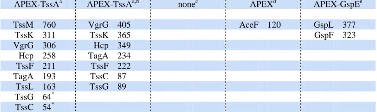
cell lysis. The total eluate was subjected to mass spectrometry for protein identification (Table 107
1, first column). The highest hits correspond to T6SS subunits, including the TssM and TssL 108
membrane proteins, the TssK, VgrG, TssF and TssG baseplate components, the Hcp and TssC 109
tail proteins, as well as a protein of unknown function, encoded by the EC042_4550 gene 110
(accession number GI: 284924271; hereafter named TagA). Interestingly, TssK, VgrG, Hcp 111
and TssC are known interacting partners of TssA18 whereas TssF and TssG form a stable 112
complex with TssK24,25 and are likely at close proximity to the TssK-bound TssA. Finally, 113
while no direct contacts have been identified between TssA and TssM and TssL, the TssA 114
protein was previously shown to co-purify with the TssJLM complex18. The EC042_4550 115
gene, located at close proximity to the T6SS core component genes (Supplementary Fig. 1b), 116
encodes a protein of the TssA family, which shares the N-terminal ImpA domain but has a 117
different C-terminal extension and was thus named TagA (Type VI accessory gene with 118
ImpA domain)36. The two T6SS integral inner membrane proteins, TssM and TssL, were not 119
retrieved when detergent was omitted during preparation of the lysate sample prior to 120
streptavidin enrichment (Table 1, second column). No significant hits were recovered from 121
mass spectrometry analysis of streptavidin-enriched samples from wild-type cells that do not 122
produce APEX2-TssA (Table 1, third column). In addition, no T6SS proteins were 123
biotinylated when APEX2 was produced from the tssA locus but not fused to TssA (Table 1, 124
fourth column). Finally, we generated a EAEC strain producing a fusion between APEX2 and 125
GspE, a cytosolic ATPase that transiently associates with the membrane-anchored Type II 126
secretion system (T2SS)48. Although two known T2SS partners of GspE, GspF and GspL49, 127
were labeled, none of the T6SS subunit was recovered in the enriched fraction (Table 1, fifth 128
column). All these controls experiments demonstrate the specificity of APEX2 labeling when 129
fused to TssA. 130
131
Stage-specific blocking mutations define the temporal TssA contacts during T6SS 132
biogenesis. 133
Previous data have suggested that TssA mediates sequential contacts with the 134
membrane complex, the baseplate and the tail, during T6SS biogenesis18,36. We sought to test 135
this model by arresting T6SS assembly at each stage, using non-polar mutants we previously 136
generated. In absence of TssL, the membrane complex does not assemble and T6SS assembly 137
is blocked at an early stage21. Indeed, no significant hit was recovered in a tssL strain (Table 138
2; first column). The observation that both TssM and TssL are biotinylated in tssK and vgrG 139
cells (Table 2, second and third columns) is in agreement with the fact that the membrane 140
complex is assembled in absence of baseplate components25 and with the proposal that TssA 141
is bound at the cytosolic face of the membrane complex18,36. Finally, the biotinylation of 142
membrane complex and baseplate components in hcp cells (Table 2, fourth column) correlates 143
with the obervation that the absence of Hcp prevents polymerization of the tail but does not 144
impact membrane complex and baseplate assembly25. A schematic summary of proteins 145
biotinylated by APEX2-TssA during T6SS biogenesis is shown in Figure 1. 146
147
TagA associates at the distal end of the sheath. 148
The identification of TagA, a protein of unknown function, as a proximity partner of 149
TssA and the location of the tagA gene at close proximity to T6SS core component genes led 150
us to further characterize this protein. Interestingly, TagA was not biotinylated in tssL, tssK, 151
vgrG or hcp cells (Table 2), suggesting that it contacts TssA in the late stage (i.e., tail
152
polymerization) of T6SS biogenesis. Bacterial two-hybrid and co-immunoprecipitation assays 153
demonstrated that TagA directly interacts with TssA (Fig. 2a and 2b). Competition assays 154
further showed that the tagA mutant strain retains 12 ± 4 % of activity (Fig. 2c). Anti-bacterial 155
activity was restored to wild-type levels upon cis-complementation with wild-type TagA or 156
VSV-G-tagged TagA (VTagA; Fig. 2c). Hence, TagA is not an essential protein for T6SS 157
action but rather increases the efficiency of the Type VI apparatus. To gain further insights 158
into TagA localization, we performed fractionation and fluorescence microscopy experiments. 159
In EAEC wild-type and ∆sci1 (i.e., a strain deleted of the T6SS gene cluster, from tssB to 160
tssJ) cells, as well as in E. coli K-12 (devoid of T6SS gene cluster) cells, VTagA co-161
fractionnates with the EF-Tu cytoplasmic elongation factor and with membrane-associated 162
proteins such as OmpA, TolA and TolB (Fig. 2d). However, by contrast to OmpA and TolA, 163
VTagA behaves similarly to the peripherally-associated membrane protein TolB: VTagA is 164
partly released from the membrane upon urea treatment (Fig. 2d), suggesting that TagA is a 165
cytosolic protein that associates with the membrane independently of T6SS subunits. In 166
agreeement with this conclusion, fluorescence microscopy imaging of a functional TagA 167
fusion to superfolder GFP (sfGFP-TagA; Supplementary Fig. 3) revealed that TagA 168
assembles sub-membrane punctate foci (Fig. 3a and 3b). The association of these punctate 169
foci with the membrane was confirmed by deconvolution analyses (Supplementary Fig. 4a). 170
Interestingly, these foci are not randomly distributed but rather cluster at the ¼ of the cell (Fig. 171
3c). Time-lapse recordings in wild-type showed that these foci dynamically associate - or 172
appear close to the ¼ and ¾ of the cell (Fig. 3d). Interestingly, the formation of static or 173
dynamic sfGFP-TagA foci is dependent upon the T6SS subunits, as sfGFP-TagA localizes at 174
diffuse membrane patches in Δsci1 cells (Supplementary Fig. 4b). 175
To better define the localization of TagA, we performed co-localization studies with a 176
mCherry-tagged TssB sheath subunit. Fig. 3e shows that TagA localizes at one extremity of 177
the sheath. Because the sheath extends from the baseplate to the opposite side of the cell, we 178
next asked whether TagA partitions with the baseplate or locates at the distal end of the sheath. 179
Co-localization experiments with the TssK baseplate subunit fused to the mCherry defined 180
that sfGFP-TagA and TssK-mCherry do not co-localize, demonstrating that TagA is not a 181
component of the baseplate (Fig. 3f and Supplementary Fig. 5). From these results, we 182
conclude that TagA associates with the distal end of the sheath. This conclusion is in 183
agreement with the APEX2-TssA-mediated biotinylation of TagA only in cells authorizing 184
sheath polymerization. To gain further insights into TagA dynamics, we imaged sheath 185
assembly in cells producing sfGFP-TagA. Examination of full cycles of sheath 186
extension/contraction (see blue arrow in Fig. 3g) established that TagA is captured by the 187
distal end of the sheath once it approaches the membrane (Fig. 3g, Supplementary Fig. 5, 188
Supplementary Video 1). 189
190
TagA stops sheath polymerization and stabilizes the extended sheath conformation. 191
To determine the role of TagA in sheath dynamics, we imaged wild-type, tagA and 192
complemented tagA cells producing TssB-sfGFP. Time-lapse fluorescence recordings 193
revealed some remarkable sheath dynamics in tagA cells: about fifty percent of the sheaths 194
extend toward the opposite side of the membrane but do not stop, and hence undergo a 195
distortion event allowing their extension parallel to the membrane (Fig. 4a-b; Supplementary 196
Video 2), ultimately leading to sheaths break or detachment from the membrane 197
complex/baseplate (see red arrowheads in Fig. 4a). This observation suggests that in absence 198
of TagA, the polymerization of some of the sheaths does not stop at the opposite membrane 199
(i.e., the location of TagA) and thus that TagA may constitute the stopper – or part of the 200
stopper – for T6SS sheath extension. These sheaths are likely to be non-productive, 201
explaining the decreased T6SS activity in tagA cells. The remaining sheaths undergo usual 202
extension/contraction cycles. However, we noticed that most of these sheaths contract rapidly 203
after completion of their extension. Comparison of the residence time of extended sheaths (i.e., 204
duration of the extended sheath conformation prior to contraction) in wild-type, tagA and 205
complemented tagA cells revealed that sheaths contract significantly more rapidly after tail 206
competion in absence of TagA (Fig. 4c, Supplementary Fig. 6). Interestingly, the distribution 207
of the residence time differs considerably between wild-type and tagA cells: the number of 208
sheaths that contract immediatly after extension is significantly increased in tagA cells (> 209
60% of the events) compared to wild-type of complemented tagA cells (∼ 5% of the events) 210
(Fig. 4c, Supplementary Fig. 6). These results provide support to an additional role of TagA 211
as a clamp to stabilize the extended sheath and/or to maintain the sheath under the extended 212 conformation. 213 214 Concluding remarks 215
In this study we have adapted the APEX2-dependent biotin ligation technology 216
initially developed in eukaryotic cells to study bacterial complexes. We report the 217
identification of the in vivo proximity partners of the T6SS-associated TssA protein and use 218
this approach in stage-specific blocking mutant cells to temporally resolve the TssA contacts 219
during T6SS biogenesis. We show that TssA successively engages with different complexes: 220
it interacts first with the inner membrane proteins of the membrane complex (TssL, TssM), 221
and then the baseplate (TssF, TssG, TssK and VgrG,) and the tail (Hcp, TssC). This powerful 222
approach recapitulates the known interacting partners of TssA previously identified by 223
bacterial two-hybrid, co-precipitation and surface-plasmon resonance assays18. This 224
technology also detected two known partners of the T2SS-associated GspE ATPase, GspF 225
and GspL49-52. To our knowledge, this is the first report of the use of APEX2-dependent 226
proximity biotinylation inside living bacterial cells. This approach is specifically powerful to 227
dissect contacts of a dynamic protein that engages in different complexes during assembly of 228
a multiprotein system or that is involved in different processes during the cell cycle. 229
Interestingly, APEX2 fusion to TssA revealed an additional player in Type VI secretion in 230
EAEC, TagA. TagA could not be considered as a T6SS core component as the tagA gene is 231
found associated with a limited number of T6SS gene clusters, including that of Xenorhabdus 232
and Photorhabdus species as well as Aeromonas hydrophila and Vibrio cholerae36. In
233
agreement with the phenotypes associated with the deletion of the tagA gene in V. cholerae, 234
VCA012153, competition experiments provided evidence that TagA is not essential for T6SS
235
activity in EAEC. However, the presence of TagA optimizes T6SS efficiency as a tagA 236
mutant retains 12 ± 4 % of anti-bacterial activity compared to the wild-type strain. Domain 237
analyses of TagA showed that it shares the N-terminal ImpA domain found in proteins of the 238
TssA family, including EAEC TssA and P. aeruginosa TssA118,36,54, and a C-terminal 239
extension of unknown function. These two domains are separated by a long linker comprising 240
a stretch of hydrophobic residues predicted to be arranged as an amphipatic α-helix36. Indeed, 241
TagA is a cytosolic protein that peripherally but tightly associates with the membrane. 242
Interestingly, TagA localizes at specific positions in the cell, halfway between midcell and the 243
cell poles. Although we do not know the cellular determinants that control this specific 244
positioning, TagA is captured by the distal end of the sheath, likely by directly interacting 245
with the TssA cap protein. TagA-TssA contacts might be mediated by their N-terminal ImpA 246
domains, which have been previously shown to dimerize18. It is noteworthy that the tagA gene 247
is usually present within T6SS gene clusters encoding TssA, but not TssA1. This observation, 248
the interaction between the TssA and TagA, and the presence of TagA at proximity of TssA 249
when sheath extension is completed as revealed by the in vivo APEX2 assay, support a 250
functional relationship between TssA (and the distal extremity of the sheath) and TagA. 251
Indeed, sfGFP-TagA was shown to present a diffuse membrane pattern in absence of T6SS 252
subunits and to assemble foci at the site of contact of the distal end of the sheath with the 253
opposite membrane. The observation that TagA is captured by the distal end of the sheath 254
suggests a role of this accessory protein during the late stages of sheath assembly. In 255
agreement with this suggestion, fluorescence microscopy recordings of the T6SS sheath 256
showed two different consequences of the absence of TagA. First, aberrant sheath 257
polymerization was observed, in which sheath polymerization does not stop at the membrane, 258
leading to sheath distortion, bending and, ultimately, breaking. These sheaths do not contract 259
and hence are unfruitful to destroy rival bacteria, likely explaining the decreased T6SS 260
activity in tagA cells. The second consequence of the absence of TagA is the rapid contraction 261
after sheath extension. One explanation to these two distinct events might be how the sheath 262
hits the membrane. It has been proposed that incorporation of new Hcp hexamers in the 263
growing tail occurs by opening the central hexaflexagon structure of TssA18,36. Therefore, one 264
may hypothesize that if the sheath arrives perpendicular to the membrane, there is no room to 265
allow incorporation of new Hcp hexamers, hence arresting sheath polymerization. In absence 266
of TagA, these sheaths will contract rapidly. By contrast, if the sheath does not arrive 267
perpendicular, new Hcp hexamers can enter the TssA central lumen, allowing the sheath 268
polymerization to proceed and causing sheath distortion and bending. One alternative 269
hypothesis is that the absence of TagA is partly compensated by an unknown factor for 270
preventing uncontroled sheath extension. Using the APEX2 technology to identify proteins at 271
proximity of TagA may provide further insights into T6SS tail completion. 272
Based on our results, we propose that TagA associates with the cytoplasmic side of the 273
inner membrane. Once the distal end of the sheath approaches the opposite membrane, TagA 274
is captured, stops sheath polymerization and clamps the extended sheath to the membrane. By 275
linking both the sheath distal extremity and the membrane, TagA prevents the immediate 276
contraction of the sheath and maintains the tensile forces required for efficient killing. TagA 277
is thus employed by TssA+ T6SS as a sheath stopper and clamp. 278
279
METHODS 280
Bacterial strains, media and chemicals 281
Enteroaggregative E. coli was used as model micro-organism. Escherichia coli K-12 strains DH5α, W3110 and
282
BTH101 were used for cloning procedures and fractionation, and bacterial two-hybrid assay, respectively.
283
Enteroaggregative E. coli (EAEC) strains used in this work are isogenic derivatives of the wild-type O3:H2 17-2
284
strain. E. coli K-12 and EAEC cells were routinely grown in LB broth at 37°C, with aeration. For induction of
285
the sci1 T6SS gene cluster, cells were grown in Sci1-inducing medium [SIM: M9 minimal medium
286
supplemented with glycerol (0.2%), vitamin B1 (1 μg.mL−1), casaminoacids (40 μg.mL−1), LB (10% v/v)]55. 287
Plasmids and chromosomal deletions and insertions were maintained by the addition of ampicillin (100 μg.mL−1 288
for K-12), kanamycin (50 μg.mL−1 for K-12, 50 μg.mL−1 for chromosomal insertion on EAEC, 100 μg.mL−1 for 289
plasmid bearing EAEC), or chloramphenicol (40 μg.mL−1). Expression of genes from pBAD or pASK-IBA 290
vectors was induced at A600nm ≈ 0.6 with 0.02% of L-arabinose (Sigma-Aldrich) for 1 h or with 0.1 μg.mL-1 of 291
anhydrotetracyclin (IBA Technologies) for 45 min, respectively. For BACTH experiments, gene expression was
292
induced by the addition of iso-propyl-β-D-thio-galactopyranoside (IPTG, Sigma-Aldrich, 0.5 mM) and plates
293
were supplemented with 5-bromo-4-chloro-3-indolyl-β-D-galactopyranoside (X-Gal, Eurobio, 40 μg.mL−1). 294
Strain construction 295
The tagA gene (EC042_4550) was deleted in the enteroaggregative E. coli 17-2 strain using λ-red
296
recombination47 as previously described19 using plasmid pKOBEG56. In brief, a kanamycin cassette was 297
amplified from plasmid pKD4 using oligonucleotides carrying 50-nucleotide extensions homologous to regions
298
adjacent to tagA. After electroporation of 600 ng of column-purified PCR product, kanamycin-resistant clones
299
were selected and verified by colony-PCR. The kanamycin cassette was then excised using plasmid pCP2047 and 300
confirmed by colony-PCR. The same procedure was used to introduce APEX2 on the chromosome. The
APEX2-301
coding sequence was amplified using the pKD4-Nter-APEX2 vector and inserted downstream of the start codon
302
of the tssA (APEX2-TssA) or gspE (APEX2-GspE) genes or upstream the start codon of tssA (APEX2 in the
303
T6SS gene cluster). Fluorescent reporter genes were amplified from the pKD4-Nter-sfGFP (N-terminal sfGFP
304
fusions) or pKD4-Cter-mCherry (C-terminal mCherry fusions) vectors18,25 and inserted on the chromosome 305
downstream of the start codon of the tagA gene (sfGFP-TagA fusion) or upstream of the stop codon of the tssK
306
gene (TssK-mCh fusion). For cis-complementation, a pKD4 derivative vector was engineered in which the tagA
307
gene (or the tagA gene encoding an N-terminally VSV-G-tagged version of tagA) was placed under the control
of the sci1 T6SS promoter. The cassette was then introduced on the chromosome of the ∆tagA and ∆tagA
309
tssBsfGFP strains, at the lacZ locus, by λ-red recombination using pKOBEG.
310
Plasmid construction 311
PCR was performed using a Biometra thermocycler using the Q5 DNA polymerase (New England Biolabs).
312
Enteroaggregative E. coli 17-2 chromosomal DNA or the pcDNA3-APEX2-NES plasmid (Addgene #49386)38 313
were used as template for PCR amplification. All the plasmids have been constructed by restriction-free
314
cloning57. Primers used in this study are listed in Supplementary Table 1. Briefly, the gene of interest was 315
amplified using oligonucleotides introducing extensions annealing to the target vector. The double-stranded
316
product of the first PCR has then been used as oligonucleotides for a second PCR using the target vector as
317
template. PCR products were then treated with DpnI to eliminate template plasmids. All constructs have been
318
verified by PCR and DNA sequencing (Eurofins Genomics). Plasmid pKD4-Nter-APEX2 was deposited in the
319
Addgene plasmid repository under accession number 112868.
320
In vivo APEX2-dependent biotin labeling and identification of biotinylated proteins by mass spectrometry 321
Biotin proximity labeling. Experiments were initially conducted to optimize the conditions of biotin proximity
322
labeling, by varying the concentration of biotin phenol, the concentration of H2O2, and the duration of the H2O2 323
pulse. EAEC wild-type and APEX2 derivative cells were mixed with E. coli K-12 competitor cells and spotted
324
on SIM plates supplemented with 10 mM biotin phenol (Biotine Tyramide; BP, Iris Biotech). After 4 h at 37°C,
325
cells were washed with 1 mL of SIM medium to eliminate residual BP, and treated with 1 mM hydrogen
326
peroxide (H2O2; Sigma Aldrich) for 1 min before quenching by washing with TSEN buffer (Tris-HCl 20 mM pH 327
8, sucrose 30%, EDTA 1 mM, NaCl 100 mM) supplemented with egg-white lysozyme 10 μg.mL−1, 10 mM 328
sodium ascorbate and 10 mM sodium azide. Cells were either analyzed by SDS-PAGE and Streptavidin
western-329
blotting or lysed by resuspension in CellLyticTM B (Sigma-Aldrich) supplemented or not with 0.2% Igepal® CA-330
630 (Sigma-Aldrich). After 1 hour on a wheel, lysates were clarified by centrifugation at 20,000×g for 10 min.
331
Streptavidin pull-down of biotinylated proteins. The clarified cell lysate was incubated for 30 min at room
332
temperature with 2 mg of Streptavidin-coated magnetic beads, equilibrated in CellLyticTM B buffer 333
supplemented with Igepal CA-630. After three washes with CellLyticTM B supplemented with Igepal CA-630, 334
the beads were resuspended in Laemmli loading dye and loaded on a SDS-PAGE gel. The migration was
335
stopped when the sample reached the interface between concentrating and separating gels, and the band
336
containing the total biotinylated proteins was cut out.
337
Mass spectrometry analyses. The protein-containing SDS-PAGE gel bands were washed with 100 mM
338
acetonitrile/ammonium bicarbonate pH 7.5, reduced by 10 mM dithiothreitol in 100 mM ammonium bicarbonate
339
pH 7.5, alkylated by 55 mM iodoacetamide in 100 mM ammonium bicarbonate pH 7.5, and overnight digested
340
at 37 °C by Trypsin/Lys-C Mix from Pseudomonas aeruginosa (Promega) at 10 ng.µL-1 in 25 mM ammonium 341
bicarbonate pH 7.5/proteaseMAXTM surfactant 0.025% (v/v). Tryptic peptides were extracted from gels by 0.1% 342
(vol/vol) trifluoroacetic acid (TFA)/0.01% (vol/vol) proteaseMAXTM/50% (vol/vol) acetonitrile, and dried by 343
speed vacuum. Solubilized samples in 0.05% (vol/vol) TFA /2 % (vol/vol) acetonitrile were analyzed on a
ESI-344
Q-Exactive Plus (ThermoFisher) mass spectrometer coupled to a nanoliquid chromatography (Ultimate 3000,
345
Dionex). Peptides were eluted from a C18 column (Acclaim PepMap RSLC, 75 µm × 150 mm, 2 µm, 100 Ǻ,
346
Dionex) by a 6-40% linear gradient of mobile phase B (0.1% (vol/vol) formic acid (FA)/80% (vol/vol)
347
acetonitrile) in mobile phase A (0.1% (vol/vol) FA) for 52 min. The peptides were detected in the mass
348
spectrometer in a positive ion mode using a Top 10 Data Dependent workflow. One scan event full MS in the
349
Orbitrap at 70,000, in a 350-1900 m/z range was followed by a fragmentation MS/MS step, at 17,500, of the 10
350
top ions, in the Higher Energy Collisional Dissociation cell set at 27. The spectra were processed by Proteome
351
Discoverer software (ThermoFisher, version: 2.1.0.81) using the Sequest HT algorithm with the search following
352
parameters: enteroaggregative E. coli 042 database (Taxonomy ID 216592 downloaded from NCBI by Protein
353
Center, 4921 entries); trypsin enzyme (maximum 2 missed cleavages); fixed modification: carbamidomethyl
354
(Cys); variable modification: oxidation (Met); mass values specific for monoisotopic; precursor mass tolerance:
355
± 10 ppm; fragment mass tolerance: ± 0.02 Da. Peptide validation was based on the best Peptide Spectrum
356
Match (PSM) defined at a 0.05 maximum Delta Cn and a 0.01 Strict Target False Discovery Rate. Proteins were
357
identified if minimum 2 unique peptide sequences more than 6 amino-acids passed the high confidence filter.
358
Human keratin being considered as a common contaminant in protein identification by mass spectrometry, only
359
hits with a number of PSM higher than keratin were considered significant. Mass spectrometry datasheets are
360
available in Supplementary Datasheets 1-9. The experiments were done in triplicate and a representative result is
361
shown.
362
Cell fractionation 363
Cell fractionation assay was performed as previously described15,23,25. Briefly, 5×10¹⁰ exponentially growing 364
cells were resuspended in 750 μl of TSEN buffer and incubated for 10 min on ice. After addition of 100 μg.mL-1 365
of lysozyme and further incubation for 20 min on ice, 750 μL of TN buffer (Tris-HCl 10 mM, pH 8.0, NaCl 100
366
mM) was added, and cells were lysed by three cycles of freezing and thawing and four cycles of sonication.
367
Unbroken cells were removed by centrifugation, and soluble and membrane fractions were separated by
368
ultracentrifugation for 45 min at 45,000×g. Membranes were washed once with TE buffer and resuspended in 1
369
mL of TN buffer supplemented with 8 M urea, incubated on a wheel for 1 h at 25°C, and then centrifuged for 45
370
min at 45,000×g to separate integral membrane and peripherally membrane associated proteins. Soluble and
371
membrane-associated fractions were resuspended in loading buffer and subjected to SDS-PAGE and
372
immunoblotting. Anti-EF-Tu (HyCult Biotech, clone mAb900), VSV-G (Sigma-Aldrich, clone P5D4),
anti-373
OmpA, anti-TolA and anti-TolB (laboratory collection) antibodies were used to identify the cytoplasmic Tu
374
elongation factor, the VSV-G-tagged TagA protein, the outer membrane OmpA protein, the inner membrane
375
TolA protein, and the outer membrane-peripherally-associated periplasmic TolB protein, respectively. The
376
experiments were done in triplicate and a representative result is shown.
377
Bacterial two-hybrid assay (BACTH) 378
The adenylate cyclase-based bacterial two-hybrid technique58 was used as previously published59. Briefly, the 379
proteins to be tested were fused to the isolated T18 and T25 catalytic domains of the Bordetella adenylate
380
cyclase. After introduction of the two plasmids producing the fusion proteins into the BTH101 reporter strain,
381
plates were incubated at 30°C for 24 h. Three independent colonies for each transformation were inoculated into
382
600 μL of LB medium supplemented with ampicillin, kanamycin, and IPTG (0.5 mM). After overnight growth at
383
30°C, 10 μL of each culture was spotted onto LB plates supplemented with ampicillin, kanamycin, IPTG, and
X-384
gal and incubated at 30°C. Controls include interaction assays with TolB and Pal, two protein partners unrelated
385
to the T6SS. The experiments were done in triplicate and a representative result is shown.
386
Co-immuno-precipitation 387
Soluble lysates from 2×10¹⁰ cells producing VSV-G-tagged TagA or VSV-G-tagged TagA and FLAG-tagged 388
TssA were obtained using the cell fractionation procedure, and subjected to immuno-precipitation on anti-FLAG
389
M2 affinity gel (Sigma-Aldrich) for 1 h at 20°C. Beads were washed three times with TN buffer, resuspended in
390
non-reducing Laemmli loading dye, and subjected to SDS-PAGE and immuno-blot analyses using monoclonal
391
anti-VSV-G (Sigma-Aldrich, clone P5D4) and anti-FLAG (Sigma-Aldrich, clone M2) antibodies. The
392
experiments were done in triplicate and a representative result is shown.
393
Inter-bacterial competition 394
The anti-bacterial growth competition assay was performed as described60. The WT E. coli strain W3110 bearing 395
the kanamycin resistant pUA66-rrnB plasmid61 was used as prey in the competition assay. The pUA66-rrnB 396
plasmid provides a strong constitutive green fluorescent (GFP+) phenotype. Attacker and prey cells were grown 397
in SIM medium to a A600nm ≈ 0.6-0.8, harvested and normalized to a A600nm of 10 in SIM. Attacker and prey cells 398
were mixed to a 4:1 ratio and 15-μL drops of the mixture were spotted in triplicate onto a prewarmed dry SIM
399
agar plate. After 4-hour incubation at 37°C, fluorescent images were recorded with a LI-COR Odyssey imager.
400
The bacterial spots were scratched off, and cells were resuspended in LB medium and normalized to a A600nm of 401
0.5. For fluorescence measurements, triplicates of 200 µl were transferred into wells of a black 96-well plate
402
(Greiner) and the A600nm and fluorescence (excitation: 485 nm; emission: 530 nm) were measured with a TECAN 403
infinite M200 microplate reader (9 measures per mixture per experiment). The relative fluorescence was
404
expressed as the intensity of fluorescence divided by the A600nm, after subtracting the values of a blank sample. 405
For enumeration of viable E. coli K-12 cells, serial dilutions were plated on kanamycin plates and the number of
406
colonies were counted after 16-h incubation at 37°C. The experiments were done in triplicate and a
407
representative result is shown. Statistical analyses of inter-bacterial competition assays were performed by
408
Student's t-test. Significant differences were defined as p < 0.05 (*), p < 0.01 (**), and p < 0.001 (***).
409
Fluorescence microscopy. 410
Cells were grown in SIM to a A600 nm ≈ 0.4–0.6, harvested and resuspended in fresh SIM to a A600 nm ≈ 10. Cell 411
mixtures were spotted on a thin pad of SIM supplemented with 2% agarose, covered with a cover slip and
412
incubated for 20-30 min at room temperature before microscopy acquisition. Fluorescence microscopy was
413
performed with a Nikon Eclipse Ti microscope equipped with an Orcaflash 4.0 LT digital camera (Hamamatsu)
414
and a perfect focus system (PFS) to automatically maintain focus so that the point of interest within a specimen
415
is always kept in sharp focus at all times despite mechanical or thermal perturbations. All fluorescence images
416
were acquired with a minimal exposure time to minimize bleaching and phototoxicity effects. Exposure times
417
were typically 20 ms for phase contrast, 100 ms for GFP, 2000 ms for sfGFP-TagA and 800 ms for
TssB-418
mCherry and TssK-mCherry. The experiments were done in triplicate and a representative result is shown. For
419
fluorescence microscopy, statistical analyses were performed using several representative fields from three
420
independent biological replicates. Images were analyzed using ImageJ (http://imagej.nih.gov/ij/) and the
MicrobeJ plugin (http://www.microbej.com/)62 or Zen (Carl Zeiss). Statistical dataset analysis was performed 422
using Excel and the R software environment (https://www.r-project.org/). Contractile sheath residence time was
423
determined as the number of frames during which the sheath remained fully extended. Only sheaths that
424
performed visible extension and contraction within the movie period were considered.
425
Data availability. 426
Excel sheets with the raw mass-spectrometry data are provided as Supplementary Datasheets 1-9. Plasmid
427
pKD4-Nter-APEX2 has been deposited in the Addgene plasmid repository under accession number 112868. All
428
data that support the findings of this study are available from the corresponding author upon request.
429 430
References 431
432
1. Basler, M., Pilhofer, M., Henderson, G.P., Jensen, G.J., & Mekalanos, J.J. Type VI secretion 433
requires a dynamic contractile phage tail-like structure. Nature 483, 182–186. (2012). 434
2. Brunet, Y.R., Espinosa, L., Harchouni, S., Mignot, T., & Cascales, E. Imaging type VI secretion-435
mediated bacterial killing. Cell Rep. 3, 36–41. (2013). 436
3. Basler, M. Type VI secretion system: secretion by a contractile nanomachine. Philos Trans R Soc 437
Lond B Biol Sci 370, 1679. (2015). 438
4. Zoued, A., et al. Architecture and assembly of the Type VI secretion system. Biochim Biophys Acta 439
1843, 1664–1673. (2014).
440
5. Durand, E., Cambillau, C., Cascales, E., & Journet, L. VgrG, Tae, Tle, and beyond: the versatile 441
arsenal of Type VI secretion effectors. Trends Microbiol. 22, 498–507. (2014). 442
6. Ho, B.T., Dong, T.G., & Mekalanos, J.J. A view to a kill: the bacterial type VI secretion system. 443
Cell Host Microbe 15, 9–21. (2014). 444
7. Cianfanelli, F.R., Monlezun, L., & Coulthurst, S.J. Aim, load, fire: the type VI secretion system, a 445
bacterial nanoweapon. Trends Microbiol 24, 51–62. (2016). 446
8. Brackmann, M., Nazarov, S., Wang, J., & Basler, M. Using force to punch holes: mechanics of 447
contractile nanomachines. Trends Cell Biol 27, 623–632. (2017). 448
9. Russell, A.B., Peterson, S.B., & Mougous, J.D. Type VI secretion system effectors: poisons with a 449
purpose. Nat Rev Microbiol 12, 137–148. (2014). 450
10. Alcoforado Diniz, J., Liu, Y.C., & Coulthurst, S.J. Molecular weaponry: diverse effectors 451
delivered by the Type VI secretion system. Cell Microbiol 17, 1742–1751. (2015). 452
11. Chassaing, B., & Cascales, E. Antibacterial weapons: targeted destruction in the microbiota. 453
Trends Microbiol. 26, 329-338 (2018). 454
12. Hachani, A., Wood, T.E., & Filloux, A. Type VI secretion and anti-host effectors. Curr Opin 455
Microbiol. 29, 81-93 (2016). 456
13. Ryu, C.M. Against friend and foe: type 6 effectors in plant-associated bacteria. J Microbiol. 53, 457
201-8 (2015). 458
14. Si, M., et al. The type VI secretion system engages a redox-regulated dual-functional heme 459
transporter for zinc acquisition. Cell Rep. 20, 949-959 (2017). 460
15. Aschtgen, M.S., Gavioli, M., Dessen, A., Lloubès, R., & Cascales, E. The SciZ protein anchors the 461
enteroaggregative Escherichia coli Type VI secretion system to the cell wall. Mol Microbiol 75, 462
886–899. (2010). 463
16. Shneider, M.M., et al. PAAR-repeat proteins sharpen and diversify the type VI secretion system 464
spike. Nature 500, 350–353. (2013). 465
17. Weber, B.S., et al. Genetic dissection of the type VI secretion system in Acinetobacter and 466
identification of a novel peptidoglycan hydrolase, TagX, required for its biogenesis. MBio 7, pii: 467
e01253-16. (2016). 468
18. Zoued, A., et al. Priming and polymerization of a bacterial contractile tail structure. Nature 531, 469
59–63. (2016). 470
19. Aschtgen, M.S., Bernard, C.S., de Bentzmann, S., Lloubès, R., and Cascales, E. SciN is an outer 471
membrane lipoprotein required for type VI secretion in enteroaggregative Escherichia coli. J 472
Bacteriol 190, 7523–7531. (2008). 473
20. Felisberto-Rodrigues, C., et al. Towards a structural comprehension of bacterial type VI secretion 474
systems: characterization of the TssJ-TssM complex of an Escherichia coli pathovar. PLoS 475
Pathog 7, e1002386. (2011). 476
21. Durand, E., et al. Biogenesis and structure of a type VI secretion membrane core complex. Nature 477
523, 555–560. (2015).
478
22. Santin, Y.G., & Cascales, E. Domestication of a housekeeping transglycosylase for assembly of a 479
Type VI secretion system. EMBO Rep 18, 138–149. (2017). 480
23. Zoued, A., et al. TssK is a trimeric cytoplasmic protein interacting with components of both 481
phage-like and membrane anchoring complexes of the type VI secretion system. J Biol Chem 482
288, 27031–27041. (2013).
483
24. English, G., Byron, O., Cianfanelli, F.R., Prescott, A.R., & Coulthurst, S.J. Biochemical analysis 484
of TssK, a core component of the bacterial Type VI secretion system, reveals distinct oligomeric 485
states of TssK and identifies a TssK-TssFG subcomplex. Biochem J 461, 291–304. (2014). 486
25. Brunet, Y.R., Zoued, A., Boyer, F., Douzi, B., & Cascales, E. The type VI secretion TssEFGK-487
VgrG phage-like baseplate is recruited to the TssJLM membrane complex via multiple contacts 488
and serves as assembly platform for tail tube/sheath polymerization. PLoS Genet 11, e1005545. 489
(2015). 490
26. Taylor, N.M., et al. Structure of the T4 baseplate and its function in triggering sheath contraction. 491
Nature 533, 346–352. (2016). 492
27. Leiman, P.G., et al. Type VI secretion apparatus and phage tail-associated protein complexes share 493
a common evolutionary origin. Proc Natl Acad Sci U S A 106, 4154–4159. (2009). 494
28. Büttner, C.R., Wu, Y., Maxwell, K.L., & Davidson, A.R. Baseplate assembly of phage Mu: 495
defining the conserved core components of contractile-tailed phages and related bacterial 496
systems. Proc Natl Acad Sci U S A 113, 10174–10179. (2016). 497
29. Nguyen, V.S., et al. Type VI secretion TssK baseplate protein exhibits structural similarity with 498
phage receptor-binding proteins and evolved to bind the membrane complex. Nat Microbiol 2, 499
17103. (2017). 500
30. Nazarov, S., Schneider, J.P., Brackmann, M., Goldie, K.N., Stahlberg, H., & Basler, M. Cryo-EM 501
reconstruction of Type VI secretion system baseplate and sheath distal end. EMBO J. in press 502
(pii: e201797103). (2018). 503
31. Logger, L., Aschtgen, M.S., Guérin, M., Cascales, E., & Durand, E. Molecular dissection of the 504
interface between the type VI secretion TssM cytoplasmic domain and the TssG baseplate 505
component. J Mol Biol 428, 4424–4437. (2016). 506
32. Zoued, A., et al. Structure-function analysis of the TssL cytoplasmic domain reveals a new 507
interaction between the type VI secretion baseplate and membrane complexes. J Mol Biol 428, 508
4413–4423. (2016). 509
33. Brunet, Y.R., Hénin, J., Celia, H., & Cascales, E. Type VI secretion and bacteriophage tail tubes 510
share a common assembly pathway. EMBO Rep 15, 315–321. (2014). 511
34. Kudryashev, M., et al. Structure of the type VI secretion system contractile sheath. Cell 160, 952– 512
962. (2015). 513
35. Wang, J., et al. Cryo-EM structure of the extended type VI secretion system sheath-tube complex. 514
Nat Microbiol 2, 1507–1512. (2017). 515
36. Zoued, A., et al. TssA: the cap protein of the Type VI secretion tail. Bioessays 39, 00262. (2017). 516
37. Vettiger, A., Winter, J., Lin, L., & Basler, M. The type VI secretion system sheath assembles at the 517
end distal from the membrane anchor. Nat Commun 8, 16088. (2017). 518
38. Lam, S.S., et al. Directed evolution of APEX2 for electron microscopy and proximity labeling. 519
Nat Methods 12, 51–54. (2015). 520
39. Lobingier, B.T., et al. An approach to spatiotemporally resolve protein interaction networks in 521
living cells. Cell 169, 350–360. (2017). 522
40. Rhee, H.W., et al. Proteomic mapping of mitochondria in living cells via spatially restricted 523
enzymatic tagging. Science 339, 1328–1331. (2013). 524
41. Hung, V., et al. Proteomic mapping of the human mitochondrial intermembrane space in live cells 525
via ratiometric APEX tagging. Mol Cell 55, 332–341. (2014). 526
42. Hung, V., et al. Spatially resolved proteomic mapping in living cells with the engineered 527
peroxidase APEX2. Nat Protoc 11, 456–475. (2016). 528
43. Lee, S.Y., Kang, M.G., Park, J.S., Lee, G., Ting, A.Y., & Rhee, H.W. APEX fingerprinting reveals 529
the subcellular localization of proteins of interest. Cell Rep 15, 1837–1847. (2016). 530
44. Hung, V., et al. Proteomic mapping of cytosol-facing outer mitochondrial and ER membranes in 531
living human cells by proximity biotinylation. Elife 6, e24463. (2017). 532
45. Paek, J., et al. Multidimensional tracking of GPCR signaling via peroxidase-catalyzed proximity 533
labeling. Cell 169, 338–349. (2017). 534
46. Rucks, E.A., Olson, M.G., Jorgenson, L.M., Srinivasan, R.R., & Ouellette, S.P. Development of a 535
proximity labeling system to map the Chlamydia trachomatis inclusion membrane. Front Cell 536
Infect Microbiol 7, 40. (2017). 537
47. Datsenko, K.A., & Wanner, B.L. One-step inactivation of chromosomal genes in Escherichia coli 538
K-12 using PCR products. Proc Natl Acad Sci U S A 97, 6640–6645. (2000). 539
48. Chen, Y.L., & Hu, N.T. Function-related positioning of the type II secretion ATPase of 540
Xanthomonas campestris pv. campestris. PLoS One 8, e59123. (2013). 541
49. Py, B., Loiseau, L., & Barras F. An inner membrane platform in the type II secretion machinery of 542
Gram-negative bacteria. EMBO Rep 2, 244–248. (2001). 543
50. Py, B., Loiseau, L., & Barras, F. Assembly of the type II secretion machinery of Erwinia 544
chrysanthemi: direct interaction and associated conformational change between OutE, the 545
putative ATP-binding component and the membrane protein OutL. J Mol Biol 289, 659–670. 546
(1999). 547
51. Abendroth, J., Murphy, P., Sandkvist, M., Bagdasarian, M., & Hol, W.G. The X-ray structure of 548
the type II secretion system complex formed by the N-terminal domain of EpsE and the 549
cytoplasmic domain of EpsL of Vibrio cholerae. J Mol Biol 348, 845–855. (2005). 550
52. Arts, J., et al. Interaction domains in the Pseudomonas aeruginosa type II secretory apparatus 551
component XcpS (GspF). Microbiology 153, 1582–1592. (2007). 552
53. Zheng, J., Ho, B., & Mekalanos, J.J. Genetic analysis of anti-amoebae and anti-bacterial activities 553
of the type VI secretion system in Vibrio cholerae. PLoS One 6, e23876. (2011). 554
54. Planamente, S., Salih, O., Manoli, E., Albesa-Jové, D., Freemont, P.S., & Filloux, A. TssA forms a 555
gp6-like ring attached to the type VI secretion sheath. EMBO J 35, 1613–1627. (2016). 556
55. Brunet, Y.R., Bernard, C.S., Gavioli, M., Lloubès, R., & Cascales, E. An epigenetic switch 557
involving overlapping fur and DNA methylation optimizes expression of a type VI secretion gene 558
cluster. PLoS Genet 7, e1002205. (2011). 559
56. Chaveroche, M.-K., Ghigo, J.-M., & d’ Enfert, C. A rapid method for efficient gene replacement in 560
the filamentous fungus Aspergillus nidulans. Nucleic Acids Res 28, e97. (2000). 561
57. van den Ent, F., & Löwe, J. RF cloning: a restriction-free method for inserting target genes into 562
plasmids. J. Biochem. Biophys. Methods 67, 67–74. (2006). 563
58. Karimova, G., Pidoux, J., Ullmann, A., & Ladant, D. A bacterial two-hybrid system based on a 564
reconstituted signal transduction pathway. Proc Natl Acad Sci U S A 95, 5752–5756. (1998). 565
59. Battesti, A., & Bouveret, E. The bacterial two-hybrid system based on adenylate cyclase 566
reconstitution in Escherichia coli. Methods 58, 325–334. (2012). 567
60. Flaugnatti, N., et al. A phospholipase A1 antibacterial Type VI secretion effector interacts directly 568
with the C-terminal domain of the VgrG spike protein for delivery. Molecular Microbiology 99, 569
1099–1118. (2016). 570
61. Zaslaver, A., et al. A comprehensive library of fluorescent transcriptional reporters for Escherichia 571
coli. Nat. Methods 3, 623–628. (2006). 572
62. Ducret, A., Quardokus, E.M., & Brun, Y.V. MicrobeJ, a tool for high throughput bacterial cell 573
detection and quantitative analysis. Nat Microbiol 1, 16077. (2016). 574
575
Correspondence and requests for materials should be addressed to E.C. (cascales@imm.cnrs.fr).
576
Acknowledgments
577
This work was funded by the Centre National de la Recherche Scientifique, the Aix-Marseille 578
Université, and grants from the Agence Nationale de la Recherche (ANR-14-CE14-0006-02, ANR-17-579
CE11-0039-01). Y.G.S. is supported by a doctoral fellowship from the French ministry of research. 580
We thank Hugo Le Guenno of the IMM microscopy facility for helpful advices regarding 581
deconvolution analyses, James Sturgis (IMM, Marseille), Elizabeth (Lisa) A. Rucks and Scott 582
Ouellette (University of South Dakota, Vermillion, USA) for initial discussions on biotin-dependent 583
ligation; the members of the Cascales, Lloubès, Sturgis and Bouveret research groups for discussions; 584
M. Ba, I. Bringer, A. Brun and O. Uderso for technical assistance, and the three anonymous reviewers 585
for their constructive comments. 586
Authors Contributions
587
Y.G.S. & E.C. designed and conceived the experiments. Y.G.S., T.D., R.L., L.E. performed the 588
experiments. Y.G.S. performed all the experiments, with the help of T.D. and L.E. for fluorescence 589
microscopy. R.L. performed the mass spectrometry analyses. E.C supervised the execution of the 590
experiments. L.J. & E.C. provided tools. E.C. wrote the paper with contributions of Y.G.S, T.D., R.L. 591
and L.J. 592
Additional Information 593
Supplementary Information is available for this manuscript. It includes one Supplementary Table,
594
seven Supplementary Figures, two Supplementary Videos, and nine Supplementary Datasheets. 595
596
Competing Interests
597
The authors declare no competing financial interests. 598
Legend to Figures 600
Figure 1 | Summary of TssA proximity partners. The different stages of T6SS assembly 601
are depicted (1, assembly of the membrane complex; 2, baseplate recruitment and docking; 3, 602
polymerization of the tail), as well as the stage-blocking mutations used in this study (deletion 603
of the tssK or vgrG gene stops T6SS assembly at stage 1 whereas deletion of the hcp gene 604
stops T6SS assembly at stage 2). The TssA protein is shown in red and the other T6SS 605
subunits are indicated (J corresponds to TssJ). The blue color highlights the subunits that are 606
biotinylated by the functional APEX2-TssA fusion protein. 607
Figure 2 | TagA is an accessory T6SS cytosolic component that associates with the 608
membrane. a and b, TagA interacts directly with TssA. a, BACTH assay. BTH101 reporter 609
cells producing the indicated proteins fused to the T18 and T25 domain of the Bordetella 610
adenylate cyclase were spotted on X-Gal-IPTG reporter LB agar plates. The BACTH 611
experiment was performed in triplicate with identical results. b, Co-immunoprecipitation 612
assay. Soluble lysates of E. coli cells producing VSV-G-tagged TagA (VTagA) alone or 613
VTagA with FLAG-tagged TssA (TssAF) were subjected to immunoprecipitation with anti-614
FLAG-coupled beads. The total lysates (T) and immunoprecipitated (IP) material were 615
separated by 12.5% acrylamide SDS-PAGE and immunodetected with anti-FLAG (upper 616
panel) and anti-VSV-G (lower panel) monoclonal antibodies. Molecular weight markers (in 617
kDa) are indicated on left. The co-immunoprecipitation experiment was performed in 618
triplicate with identical results. c, TagA optimizes T6SS activity. Antibacterial competition 619
assay. E. coli K-12 competitor cells (W3110 gfp+, kanR) were mixed with the indicated 620
attacker cells, spotted onto SIM agar plates, and incubated for 4 h at 37 °C. The fluorescence 621
of the bacterial spot (in arbitrary units, bars represent the average, standard deviation are 622
indicated, dot plots (grey circles) are overlaid) is shown on top. The number of surviving E. 623
coli competitor cells (counted on selective kanamycin medium) is indicated in the lower
624
graph (in log10 of colony forming units). The circles indicate the values from three 625
independent assays, and the average is indicated by the bar. Statistical significance relative to 626
the wild-type strain is indicated (p-values; NS, non significant; *, p < 0.05; **, p < 0.01, two-627
sided Student's t-test; p-values for fluorescence measurements (upper graph): ∆tssA, 628
0.000196; ∆tagA, 0.0086; VtagA+, 0.119; tagA+, 0.746; p-values for E. coli K-12 survival 629
(lower graph): ∆tssA, 0.00219; ∆tagA, 0.0194; VtagA+, 0.531; tagA+, 0.831). d, TagA 630
fractionates with soluble and peripherally-associated membrane proteins. Fractionation assay. 631
Total extracts (T) of WT or ∆sci1 EAEC, or E. coli K-12 cells producing VTagA were 632
subjected to fractionation to separate the soluble (S) and membrane (M) fractions. 633
Peripherally-associated (pM) and integral (iM) membrane proteins were separated by 634
treatment with 8 M urea. Control markers include the integral outer membrane OmpA protein, 635
the integral inner membrane TolA protein, the peripherally-associated membrane TolB 636
protein, and the EF-Tu cytoplasmic elongation factor. Molecular weight markers (in kDa) are 637
indicated on left. Fractionation experiments were performed in triplicate with identical results. 638
Uncropped blots are shown in Supplementary Fig. 7. 639
Figure 3 | TagA localizes at the cell quarters and binds the distal end of the sheath. a, 640
Fluorescence microscopy recording of wild-type EAEC cells producing sfGFP-TagA (upper 641
panel, phase channel; lower panel, GFP channel). Localization of TagA clusters are indicated 642
by white arrowheads. Scale bar, 1 μm. Fluorescence microscopy recordings have been 643
performed thirty times with identical results. A deconvolution analysis of WT EAEC cells 644
producing sfGFP-TagA is shown in Supplementary Fig. 4a. b, Number of sfGFP-TagA foci 645
per cell. The percentage of cells with 0, 1, 2 or >2 foci is indicated (n= 1171 cells from three 646
biological replicates, bars represent the average, standard deviation are indicated, dot plots 647
(grey circles) are overlaid). The mean number of foci per cell is 0.74 ± 0.24. c, Spatial 648
repartition of sfGFP-TagA foci. Shown is a projection of the foci from n = 316 cells on a 649
single cell (from blue (low abundance) to yellow (high abundance)). d, Fluorescence 650
microscopy time-lapse recording of EAEC cells producing sfGFP-TagA. Individual images 651
were taken every 40 s. The localization of TagA is indicated by the white arrowhead. A 652
schematic diagram representating the dynamics of TagA is shown below. Scale bar, 1 μm. 653
Time-lapse recordings have been performed thirty times with identical results. A statistical 654
analyses of the distribution of sfGFP-TagA dynamics in wild-type and Δsci1 cells is shown in 655
Supplementary Fig. 4b. e and f, Co-localization of sfGFP-TagA with TssB-mCherry (e) or 656
TssK-mCherry (f). From top to bottom are shown the GFP, and mCherry channels, an overlay 657
of the GFP and mCherry channels and a schematic representation. Scale bar, 1 μm. Statistical 658
analyses of TagA co-localization with TssK and TssB are shown in Supplementary Figure 5. 659
Co-localization recordings have been performed three and five times for TssK-mCherry and 660
TssB-mCherry, respectively, with identical results. g, Fluorescence microscopy time-lapse 661
recording of EAEC cells producing sfGFP-TagA and TssB-mCherry. Individual images were 662
taken every 30 s. White arrowheads indicate assembly or contraction events whereas the blue 663
arrowheads point at a complete assembly-contraction cycle. A schematic diagram 664
representating a complete cycle is shown below. Scale bar, 1 μm. See also Supplementary 665
Video 1. Statistical analyses of co-localization of sfGFP-TagA with TssK-mCherry or with 666
the distal end of the sheath are shown in Supplementary Fig. 5. Time-lipse co-localization 667
recordings have been performed five times with identical results. 668
Figure 4 | TagA stops sheath elongation and maintains the sheath under the extended 669
conformation. a, Sheath dynamics. Fluorescence microscopy time-lapse recording of EAEC 670
wild-type (WT), ΔtagA cells or ΔtagA cells expressing tagA (ΔtagA tagA+), producing TssB-671
sfGFP. Individual images were taken every 40 s. White arrowheads indicate complete 672
assembly-contraction cycles whereas blue arrowheads indicate sheaths that do not stop at the 673
opposite membrane and bend. Red arrowheads point sheath detachment from the membrane 674
complex/baseplate. Scale bar, 1 μm. See also Supplementary Video 2. Time-lapse recordings 675
have been performed in triplicate with identical results. b, Quantification of the different 676
sheath assembly events (blue, sheaths that stop at the opposite membrane; green, sheaths that 677
do not stop at the opposite membrane, bend and break). The number of sheath events 678
analyzed for each strain (n) is indicated on top, standard deviation are indicated by the vertical 679
bar, dot plots (grey circles) are overlaid. c, Violin plot representation of the sheath residence 680
time. The distribution of the sheath residence time in wild-type (WT, blue), ∆tagA (red) cells 681
and ∆tagA expressing tagA (∆tagA tagA+, green) is represented as a violin plot. The bold 682
horizontal bar represents the median values; the closed circle represents the mean: the lower 683
and upper boundaries of the internal box plot correspond to the 25% and 75% percentiles 684
respectively; the whiskers represent the 10% and 90% percentiles. Outliers are shown as black 685
stars. The distribution is indicated by the outer shape. The number of sheath 686
assembly/contraction events (n) is indicated on right, as well as the mean and the standard 687
deviation, and the statistical significance relative to WT (NS, non significative; ****, p < 688
0.0001; two-sided Wilcoxon’s t-test; p-values: ∆tagA, 2.2×10⁻¹⁶; tagA+, 0.62). The 689
distribution is shown as a graph in Supplementary Fig. 6. 690
Table 1. Mass spectrometry identification of proteins after APEX2-dependent proximity biotinylation.
The highest ranked proteins are indicated, as well as the number of Peptide Spectral Matches (PSM), i.e., the number of validated peptides for the corresponding protein, indicating its relative abundance. The mass spectrometry datasheets are available in supplemental data files.
APEX-TssAa APEX-TssAa,b nonec APEXd APEX-GspEe
TssM 760 VgrG 405 AceF 120 GspL 377 TssK 311 TssK 365 GspF 323 VgrG 306 Hcp 349 Hcp 258 TagA 234 TssF 211 TssF 222 TagA 193 TssC 87 TssL 163 TssG 89 TssG 64* TssC 54* a
apex2 fused at the 5' of tssA. b
no detergent used in the preparation of cell lysate. c
no apex2 inserted on the chromosome. d
apex2 inserted in the T6SS gene cluster. e
apex2 fused at the 5' of gspE.
Table 2. Mass spectrometry identification of proteins after APEX2-dependent proximity biotinylation in stage-blocking mutant cells.
The highest ranked proteins are indicated, as well as the number of Peptide Spectral Matches (PSM), i.e., the number of validated peptides for the corresponding protein, indicating its relative abundance. The mass spectrometry datasheets are available in supplemental data files.
∆tssL ∆tssK ∆vgrG ∆hcp AceF 39 TssM 893 TssM 1343 TssM 1173 TssL 222 TssL 296 TssK 460 AceF 76 AceF 67 VgrG 448 TssF 241 TssL 235 TssG 102
M L K! FG! VgrG! E Hcp! BC! TagA!
1!
3!
ΔtssK!
ΔvgrG!
Δhcp!
2!
TssA! JT18-Pal! T18-TagA!
b!
a!
c!
T!
IP!
T!
IP!
TssAF VTagA ! VTagA ! 50! 64! 50! 64! TssAF! VTagA! R e la ti ve flu o re sce n ce (1 0 4 AU )! 1! 2! 3! 0! NS! NS!**!
**!
d!
iM!
T!
S!
M!
pM!
OmpA! 36! 30! VTagA! 36! 64! 50! VTagA! Δsci1! 36! 64! 50! VTagA! K-12! 36! 64! 50! TolA! 50! 64! EF-Tu! 36! 50! TolB! 36! 50! NS! NS!*!
**!
8! 6! 5! 4! 7! E. co li K-1 2 su rvi va l (l o g cf u ) !WT! ∆tssA! ∆tagA! ∆tagA!
VtagA+!
∆tagA!
c!
a!
n=316! 40! 0! 80! 120!d!
150! 180! 210! 240! 270! 0! 30! 60! 90! 120! sfGFP-TagA / TssB-mCh! extension! contraction! sfGFP-TagA / TssK-mCh! sfGFP-TagA / TssB-mCh!g!
f!
e!
b!
80! 60! 40! 20! > 2! 0! 1!foci per cell! 2! % of ce lls ! 0.74 ± 0.24! n=1171!
a!
c!
n=170! n=214! n=322! ΔtagA tagA+! WT! ΔtagA! % of sh e a th a sse mb ly e ve n ts ! 100! 60! 40! 0! 20! 80!sheath bending and breaking!
normal sheath extension!
0! 40! 80! 120! 160! 200! 240! 280! 320! 360! 400! 440! TssB-sfGFP! 0! 40! 80! 120! 160! 200! 0! 40! 80! 120! 160! 200! ΔtagA tagA+! WT! ΔtagA! WT! ΔtagA! ΔtagA ! tagA+! residence time (s)! 0! 100!200! 1000! 0 300 600 900 A B C T yp e RT 0 300 600 900 A B C T yp e RT 0 300 600 900 A B C T yp e RT 0 300 600 900 A B T yp e RT 500!