HAL Id: tel-02873486
https://tel.archives-ouvertes.fr/tel-02873486
Submitted on 18 Jun 2020
HAL is a multi-disciplinary open access
archive for the deposit and dissemination of sci-entific research documents, whether they are pub-lished or not. The documents may come from teaching and research institutions in France or abroad, or from public or private research centers.
L’archive ouverte pluridisciplinaire HAL, est destinée au dépôt et à la diffusion de documents scientifiques de niveau recherche, publiés ou non, émanant des établissements d’enseignement et de recherche français ou étrangers, des laboratoires publics ou privés.
Failure of normal intestinal homeostasis under the
influence of tumour cell-derived paracrine signals
Guillaume Jacquemin
To cite this version:
Guillaume Jacquemin. Failure of normal intestinal homeostasis under the influence of tumour cell-derived paracrine signals. Cellular Biology. Université Paris sciences et lettres, 2019. English. �NNT : 2019PSLET017�. �tel-02873486�
Préparée à l’Institut Curie
Failure of normal intestinal homeostasis under the
influence of tumour cell-derived paracrine signals
L'échec de l'homéostasie intestinale normale sous l'influence
de signaux de paracrine dérivés de cellules tumorales
Soutenue par
Guillaume JACQUEMIN
Le 26/09/2019 Ecole doctorale n° 515Complexité du vivant
SpécialitéBiologie Cellulaire
Composition du jury :Danijela, VIGNJEVIC Président
DR2, Institut Curie – INSERM
Kim Bak JENSEN Rapporteur
Assoc. Prof., Biotech Research & Innovation Centre – University of Copenhagen
Julie, PANNEQUIN Rapporteur
DR2, Institut de Génomique Fonctionnelle – CNRS
Michel, COHEN-TANNOUDJI Examinateur
DR2, Institut Pasteur – CNRS
Audrey, FERRAND Examinateur
CR, Institut de Recherche en Santé Digestive – INSERM
Silvia, FRE Directeur de thèse
1 | P a g e
L'homéostasie épithéliale et la tumorigénèse sont deux concepts étroitement liés. En effet, la formation d’une tumeur et son évolution vers le cancer sont la conséquence d’une perte de contrôle des interactions spatiales et mécaniques des cellules épithéliales avec leur environnement. Ces altérations de l’homéostasie peuvent avoir deux origines : une origine intrinsèque, souvent due à des mutations, où la cellule perd sa capacité à interpréter correctement les signaux environnementaux et une origine extrinsèque où l’environnement lui-même ne fournit plus d’informations cohérentes capables d’organiser correctement le tissu. De précédents travaux ont montré des similarités sur le plan transcriptionnel entre certaines cellules souches intestinales tumorales et normales. L’objectif de cette thèse était d'étudier le processus d’initiation tumorale et l'hétérogénéité intra tumorale centrée sur les cellules épithéliales dans le cadre du cancer colorectal.
Grâce au modèle organoïde, permettant d'étudier in vitro des cellules épithéliales évoluant dans un micro-environnement contrôlé exempt de cellules stromales, nous avons identifié un effet « transformant » des cellules tumorales sur des cellules sauvages. Nos travaux ont montré que cette transformation était médiée par des protéines sécrétées. En utilisant la spectrométrie de masse SILAC, nous avons identifié les protéines exclusivement synthétisées par les organoïdes tumoraux. Cette analyse nous a permis d'identifier un facteur nécessaire à la transformation observée : la thrombospondine-1 (Thbs1). En effet, la neutralisation de Thbs1 par anticorps ou par ablation génétique dans les organoïdes tumoraux a été suffisante pour abolir l’effet transformant. La transformation des organoïdes sauvages se manifeste par un changement morphologique : une perte de polarisation cellulaire, la formation de kystes vides et une perte de compartimentation des cellules prolifératives normalement limitées à la crypte intestinale. L’analyse du transcriptome des organoïdes sauvages transformés par séquençage d'ARN a révélé une activation de la voie de signalisation Hippo, récemment décrite dans le développement et la régénération de l’épithélium intestinal. Nos travaux ont permis de montrer comment les cellules tumorales sont capables de conduire les cellules saines à adopter un programme génétique de régénération les rendant ainsi aptes à survivre et à proliférer dans un contexte tumoral.
Ce travail contribue à une meilleure compréhension du remodelage précoce des tumeurs et de la communication entre les cellules épithéliales indépendamment des cellules stromales. Le mécanisme moléculaire que nous avons mis en évidence appuie l'hypothèse selon laquelle les cellules souches sauvages coexistent avec des cellules mutantes au sein des tumeurs et contribuent à la croissance tumorale.
2 | P a g e
Epithelial homeostasis and tumorigenesis are two intertwined concepts. Indeed, the formation of a tumour and its progression to aggressive stages are the consequence of a loss of control of the spatial and mechanical interactions of epithelial cells with their environment. Such perturbed tissue homeostasis can have two origins: an intrinsic one, often due to genetic mutations, causing mutant cells to lose the ability to correctly interpret environment signals, and an extrinsic or non-cell autonomous cause, as the environment surrounding mutant cells can no longer provide coherent information to correctly orchestrate tissue homeostasis. Previous results in the lab indicated that some tumour cells transcriptionally resemble normal stem cells. I was intrigued by this observation and decided to study the molecular basis of intratumoral heterogeneity, keeping in mind that normal-like stem cells could be present within tumours. My PhD was focused on examining interactions between normal and tumour epithelial cells, using the stroma-free model of organotypic cultures. During these studies, I discovered and characterised a hitherto unknown mechanism of cellular communication between tumour epithelial cells and genetically wild type epithelial cells in the context of colorectal cancer.
Taking advantage of the intestinal organoid model system, allowing in vitro study of epithelial cells organising in a defined micro-environmental context devoid of stromal cells, I identified a rapid "transforming" effect of tumour cells on genetically wild type intestinal stem cells. I then demonstrated that this fast and reversible transformation was mediated by a secreted protein, and evaluated by SILAC mass spectrometry which proteins were specifically secreted by tumour but not normal organoids. This high-throughput quantitative analysis allowed us to identify a factor that was necessary for the observed transformation: thrombospondin-1 (Thbs1). Indeed, inhibition of Thbs1 by neutralizing antibodies or by genetic knock-out was sufficient to abolish the transforming capacity of tumour organoids. Transformation of wild type organoids by tumour organoids is manifested by a morphological change resulting in loss of cell polarisation and formation of hollow cysts, but also by a loss of compartmentalisation of proliferative cells, normally restricted to the crypt regions of organoids. In order to understand how Thbs1 induced such a change, I then analysed the transcriptome of transformed organoids by RNA sequencing and showed a specific activation of the Hippo signalling pathway in response to tumour-derived conditioned medium. This study shows how tumour cells can induce genetically wild type cells to switch to a tumour-like behaviour, using signal such as Hippo pathway activation normally employed during regeneration, making non-mutant cells dangerously adapted to survive and proliferate in a tumoral context.
This work provides original mechanistic insights into the processes of early tumour remodelling and epithelial cell communication independently of stromal cells. The molecular mechanisms we have unveiled support the hypothesis that wild type stem cells can co-exist with mutant cells in tumours and contribute to tumour growth and clonal expansion, thanks to paracrine factors (like Thbs1) secreted by surrounding tumour cells, which allow them to thrive in the tumour environment.
3 | P a g e
RESUME (FR) ... 1
ABSTRACT ... 2
ACKNOLEDGMENTS ... 7
RESUME DETAILLE (FR) ... 8
1. Interactions paracrines entre organoïdes tumoraux et sauvages ... 8
2. Effet du T-cM sur les organoïdes génétiquement sauvages ...10
3. Mécanisme d’interaction ...12
4. Discussion ...12
INTRODUCTION ...15
1. Intestinal homeostasis ...15
1.1. Anatomy of the gastrointestinal tract ...15
1.2. Intestinal histology ...17
1.3. Intestinal Stem Cell ...20
2. Cell signalling ...20
2.1. Wnt pathway ...21
2.2. Notch pathway ...24
2.3. EGFR pathway ...28
2.4. Bone Morphogenetic Proteins Pathway ...31
2.5. Hippo Pathway ...33
3. Intestinal tumorigenesis ...36
3.1. Signalling pathways in intestinal tumorigenesis ...37
3.2. Tumour micro-environment and tumour heterogeneity ...39
3.3. Thrombospondin-1 ...40
4. Intestinal organoids ...42
4.1. Intestinal organoids ...43
4 | P a g e
OBJECTIVES ...44
RESULTS ...46
1. Paracrine interactions between intestinal tumour and normal organoids ...46
1.1. Intestinal tumour organoids can “transform” genetically wild type organoids through secreted proteins. ...46
1.2. SILAC secretome of T-cM and WT-cM ...54
1.3. Validation of the mass spectrometry candidates ...55
2. Effect of T-cM on genetically wild type intestinal organoids. ...62
2.1. Impact of T-cM on wild type organoid homeostasis ...62
2.2. Transformed organoids transcriptomic analysis ...64
2.3. Hippo pathway activity in transformed organoids ...68
3. Organoids interaction mechanism ...70
3.1. Yap1 nuclear translocation is required for organoids transformation ...70
3.2. Yap1 nuclear translocation is uncoupled from the action of Thbs1 ...71
CONCLUSIONS ...73
DISCUSSION & PERSPECTIVES ...74
1. Heterogeneity in signal reception and transmission ...74
2. Controversy on Thbs1 in tumorigenesis ...76
3. Inter-relationship between Thbs1 and Hippo ...77
4. Angiogenin ...77
5. From ex vivo organoids to in vivo tumours… ...77
6. From mouse to human… ...78
OUR MODEL ...79
MATERIAL AND METHODS ...82
1. Organoids Culture ...82
1.1. Transgenic mouse model ...82
1.2. Wild type normal organoids ...82
5 | P a g e 1.4. Medium replacement ...84 1.5. Organoids passage ...85 1.6. Organoids freezing ...85 1.7. Organoids thawing ...86 1.8. Organoids infection ...86 1.9. Coculture assay ...87
1.10. Conditioned medium assay ...87
2. Medium conditioning ...88
2.1. Tumour organoids conditioned medium ...88
2.2. Wild type organoids conditioned medium ...88
2.3. SILAC conditioned medium ...88
3. Lentivirus ...89
3.1. Lenti-sgRNA-mTomato (LRT) ...89
3.2. LentiCRISPRv2-GFP (LCG)...89
3.3. sgRNA cloning ...89
3.4. Virus production ...89
4. Mass Spectrometry & Western-blot ...90
4.1. Sample preparation ...90
4.2. Western-blot ...91
4.3. LC-MS Sample processing ...91
4.4. LC-MS/MS analysis ...91
4.5. Data Processing ...92
5. Immunostainings & imaging ...92
5.1. Whole mount organoids staining ...92
5.2. Yap nuclear quantification ...93
6. RNA-seq ...93
6 | P a g e 6.2. Exploratory analysis ...94 6.3. Differential analysis ...94 7. Statistics ...94 REFERENCES ...98 FIGURES LIST ... 109 ANNEXES ... 111
7 | P a g e
I would like to thank Silvia, my boss, for her support in my projects, for her receptivity and discussion to my -maybe too often- crazy ideas. I thank the fact that she never refuses an experiment to explore path or idea, and that she sacrifices herself to obtain funding for it. I thank the fact that she is a scientific uprightness example, producing science as it should be. Silvia, I am sure that you do not care about this kind of acknowledgment but I really think that you are a good boss and it was a pleasure to work in your team!
I would like to thank Bethan for our -secret- project, your creativity (even if you say no), your time and our discussions. Thank to be true (but what is true?)
I would like to thank “The Dream Breaker” for her permanent pessimism but above all her unwavering rigour, pedagogy, availability and to still try to teach me French. Thank Mathilde. I would like to thank Annabelle & Fairouz, my Master students for their amazing amount of work that they did and their autonomy. I hope that I teach you something else than “You're on your own if you do not ask help”.
I would like to thank Anna for her open mind, her calm and our discussions.
I would like to thank Larix for her craziness, it was always refreshing to discuss about everything. You managed the social life of the lab.
I would like to thank Meghan for her help, her false negativity about the world and so, fun discussions.
I would like to thank Claudia to be always smiling.
I would like to thank Raphaël and the Margueron team for their help during my molecular biology nightmare. The gut meeting core for their concrete and conceptual discussion about gut in general. People from work that I forgot to acknowledge.
I also thank Deepl.com for its help during this thesis redaction. You are my best friend now even if you are getting worst and worst.
Je remercie Martine et Philippe pour leurs soutiens inconditionnels dans TOUTES mes activités, m’ayant permis de développer un esprit curieux et créatif, souvent quel qu’en soit le sujet ou le coût. Je remercie également le fonds d’investissement Jacquemin pour la conséquente subvention accordée au cours de ces… nombreuses années.
Je remercie mon Hérisson pour tout son soutien, pour notre vie. Pour les aventures crapissonesques, qu’elles aient été ici ou ailleurs, dans la réalité ou non. Pour son infini patience et tout ce qu’elle fait tous les jours pour me rendre meilleur sans jamais me changer.
8 | P a g e
L'hétérogénéité tumorale est à la fois une cause et une conséquence de la progression du cancer. En effet, l'enrichissement de la tumeur primaire par différents types cellulaires conduit à la génération de cellules capables de métastases, mais aussi de sous-populations de cellules capables d'échapper au traitement et responsables de la récidive tumorale. Si l'étude de l'hétérogénéité tumorale nécessite avant tout la découverte de nouveaux marqueurs permettant de distinguer différents types cellulaires tumorales, il est également essentiel d'élucider les mécanismes moléculaires impliqués dans l'acquisition et la diffusion de cette hétérogénéité.
De précédents travaux réalisés au sein de notre laboratoire visant à caractériser l'identité et le comportement clonal des cellules tumorales exprimant le récepteur NOTCH1 ont montré que ces cellules présentent des caractéristiques de cellules souches cancéreuses (CSC), contribuant à l'hétérogénéité tumorale (Mourao et al. 2019). De manière inattendue, nous avons constaté que les CSC Notch1+ étaient transcriptomiquement similaires aux cellules souches intestinales de type sauvage exprimant le marqueur Lgr5+ (Schepers et al. 2012), cible de la voie de signalisation Wnt (Van de Wetering et al. 2002, p. 4). En effet, dans le contexte du cancer colorectal, il est communément admis que les mutations Wnt, conduisant soit à la perte du suppresseur de tumeur Adenomatous polyposis coli (Apc) ou à la stabilisation de la b-caténine, représentent le premier événement tumorigène dans la polypose adénomateuse familiale (FAP) (Kinzler et al, 1991) et dans le cancer colorectal sporadique (Powell et al, 1992). Cependant, les données que nous avons obtenues indiquent l'existence d'une population de cellules cancéreuses ne correspondant pas aux cellules Wnt activées. Sur la base de ces résultats, nous avons émis l'hypothèse qu'à la suite de la perte d’hétérozygotie du gène Apc (LOH) et de la subséquente hyperplasie épithéliale, les cellules sauvages s'agrègent à la tumeur naissante et adoptent un comportement tumoral grâce à la génération d’un micro-environnement par les cellules épithéliales mutantes.
Afin d’étudier les interactions paracrines entre cellules épithéliales mutantes et normales (sauvages), nous avons co-cultivé des organoïdes dérivés de tumeurs intestinales de souris
9 | P a g e
normaux issus de souris sauvages exprimant une protéine fluorescente verte (GFP). Les organoïdes normaux présentent une morphologie caractéristique « bourgeonnante » du fait de la présence de cryptes intestinales (bourgeons). Au contraire, les organoïdes tumoraux présente une morphologie classiquement cystique. Lors de la co-culture de ces deux types d’organoïdes, nous avons pu observer grâce à la GFP qu’une partie des organoïdes génétiquement sauvage adoptait une morphologie cystique, phénocopiant les organoïdes tumoraux par le biais d’une transformation morphologique.
Grâce à l’utilisation de milieux conditionnés tumoraux (T-cM), nous avons également pu mettre en évidence que ces interactions étaient d’origine secrétée, et ainsi, que la présence directe des cellules mutantes tumorales était dispensable. De plus, l’utilisation des T-cM nous a permis d’observer une cinétique de transformation très rapide où la transformation des organoïdes normaux était observable dès 7h d’exposition. Une totale réversion de ce phénotype après 48 à 72h nous a également permis de conclure en l’absence de mutation ou de modification épigénétique, indiquant l’implication d’un mécanisme de signalisation cellulaire classique de type facteur-récepteur.
Afin de découvrir les facteurs impliqués dans cette communication paracrine, nous avons réalisé une spectrométrie de masse exploratoire qui a retourné un enrichissement en vésicules extra-cellulaires (exosomes, microvésicules, …). Grâce à un protocole d’ultracentrifugations successives permettant la séparation des différentes vésicules et ultimement, des facteurs solubles, nous avons pu mettre en évidence que l’effet transformant était contenu dans la fraction soluble. Cependant, ces facteurs solubles pouvaient être d’origine protéique mais également métabolique. Afin d’exclure une origine transformante des métabolites tumoraux, nous avons traité par protéinase K et par dénaturation thermique nos milieux conditionnés tumoraux et observé une perte d’effet, suggérant une origine protéique.
De manière à identifier les facteurs protéiques responsables de l’effet transformant, nous avons réalisé une spectrométrie de masse couplée à la méthode SILAC (Stable Isotope Labeled Amino-Acids in Culture) consistant à incorporer dans les protéines nouvellement synthétisées des acides aminés isotopiques, identifiables par le spectromètre de masse. A défaut de protéines secrétées de manière constitutive, cette technique nous a également permis de réaliser une normalisation inter-échantillons basée sur la quantité de protéines secrétées. Dans le but de filtrer les candidats impliqués dans l’effet observé, nous avons analysé les milieux conditionnés de deux lignées d’organoïdes tumoraux « hautement transformantes » et d’une lignée « faiblement transformante », relativement à un milieu
10 | P a g e
conditionné d’organoïdes sauvages. L’analyse protéomique a retourné 47 protéines significativement sur-exprimées. Nous avons extrait une liste de 9 candidats « prioritaires » basée sur leurs niveaux de représentation absolue, leurs niveaux relatifs entre T-cM « hautement » et « faiblement » transformant et la littérature.
Grâce à des anticorps neutralisants, nous avons pu identifier l’un de ces candidats, la Thrombospondin-1 (THBS1), comme étant impliqué dans l’effet transformant. En effet, sa neutralisation a permis une abrogation de la formation de cystes suite à l’exposition d’organoïdes sauvages aux milieux conditionnés tumoraux. Cependant, nous avons observé que l’ajout de THBS1 recombinante seule n’a pas suffi à induire la transformation. Nous avons ainsi identifié THBS1 comme nécessaire mais insuffisante à la transformation.
Notre hypothèse de travail propose que des cellules non mutées sur la voie Wnt, voire sauvages, adoptent un comportement tumoral dans les tumeurs spontanées grâce au microenvironnement tumoral d’origine épithéliale, incluant notamment THBS1. Nous avons donc émis une seconde hypothèse consistant au fait que cette hétérogénéité cellulaire puisse être conservée au niveau des organoïdes tumoraux, et que de ce fait, ces derniers seraient susceptibles de répondre à la neutralisation de THBS1. Ainsi, nous avons pu observer qu’en présence d’anticorps inhibiteur anti-THBS1, l’efficacité de formation des organoïdes tumoraux sous 48h était divisée par 2 et leur capacité à croître par 5.
Nous avons également pu montrer que l’ablation génétique (KO) du gène Thbs1 dans les organoïdes tumoraux permettait une réduction de moitié de l’effet transformant de son milieu conditionné sur les organoïdes sauvages. Contre toute attente, la déplétion génétique n’a produit qu’une réduction partielle du phénotype contrairement à l’abrogation observée lors de l’utilisation d’anticorps inhibiteur. Ce résultat est cependant à mettre au regard du fait que la technologie CRISPR n’est pas efficace à 100% et que, ayant montré un rôle de THBS1 dans la viabilité des organoïdes tumoraux, il est probable qu’un niveau basal de THBS1 soit maintenu par compétition et survie des cellules non-KO.
Nous avons observé que l’exposition des organoïdes intestinaux génétiquement sauvages aux milieux conditionnés d’organoïdes tumoraux (T-cM) induisait une transformation morphologique, conduisant les organoïdes sauvages à phénocopier les organoïdes tumoraux. Il nous a donc paru intéressant d’explorer cette transformation à l’échelle moléculaire.
11 | P a g e
S’agissant de comportement pseudo-tumoral, nous avons tout d’abord analysé les cellules prolifératives. Alors que les cellules prolifératives (EdU ou Ki67) sont restreintes à la crypte intestinale dans les organoïdes génétiquement sauvages, tel que publié et attendu, nous avons observé une perte de compartimentation de ces cellules dans les organoïdes transformés et, dans une moindre mesure, dans les organoïdes non-transformés exposés au T-cM.
Dans le but de découvrir les voies de signalisation impliquées dans ce changement morphologique mais également d’expliquer la présence ectopique de cellules prolifératives hors de la crypte (ou d’un pôle de niche lors de la transformation), nous avons réalisé une analyse transcriptomique par séquençage d’ARN. Nous avons observé que les organoïdes sauvages exposés au T-cM (relativement à ceux exposés au WT-cM) étaient enrichis pour le terme « Adénocarcinome colorectal » (Gene Atlas) et pour la voie de signalisation Hippo (KEGG). Afin de visualiser ces enrichissements sous forme de réseau d’interactions, nous avons réalisé une clustérisassions des gènes surexprimés à l’aide de String-db. L’analyse de ce réseau nous a permis de mettre en évidence l’implication de la voie de signalisation Hippo/Yap et deux différents clusters de protéines jouant un rôle dans les fonctions cellulaires des muscles striés et de la cicatrisation, deux processus impliquant la voie Hippo. Finalement, nous avons souhaité connaitre l’état cellulaire des organoïdes transformés et tumoraux à l’aide de GSEA. Nous avons observé qu’alors que les organoïdes tumoraux présentent une forte corrélation avec la signature des ISC, de Wnt et du processus de développement fœtal et de régénération via Hippo, les organoïdes sauvages exposés au T-cM ne présentent de corrélation qu’avec les processus de développement fœtal et de régénération via Hippo. En effet, les organoïdes transformés sont anti-corrélés avec la signature des ISC et des cellules TA tardives, découplés de la voie Wnt.
Afin de vérifier par une approche visuelle l’activation de Hippo dans les organoïdes, nous avons réalisé des immunomarquages de l’effecteur principal de Hippo : YAP1. Alors que YAP1 est principalement cytoplasmique (Hippo off) dans les organoïdes sauvages exposés à WT-cM, les organoïdes exposés au T-cM présentent un enrichissement en YAP1 nucléaire (Hippo on), massif pour les organoïdes transformés et notable pour les organoïdes non transformés. De manière à quantifier de manière non biaisée le pourcentage de cellules Hippo activé (YAP1 nucléaire), nous avons réalisé une macro d’analyse calculant par petit cluster de cellules (1-3) le ratio nYap/cYap : de manière cohérente avec ce que nous avons pu observer, les organoïdes sauvages exposés au WT-cM présentent en moyenne 25% de cellules Hippo on.
12 | P a g e
Cependant, en présence de T-cM, les organoïdes non-transformés présentent en moyenne 40% de cellules Hippo on, pourcentage atteignant 82% pour les organoïdes transformés. Alors qu’il aurait été intuitif de penser que l’activation de Hippo résultait simplement de la transformation morphologique des organoïdes, le fait que les organoïdes non-transformés présentent une augmentation significative de cellules avec Hippo nucléaire suggère au contraire, qu’un facteur présent dans T-cM est responsable de cette activation.
!
"
Nous avons pu observer précédemment que la neutralisation de THBS1 dans les milieux conditionnés par le biais d’anticorps ou sa déplétion partielle par ablation génétique permettait l’abrogation de la transformation morphologique des organoïdes sauvages.
De la même manière, nous avons souhaité savoir si la voie Hippo était impliquée dans la transformation induite par T-cM. Pour ce faire, nous avons génétiquement ablaté Yap1 et deux de ses effecteurs retrouvés dans la RNASeq : Tead4 (co-TF) et Ajuba (régulateur négatif de Yap). Nous avons observé une transformation minime (0.6%) des organoïdes Yap1-KO sous l’effet de T-cM. De même, l’ablation génétique du TF Tead4 a entrainé une forte réduction d’organoïdes transformés (2.4%) alors qu’Ajuba-KO ne semblait pas avoir d’effet évident (7.8%) par rapport au contrôle (11.5%).
Afin de savoir si THBS1 était responsable de l’activation de Hippo, nous avons réalisé un immunomarquage de YAP1 dans une condition où la THBS1 du T-cM est neutralisée par anti-corps. De façon surprenante, nous avons observé qu’en moyenne, 61% des organoïdes étaient Hippo on.
#
$
Nous avons observé que la transformation des organoïdes sauvages en présence d’organoïdes tumoraux (co-culture) ou de leurs milieux conditionnés (cM assay) n’atteignait jamais 100% de pénétrance. Ce résultat suggère que les organoïdes sauvages présentent une hétérogénéité intrinsèque comme par exemple l’expression de récepteurs répondant aux facteurs secrétés par les organoïdes tumoraux. Ayant découvert au moins deux mécanismes indépendants impliqués dans le phénotype transformant observé, nous pouvons émettre l’hypothèse que soit THBS1 soit l’activation de YAP1 peut officier comme facteur régulant. De
13 | P a g e
même, nous avons observé que toutes les cultures d’organoïdes tumoraux, et de ce fait, toutes les tumeurs ne disposent pas de capacités transformantes égales. L’identification par hybridation fluorescente in situ (smFISH) des cellules tumorales exprimant le facteur THBS1 est alors déterminant pour la poursuite de l’étude in vivo. En effet, l’identification des cellules sécrétantes permettrait une étude in vitro et in vivo approfondie quant à leur rôle dans la tumorigénèse.
La littérature traitant du rôle de Thbs1 dans le cancer est contradictoire. En effet, la THBS1 présentant de nombreuses fonctions et notamment un rôle angiostatique, son absence est souvent associée à un processus de néoangiogénèse et donc à une meilleure perfusion de la tumeur. Ainsi, le rôle anti-tumoral de Thbs1 dans le CRC a été étudié dans le modèle ApcMIN
montrant qu’en son absence (Thbs1-/-), le nombre de tumeurs et leurs agressivités, associés
à une hyper-perfusion, augmentaient (Gutierrez et al., 2003). Cependant, une étude étroitement similaire réalisée dans le modèle de tumeurs spontanées par induction chimique (AOM-DSS) a montré que malgré l’effet angiostatique de Thbs1, son absence (Thbs1-/-)
permettait de réduire drastiquement la croissance tumorale mais pas le nombre de lésions, suggérant un rôle de Thbs1 dans la croissance (fonction de support) mais pas dans l’initiation tumorale. J’ai pu observer lors d’expériences non présentées dans cette thèse que les milieux conditionnés issus d’organoïdes Apcflox/flox (n=2) ou d’organoïdes Ctnnb1ACT (n=1) ne
présentaient aucune capacité transformante, contrairement à des organoïdes issus de tumeurs spontanées N1IC ;p53-/- (Chanrion et al., 2014) hautement transformants (n=2).
Considérant les résultats obtenus par neutralisation de THBS1 sur les organoïdes tumoraux, ces résultats doivent être revisités afin d’explorer la relation entre facteurs secrétés et activation de Wnt.
Nous avons observé que malgré leur implication commune dans la transformation des organoïdes sauvages, THBS1 et YAP1 ne présentent aucune relation épistatique directe. Afin de comprendre leur interconnexion, il serait intéressant de réaliser des analyses transcriptomiques des organoïdes sauvages exposés au T-cM soit dans la condition Thbs1-KO soit dans la condition Yap1-Thbs1-KO. De même, une analyse plus poussée du facteur Angiogenin, dont la neutralisation a permis une baisse de l’effet transformation, et donc potentiellement impliqué dans ce processus permettrait une vision plus large de la complexité du T-cM.
Malgré la qualité du modèle organoïde, notre étude a été exclusivement réalisée in vitro. Afin de valider, fonctionnellement, notre mécanisme in vivo, nous souhaitons greffer
14 | P a g e
orthopiquement (colon) à des souris Thbs1-null des organoïdes tumoraux fluorescents exprimant ou génétiquement dépletés pour THBS1 afin d’évaluer leurs capacités de croissance mais aussi l’hétérogénéité des tumeurs générées. En effet, cette expérience permettra d’évaluer la composition cellulaire des tumeurs et donc le recrutement de cellules WT (hôte, non fluorescentes) de manière indépendante de la THBS1 d’origine stromale (hôte
Thbs1-null). Le colon présentant tout de même de nombreuses différences avec l’intestin
grêle, il est possible que nous n’observions pas un effet identique. De fait, cette expérience pourra également être réalisée par greffe d’organoïdes en co-culture afin de tester nos hypothèses in vivo comme nous avons pu le faire in vitro.
La présence de THBS1 tout comme la localisation nucléaire de YAP1 dans le CRC n’est plus à démontrer (ProteinAtlas). Cependant, le mécanisme que nous avons pu mettre en évidence dans des organoïdes murins doit être également vérifié sur du matériel d’origine humaine. Les organoïdes humains ne présentant pas une morphologie « bourgeonnante » reproductible dû à l’ajout de facteurs additionnels, il n’est pas envisageable de reproduire nos expériences directement sur des organoïdes humains. Cependant, les facteurs impliqués peuvent être conservés. Nous allons donc tester les milieux conditionnés d’organoïdes issus de cultures primaires de CRC sur des organoïdes sauvages murins, mieux définis.
15 | P a g e
Cancer, due to an aging population, increased demographics and lifestyle, is the disease of the 21st century. Indeed, it is the second leading cause of death worldwide and accounts for more than 8.8M deaths per year (OMS 2018). Colorectal cancer (CRC) is one of the most common and therefore one of the deadliest. Despite increasingly early treatment thanks to the introduction of systematic CRC screening, new drugs and new post-surgical therapies, cancer still escapes and relapses, limiting its total remission. For many years, tumours were visualised as an accumulation of identical cells originating from a single mutated cell. However, advances in genomic techniques rise a new and long suspected concept: the tumour heterogeneity. Indeed, tumours are not a unique clone of cells but rather an intertwined aggregation of sub clones of different tumour cells able to give rise to differentially mutated and/or behaving cancer cells.
Defining the identity of these cells and their contribution to tumour evolution implies first to understand where they originate from and what makes different types of cells persist and expand within the same tumour. Therefore, examining colorectal cancer begins by studying the biology and homeostasis of the normal intestine.
%
! " # ! # $ " %
The gastrointestinal (GI) tract (Figure 1) is the system of organs dedicated to food processing and water absorption. It is composed of the digestive tract (oral cavity, pharynx, oesophagus, stomach, intestines, and anus) and the annexed glands allowing digestion (salivary glands, liver, and pancreas). The ends of the digestive tract are composed by the oral cavity on one side and the anus on the other, both derived from the ectoderm, unlike the rest of the GI tract which has an endodermic origin (Collège des universitaires en hépato-gastro-entérologie et al., 2014).
The digestive tract, and more particularly the intestine, is a selective barrier between the external environment and the body. Indeed, while it is imperative to allow the passage of nutrients, the digestive tract is exposed to many pathogens and toxic substances that must remain outside. This single objective dedicated to the assimilation of nutrients actually includes complex defence and sensitivity functions involving many cell types specific of the digestive
16 | P a g e
system and capable of interacting with cells from other systems in the body, such as the immune system or the central and vegetative nervous systems. The preparation of food for absorption is ensured by the chewing and initial digestion steps (oral cavity, stomach). The digestion of food into nutrients continues in the small intestine through the release of digestive enzymes by the liver and pancreas before their absorption through the intestinal epithelium. Finally, the elements not assimilated by the small intestine continue their progression into the colon where the intestinal flora continues the digestion and where the colonic epithelium absorbs water, minerals and certain sub-products resulting from microbial fermentation (sugars, vitamins, metabolites) forming the faeces.
The mammalian small intestine is divided into 3 anatomical segments: the duodenum, the jejunum and the ileum. The duodenum is the shortest segment, connected to the pancreatic ducts. The first two proximal thirds of the duodenum come from the primitive anterior intestine (foregut) while the last third comes from the cranial part of the middle intestine (midgut). Jejunum is the central part of the small intestine and together with ileum is the main segment responsible for digestion-absorption. As a result, it is highly vascularized and, like all segments of the small intestine, has a larger internal surface area due to the presence of numerous anatomical folds (Kerkring folds), epithelial villi and cellular micro-villi. Jejunum is derived from the cranial branch of the primitive middle intestine. Finally, the ileum connects the jejunum to the colon through the ileo-caecal junction (the caecum being atrophied in humans). The ileum is particular due to the presence of Peyers plaques in the sub mucosa, sites of induction of the immune response and part of the lymphoid tissue associated with the intestine. The ileum is derived from both the cranial and caudal branches of the primitive middle intestine.
The human colon is divided into 4 anatomical segments: ascending, transverse, descending and sigmoid colon. Unlike the small intestine, the colon has few or no anatomical folds and the epithelium is not organized into villi. This difference is explained by a functional reason where the colon has only a minor absorption action, mainly restricted to water. The ascending colon and the first two thirds of the transverse colon derive from the caudal branch of the primitive middle intestine, explaining a common vascular territory with the small intestine (upper mesenteric vascularization). The rest of the colon (transverse, descending and sigmoid) is derived from the primitive posterior intestine, as are the rectum and upper anal canal.
17 | P a g e
Figure 1. Digestive tract tissue organisation
(Adapted from « LES FONDAMENTAUX DE LA PATHOLOGIE DIGESTIVE », illustration by Carole Fumat)
The digestive tract is surrounded by a smooth muscle structure, except for the striated muscles of the buccal region, oesophagus and anus. These muscle layers, the muscularis, are responsible for coherent peristalsis, which is controlled by the Cajal pacemaker cells, communicating through gap junctions, whose function is directed by the enteric nervous system and modulated by the vegetative nervous system (parasympathetic and orthosympathetic).
& ! # $ #! $
The digestive tract is a hollow tube formed by 4 concentric layers (Figure 2): the mucosa, the sub mucosa, the muscularis and the adventice. The mucosa is delimited by an epithelium characteristic of the organ or intestinal segment and ensure its proper function. The epithelium has a basal blade above a chorion, a loose and vascularized connective tissue, innervated and rich in immune cells. The most concentric part of the mucosa forms the muscularis mucosae, the smooth muscle layer of the mucosa. The sub mucosa, unlike the chorion, is a dense connective tissue that is also vascularized and innervated by the enteric nervous system and the vegetative nervous system. The digestive tract muscular system is composed of smooth muscle cells in two layers: inner circular and outer longitudinal, with a third inner layer oblique for the stomach, innervated between these layers by the vegetative nervous system. The adventice (sub serosa) is a dense, vascularized and innervated connective tissue, covered by the visceral sheet of peritoneum (peritoneal serosa).
18 | P a g e
Figure 2. Intestine histology
(adapted from « LES FONDAMENTAUX DE LA PATHOLOGIE DIGESTIVE », illustration by Carole Fumat)
The epithelium of the small intestine (Figure 3) is a monolayer and forms two spatially defined cell compartments: the villi and the intestinal crypts, otherwise known as the crypts or glands of Lieberkühn. The epithelium of the villus rests on a chorion finger and is composed exclusively of differentiated cells, mainly enterocytes (absorptive), calceiform or Goblet cells (secreting mucus) and rare neuroendocrine and tuft cells (devoted to innervation, hormonal regulation and immunity of the intestinal epithelium). The crypts are the site of cell proliferation and are composed mainly of proliferative stem cells and cells in the process of differentiation (transient amplifying cells). The intestinal stem cells (ISC), also known as Columnar Base Cells (CBC) at the base of the crypt are interspersed with Paneth cells, the only fully differentiated cell type of the crypt. Paneth cells have a dual role: they serve as a niche for stem cells by producing signals essential for stemness and protect the crypt from bacteria by producing defensins. Stem cells, by division, give rise to progenitors along the crypts-villus axis which in turn divide and generate differentiated cells along the crypt-villus axis. Cells continuously migrate apically and when they reach the top of the villi they are extruded from the chorion and enter apoptosis by anoïkis.
The colonic epithelium is also a mono stratified epithelium, similarly organised like the intestinal epithelium with the important difference that it does not form villi. Indeed, the colonic epithelium is exclusively composed of crypts. The apical part of such crypts (plateau) contains differentiated cells, in particular calceiform cells (secreting mucus), colonocytes (absorptive) and neuroendocrine cells. The proportion of colonocytes decreases along the colon. With the exception of Paneth cells, which are absent in the colon, colonic crypts are organized similarly to intestinal crypts: stem cells are at the base of the crypts and give rise to progenitors for the distinct types of differentiated cells.
19 | P a g e
Figure 3. Schematic of the small intestinal epithelium.
The intestinal epithelium is monostratified and can be divided in two compartments: the crypt and the villus. The intestinal crypt contains Intestinal Stem Cells (ISC, shown in green) intermingled with the only differentiated cells of the crypt, the Paneth cells (in red). ISC rapidly proliferate and give rise to progenitor cells forming the Transit Amplifying (TA) cells (in blue). TA cells migrate upward while progressively differentiating into two different cell lineages. The main lineage is absorptive and represented by enterocytes (brown). The second lineage is secretory and it is composed by the Paneth cells (red), the goblet cells (yellow), the Tuft cells (purple) and the enteroendocrine cells (green). Continuous apical migration moves cells along the crypt-villus axis; at the top of the villus, cells are expulsed in the intestinal lumen.
20 | P a g e ' ! # $ $$
ISC is a continuously proliferative stem cell making it an ideal model for adult stem cell research since its discovery (Cheng and Leblond, 1974). ISC are conventionally divided in two groups: the columnar based cells (CBC) and the label retaining cells (LRC). Indeed, the ISC sub-population LRC, also known as the +4 cells due to their localisation, are in fact quiescent stem cells able to regenerate the crypt upon injury and therefore, expressing different and similar markers that CBC. The makers of CBC are as the Lgr5+ (Leucine-rich repeat-containing G-protein coupled receptor 5) cells (Barker et al., 2008) while several other markers were proposed such as Notch1 (Fre et al., 2011), Olfm4 (olfactomedin-4) (van der Flier et al., 2009a), Ascl2 (Achaete-Scute Family BHLH Transcription Factor 2) (van der Flier et al., 2009b), EphB2 (Ephrin type-B receptor 2) (Merlos-Suárez et al., 2011), Lrig1 (Leucine-rich repeats and immunoglobulin-like domains 1) (Powell et al., 2012; Wong et al., 2012), Sox9 (Formeister et al., 2009) and many more. CBC share some of these markers with LRC such as Notch1 and Lrig1. However, LRC express also specific markers characteristic of highly-resilient cell: Tert (Telomerase reverse transcriptase) (Montgomery et al., 2011), Bmi1 (polycomb group RING finger protein 4) (Sangiorgi and Capecchi, 2008) or Hopx (Homeodomain-Only Protein) (Takeda et al., 2011).
Beyond being simple markers, these genes display the cell properties ruling their behaviour and their response to their environment.
Intestinal epithelium homeostasis is crucial to ensure the function of the gastrointestinal tract. Indeed, this is the epithelium with the largest surface area for exchange with the external environment and is therefore the most exposed to both mechanical and chemical aggressions. Breaking the intestinal barrier would expose the organism to pathogens, and thus requires rapid and permanent renewal. The renewal of the intestinal epithelium is estimated to be about 3-4 days in mice and a week in humans (Bjerknes and Cheng, 1999). However, if the intestinal barrier has to be maintained and renewed, it is essential that the cells that form it are fully differentiated and functional in order to ensure their function in digestion. Thus, the key to homeostasis lies in the balance between crypt and villus, proliferation and differentiation. This balance is ensured by numerous signalling pathways differentially acting along the crypt-villus axis and controlled both by neighbouring stromal cells and directly by the epithelial cells.
21 | P a g e
Among the signalling pathways, three are crucial to ensure the proliferation and maintenance of stem cells: Notch, Wnt and EGFR pathway. Cell differentiation is orchestrated by several pathways including Notch and Wnt, but also BMPs and Ephrins. Finally, regeneration upon injury also requires the Hippo signalling pathway.
Due to their relevance for my PhD work, I will introduce below five signalling pathways: Wnt, Notch, EGFR, BMP and Hippo.
& ( ) *
The Wnt pathway is a well-conserved signalling pathway that plays a role in mammals from embryogenesis and throughout adult life. The Wingless proto-oncogene (wg), the first ligand discovered in the Wnt signalling pathway, was discovered in flies (Nüsslein-Volhard and Wieschauss in 1980). Two years later, the ligand was rediscovered as Int1 in mice in a viral insertion screening for breast cancer (Nusse and Varmus, 1982). In 1987, Rijsewijk et al. showed that Int1 was the mammalian homologue of the wg gene and was renamed Wnt1 (Rijsewijk et al., 1987).
Today, 19 Wnt ligands divided into 12 groups have been discovered in humans. These cystein-rich proteins contain between 22 and 24 cysteines, allowing the acquisition of their secondary conformation through the formation of disulphide bridges. Wnt ligands have the particularity of being systematically associated with mono-unsaturated lipids (Mulligan et al., 2012; Takada et al., 2006) thanks to the protein-serine O-palmitoleoyltransferase Porcupine (PORCN) (Hofmann, 2000; Kadowaki et al., 1996). These co/post-translational changes occur in the endoplasmic reticulum. Wntless (WLS), a receptor with 7 transmembrane domains, sorts the Wnt ligand across the Golgi.
The hydrophobic properties brought by the lipid modifications allow Wnt ligands not only to interact with Frizzled receptors (Janda et al., 2012), but also to limit their range through Wnt ability to interact with the extracellular matrix (Reichsman et al., 1996). This characteristic is particularly important as the Wnt signalling pathway is known to be involved in morphogenesis where the formation of a functional gradient determining cell fate is crucial. However, if this gradient is normally spread over a range of a few cells, thanks to proteins such as Swim, long-range effect exists in Drosophila, for example at the dorso-ventral boundary of the imaginal wing disc (Mulligan et al., 2012). Indeed, thanks to a transverse band of cells expressing
22 | P a g e
Wingless, the ligand diffuses and controls the formation of the future wing by long gradient activation of gene expression, particularly Distalless (dll) and vestigial (vg) (Zecca et al., 1996).
Frizzled (FZ) is a family of 10 receptors capable of interacting with Wnt ligands, among other factors. These are G-protein coupled receptors with 7 transmembrane domains. Under the stimulus of WNT ligands, Frizzled associates with its co-receptors LRP5 or LRP6 leading to the phosphorylation cascade necessary for Wnt pathway activation. The FZ-LRP heterodimer is capable of recruiting and phosphorylating the Dishevelled protein (DVL).
Wnt pathway core
The canonical Wnt pathway involves a regulation of nuclear b-catenin as well as the core complex formed by the DVL-AXIN-CK1-APC-GSK3b proteins. In the absence of Wnt ligand, b-catenin (CTNNB1) is degraded by this complex: AXIN interacts with CTNNB1 allowing a first phosphorylation event on Ser45 by Casein Kinase 1 (CK1), inducing a second wave of phosphorylation by GSK3beta at residues 33, 37 and 44 CTNNB1 (Yost et al., 1996). Once phosphorylated, CTNNB1 is released from the complex by Adenomatous Polyposis Coli (APC) and then delivered to the proteasome by the polyubiquitination of E3-ligase b-TrCP (Hart et al., 1999). In an active state, following the formation of the FZ-WNT-LRP5/6, GSK3 and CK1 phosphorylate LRP6 receptor complex. This modification of the LRP co-receptor allows the phosphorylation of DVL as well as its recruitment with the rest of the membrane complex. The sequestered and phosphorylated complex is no longer able to ensure the phosphorylation of CTNNB1, which is then accumulated in the cytoplasm and translocated in the nucleus. CTNNB1 can then associate with the transcription factor TCF/LEF replacing Groucho/TLE (Gro) co-repressors and induce the expression of Wnt target genes.
Wnt non-canonical pathway or b-catenin independent has been described in other processes such as planar cell polarity (PCP) or calcium signalling. Loss of Wnt through genetic ablation or Wnt inhibitors results in a PCP defect in mouse inner ear (Dabdoub et al., 2003) or calcium modulation in zebrafish embryo driven by Wnt5a (Slusarski et al., 1997)
Wnt regulators
The Wnt pathway is regulated through various mechanisms at different levels, from the Wnt ligand to the b-Catenin signal transducer. One of these mechanisms, which was recently characterised, involves the ligand R-Spondin (RSPO).
23 | P a g e
Rspo family members (Figure 4) share unique structural characteristics: a N-terminal signal
peptide, two furin-like domains (FU-1 and FU-2) and one Thrombospondin-1 type I repeat (TSR) (Kamata et al., 2004). The C-terminal basic amino acid-rich domain (BR) varies in size and charge depending of the Rspo member.
Figure 4. R-Spondin structure and binding partners.
Rspo proteins are composed of 4 functional domains. The signal sequence (SS) allows the secretion of the protein. The two Furin-like domains ensure protein interactions: Fu-1 with ZNRF3/RNF43 and Fu-2 with the Lgr4-6 receptors. The last region is composed of a TSR and a BR domain and allows interaction of Rspo with HSPG, notably Syndecan and Glypican (ECM).
RSPO has been described as an enhancer of Wnt pathway through its interaction with RNF43/ZNRF3 (Hao et al., 2012). Indeed, Hao et al. showed that ZNRF3/RNF43 is able to negatively regulate Wnt signalling by controlling available FZ/LRP6 Wnt receptors at the cell membrane. In the absence of RSPO, ZNRF3/RNF43 ubiquitylate FZ/LRP6 inducing their internalisation so reducing their capacity to transduce Wnt signalling. However, RSPO binds the LGR4 receptor, enhancing the binding capacity of the LGR4/RSPO complex to ZNRF3 and acting as a decoy: while LGR4/RSPO are internalised and degraded, FZ/LRP6 are maintained at the membrane allowing to transduce Wnt signalling. Rspo genetic ablation induces strong developmental defects. Indeed, Rspo2 knock out affects lungs and limbs, leading to a quick perinatal death (Bell et al., 2008), while Rspo3 knock out affects angiogenesis and placenta development inducing embryogenesis defects (Aoki et al., 2007).
Wnt signalling can also be negatively regulated by secreted antagonists. The Dickkopf (DKK) family, the sFRP family (secreted Frizzled-related proteins) or WIF1 (Wnt inhibitor factor 1) are among the best known. While sFRP can sequester Wnt ligands, DKKs make a complex with LRP5/6 and KRM (Kremen), leading to LRP internalisation and consequently to Wnt down-regulation (Glinka et al., 1998). WIF1 and sclerostin inhibit Wnt by binding LRP5/6, hence preventing Wnt activation.
24 | P a g e
Wnt cross-talk
The Wnt pathway interacts with other pathways either at the protein level or through shared target genes. Indeed, the Wnt pathway has been shown to be connected with the Hippo pathway (see below) but also with Sonic hedgehog (Shh), Notch, Insulin and BMP pathways (Foley, 2012; Kay et al., 2017; Noubissi et al., 2018; Yamamizu et al., 2010). These shared regulators make the Wnt pathway a complex network of signalling which integrates the cellular signalling state.
Wnt is a critical pathway for intestinal stem cell (ISC) proliferation and maintenance. Indeed, while constitutive b-catenin activation through Ctnnb1 stabilizing mutation or Apc loss of function is well known to induce overproliferation and hyperplasia, eventually leading to cancer, Wnt inactivation through genetic deletion of the main intestinal effector Tcf4 induces loss of the epithelial proliferative compartment (Korinek et al., 1998). Similarly, impaired stromal Wnt ligand production was shown to be deleterious for intestinal homeostasis (Kabiri et al., 2014; San Roman et al., 2014). However, if WNTs are known to be required, we still do not know which WNTs act on ISC and several ones may be involved either for different cell functions or in a redundant way (Gregorieff et al., 2005). While single Rspo knock out experiments did not present an intestinal phenotype during homeostasis, Rspo2 and 3 have been shown to be necessary for intestinal regeneration upon irradiation (Storm et al., 2016). In addition, ectopic expression of Rspo1 induced a drastic intestinal phenotype, causing extensive hyper proliferation resulting in the elongation of intestinal crypts and of the whole intestine (Kim et al., 2005). Moreover, conditional knock out of the negative regulator RNF43/ZNRF3, similarly to Rspo1 ectopic expression, induced the expansion of the crypt size due to hyper proliferation of Olfm4+ ISC (Koo et al., 2012).
& & % ) *
Like the Wnt pathway, the Notch pathway is a primitive and well-conserved cell signalling mechanism involved in embryogenesis and throughout adult life, essential for the formation and maintenance of most Eukaryotic tissues.
The Notch gene takes its name from a Drosophila wing phenotype described at the beginning of the 20th century by John S. Dexter who observed, on Notch heterozygous flies, notches on
the wing borders (Figure 5). Some years later (1917) the notch alleles were identified by Thomas Hunt Morgan but it was not until the 80s that Spyros Artavanis-Tsakonas (Wharton et
25 | P a g e
al., 1985) and Michael Young (Kidd et al., 1986) sequenced and molecularly defined the Notch receptor.
Figure 5. Notch phenotype on Drosophila melanogaster wings
The Notch receptor was initially described in Drosophila melanogaster and deciphering of its molecular modes of action still mainly relies on fruit fly studies. However, four Notch receptors paralogs exist in mammals, commonly grouped in the Notch receptor family.
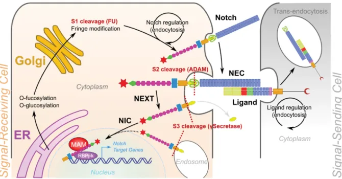
NOTCH receptors (Figure 6) are large single-pass transmembrane proteins of approximately 300kDa, which are post-translationally modified by a cleavage in the trans-golgi network, thanks to a furin-like convertase (Logeat et al., 1998), giving rise to the mature heterodimeric NOTCH receptor, composed by the NOTCH extracellular domain (NEC) of ~180kDa and the NOTCH transmembrane domain (NTM) of ~110-120kDa (Blaumueller et al., 1997). NEC is composed of 33 EGF-like repeats and a negative regulatory region (NRR) consisting of three Lin-12-and-Notch repeats (LNR) and a heterodimerization domain (HD). NEC-HD is able to dimerize with NTM through a similar HD. NTM contains a hydrophobic transmembrane domain (TM), a RAM domain through it binds its molecular effector RBPJ, an ankyrin repeats (AKR) region and a trans-activation domain (TAD) at the C-terminus. TAD contains 3 nuclear localisation sequences (NLS) and a PEST degron domain (Kovall et al., 2017).
During maturation, the EGF-like repeats region of the NEC is modified through O-fucosylation by the O-fucosyltransferase-1 (POFUT1) and by the O-fucose specific -1,3-N-acetylglucosaminyltransferases glycosyltransferases Fringe members (Manic, Lunatic and Radical). These modifications of the extracellular domain are required to modulate the
26 | P a g e
interaction of NOTCH with its ligands and control which ligands can interact with it (Haines and Irvine, 2003; Haltiwanger and Stanley, 2002).
Figure 6. Notch and its ligands structures.
The DSL ligands (for Delta/Serrate/LAG-2) are the three original Notch ligands discovered either in drosophila (Delta and Serrate) or in C. elegans (Lag-2). Their orthologues in vertebrates are Delta-like (Dll1,3,4) and Jagged (Jag1,2). These ligands (Figure 6), like the NOTCH receptors, are transmembrane proteins composed of an N-ter domain, a DSL and DOS domain and a various number of EGF-like repeats. Only Jagged ligands have a juxtamembrane cysteine-rich region. DLL1, DLL3, DLL4 and JAG1 carry in their C-ter a PSD-95/Dlg/ZO-1-ligand (PDZL) motif involved in Neotch independent cell signalling (D’Souza et al., 2010). Some non-canonical ligands were also described, such as scabrous, wingless or Thrombospondin-2 (Meng et al., 2009; Powell et al., 2001; Wesley, 1999).
The Notch pathway core
The Notch pathway is a cell-cell contact signalling pathway (Figure 7). Thus, it requires the interaction of a DSL ligand presented by a signal-sending cell. Upon ligand binding, a pulling force, induced by the endocytosis of the ligands (Itoh et al., 2003) seems to be necessary to induce a conformational change of the NNR, allowing a cleavage by ADAM10
(A-Disintegrin-27 | P a g e
And-Metalloprotease 10). This first cleavage releases the NEC bound to the ligand, which is internalised by the signal-sending cell. The NEXT fragment (NOTCH extracellular truncation) left in the signal-receiving cell is then cleaved a second time, either immediately or after internalisation, by g-secretases, releasing cytoplasmic NIC (Notch intracellular), able to translocate in the nucleus thanks to the 3 NLS motifs. Nuclear NIC can then recruit co-activators (such as Mastermind-like 1) and bind RBPJ.
Figure 7. The Notch signalling pathway.
The Notch receptor is synthetized in the Endoplasmic Reticulum (ER) where it is modified by O-fucosylation and O-glucosylation. NOTCH is cleaved in the Golgi by a Furin-like convertase, leaving the Notch extracellular domain (NEC) and NOTCH transmembrane (NTM) to assemble through their heterodimerisation domain (HD). The NOTCH receptor can also be modified by Fringe family members by extra O-fucosylation. Finely tuned intracellular trafficking controls the availability of NOTCH receptors at the membrane, where they can bind their ligands, presented by a neighbouring cell (the signal-sending cell). A pulling force generated by ligand endocytosis drives a conformational change of NRR, exposing the S2 cleavage site, targeted by the metalloprotease ADAM10, and releasing the NOTCH extracellular truncated (NEXT) domain. A second cleavage on the S3-site by gSecretase releases NIC, which enters the nucleus. There, NIC binds RBPJ and MAM-1 and drives the expression of the Notch target genes in the signal-receiving cell.
Notch modulation
As previously mentioned, NOTCH receptor post-translational modifications can modulate its response, depending on ligand affinity. However, co-expression of the ligand and the receptor on the same cell can switch off Notch signalling by cis-inhibition (Sprinzak et al., 2010). This molecular mechanism represents the base of the lateral inhibition process, used for cell
28 | P a g e
patterning and border definition (hence the original wing border phenotype) but also a source of cross-talk with other pathways able to induce the expression of Notch ligands in Notch activated cells, thus silencing Notch by cis-inhibition.
The Notch pathway is essential for intestinal homeostasis. The expression of NOTCH receptors, DSL ligands, and Notch target genes was broadly analysed in the embryonic and mouse adult intestinal epithelium. Of the four NOTCH receptors, it has been reported that
Notch1 is expressed in ISC and TA cells, while some crypts also express Notch2 (Carulli et
al., 2015; Fre et al., 2011). The ligands Dll1 and Dll4 are expressed by secretory cells, notably Paneth cells intercalating the ISC, providing the Notch niche signal. However, the Jag1 and
Jag2 ligands were reported to be co-expressed by Notch1+ cells (Sander and Powell, 2004).
Functionally, Notch has been first reported to have a role in cell fate specification when Milano and collaborators observed secretory cells expansion upon administration of the g-secretase inhibitor DAPT (Milano et al., 2004). Subsequently, the role of Notch in both ISC maintenance and lineage commitment were described through either loss of function (van Es et al., 2005) or gain of function (Fre et al., 2005).
& ' ) *
Growth factor receptors were discovered in the 80s by the characterisation of the EGFR which displayed an intrinsic protein kinase activity (Cohen et al., 1980). This discovery preceded the description of a super family of growth factor receptors with a tyrosine kinase activity, the growth factor receptors tyrosine kinase (GF-RTK). Among them, four EGFRs were described in mammals, referred to as ERBB1-4 (HER1-4).
EGFR pathway, and more widely, receptor tyrosine kinase (RTK) pathways are complex signalling networks regulating cell proliferation. Indeed, the RTK system is connected to MAPK and PI3K/mTOR signalling, able to sense and regulate all cell functions, in different manners depending on the interconnection of these pathway with cell state and the signalling kinetics Moreover, this pathway can get activated at the cell membrane but also during endocytosis, or more widely during receptor trafficking, and it is suspected to display different responses depending of the RTK localization. So, there is for now no sure way to link activation of RTK signalling with a defined genetic output. However, EGF stimulation can give an extraordinary stereotyped response, despite the numerous actors of the network (Avraham and Yarden, 2011).
29 | P a g e
Figure 8. RTK signalling is connected with PI3K/AKT, p38/MAPK, JNK, ERK, PKC and mTor. Upon binding of the ligand on one RTK subunit, the receptor assembles as a dimer, auto-phosphorylates and binds Grb2. RTK-Grb2 interacts and activates RAS and therefore the MAPK cascade. In parallel, phospho-RTK can activate, through Src, the PI3K/Akt cascade. Similarly, phospho-RTK can interact and activate, through PLC PIP2, the PKC cascade. Upon activation of these 3 kinase networks, mTOR, JNK, p38 and other kinases are activated, leading to the expression of numerous pleiotropic genes.
RTK signal transduction is mainly conducted by the MAPK/ERK pathway (Figure 8). Indeed, growth factor binding on RTK induces dimerization of the receptor and auto-phosphorylation. Phospho-RTK is recognised by growth factor receptor-bound protein 2 (GRB2), which recruits SOS and RAS, activating both the MAPK/ERK pathway (RAF-MEK-ERK) and the PI3K/AKT pathway. RAF activation by phosphorylation can activate the third rank protein kinase MAP3K (MAP kinase kinase kinase), which in turn induces, through Mitogen-activated protein kinase kinase (MKKs), the JNK or p38 pathway. In addition, phospho-AKT can also activate mTORs.
30 | P a g e
In parallel, RTK through PLC and PIP2 can activate the PKC pathway working both as retro-inhibition feedback for RTK and as nuclear transducer. Finally, depending of the growth factor, the receptor and the cellular context, MAPK/ERK, PI3K/AKT, mTOR, p38, JNK and PKC can be activated, stimulating proliferation or differentiation, survival, cell death or cell shape changes. Moreover, through these pathways RTK can modulate other signals such as the Hippo pathway.
!
In homeostasis, intestinal RTK are mainly represented by EGF receptors, IGF receptors and Ephrin receptors. EphB2 and EphB3 (receptors) are mostly expressed in the intestinal crypts in a decreasing gradient along the crypt-villus axis and they have been reported as key drivers of crypt proliferative activity and cell sorting within crypts (Batlle et al., 2002; Holmberg et al., 2006). However, while Ephrin ligands are expressed in the whole crypt, including ISC, this pathway seems to have a limited role in stem cells.
The EGFR pathway directly regulates ISC and progenitor behaviour by balancing proliferation and differentiation (Jiang et al., 2011; Suzuki et al., 2010; Yusta et al., 2009). The EGF receptor (EGFR) is highly expressed in ISC, whereas its ligands are mainly expressed by Paneth cells (Sato et al., 2011).
In normal conditions, EGFR is down-regulated by the pan-ErbB negative regulator LRIG1, a transmembrane protein expressed in ISC (Powell et al., 2012; Wong et al., 2012). Loss of Lrig1 causes EGFR activation and a concomitant rapid expansion of crypts and stem cell numbers. These lines of evidence illustrate the importance of this pathway as an inductive signal for stem cell proliferation. Although the role of EGFR/ErbB signalling for ISC proliferation is clearly established, it is not known if it is a prerequisite for ISC identity.
More generally, it is difficult to assign a defined role to the EGFR pathway in intestinal homeostasis, due to the redundancy of its receptors and their connections with MAPK, PI3K and other kinases, notably with the IGFR pathway. Indeed, many authors described similar mechanisms of action of EGFR and IGFR through myofibroblast-produced Glucagon-like peptide-2 (GLP-2) (Dubé et al., 2006; Rowland et al., 2011; Yusta et al., 2009).
During regeneration, the role of the EGFR pathway role is more straightforward. Indeed, it has been shown that loss of Egfr, Epiregulin (Ereg) or Amphiregulin (Areg), two EGF ligands upregulated during gut regeneration, caused a drastic reduction in the regeneration capacity of the intestinal epithelium following Dextran Sulfate Sodium (DSS) treatment (Egger et al.,
31 | P a g e
2000; Lee et al., 2004). Interestingly, both these studies using either a hypomorphic Egfrmut
allele or Ereg-/- mice, obtained phenotypes similar to Hippo loss-of-function, described later
(Gregorieff et al., 2015).
& + ") #% ," # ! , *
Discovery of Bone Morphogenetic Proteins (BMPs) was made possible by their ability to induce ectopic bones (Urist, 1965). The subsequent molecular characterisation of these proteins revealed that they belong to the TGFb super-family and they share many regulators with TGFbeta pathway (activins, inhibins, hormones …). Since, BMPs were found to be involved in many developmental and homeostatic programs, not only in bones but in most organs and cell types (Wagner et al., 2010).
" #
BMP signalling and more generally the TGFb super-family pathway involves receptors belonging to the RTK super-family. So, they share some RTK pathway mechanisms described above and so-called TGFb non-canonical pathways. The canonical BMP signalling operates through SMAD proteins. The BMP pathway implies private receptors such as BMPR1A, BMPR1B and BMPR2 and TGFb-shared receptors such as activins receptors, ACVR1A, ACVR2A and ACVR2B. While the private receptors act through SMAD1, 5, 8, the TGFb-shared ones act through the common effector SMAD2/3. Binding of the BMP ligand to the type 1 receptor leads to the phosphorylation of the type 2 BMP receptor. The activated receptor complex allows the phosphorylation of SMAD1, 5, 8, which become able to associate to SMAD4 and translocate into the nucleus. BMP signaling generally acts as inducer of differentiation, making it “anti-oncogenic” pathway. However, through its ability to regulate metabolism, ECM and through its shared receptors, BMPs are also reported to be involved in cancer, notably oesophageal cancer (Milano et al., 2007; Taylor et al., 2015).
" # $ %
In the intestinal epithelium, the main function of the BMP pathway is to promote differentiation and silence stemness, through the two ligands BMP-2 and BMP-4, expressed in the differentiated villi (Haramis, 2004; Hardwick et al., 2003). Thus, BMP signalling negatively controls proliferation and stemness in a fine gradient along the crypt-villus axis, allowing the progressive differentiation of the ISC and progenitors to feed the villus with functional differentiated cells (Auclair et al., 2007; Batts et al., 2006).
32 | P a g e
Figure 9. Simplified BMP and TGFb pathways
(Adapted from KEGG). BMP belong to the TGFb super family and share mechanisms and
regulators. Upon binding of BMP to the BMPR1 receptor, this phosphorylates the co-receptor BMPR2. The activated BMPR1/2 complex phosphorylates Smad1,5 and 8, allowing them to binds to Smad4. The complex SMAD1,5,8,4 translocates into the nucleus and, depending of the transcription factor (TF) and co-transcription factor (coTF) association, regulates genes involved in differentiation (adult), lineage specification (embryogenesis) and metabolism (iron and energy). TGFbRs act similarly through SMAD2/3 and depending of the TF and CoTF, TGFb pathway can induce apoptosis, G1 phase arrest, ECM remodelling or angiogenesis. BAMBI, a pseudo-receptor lacking the serine-threonine kinase domain, is one of the shared upstream negative regulators of TGFb and BMP pathways through a decoy function. SMAD6/7 is a negative regulator of SMADs interaction by direct competition with SMAD4. On the contrary, some regulators such as SARA, involved in the EMT process, are specific to the TGFb pathway.