HAL Id: hal-02145351
https://hal.archives-ouvertes.fr/hal-02145351
Submitted on 4 Jun 2019
HAL is a multi-disciplinary open access
archive for the deposit and dissemination of
sci-entific research documents, whether they are
pub-lished or not. The documents may come from
teaching and research institutions in France or
abroad, or from public or private research centers.
L’archive ouverte pluridisciplinaire HAL, est
destinée au dépôt et à la diffusion de documents
scientifiques de niveau recherche, publiés ou non,
émanant des établissements d’enseignement et de
recherche français ou étrangers, des laboratoires
publics ou privés.
Deciphering the reading of the genetic code by
near-cognate tRNA
Sandra Blanchet, David Cornu, Isabelle Hatin, Henri Grosjean, Pierre Bertin,
Olivier Namy
To cite this version:
Sandra Blanchet, David Cornu, Isabelle Hatin, Henri Grosjean, Pierre Bertin, et al.. Deciphering
the reading of the genetic code by near-cognate tRNA. Proceedings of the National Academy of
Sciences of the United States of America , National Academy of Sciences, 2018, 115 (12),
pp.3018-3023. �10.1073/pnas.1715578115�. �hal-02145351�
Deciphering the reading of the genetic code by
near-cognate tRNA
Sandra Blancheta,1, David Cornua, Isabelle Hatina, Henri Grosjeana, Pierre Bertina, and Olivier Namya,2
aInstitute for Integrative Biology of the Cell (I2BC), Commissariat à l‘énergie atomique et aux énergies alternatives, CNRS, Université Paris‐Sud, Université
Paris-Saclay, 91198 Gif‐sur‐Yvette cedex, France
Edited by Ada Yonath, Weizmann Institute of Science, Rehovot, Israel, and approved February 11, 2018 (received for review September 3, 2017)
Some codons of the genetic code can be read not only by cognate, but also by near-cognate tRNAs. This flexibility is thought to be conferred mainly by a mismatch between the third base of the codon and the first of the anticodon (the so-called“wobble” po-sition). However, this simplistic explanation underestimates the importance of nucleotide modifications in the decoding process. Using a system in which only near-cognate tRNAs can decode a specific codon, we investigated the role of six modifications of the anticodon, or adjacent nucleotides, of the tRNAs specific for Tyr, Gln, Lys, Trp, Cys, and Arg inSaccharomyces cerevisiae. Modifica-tions almost systematically rendered these tRNAs able to act as near-cognate tRNAs at stop codons, even though they involve noncanonical base pairs, without markedly affecting their ability to decode cognate or near-cognate sense codons. These findings reveal an important effect of modifications to tRNA decoding with implications for understanding the flexibility of the genetic code.
translation fidelity
|
stop codon readthrough|
tRNA modification|
genetic code|
near-cognate tRNAT
he ribosome uses transfer RNAs (tRNAs) to decode codons of mRNAs into a sequence of amino acids in a polypeptide. The specificity of this process depends on two main functions of the tRNA: its recognition by cognate aminoacyl-tRNA synthe-tases (1) and its anticodon pairing with the mRNA codons (2). In all living organisms, correct reading of the genetic code by tRNAs is essential, to prevent the misincorporation of amino acids and premature termination. However, translation fidelity is not maximal, and misreading rates can vary from 10−3to 10−6, depending on the tRNA and the organism considered (3, 4). Accurate decoding depends on the ability of the ribosome to dis-criminate between correct (cognate) and incorrect (near-cognate or noncognate) codon–tRNA interactions, while allowing the tRNA to decode synonymous codons. It was long thought that this ability was conferred by monitoring of exclusively the stability of the codon–anticodon complex, mainly based on the number of hydrogen bonds. However, recent data suggest that the ribosome plays an active role in decoding and thereafter accommodates both cognate and near-cognate tRNAs in a similar manner by forcing mismatched base pairs to adopt a Watson–Crick geometry as normal A-U/U-A or C-G/G-C base pairs (5).More than a 100 base and ribosome modifications in tRNAs of different organisms have been shown to contribute to various aspects of gene expression (6). They can be classified into two groups: one group of modifications that serves to stabilize the core of the tRNA and its overall L-shaped structure, and a second group of modifications that are crucial for correct mRNA decoding and fine-tuning of the translation process (7). The latter modifications are mainly present within the anticodon loop, in particular at positions 34, the first nucleotide of the anticodon, and 37, located immediately 3′ to the anticodon. These modifications have a direct impact on the capacity of the tRNA to read synonymous codons and to prevent decoding of near-cognate codons (8), although this view has recently been challenged by studies revealing that, in some cases, these modi-fications increase the misreading of codons (3). For example, it
has been suggested that the combination of 5-methylene deriv-atives and 2-thiolation modifications of U34restrict the decoding of codons ending with A (9, 10); moreover, 2-thiolation increases affinity of binding to the cognate codon and reduces tRNA re-jection (11). These modifications have also been implicated in protein homeostasis (12), in reading-frame maintenance (13), and in the enhancement of recognition by aminoacyl-tRNA synthetases (14).
However, data are not easily transposable from one organism to another because notable differences exist in the decoding strategies of distantly evolutionary related organisms (15). The impact of nucleotide modifications in the anticodon loop on the likelihood of near-cognate tRNA being used by the ribosome has yet to be analyzed in eukaryotes. Here, we used a system in which cytoplasmic near-cognate tRNAs do not compete with cognate tRNAs to study the impact of these modifications on the rec-ognition of naturally occurring stop codons (Fig. 1). We con-structed yeast mutant strains with deletions of genes encoding specific modification enzymes and used LC-MS/MS and ribo-some profiling techniques to investigate the impact of these deletions on the selection of natural suppressor tRNAs, which are, by definition, near-cognate tRNAs. We found that modifi-cations of the tRNA anticodon loop are required for decoding a near-cognate stop codon, but that they have no marked impact
Significance
Protein translation is a key cellular process in which each codon of mRNAs has to be accurately and efficiently recognized by cognate tRNAs of a large repertoire of noncognate tRNAs. A successful decoding process is largely dependent on the prence of modified nucleotides within the anticodon loop, es-pecially of tRNAs having to read A/U-rich codons. In this latter case, their roles appear to stabilize the codon–anticodon in-teraction, allowing them to reach an optimal energetic value close to that of other interacting tRNAs involving G/C-rich an-ticodons. In this work we demonstrate that, while helping an efficient translation of A/U-rich codons, modified nucleotides also allow certain unconventional base pairing to occur, as evidenced in the case of stop codon suppression.
Author contributions: S.B., D.C., and O.N. designed research; S.B., D.C., and I.H. performed research; P.B. contributed new reagents/analytic tools; S.B., D.C., I.H., H.G., P.B., and O.N. analyzed data; and H.G. and O.N. wrote the paper.
The authors declare no conflict of interest. This article is a PNAS Direct Submission.
This open access article is distributed underCreative Commons Attribution-NonCommercial-NoDerivatives License 4.0 (CC BY-NC-ND).
Data deposition: The data reported in this paper have been deposited in the Gene Ex-pression Omnibus (GEO) database,https://www.ncbi.nlm.nih.gov/geo(accession no.
GSE108772).
1Present address: Department of Physical Biochemistry, Max Planck Institute for Biophys-ical Chemistry, 37077 Goettingen, Germany.
2To whom correspondence should be addressed. Email: [email protected]. This article contains supporting information online atwww.pnas.org/lookup/suppl/doi:10. 1073/pnas.1715578115/-/DCSupplemental.
BIO
on the decoding of cognate codons. We also showed that overall stop codon suppression efficiency appears as a poor indicator of the ability of individual tRNAs to decode stop codons, due to adjustments between the various competing near-cognate cellu-lar tRNAs at reading the same stop codon. Taken together, our results highlight the flexibility of the genetic code and reveal an unexpected capacity of theSaccharomyces cerevisiae translation machinery to discriminate between sense and nonsense near-cognate codons. Although one cannot rule out that increased readthrough due toΨ35is a direct consequence of the increased stability of the codon:anticodon pairing, one appealing hypoth-esis is that genes coding for enzyme catalyzing Ψ35 have been conserved through evolution to regulate stop codon readthrough according to the cellular needs.
Results
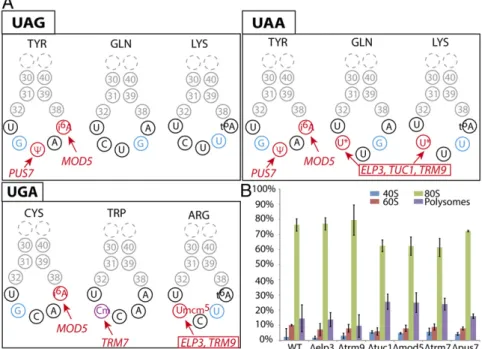
Deletion of Genes Coding Enzymes Responsible for tRNA Modifications Have No Major Impact on Cell Growth and Polysomes.To study the impact of several yeast tRNA modifications on the decoding of near-cognate codons, we used a reporter system based on the stop codon readthrough-dependent expression of a gene encoding a GST protein. This system provides a precise quantitative analysis of the incorporation of natural suppressor tRNAs at stop codons. In a previous study, we identified tyrosine, glutamine, and lysine tRNAs as the cytoplasmic tRNAs incorporated at UAA and UAG codons, and tryptophan, cysteine, and arginine tRNAs as the tRNAs incorporated at UGA codons (Table S1) (16).
Here, we systematically deleted six genes encoding enzymes responsible for posttranscriptionnal modification of bases or hydroxyl-riboses in the anticodon loop of the above-identified suppressor tRNAs (Fig. 1A). Three of these genes (Elp3p, Trm9p, and Tuc1p) are involved in conversion of U34residue in the glutamine tRNAUUG, lysine tRNAUUU, and arginine tRNAUCU into a hypermodified 5-methoxycarbonylmethyl-2-thiouridine residue (mcm5s2U) (6). The deletion of ELP3 or TRM9 results in glutamine and lysine tRNAs carrying only s2U34or cm5s2U34, respectively (U34and cm5s2U34for arginine tRNA), whereas the deletion of TUC1 results in glutamine and lysine tRNAs with mcm5U
34 (17, 18). The anticodons of the unique tyrosine and tryptophan suppressor tRNAs are also modified by two other enzymes: Pus7p, which catalyses the pseudouridylation of U35in
the tyrosine tRNA and in U2 snRNA (19); and Trm7p, which methylates C34 and C32 in the tryptophan tRNA (19, 20). An-other enzyme of interest is Mod5p, which converts the nucleo-tide at position 37, 3′-adjacent to the anticodon, to i6A in the tyrosine and cysteine tRNAs (21). We first confirmed that de-letions of these genes have no severe effect on cell viability (Fig. S1). Doubling times were similar for the parental and deletion strains, except for theΔTRM9 and ΔTRM7 strains, which grew more slowly, as reported in previous studies (20). Polysome profile analysis revealed that the proportions of 40S, 60S, and 80S ribosomes were unaffected in each strain carrying the above-selected deletion of modification enzyme genes (Fig. 1B).
U34Modification Affects Stop Codon Recognition by the Gln, Lys, and
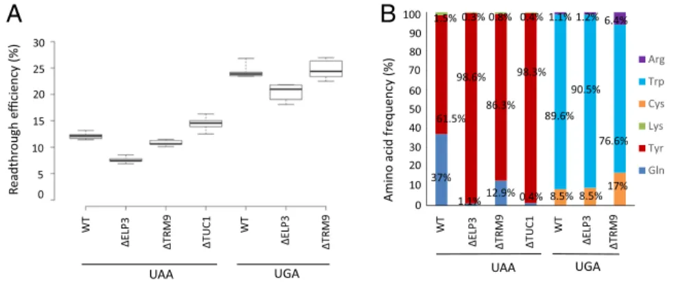
Arg tRNAs.In yeast, most of the uridine residues in position 34 of tRNAs are hypermodified. We investigated the influence of different elements of mcm5s2U modification on stop codon decoding by natural suppressor tRNAs by deleting the ELP3, TRM9, and TUC1 genes separately in the parental strain. Be-cause these enzymes modify U34 only in glutamine tRNAUUG, lysine tRNAUUU(reading UAA), and arginine tRNAUCU(reading UGA) (Fig. 1A), we assessed their impact on these two stop co-dons. We first used a dual reporter system (22) to analyze the effect of each deletion on the levels of stop codon suppression. We found no significant effect ofTRM9 deletion, but a slight decrease in readthrough efficiency in Δelp3 mutants, for the UAA and UGA codons only (Fig. 2A).
Next, we assessed the contributions of the glutamine tRNAUUG, lysine tRNAUUU, and arginine tRNAUCU to the levels of stop codon expression observed, by transforming theΔelp3, Δtrm9, and Δtuc1 strains with the GST reporter system (16) and performing mass spectrometry on the GST proteins generated by stop codon suppression, to quantify the frequency of incorporation of the amino acids corresponding to the three tRNAs, as previously described (Fig. S2B) (16). Quantification of the proportions of amino acid present at UAA codon revealed that incorporation of the glutamine tRNAUUG occurred less frequently in the three deletion strains than in the WT, this tRNA being replaced by the tyrosine tRNA, the incorporation of which was not affected by the deletions (Fig. 2B). We also found that deletions of ELP3 (resulting in mcm5-lacking s2U34) and TUC1 (resulting in the
Fig. 1. (A) A schematic diagram of the anticodon loops from the three near-cognate tRNAs inserted at the various stop codons. Modifications studied in this work are shown in red, with the name of the cor-responding gene (*= mcm5s2). The position of the
mismatch (or nonconventional base pairing) be-tween the anticodon and the stop codon is high-lighted in blue (or in purple if this position is also modified). Lysine tRNACUU and arginine tRNAUCU
also carry t6A
37modification that is not studied in
this work because of the strong growth defect link to the absence of this modification. (B) The quanti-fication of the various fractions of the polysome profiles obtained for each mutant. Three to five in-dependent experiments were performed for each mutant.
absence of the 2-thio group of uracil at position 34 bearing the 5-mcm group) decreased glutamine incorporation more strongly than the deletion of TRM9 (resulting in m-lacking cm5s2U34). Quantitative analyses for the UGA codon showed that deletion of ELP3 had no effect on the ratios of the various amino acids, whereas the deletion ofTRM9 strongly stimulated incorporation of arginine and also slightly increased cysteine incorporation, even though the corresponding cysteine tRNA is not thought to be modified byTRM9 (Fig. 2B). Clearly, 5-mcm modification does not have the same effect on the ability of glutamine, lysine, and arginine tRNAs to read UAA and UGA stop codons.
Modification of the Nucleotides Adjacent to the Anticodon Plays an Important Role in Near-Cognate Decoding. Beside the arginine tRNAUCU, the two main other suppressor tRNA of the UGA codon are the tryptophan and cysteine tRNAs, both of which carry nucleotide modifications. The tryptophan tRNA anticodon loop is modified at C32and C34into 2′-methyl cytosine (Cm) by Trm7p, while the Cys tRNAGCAis modified at A37into i6A37by Mod5p (Fig. 1A). They both belong to the same four-codon family boxes. Suppression involved a mismatch base pair at po-sition 34 of the anticodon and the third base (A) of the codon (respectively, Cm34-A3 for tryptophan tRNA and G34-A3 for cysteine tRNA). As shown in Fig. S3A, efficiency of UGA readthrough is not affected byTRM7 deletion (Fig. S3A), which also had no effect on the proportions of tryptophan, cysteine, and arginine incorporated (Fig. S3B). In contrast, the absence of Mod5p affected only the relative proportions of suppressor tRNAs not the level of the UGA suppression. Indeed, cysteine was not incorporated anymore at the UGA codon in the ΔMOD5 strain (Fig. S3B).
Like the cysteine tRNA, the tyrosine tRNA is modified by Mod5p, resulting in an i6A residue at position 37. We analyzed the effect of the elimination of this modification from the tyrosine tRNA on the level of stop codon suppression. Cysteine tRNA harbors a G34-A3mismatch when reading UAA and tyrosine tRNA harbors a G34-G3 mismatch when reading UGA). Quantitative analyses for theΔMOD5 strain showed that stop codon suppression efficiency at both the UAA and UAG codons was much lower than in the parental strain (Fig. 3A). Contribution of the tyrosine tRNA to the global suppression observed in theΔMOD5 strain was next investigated by quantifying the incorporation of suppressor tRNAs at UAA and UAG codons. Levels of tyrosine incorporation were slightly lower at the UAG codon and much lower at the UAA codon (Fig. 3B). Thus, the effect of the i6A modification of the tyrosine tRNA depends on the codon decoded.
The Presence of a Pseudouridine Residue at Position 35 of the Tyr tRNA Is Important for Stop Codon Suppression.InS. cerevisiae, there is only one cytosolic tyrosine tRNA, which acts as the major suppressor at the UAA and UAG stop codons, despite the cre-ation of a G34-A3 and a G34-G3 mismatch, respectively. This
tyrosine tRNA is unique because, in addition to being modified at position 37 (i6A), it is the only a suppressor in yeast that also has a pseudouridine (Ψ) instead of a usual uridine at the center (position 35) of the anticodon. This modification is post-transcriptionnally catalyzed by Pus7p (Fig. 1A).
The lack of this pseudouridine residue on suppression of the UAA and UAG stop codons was investigated as above for the other tRNA modifications.PUS7 deletion did not change tyro-sine tRNA stability as measured by four-leaf clover (FL)–qRT-PCR (23) (Fig. S5), but resulted in lowering the level of stop codon suppression at UAG with almost no effect on stop codon suppression at the UAA codon (Fig. 3A). However, a quantifi-cation of amino acids incorporated at UAA and UAG codons revealed that the frequency of tyrosine tRNA incorporation at UAG was strongly decreased at the benefit of the noncognate glutamine or lysine tRNAs (Fig. S2A), even more significantly than in theΔMOD5 strain (resulting in i6-lacking A37). While in the case of UAA suppression, the incorporation of tyrosine tRNA is now almost exclusively replaced by the noncognate glutamine tRNAUUG (Fig. 3B). We next checked whether the observed phenotype was solely due to the absence of Pus7p, by reexpressing either the WT protein or a catalytic mutant of Pus7p, Pus7-D256A [which has been reported to be inactive (19)] in the ΔPUS7 strain. Results indicate that reexpression of the WT protein restored parental amino acid levels, whereas expression of the single mutant did not (Fig. S4). Thus, both modifications of the tyrosine tRNA (i6A37andΨ35) are impor-tant for stop codon suppression, the i6A37 modification being important only for UAA readthrough, whereas the Ψ35 modifi-cation appears crucial mainly for the recognition of UAG codon. We also tested the absence of both modifications by constructing a strain in which the PUS7 and MOD5 genes were deleted. Subsequent quantification of stop codon readthrough efficiency (Fig. 3A) and amino acid insertion at the UAA and UAG codons (Fig. 3B) demonstrated a significant additive effect on stop co-don readthrough, especially on the UAG coco-don.
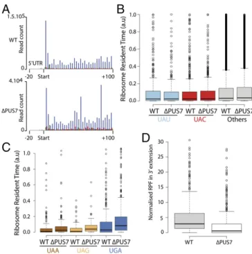
Absence of the Pseudouridine Residue at Position 35 in the Tyr tRNA Does Not Prevent the Decoding of Tyrosine Codons.InS. cerevisiae, synonymous tyrosine codons UAU and UAC are almost equally used in cytoplasmic mRNAs. Only one tyrosine tRNA isoacceptor containing a GΨA anticodon exists for reading each of these two codons. Because, tyrosine tRNA is dependent onΨ35for decoding the two near-cognate stop codons (UAA and UAG, see above), it was of interest to determine how tyrosine tRNA lacking ofΨ35 behaves for decoding the two tyrosine sense codons (UAU/C). We used a ribosome profiling technique that allows in vivo quantifi-cation of translation speed through calculation of the genome-wide ribosome residence time at each codon (24). We first checked the periodicity of the signal to demonstrate that the sig-nals obtained corresponded to translating ribosomes (Fig. 4A). Then, in both the parental andΔPUS7 strains, we compared the 0% 10% 20% 30% 40% 50% 60% 70% 80% 90% 100% Arg Trp Cys Lys Tyr Gln 61.5% Amino a cid fr e quency (%)
A
30 25 15 5 0 WT ∆ELP3 ∆TRM9 ∆TUC1 ∆TRM9 UAA UGA 20 10 WT ∆ELP3 R e adthr o ugh e fficiency (%) WT ∆ELP3 ∆TRM9 ∆TUC1 ∆TRM9 UAA UGA WT ∆ELP3 90 80 70 60 50 40 30 20 10B
0 100 37% 1.5% 0.3% 98.6% 1.1% 0.8% 86.3% 12.9% 0.4% 98.3% 0.4% 1.1% 1.2% 6.4% 90.5% 89.6% 76.6% 8.5% 8.5%17%Fig. 2. Consequences on stop codon suppression of deletion genes coding for U34 modification in vari-ous tRNAs. (A) Quantification of readthrough effi-ciency at UAA and UGA codons in three mutants in which mcm5s2U
34formation was abolished. At least
five independent experiments were performed for each value. The central lines show the medians; the box limits indicate the 25th and 75th percentiles. (B) Corrected intensities of readthrough peptides nor-malized according to the amount of GST were quantified by determining the mean value for three experiments. Quantification was performed as de-scribed in Materials and Methods.
BIO
normalized ribosome density at each tyrosine and stop codons in the A site for each gene (Materials and Methods). We found that UAU and UAC were decoded with similar efficiency in both Δpus7 and parental strains (P = 0.4 and 0.6, respectively) (Fig. 4B) and that this efficiency was not different from that for all of the other codons. We concluded thatΨ35in Tyr tRNA does not allow a better decoding of the near-cognate UAU codon, despite it playing an important role for decoding near-cognate UAA and UAG codons. Interestingly, we did not observe any significant difference in ribosome occupancy at stop codons (Fig. 4C), whereas a reduction of in-frame ribosome footprints downstream the stop codon is evident (P = 1.5 × 10⁻⁴) (Fig. 4D). This confirms that stop codon readthrough is impaired in the Δpus7 strain without accumulation of ribosomes at natural stop codons when tyrosine tRNA is lackingΨ35.
Discussion
Decoding of mRNAs by tRNAs into polypeptides on the ribosome was considered for a long time as a relatively simple process, where formation of a set of H-bonds between three complemen-tary base pairs of codons and anticodons within a minihelix con-figuration plays a major role. However, it is now clear that other elements of tRNA molecules, as well as of rRNA of the ribosome, are also important for accurate and efficient mRNA decoding (5, 8, 25, 26). The biochemical properties of tRNAs depend strongly on posttranscriptional modifications of nucleotides within the anticodon (positions 34 and 35) and the so-called“proximal an-ticodon loop” of tRNA (positions 37, 38, and/or 32) (Fig. S6) (6, 15). The main role of these nucleotide modifications is generally argued to notably fine-tune the accuracy of decoding: that is, to restrict the decoding of split box codons (2:2 or 3:1 decoding boxes) or to extend the decoding of nonsplit four-codon boxes. However, it is now clear that, together with other bases of the anticodon loop, their roles are to mainly achieve a uniform ribo-some binding by stabilizing the codon–anticodon interaction of tRNAs (27). This is especially important for tRNA harboring an A/U-rich anticodon triplet that have to reach about the same optimal interaction energies than tRNAs harboring G/C-rich an-ticodon. Base or ribose modifications lead to various chemical and physical consequences, such as keto-enol tautomerism, base pro-tonation, uridine isomerization to pseudouridine (Ψ), anti to syn base transconformation, freezing a 2′-O-glycosidyl bond into its 3′-endo configuration, improvement of base stacking with neighbor-ing bases, or additional interactions with ribosomal elements. One corollary of these multiple stabilizing effects is to allow certain noncanonical base pairs, which are isosteric with standard Wat-son–Crick pairs, to occur during decoding (28, 29).
In a preceding work, we and others identified in yeast eight naturally occurring tRNAs that misread stop codons as sense
codons under in vivo conditions (Fig. 1) (16, 30). Although we are using a [PSI+] strain, it has been previously shown that this does not impact tRNA ratios found at the stop codon during read-through (30). Here, we investigated the consequences of the ab-sence of a given modification normally present in the anticodon loops (at positions 34, 35, and/or 37) on their relative efficacy to readthrough stop codons. This is a different situation from studies of missense errors at sense codons during the elongation process (3), because the near-cognate tRNAs of a stop codon will never be in competition with cognate tRNA that normally does not exist.
Lys Tyr Gln
WT
∆MOD5 ∆PUS7 ∆PUS7 ∆MOD5
WT ∆ MOD5 ∆PUS7 ∆PUS7 ∆MOD5 37% 61.5% 1.5% 87.4% 9.9% 2.8% 92.1% 2.2% 5.7% 90.3% 7.5% 2.2% 3.3% 95.4% 1.3% 10.8% 79.3% 9.9% 57.8% 23.4% 18.8% 73.1% 5.8% 21% UAA UAG Amino a cid fr e quency (%) 100 90 80 70 60 50 40 30 20 10
B
A
40 30 20 10 0 0 WT∆MOD5 ∆PUS7 ∆PUS7 ∆MOD5
WT ∆ MOD5 ∆PUS7 ∆PUS7 ∆MOD5 UAA UAG R e adthr o ugh e fficiency (%)
Fig. 3. Consequences on stop codon suppression of deletion genes coding for enzymes catalyzing for-mation of i6A
37andΨ35in various tRNAs. (A)
Quan-tification of readthrough efficiency at UAA and UAG codons in the two mutants lackingΨ35and i6A37
formation. At least five independent experiments were performed for each value. The box plot is done as in Fig. 2A. (B) Quantification of the relative pro-portions of the amino acids identified at the two stop codons as in Fig. 2B.
Fig. 4. Ribosome profiling and codon occupancy in the WT andΔPUS7 strains. (A) Metagene analysis of 28-mers in the A site of the ribosome, be-tween−20 and +100 nt from the start codon of each CDS. Phasing is in-dicated by a color scheme: phase 1 (blue), 2 (red), 3 (green). Periodicity is clearly visible for both strains, confirming that the footprints correspond to active ribosomes. (B and C) Quantification of the ribosome occupancy at tyrosine and stop codons in the A site. The lines in the center correspond to medians.“Other” corresponds to all codons other than UAU, UAC, and stop codons. n = 1,557 (UAU), 2,143 (UAC), 209 (UAA), 75 (UAG), 344 (UGA), 169,210 (Other) sample points. (D) Normalized ribosome-protected mRNA fragments (RPF) counts found downstream of the CDS stop codon and up-stream the next in-frame stop codon (3′ extension). Box plot is done as previously indicated. Differences are significant according to a student test (P = 1.5 × 10⁻⁴). n = 593 WT and Δpus7.
All tRNAs inFig. S6belong to the so-called“intrinsically weak, A/ U-rich” interacting tRNAs (Lys, Tyr) or “intermediate” interact-ing tRNAs (Gln, Arg, Trp, Cys), which are supposed to be among the most prone for miscoding (for details, see ref. 25). First, in the glutamine and lysine tRNAs, uridine-34 is modified to mcm5s2U (symbolized as sU*) and to mcm5U only (U*) in the arginine tRNAUCU. The chemical adduct at C5of U34has been shown to favor binding to G3over A3, via enol-keto tautomerism, while the thiol (s2) group, beside reinforcing base pairing mostly with A3, also reinforces stacking, and thus stability and conformational rigidity of both base 34 and the neighboring base pair N35-N2(Fig. S6) (9, 10). Deletion (one by one) of each of the three genes responsible for U34 modification into mcm5s2U34 inS. cerevisiae (ELP3, TRM9, andTUC1) shows a slight growth defect for only the deletion of TRM9 (affecting only the last methylation step of cm5U34) (Fig. S1). Deletion of these genes had only a slight impact on UAA readthrough efficiency, the most pronounced effect being observed for theΔelp3 mutant corresponding to the total lack of 5-mcm group on U34(Fig. 2A). The interesting observation is that, while the efficacy is not much affected in both the ΔELP3 (lacking 5-mcm but not 2-thio) andΔtuc1 (lacking 2-thio but not 5-mcm) strains, glutamine tRNAUUGwas no longer able to compete with tyrosine tRNA, now becoming almost the only suppressor tRNA for UAA readthrough, whereas in theΔTRM9 strain (resulting in the lack of one methyl group on cm5s2U) a weak competition persists between glutamine and tyrosine tRNAs (Fig. 2B). These results demonstrate that both the fully methylated 5-mcm and 2-thio adducts of U34greatly improve the stability of the codon– anticodon interaction so that a noncanonical G36:U1can occur, allowing glutamine tRNAUUGto be stably incorporated at UAA. Because all of the base pairs of the minihelix have to adopt a strict Watson–Crick geometry (5, 26), this G36:U1pair probably fulfill this requirement through a keto-enol tautomerization of either base (31). The data obtained with lysine tRNA reading the same UAA are going in the same direction. Only when U34is fully mod-ified to mcm5s2U34, a U36:U1opposition in this case can be accom-modated, yet obviously less efficiently than for a G36:U1. Such a U:U opposition, with possibly only one single H-bond within a Watson– Crick helix geometry, seems to require a water molecule (28).
Slightly different results were obtained for reading UGA stop codon, involving a strong middle C-G pair, by the minor arginine tRNAUCUharboring also a 5-mcm residue but not a 2-thio group at U34(U*).ELP3 deletion (absence of 5-mcm on U34) did not alter significantly the ratio of amino acids inserted at UGA (Fig. 2B), whereas both arginine and cysteine incorporation levels were a bit higher in theΔtrm9 strain (lacking of a methyl group on cm5U34). The better incorporation of arginine tRNA at UGA may originate from the generation of a free carboxyl group due to the lack of the methyl on cm5U34. However, this conclusion has to be taken with caution because a slight increase in cysteine incorporation has also been observed, while the cysteine tRNA is not normally modified by Trm9p. We believe instead that the strong C35-G2 pair is the main stabilization element and the adduct on U34 only slightly contributes to the ability of arginine tRNA to play a role of UGA suppressor. These results point out the importance of considering the global energetic and stability of a codon–anticodon interacting system, including the proximal extended anticodon (25), before generalizing any observation.
The opal UGA codon is decoded mostly by tryptophan tRNA, followed by cysteine tRNA, both belonging to the same four-codon box. In tryptophan tRNA, A37 is unmodified while in cysteine tRNA it is modified by Mod5p to i6A37. Conversely, in tryptophan tRNA, C34is modified to Cm34(as well as Cm32) by Trm7p, while G34in cysteine tRNA is not modified. The pres-ence of a hydrophobic isopentenyl group on A37 is known to reinforced the stacking power of the purine adenine on the ad-jacent base pair A36-U1(both by lateral and interstrand stacking, thus with the first codon base) (32). A 2′-O-metyl group on the
hydroxyl of C34is known to favor a more rigid 3′-endo confor-mation of the ribose, confines the H-bound forconfor-mation, and in-creases hydrophobic surface, hence also stacking with neighboring bases (33). No significant effect ofTRM7 deletion (lacking Cm34) is found on either global stop codon readthrough efficiency (Fig. S3A) or the nature of the amino acids incorporated, attesting again that the strong central C35-G2pair rather than the 2′-O-methylation of C34 (and C32) is probably the main stabilizing element (Fig. S3B). An isosteric C34or Cm:A3opposition prob-ably occurs through an amino ionization of either C/Cm or A; alternatively, Cm:A3can also occur in the absence of a hydrogen bond (28, 29).
In contrast, deletion of the isopentenyl group on A37of cysteine tRNA in the ΔMOD5 strain prevents incorporation of cysteine at UGA stop codon (Fig. S3B). The same trend was observed with i6A37containing tyrosine tRNA, the suppression efficiency being more severely affected in the case of UAA than of UAG (Fig. 3B). Thus, lowering the stability of a A36-U1Watson–Crick pair by the lack of an isopentenyl group on A37in cysteine or tyrosine tRNA affects the efficacy of a triplet pairing involving a G34mispair with A3of stop codon UGA and UAA, respectively, or with G3in the case of stop codon UAG. Formation of an isosteric base pair in-volving two purines requires that one of the two purines switches conformation fromanti to syn, probably the one of the codon, to form a Watson–Crick/Hoogsteen base pair (28, 29). Notice that codon–anticodon involving noncanonical G34:A3or G34:G3have been demonstrated for several tRNAs involving G/C-rich anticodons and reading codons of the unsplit four-decoding boxes. For a long time, this type of decoding process has been designated as a“two-out-of-three” decoding rule (34).
Tyrosine tRNA carries an unusualΨ at the central position of the anticodon that is catalyzed by Pus7p. Pseudouridine enables engaging a strong base pairing (almost as strong as C-G), rigid-ifies the sugar-phosphate backbone, and improves stacking with a neighboring base pair, forcing aΨ-A pair to adopt an A-form conformation, as in a genuine Watson–Crick helix (35, 36). In agreement with an earlier similar observation in a plant trans-lation system (37), we found that tyrosine tRNA in a ΔPus7 strain (lacking Ψ35) is a less-efficient suppressor at both UAA and UAG stop codons, but mainly at UAA (Fig. 3B), although the tyrosine tRNA is equally stable in WT and ΔPus7 strains (Fig. S5). The effect of Ψ35 appears independent of A37 iso-pentenylation, as shown by the additive effect of thePUS7 and MOD5 double deletion on UAG (Fig. 3B). The level of UAA readthrough was not affected byPUS7 deletion, even though this deletion prevented the incorporation of the tyrosine tRNA at UAA (Fig. 3). These results illustrate that a strong stabilization of codon–anticodon interaction allows a noncanonical G34:A3 base opposition to occur. Interestingly, a ribosome profiling (RiboSeq) experiment performed in parental WT and ΔPUS7 strains allows us to demonstrate that a tyrosine tRNA iso-acceptor (with or withoutΨ35) reads equally well both synony-mous tyrosine codons, UAU and UAC (Fig. 4). This indicates the lack of involvement ofΨ35in discrimination between these two tyrosine codons in a situation where there is no real mis-match between the anticodon and the codon (whereas a G34:A3 or G34:G3base opposition systematically occurs when tyrosine tRNA reads stop codons). A similar situation exists with echi-noderm asparagine tRNAGUUharboring aΨ35in reading a lysine near-cognate codon AAA, while not affecting the normal read-ing of asparagine codons AAC/U (38).
In conclusion, our findings reveal the importance of certain modifications for the suppression of stop codons. Consistent with the findings of another recent study (3), we found that tRNA modifications did not solely serve to restrict the decoding ca-pacity of the tRNA to its cognate codon, but also allow the decoding of near-cognate stop codons. This involves an expected mismatch at the wobble position and also noncanonical base
BIO
pairs at the third anticodon position and the first codon position. The allowed base mispairs or base oppositions are, however, only those that would mold within a minihelix of the Watson–Crick type of geometry that is mandatory for the aminoacyl-tRNA to be accepted and finally captured by the mRNA-ribosome ma-chinery (26). We observed that mismatch with a stop codon never occurred at the middle position of the anticodon, while such possibilities have been demonstrated at sense codons (6, 7). The reason is that there is no naturally occurring tRNA that could sustain such base opposition (like U:G) within a Watson– Crick minihelix in the whole tRNA repertoire ofS. cerevisiae.
Only a small subset of tRNAs have been analyzed and it is likely that studies of other tRNAs in other translation systems, as well as organisms, will turn up other surprises. In this work, we propose that the activities of certain tRNA modification enzymes can be a regulatory device for the production of certain functional “read-through” proteins. This work should help at elaborating synthetic or mutated tRNAs able to introduce nonproteinous amino acids at specific locations of a mRNA where a sense codon has been appropriately mutated into a stop codon (39).
Materials and Methods
Detailed information on materials and methods used in this study is provided inSI Materials and Methods.
Strains and Plasmids. All of the strains used in this study were derived from 74-D694 (mata ade1-14 [UGA] ura3-52 trp1-289 [UAG] his3Δ200 leu2-3,112 [PSI+]).
Mass Spectrometry.
LC-MS/MS analyses. Proteolytic peptides were identified and quantified with a Triple-TOF 4600 mass spectrometer (ABSciex) coupled to the nanoRSLC system (Thermo Fisher Scientific) equipped with a trap column (Acclaim PepMap100C18, 75μm i.d. × 2 cm, 3 μm) and an analytical column (Acclaim PepMapRSLCC18, 75μm i.d. × 25 cm, 2 μm, 100 Å).
Relative quantification of readthrough peptides. The intensity of each chro-matographic peak was corrected by a factor taking the ionization and di-gestion efficiencies of each readthrough peptide into account (16). The means of three technical replicates are reported.
RNA-Seq and Ribosome Profiling. Cells were grown to an OD600of 0.6 in 1 L of
liquid glucose-YNB supplemented with CSM and 2× adenine and flash-frozen. Total RNA and polysomes were extracted as previously described (40). ACKNOWLEDGMENTS. We thank Eric Westhof for his useful advice and stimulating discussions; Ammara Mohammad and Stéphane Le Crom from the Genomic Paris Centre for assistance with sequencing; and Alex Edelman & Associates for correcting English usage in the work. This work was sup-ported by the Association pour la Recherche sur le Cancer Grants (SFI20101201647 and PJA 20131200234); French association“Ligue Nationale contre le cancer” Grant 3FI10167LVCY (to O.N.); and a fellowship from the Association pour la Recherche sur le Cancer (to S.B.). P.B. is supported by Agence Nationale de la Recherche (ANR) RiboTerm and O.N. has received support from the ANR rescue_ribosome Grant (ANR-17-CE12-0024-01). This work benefited from the facilities and expertise of the SICaPS platform of IMAGIF for the mass spectrometry analysis and from the High-throughput Sequencing Platform of I2BC for library preparation. This work was supported by the France Génomique national infrastructure, funded as part of the‘Investissements d’Avenir’ program managed by the ANR (Contract ANR-10-INBS-0009).
1. Yadavalli SS, Ibba M (2012) Quality control in aminoacyl-tRNA synthesis its role in translational fidelity. Adv Protein Chem Struct Biol 86:1–43.
2. Parker J (1989) Errors and alternatives in reading the universal genetic code. Microbiol Rev 53:273–298.
3. Manickam N, Joshi K, Bhatt MJ, Farabaugh PJ (2016) Effects of tRNA modification on translational accuracy depend on intrinsic codon-anticodon strength. Nucleic Acids Res 44:1871–1881.
4. Kramer EB, Vallabhaneni H, Mayer LM, Farabaugh PJ (2010) A comprehensive analysis of translational missense errors in the yeast Saccharomyces cerevisiae. RNA 16: 1797–1808.
5. Demeshkina N, Jenner L, Westhof E, Yusupov M, Yusupova G (2012) A new un-derstanding of the decoding principle on the ribosome. Nature 484:256–259. 6. Machnicka MA, et al. (2013) MODOMICS: A database of RNA modification pathways—
2013 update. Nucleic Acids Res 41:D262–D267.
7. Yarian C, et al. (2002) Accurate translation of the genetic code depends on tRNA modified nucleosides. J Biol Chem 277:16391–16395.
8. Agris PF, Narendran A, Sarachan K, Väre VYP, Eruysal E (2017) The importance of being modified: The role of RNA modifications in translational fidelity. Enzymes 41:1–50. 9. Takai K, Yokoyama S (2003) Roles of 5-substituents of tRNA wobble uridines in the
recognition of purine-ending codons. Nucleic Acids Res 31:6383–6391.
10. Sochacka E, et al. (2017) C5-substituents of uridines and 2-thiouridines present at the wobble position of tRNA determine the formation of their keto-enol or zwitterionic forms—A factor important for accuracy of reading of guanosine at the 3´-end of the mRNA codons. Nucleic Acids Res 45:4825–4836.
11. Ranjan N, Rodnina MV (2017) Thio-modification of tRNA at the wobble position as regulator of the kinetics of decoding and translocation on the ribosome. J Am Chem Soc 139:5857–5864.
12. Nedialkova DD, Leidel SA (2015) Optimization of codon translation rates via tRNA modifications maintains proteome integrity. Cell 161:1606–1618.
13. Tükenmez H, Xu H, Esberg A, Byström AS (2015) The role of wobble uridine modifi-cations in +1 translational frameshifting in eukaryotes. Nucleic Acids Res 43: 9489–9499.
14. Rogers KC, Crescenzo AT, Söll D (1995) Aminoacylation of transfer RNAs with 2-thi-ouridine derivatives in the wobble position of the anticodon. Biochimie 77:66–74. 15. Grosjean H, de Crécy-Lagard V, Marck C (2010) Deciphering synonymous codons in the
three domains of life: Co-evolution with specific tRNA modification enzymes. FEBS Lett 584:252–264.
16. Blanchet S, Cornu D, Argentini M, Namy O (2014) New insights into the incorporation of natural suppressor tRNAs at stop codons in Saccharomyces cerevisiae. Nucleic Acids Res 42:10061–10072.
17. Karlsborn T, et al. (2014) Elongator, a conserved complex required for wobble uridine modifications in eukaryotes. RNA Biol 11:1519–1528.
18. Leidel S, et al. (2009) Ubiquitin-related modifier Urm1 acts as a sulphur carrier in thiolation of eukaryotic transfer RNA. Nature 458:228–232.
19. Behm-Ansmant I, et al. (2003) The Saccharomyces cerevisiae U2 snRNA:pseudouridine-synthase Pus7p is a novel multisite-multisubstrate RNA:Psi-snRNA:pseudouridine-synthase also acting on tRNAs. RNA 9:1371–1382.
20. Pintard L, et al. (2002) Trm7p catalyses the formation of two 2′-O-methylriboses in yeast tRNA anticodon loop. EMBO J 21:1811–1820.
21. Dihanich ME, et al. (1987) Isolation and characterization of MOD5, a gene required for isopentenylation of cytoplasmic and mitochondrial tRNAs of Saccharomyces cerevisiae. Mol Cell Biol 7:177–184.
22. Namy O, et al. (2002) Gene overexpression as a tool for identifying new trans-acting factors involved in translation termination in Saccharomyces cerevisiae. Genetics 161:585–594. 23. Honda S, Shigematsu M, Morichika K, Telonis AG, Kirino Y (2015) Four-leaf clover
qRT-PCR: A convenient method for selective quantification of mature tRNA. RNA Biol 12:501–508.
24. Ingolia NT (2016) Ribosome footprint profiling of translation throughout the ge-nome. Cell 165:22–33.
25. Grosjean H, Westhof E (2016) An integrated, structure- and energy-based view of the genetic code. Nucleic Acids Res 44:8020–8040.
26. Rozov A, Demeshkina N, Westhof E, Yusupov M, Yusupova G (2016) New structural insights into translational miscoding. Trends Biochem Sci 41:798–814.
27. Olejniczak M, Dale T, Fahlman RP, Uhlenbeck OC (2005) Idiosyncratic tuning of tRNAs to achieve uniform ribosome binding. Nat Struct Mol Biol 12:788–793.
28. Westhof E (2014) Isostericity and tautomerism of base pairs in nucleic acids. FEBS Lett 588:2464–2469.
29. Westhof E, Yusupov M, Yusupova G (2014) Recognition of Watson-Crick base pairs: Constraints and limits due to geometric selection and tautomerism. F1000Prime Rep 6:19.
30. Roy B, Leszyk JD, Mangus DA, Jacobson A (2015) Nonsense suppression by near-cognate tRNAs employs alternative base pairing at codon positions 1 and 3. Proc Natl Acad Sci USA 112:3038–3043.
31. Demeshkina N, Jenner L, Westhof E, Yusupov M, Yusupova G (2013) New structural insights into the decoding mechanism: Translation infidelity via a G·U pair with Watson-Crick geometry. FEBS Lett 587:1848–1857.
32. Konevega AL, et al. (2004) Purine bases at position 37 of tRNA stabilize codon-anti-codon interaction in the ribosomal A site by stacking and Mg2+-dependent interac-tions. RNA 10:90–101.
33. Kawai G, et al. (1992) Conformational rigidity of specific pyrimidine residues in tRNA arises from posttranscriptional modifications that enhance steric interaction between the base and the 2′-hydroxyl group. Biochemistry 31:1040–1046.
34. Lagerkvist U (1978)“Two out of three”: An alternative method for codon reading. Proc Natl Acad Sci USA 75:1759–1762.
35. Davis DR (1995) Stabilization of RNA stacking by pseudouridine. Nucleic Acids Res 23: 5020–5026.
36. Spenkuch F, Motorin Y, Helm M (2014) Pseudouridine: Still mysterious, but never a fake (uridine)! RNA Biol 11:1540–1554.
37. Zerfass K, Beier H (1992) Pseudouridine in the anticodon G psi A of plant cytoplasmic tRNA(Tyr) is required for UAG and UAA suppression in the TMV-specific context. Nucleic Acids Res 20:5911–5918.
38. Tomita K, Ueda T, Watanabe K (1999) The presence of pseudouridine in the antico-don alters the genetic code: A possible mechanism for assignment of the AAA lysine codon as asparagine in echinoderm mitochondria. Nucleic Acids Res 27:1683–1689. 39. Mukai T, Lajoie MJ, Englert M, Söll D (2017) Rewriting the genetic code. Annu Rev
Microbiol 71:557–577.
40. Baudin-Baillieu A, et al. (2014) Genome-wide translational changes induced by the prion [PSI+]. Cell Rep 8:439–448.