A newborn was referred to our hospital because of prominent cardiac pulsations in the subxyphoid region associated with supraumbilical midline cutaneous le-sions (Fig. 1). Echocardiography revealed mesocardia with a ventricular septal defect, a secundum atrial septal defect, and an unroofed coronary sinus. The morphological correlate for the pulsatile tumor was a left ventricular diverticulum diagnosed by transtho-racic echocardiography (Fig. 2). Cardiac magnetic resonance imaging (MRI) confirmed the presence of a left ventricular diverticulum originating from the apex of the heart and ruled out the presence of a dia-phragmatic hernia (Fig. 3).
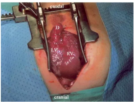
Cardiac surgical repair, performed at 4 weeks of age, confirmed the preoperative findings (Fig. 4). The left ventricular diverticulum was a continuation of the apex of the left ventricle and was totally covered by peri-cardium. We found a midline muscular defect of the abdominal wall and a missing linea alba, as well as a
missing xyphoid, but no evidence of diaphragmatic hernia or additional midline malformations.
This represents a forme fruste of Cantrell’s Syn-drome. Cantrell’s pentalogy includes a midline, sup-raumbilical abdominal wall defect, a defect of the lower sternum, a deficiency of the anterior diaphragm, a defect in the diaphragmatic pericardium, and con-genital intracardiac defects [1]. In this case, the defi-nition of the topographic localization of the diverticulum performed by MRI and of its relation to the diaphragm was essential in planning the surgical repair.
References
1. Cantrell JR, Haller JA, Ravitch MM (1958) A syndrome of congenital heart defects involving the abdominal wall, ster-num, diaphragm, pericardium and heart. Surg Gynecol Obstet 107:602–614
W. Knirsch (&) Æ E. V. Bu¨chel
Division of Pediatric Cardiology, University Children’s Hospital Zurich, Steinwiesstr-75, CH 8032, Zurich, Switzerland
E-mail: walter.knirsch@kispi.unizh.ch A. Dodge-Khatami
Division of Cardiovascular surgery, University Children’s Hospital Zurich, Switzerland
D. Bolz
Division of Pediatric Cardiology, University Children’s Hospital Basel, Switzerland
Pediatr Cardiol (2006) 27:652–654 DOI 10.1007/s00246-005-6001-2
123
I M A G E S I N P E D I A T R I C C A R D I O L O G Y
Cantrell’s Syndrome Forme Fruste in a Newborn Diagnosed by
Transthoracic Echocardiography and Cardiac Magnetic
Resonance Imaging
W. Knirsch Æ A. Dodge-Khatami Æ D. Bolz Æ E. Valsahgiacomo Bu¨chel
Fig. 1 Preoperative status showing the subxyphoid-to-supraumbilical covered midline defect of the abdominal wall
Fig. 2 (Left) Two-dimensional echocardiogram of the left ventricular diverticulum (D, arrows) and small left ventricle (LV). (Right) Color Doppler showing blood flow (red) into the diverticulum (D)
Pediatr Cardiol (2006) 27:652–654 653
Fig. 4 Intraoperative situs of the left ventricular diverticulum (D). LV, left ventricle, RV, right ventricle
Fig. 3 Contrast-enhaced MR angiography demonstrating the left ventricular diverticulum (D) with ‘‘vermiform’’ structure originating from the apex of the left ventricle (LV) and its relationship to the diaphragm (asterists) (lateral projection)
654 Pediatr Cardiol (2006) 27:652–654