S Y M P O S I U M : A B J S C A R L T . B R I G H T O N W O R K S H O P O N H I P P R E S E R V A T I O N S U R G E R Y
Surgical Technique
Second-generation Bone Marrow Stimulation via Surgical Dislocation to Treat Hip
Cartilage Lesions
Michael Leunig MD, Lisa M. Tibor MD, Florian D. Naal MD, Reinhold Ganz MD, Matthias R. Steinwachs MD
Published online: 7 July 2012
Ó The Association of Bone and Joint Surgeons1 2012
Abstract
Background Compared to knees, hips have more bony constraint and soft tissue coverage. Thus, repair of focal cartilage defects in hips requires more invasive and tech-nically complex surgeries than simple arthroscopy or arthrotomy. Autologous matrix-induced chondrogenesis (AMIC) is a second-generation bone marrow stimulation technique. Improvement in Tegner, Lysholm, International Cartilage Repair Society (ICRS), and Cincinnati scores has been reported at 1 and 2 years after AMIC in knees. AMIC is potentially useful to repair defects in hips, but it is unknown whether it relieves symptoms or results in a durable construct.
Description of Technique A surgical hip dislocation is used to access the defect. This is de´brided to stable carti-lage shoulders, necrotic bone is removed, and the lesion base is drilled. Autogenous bone graft is used for lesions with bony defects to create a level surface. Fibrin gel and a collagen membrane are placed to stabilize the superclot for fibrocartilage formation.
Methods We treated six patients with AMIC in the hip between 2009 and 2010. We obtained Oxford Hip and UCLA Activity Scores. Repair quality was assessed on 6-month postoperative MRI using the modified magnetic resonance observation of cartilage repair tissue (MOCART) system. Minimum 1-year followup data were available for four patients (range, 1–2.5 years).
Results Postoperative Oxford Hip Scores ranged from 13 to 17, UCLA Activity Scores ranged from 5 to 10, and MOCART scores ranged from 55 to 75. No complications occurred.
Conclusions We describe AMIC in the hip. Although these patients had pain relief and improved function, long-term followup is necessary to assess the duration of improvement, durability of repair, and potential for arthrosis.
Level of Evidence Level IV, therapeutic study. See Instructions for Authors for a complete description of levels of evidence.
Introduction
As the concept of femoroacetabular impingement (FAI) has become accepted [23], more hip problems are being treated with joint preservation surgeries [5,46,61]. These advances in hip preservation are fueled by better diagnos-tics and technical improvements in both hip arthroscopy Each author certifies that he or she, or a member of his or her
immediate family, has no commercial associations (eg, consultancies, stock ownership, equity interest, patent/licensing arrangements, etc) that might pose a conflict of interest in connection with the submitted article.
All ICMJE Conflict of Interest Forms for authors and Clinical Orthopaedics and Related Research editors and board members are on file with the publication and can be viewed on request. Each author certifies that his or her institution approved or waived approval for the reporting of this investigation and that all investigations were conducted in conformity with ethical principles of research.
This work was performed at Schulthess Clinic, Zu¨rich, Switzerland. M. Leunig (&), L. M. Tibor, F. D. Naal, M. R. Steinwachs Department of Orthopaedic Surgery, Schulthess Clinic, Lengghalde 2, 8008 Zu¨rich, Switzerland
e-mail: [email protected] M. Leunig, R. Ganz
University of Bern, Bern, Switzerland M. R. Steinwachs
University of Freiburg, Freiburg, Germany Clin Orthop Relat Res (2012) 470:3421–3431
DOI 10.1007/s11999-012-2466-5
and Related Research
® A Publication of The Association of Bone and Joint Surgeons®
and surgical hip dislocation. However, focal chondral and osteochondral defects in the hip remain a therapeutic challenge. Most cartilage repair techniques were intro-duced to treat lesions in the knee. These include arthroscopic de´bridement [32], bone marrow stimulation [21,32, 52, 58], chondrocyte implantation [8], and auto-graft [26] or allograft transplantation [9]. Second-generation techniques combine the use of scaffolds with bone marrow stimulation or chondrocyte implantation. Two of the more well-known second-generation techniques are autologous matrix-induced chondrogenesis (AMIC) [7,
31, 60], which combines a scaffold with bone marrow stimulation, and matrix-induced autologous chondrocyte implantation (MACI) [16], which combines a scaffold with chondrocyte implantation.
The majority of focal cartilage lesions caused by FAI are located on the acetabulum [23,46,62], although fem-oral head defects are not infrequent [40, 54]. Acetabular cartilage defects are often treated by de´bridement of loose flaps, rim trimming, and microfracture of the remaining defect. Improvements in the modified Harris hip score [10] at a minimum of 12 months’ followup have been reported with this strategy [29, 50]. Chondral and osteochondral lesions of the femoral head are more difficult to treat. Often, these are larger lesions with cystic or sclerotic subchondral bone located in a weightbearing area of the femoral head. Thus, simple types of bone marrow stimu-lation are not good treatment options and other cartilage restoration procedures should be considered [37].
Compared with the knee, the hip has more soft tissue coverage and more bony constraint, making hip arthros-copy and open approaches more technically complex and invasive than those for the knee. As such, cartilage resto-ration techniques that require a two-stage procedure (autologous chondrocyte implantation [ACI]) or an autog-enous donor site are more invasive in the hip than in the knee. Osteochondral allograft is a potential option; how-ever, it can be expensive and there are time constraints associated with the availability of a fresh allograft.
Although similar to ACI or MACI, AMIC is less expen-sive and is a one-stage procedure. AMIC involves application of a fibrin gel in a microfractured or drilled defect, with placement of a collagen Type I/III membrane over both the gel and defect. The addition of the fibrin and collagen membrane stabilizes the microfracture clot and promotes hyaline cartilage formation rather than reparative fibrocartilage [18,59,60,64]. AMIC has been used routinely at our institution to treat large cartilage defects in the knee for more than 5 years. We reasoned it might be useful for treating larger cartilage lesions ([ 2 cm2) in the hip and began using the technique in 2009. In another report [66], the potential etiology of the rare parafoveal femoral head lesions seen in three of our six patients was described in more detail.
However, it is unclear whether the approach relieves symptoms or provides a durable repair. We therefore describe the use of second-generation bone marrow stim-ulation (AMIC) for treating focal chondral and osteochondral lesions in the hip and present the results of short-term followup in a small series of patients.
Surgical Technique
The indications for AMIC were full-thickness chondral lesions larger than 2 cm2 or osteochondral lesions larger than 1 cm2 with a bony defect after de´bridement of necrotic bone in weightbearing regions of the acetabulum or femoral head that could not be unloaded by an osteot-omy in patients younger than 35 years with symptoms despite at least 3 months of nonoperative management, including activity modification, physical therapy, medical management, and injections. Contraindications to AMIC were lesions smaller than 2 cm2that could be adequately managed with microfracture, arthroscopic de´bridement, or redirectional osteotomies; patients who were unable or unwilling to comply with postoperative rehabilitation and weightbearing protocols; arthritis due to a systemic inflammatory disorder; substantially advanced arthritis involving the majority of both the femur and acetabulum; and lesions in patients older than 35 years. A lesion size of 2 cm2 or larger was chosen as the indication for AMIC because the results of microfracture in lesions beyond this size are less predictable [34,37]. Analysis of focal cartilage defects in the knee also indicate shear stress on the adjacent cartilage increases for lesions larger 1 cm2[25], increasing the risk of symptoms and further cartilage breakdown. Microfracture reportedly improves IKDC and International Cartilage Repair Society (ICRS) scores at 2-year followup when used for chondral lesions of this size [34, 42]. Because a stable bony base is required for microfracture to form fibrocartilage, we extended our indications for AMIC in the hip to osteochondral lesions larger than 1 cm2as it is unlikely durable fibrocartilage will be produced when microfracture alone is performed for these lesions.
A surgical dislocation of the hip with a stepped tro-chanteric osteotomy, as has been described in detail elsewhere [19,55], was used for all patients. After dislo-cation of the femoral head, the cartilage lesion was evaluated (Fig.1A–B). Delaminated and unstable cartilage was removed until stable cartilage shoulders were obtained, and the bottom of the defect was de´brided with a sharp curette to a stable base. In osteochondral defects, cysts and sclerotic bone were removed until healthy bleeding bone was observed. The base of the defect was drilled with an irrigated 1.6-mm Kirschner wire (instead of bone marrow stimulation performed with a microfracture awl). We chose
to drill rather than microfracture as several recent animal studies have observed more anatomic subchondral bone structure and fibrocartilage with drilling [13–15]. Drill holes were placed 2 to 3 mm apart and drilled to a depth of at least 1 cm. Smaller bone bridges were avoided to maintain the mechanical integrity of the loaded subchon-dral bone. After the sclerotic floor of the defect was
prepared, autologous cancellous bone harvested from the stable portion of the greater trochanter was impacted into the defect and secured with a commercially available fibrin glue (Tisseel1; Baxter Healthcare Corp, Deerfield, IL, USA) (Fig.1C). Removal of the unhealthy bone from the base of the osteochondral defect creates the potential for a large height difference at the chondral surface. To avoid Fig. 1A–E Intraoperative
photo-graphs show AMIC for a large femoral head lesion in a 15-year-old male patient with epiphyseal dysplasia (Patient 4). (A) After surgical dislocation, a large flap of cartilage on the weightbearing surface of the femoral head was immediately apparent. There was complete cleavage of the carti-lage from the subchondral bone, shown here with the forceps. (B) Preparation of the defect included removal of the unstable cartilage back to stable cartilage shoulders and removal of all cystic, necrotic, and degenerative subchondral bone. (C) The base of the lesion was then drilled with a 1.6-mm Kirschner wire. This lesion also required autogenous bone grafting from the greater trochanter. After bone grafting, fibrin gel was placed in the lesion to help stabilize the microfracture clot. (D) The lesion was tem-plated for the collagen Type I/III membrane using a sterile piece of aluminum foil. (E) Finally, the moistened collagen Type I/III membrane was sutured to the surrounding cartilage with 6-0 polydioxanone suture, taking care to place the knots on the mem-brane. The addition of the fibrin and collagen membrane stabilizes the microfracture clot and promotes hyaline cartilage formation rather than reparative fibrocartilage.
this, healthy autogenous bone graft was used to fill the defect to the chondral tidemark. This technique has been previously described in combination with ACI [2]. The fibrin glue increases the mechanical stability of the local environment for the mesenchymal progenitor cells released from the bone marrow [64]. In some patients, the defect may not be deep enough to require bone grafting. The lesion was then templated with aluminum foil (Fig.1D). A dry collagen Type I/III membrane (Chondro-Gide1; Geistlich Pharma AG, Wolhusen, Switzerland) was cut slightly smaller than the template because, once the membrane was moistened, it increased in size by 10% to 20%. An overlap of the membrane on the cartilage edge was avoided because it causes increased shear forces on the membrane. The membrane was moistened and placed on the defect with the porous side to the bone. The membrane was sewn into the defect with absorbable fine suture (6-0 polydioxanone). Care was taken to place the knots on the membrane and not on the cartilage. Fibrin glue was then used to cover the knots and seal the edges of the defect (Fig.1E). The combination of the collagen Type I/III mem-brane and fibrin glue over the sutures creates a smooth cartilage surface, which reduces friction over the fragile repair, and may encourage chondrocyte differentiation [59,64].
Postoperatively, all patients remained toe-touch (15-kg) weightbearing with crutches and hip flexion limited to 90° for 6 to 8 weeks postoperatively to protect the healing cartilage and the trochanteric osteotomy. Passive motion using a standard passive motion machine for 6 to 8 hours
daily with knee flexion from 0° to 70° was prescribed for the first 6 to 8 weeks. Once postoperative radiographs demonstrated healing of the intertrochanteric osteotomy, weightbearing was advanced, crutches were discontinued, and formal physical therapy was initiated for normalization of gait, ROM, and strength.
Patients and Methods
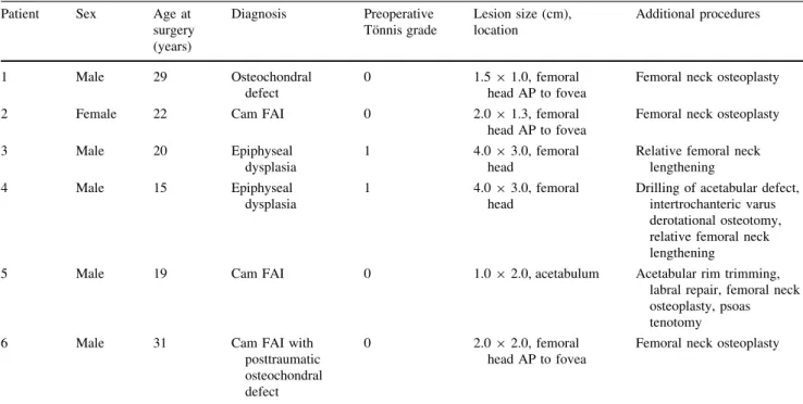
We retrospectively reviewed all six patients treated with surgical hip dislocation and AMIC for large ([ 2-cm2) femoral head or acetabular chondral or osteochondral lesions between March 2009 and December 2010 (Table 1); five femoral head and one acetabular lesions were treated, one chondral and the other five osteochon-dral. Three of these six patients were also included in a report describing the etiology of the parafoveal femoral head chondral defect [66]. At the request of that journal’s editors, the details of treatment and Tegner and Lysholm outcome scores at 1 year were included in that report although the purpose of the report was to describe the chondral lesion. All patients underwent preoperative MRI to assess the cartilage lesion and additional hip pathology; all were classified as ICRS (Table 2) Grade 3 or 4 lesions [30]. Due to different underlying diagnoses, including FAI and epiphyseal dysplasia, patients underwent a combina-tion of surgical procedures to address associated pathologic conditions. The patients were young, ranging in age from
Table 1. Patient demographics Patient Sex Age at
surgery (years) Diagnosis Preoperative To¨nnis grade Lesion size (cm), location Additional procedures 1 Male 29 Osteochondral defect 0 1.5 9 1.0, femoral head AP to fovea
Femoral neck osteoplasty
2 Female 22 Cam FAI 0 2.0 9 1.3, femoral
head AP to fovea
Femoral neck osteoplasty
3 Male 20 Epiphyseal
dysplasia
1 4.0 9 3.0, femoral head
Relative femoral neck lengthening
4 Male 15 Epiphyseal
dysplasia
1 4.0 9 3.0, femoral head
Drilling of acetabular defect, intertrochanteric varus derotational osteotomy, relative femoral neck lengthening
5 Male 19 Cam FAI 0 1.0 9 2.0, acetabulum Acetabular rim trimming, labral repair, femoral neck osteoplasty, psoas tenotomy
6 Male 31 Cam FAI with
posttraumatic osteochondral defect
0 2.0 9 2.0, femoral head AP to fovea
Femoral neck osteoplasty
15 to 31 years (Table1). All patients participating in our study were educated in detail about the surgical technique, alternatives, advantages and disadvantages, and potential complications. A minimum of 1-year postoperative fol-lowup was obtained for four patients (range, 1–2.5 years). One patient was lost to followup and did not respond to repeated attempts at both written and telephone contact. One additional patient was not available for postoperative MRI and did not return the outcome questionnaire due to incarceration. At the time of surgery, all patients gave written consent to publish their outcomes. The study was performed in compliance with our institutional review board.
Clinical and radiographic followups were scheduled at 6 weeks, 6 months, and 12 to 24 months after surgery. In November 2011, we obtained current Oxford Hip Scores and UCLA Activity Scores from the four patients who could be located [43,44]. A followup MRI was performed 6 months postoperatively to assess lesion healing and reso-lution of bone marrow edema. All MRIs were performed with a 1.5-T system using a body matrix coil (MAGNETOM1 Avanto or Espree; Siemens Medical Solutions, Erlangen, Germany). A single T2 slice 90° to the largest portion of the lesion was chosen for evaluation. Two of us (LMT, MRS) evaluated the quality of repair tissue in consensus, according to a modified magnetic resonance observation of cartilage repair tissue (MOCART) scoring system [38,39]. The MOCART system is an evaluation of nine variables on a standard MRI comparing the mor-phology and signal intensity of articular cartilage repair tissue to the adjacent native cartilage (Table3). The interobserver reliability of the MOCART system has previously been evaluated in a prospective cohort study
of patients undergoing matrix-assisted cartilage transplan-tation for articular cartilage defects larger than 2 cm2in the knee [39], with an intraclass correlation coefficient ranging from 0.76 to 1.0, depending on the variable. The score was modified from that originally reported because our insti-tution does not routinely obtain a three-dimensional gradient echo sequence with fat suppression, which is used to evaluate the signal intensity of the repair tissue. A dual T2 fast spin echo sequence is part of our routine MRI protocol. This is also part of the validated MOCART scoring system and is used to determine the repair tissue signal intensity. One of the evaluators (LMT) was a hip preservation fellow who was not involved in any way in the treatment of the patients. The second evaluator (MRS) has extensive experience with cartilage restoration in the knee and provided technical assistance with the AMIC proce-dure at the time of surgery but was not otherwise involved with the patients’ care.
Results
No intraoperative or immediate postoperative complica-tions occurred. Oxford Hip Scores and UCLA Activity Scores improved for all five patients followed until 1 year (Table 4). Patient 4 was seen 7 months postoperatively with subjectively improved hip ROM and improvement in overall hip function, but due to incarceration, 1-year postoperative outcomes scores could not be obtained (Fig.2). Patient 2 described groin pain that occurred during training for karate competitions; however, she was able to return to training and competition. On examination, pain was produced in deep flexion and with adduction and internal rotation. The ROM was, however, symmetric to the contralateral side. The patient also underwent an MR arthrogram, which showed possible adhesions [4]. As she is able to train and compete and otherwise has no symptoms at rest or with activities of daily living, the patient elected to forego further evaluation or surgical management of the adhesions. She also had a kissing lesion on the acetabular cartilage. This was repaired but may contribute to her residual symptoms.
The four lesions evaluated by MRI appeared to be completely filled with complete integration of the border zone (Table 4). The quality of the repair tissue was vari-able. The structure of the repair tissue appeared inhomogeneous for three of four patients. The cartilage signal for two of four patients was isointense when com-pared to that of the surrounding cartilage and the surface of the repaired cartilage was intact for two of four patients. The surface of the repair tissue was normal for two patients, showed mild fibrillation (\ 50%) for one patient, and was damaged in one patient. The subchondral lamina Table 2. ICRS classification [30]
Grade Description Criteria 0 Normal Normal cartilage
1 Nearly normal Superficial lesions; soft indentation, superficial fissures, or cracks 2 Abnormal Lesions extending to \ 50%
of cartilage depth 3 (A–D) Severely
abnormal
Defects extending to [ 50% of cartilage depth (A), as well as to the calcified cartilage (B), but not through subchondral bone (C); includes cartilage blistering (D)
4 (A, B) Severely abnormal
Cartilage defect that extends into the subchondral bone (A) and may have cystic changes (B) ICRS = International Cartilage Repair Society; reprinted with permission from the ICRS. ICRS Cartilage Injury Evaluation Package, 2000. Available at:http://www.cartilage.org/_files/contentmanagement/ ICRS_evaluation.pdf. Accessed November 10, 2011.
(the subchondral bone plate beneath the repair tissue) was intact for only one patient. The subchondral bone (the bone adjacent to the repair site) appeared normal for two of four patients. The subchondral bone of the other two patients appeared to consist partly of granulation tissue, but no
edema, cystic changes, or sclerotic areas were observed. One patient (Patient 2) had possible ventral adhesions; this same patient underwent MR arthrogram, making it impossible to determine whether an effusion was also present. No other patient had an effusion.
Table 3. MOCART scoring system [38,39]
Variable Classes Score
(points) Degree of defect
repair and defect filling
Complete (on a level with adjacent cartilage) 20 Hypertrophy (over the level of the adjacent cartilage) 15 Incomplete (under the level of the adjacent cartilage; underfilling)
[ 50% of the adjacent cartilage 10
\ 50% of the adjacent cartilage 5
Subchondral bone exposed (complete delamination or dislocation and/or loose body)
0 Integration to border
zone
Complete (complete integration with adjacent cartilage) 15 Incomplete (incomplete integration with adjacent cartilage)
demarcating border visible (splitlike)
10 Defect visible
\ 50% of the length of the repair tissue 5 [ 50% of the length of the repair tissue 0 Surface of the repair
tissue
Surface intact (lamina splendens intact) 10 Surface damaged (fibrillations, fissures and ulcerations)
\ 50% of repair tissue depth 5
[ 50% of repair tissue depth or total degeneration 0
Structure of the repair tissue Homogeneous 5
Inhomogeneous or cleft formation 0
Signal intensity of the repair tissue Dual T2 FSE Isointense 15 Moderately hyperintense 5 Markedly hyperintense 0 3D-GRE-FS* Isointense 15 Moderately hypointense 5 Markedly hypointense 0
Subchondral lamina Intact 5
Not intact 0
Subchondral bone Intact 5
Edema, granulation tissue, cysts, sclerosis 0
Adhesions No 5
Yes 0
Effusion No effusion 5
Effusion 0
Maximum score 100
* 3D-GRE-FS was not included in scoring because it is not routinely obtained at our institution. A dual T2 FSE sequence is part of the routine MRI protocol and was used to determine repair signal intensity. Thus, the maximum MOCART score was 85 rather than 100. MOCART = magnetic resonance observation of cartilage repair tissue; FSE = fast spin echo; 3D-GRE-FS = three-dimensional gradient echo sequence with fat suppression; reprinted from Marlovits S, Striessing G, Resinger CT, Aldrian SM, Vecsei V, Imhof H, Trattnig S. Definition of pertinent parameters for the evaluation of articular cartilage repair tissue with high-resolution magnetic resonance imaging. Eur J Radiol. 2004; 52:310–319, with permission from Elsevier.
Discussion
The bony constraint and soft tissue coverage in the hip limit the options for repair of larger cartilage defects in the femoral head and acetabulum. AMIC is a second-genera-tion bone marrow stimulasecond-genera-tion technique developed for the treatment of larger cartilage defects in the knee. We believe AMIC is a reasonable option for cartilage repair in the hip for our stated indications because it is a one-stage tech-nique that is not limited by allograft availability and does not create autograft donor site morbidity. We assessed the short-term (1- to 2.5-year) clinical and radiographic (MRI) outcomes of focal chondral and osteochondral lesions in hips treated with AMIC and surgical hip dislocation.
There are several limitations to this study. First, we are reporting a small series consisting of our initial patients treated with this approach. Second, the followup is short. Third, we do not have preoperative Oxford Hip or UCLA Activity Scores, which would have allowed comparison with our postoperative data. Fourth, five of six patients underwent bone grafting over a drilled defect. Although bone grafting with ACI has been reported [2], bone grafting an osteochondral defect combined with bone marrow stimulation has not previously been described. The effect of the bone graft on the quality of the cartilage repair tissue is unknown. The effect of the cartilage repair on the sur-gical outcome is potentially confounded by the other surgical procedures performed at the same time. Similar to the treatment of cartilage defects in the knee, however, we believe the joint should be stabilized and any correctable mechanical abnormalities addressed surgically to protect the healing cartilage and prevent further damage from occurring. The cause of chondral and osteochondral defects occurring in FAI is subject to some debate. While most agree acetabular chondral delamination occurs as a result of shear forces created by the cam deformity [11,23,29,
40,46,50,56,62], the cause of defects occurring remotely to the cam or pincer deformity is subject to more specu-lation. Charbonnier et al. [12] demonstrated impingement and secondary femoral head subluxation in dancers with morphologically normal hips, and two studies have dem-onstrated decreased hip flexion and abduction in patients with FAI [33,36]. It may be that patients participating in sports or vocations that require deeper, forceful, or repet-itive hip flexion and rotation subject their cartilage to similar episodes of dynamic instability secondary to their impingement [6]. To eliminate the potential cause of the chondral damage, we believe patients with impingement and an osteochondral defect should undergo concomitant treatment to address their bony pathomorphology. As such, femoral head osteoplasty or acetabular rim trimming and labral repair should be performed for patients with FAI, as indicated by the specific type of impingement.
We observed improved short-term function in these patients with imaging suggesting some healing after repair of these defects. It is important to remember these are complex cases in young adults with limited treatment options. Fontana et al. [22] recently reported the results of arthroscopic autologous chondrocyte transplantation (ACT) in the hip with the use of a scaffold. The study reported on patients with full-thickness chondral defects of a size ([ 2 cm2) similar to those in our series and compared their outcomes with those of a matched cohort who had under-gone arthroscopic de´bridement. The patients who underwent ACT had clearly better outcomes at 6 years as compared to the de´bridement group. In contrast to our series, all of their patients had acetabular lesions and no lesion involved the subchondral bone [22]. In the discus-sion, the authors noted simple de´bridement for lesions larger than 2 cm2 was not efficacious as there was no change in outcome scores preoperatively to postoperatively. In addition, they mentioned future studies could potentially Table 4. Results after AMIC
Patient Oxford Hip Score (points)
UCLA Activity Score (points)
MOCART score [38,39] (of 85 points)
Lesion appearance on MRI
1 13 10 60 Complete defect fill, no effusion, inhomogeneous hypointense signal, intact subchondral bone 2 17 10 55 Complete defect fill, possible effusion, inhomogeneous
hyperintense signal, granulation tissue in subchondral bone 3 13 5 60 Complete defect fill, no effusion, surface irregularity
with isointense signal, granulation tissue in subchondral bone 4*
5 14 6 75 Complete defect fill, no effusion, mild surface fibrillation, homogeneous isointense signal, intact subchondral bone 6
* Patient 4 was not available for postoperative MRI or outcome questionnaires due to incarceration; Patient 6 was lost to followup; AMIC = autologous matrix-induced chondrogenesis; MOCART = magnetic resonance observation of cartilage repair tissue.
use microfracture in combination with a scaffold or com-pare the results of their technique with microfracture [22]. Most of the studies on AMIC have been published for the knee, although there is also a single case report of AMIC performed for a talar lesion [65]. Gille et al. [24] reported their intermediate-term (2-year) results, with a stable functional result between 1 and 2 years. Knee-specific outcome and activity scores including the Lysholm, Cincinnati, Tegner, and IKDC scores all improved at 12 and 24 months. Nonetheless, the results were not flawless: three patients were not satisfied and two were referred for TKA. MRI obtained 2 years postoperatively showed bone
marrow lesions, osseous hypertrophy, and intralesional osteophytes in some patients. However, 10 of 15 patients had greater than 50% defect filling. The authors did not attempt to correlate the functional results with the radio-graphic appearance of the lesion. Other smaller cohorts with a minimum of 1-year followup have reported improvement in knee-specific outcome scores [17, 49]. Dhollander et al. [17] observed intralesional osteophytes on MRI performed 1 to 2 years postoperatively in three of five patients.
Published outcomes of other cartilage repair or resto-ration procedures in the hip are limited (Table5). Fig. 2A–D Preoperative (A) AP and
(B) adduction radiographs of the hip show the large femoral head lesion of the patient shown in Figure1. The patient also underwent a varus intertro-chanteric osteotomy to unload the majority of the defect. (C) A radiograph taken immediately postoperatively demonstrates unloading of the femoral head defect as a result of the varus osteotomy. (D) In the most recent radiograph, obtained 7 months after surgery, the bone graft has consolidated and the medial contour of the femoral head is visible. The joint space has been maintained. A small depression is vis-ible at the superolateral aspect of the femoral head, which may represent indentation of soft bone in this region.
A PubMed search using the terms ‘‘osteochondritis OR osteochondrosis OR osteochondral defect OR chondral defect AND hip[ti]’’ performed in March 2012 produced 339 results. Of these titles, 70 were published in English and pertained to humans. A review of these 70 abstracts revealed 35 were published in the last 30 years and three specifically described the outcome of a cartilage procedure in the hip [1,3,48], all of which were case reports of one or two patients. Nonetheless, there are some data regarding the treatment of cartilage lesions in the hip (Table5). In addition to the series of patients treated with ACT men-tioned earlier, there are some large series of patients who have had microfracture in the hip. The results for micro-fracture in the hip have been reported as part of the treatment for FAI [11, 29,50]. Haviv et al. [29] reported their results of patients with FAI and chondral lesions. In their cohort, patients who had microfracture had better outcomes scores in short-term followup than historical control patients who had simple de´bridement [29].
Here we describe the use of second-generation bone marrow stimulation, AMIC, as an option for treating chondral and osteochondral defects larger than 2 cm2in the hip. AMIC has not previously been described for the treatment of these lesions in the hip. The 1- to 2-year
results for AMIC in the knee, however, are generally good, with high rates of patient satisfaction [24]. Our short-term results indicate AMIC may be one practical treatment for large cartilage lesions in the hip. Nonetheless, the long-term results of AMIC, including the duration of symptom relief, durability of the repair cartilage, and potential to prevent subsequent osteoarthrosis, remain unknown. As with many other promising surgical techniques, studies with larger cohorts, longer followup, and prospective col-lection of pre-and postoperative outcomes scores are necessary to determine the answers to these questions and the efficacy of this technique.
References
1. Akimau P, Bhosale A, Harrison PE, Roberts S, McCall IW, Richardson JB, Ashton BA. Autologous chondrocyte implanta-tion with bone grafting for osteochondral defect due to posttraumatic osteonecrosis of the hip—a case report. Acta Orthop. 2006;77:333–336.
2. Bartlett W, Gooding CR, Carrington RW, Skinner JA, Briggs TW, Bentley G. Autologous chondrocyte implantation at the knee using a bilayer collagen membrane with bone graft: a preliminary report. J Bone Joint Surg Br. 2005;87:330–332.
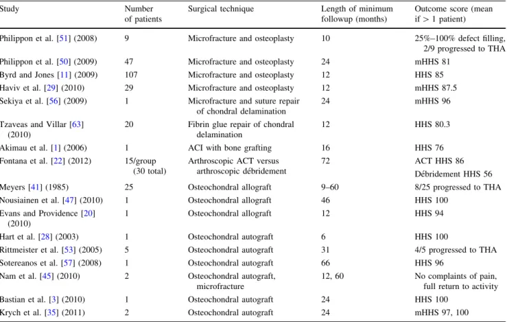
Table 5. Published results of cartilage repair procedures in the hip
Study Number
of patients
Surgical technique Length of minimum followup (months)
Outcome score (mean if [ 1 patient)
Philippon et al. [51] (2008) 9 Microfracture and osteoplasty 10 25%–100% defect filling, 2/9 progressed to THA Philippon et al. [50] (2009) 47 Microfracture and osteoplasty 24 mHHS 81
Byrd and Jones [11] (2009) 107 Microfracture and osteoplasty 12 HHS 85 Haviv et al. [29] (2010) 29 Microfracture and osteoplasty 12 mHHS 87.5 Sekiya et al. [56] (2009) 1 Microfracture and suture repair
of chondral delamination
24 mHHS 96
Tzaveas and Villar [63] (2010)
20 Fibrin glue repair of chondral delamination
12 HHS 80.3
Akimau et al. [1] (2006) 1 ACI with bone grafting 16 HHS 76 Fontana et al. [22] (2012) 15/group
(30 total)
Arthroscopic ACT versus arthroscopic de´bridement
72 ACT HHS 86
De´bridement HHS 56 Meyers [41] (1985) 25 Osteochondral allograft 9–60 8/25 progressed to THA Nousiainen et al. [47] (2010) 1 Osteochondral allograft 46 HHS 100
Evans and Providence [20] (2010)
1 Osteochondral allograft 12 HHS 94
Hart et al. [28] (2003) 1 Osteochondral autograft 6 HHS 100
Rittmeister et al. [53] (2005) 5 Osteochondral autograft 31 4/5 progressed to THA Sotereanos et al. [57] (2008) 1 Osteochondral autograft 66 HHS 96
Nam et al. [45] (2010) 2 Osteochondral autograft, microfracture
12, 60 No complaints of pain, full return to activity Bastian et al. [3] (2010) 1 Osteochondral autograft 24 HHS 100
Krych et al. [35] (2011) 2 Osteochondral autograft 24 mHHS 97, 100
ACI = autologous chondrocyte implantation; ACT = autologous chondrocyte transplantation; HHS = Harris hip score [27]; mHHS = modified Harris hip score [10].
3. Bastian JD, Bu¨chler L, Meyer DC, Siebenrock KA, Keel MJ. Surgical hip dislocation for osteochondral transplantation as a salvage procedure for a femoral head impaction fracture. J Orthop Trauma. 2010;24:e113–e118.
4. Beck M. Groin pain after open FAI surgery: the role of intraar-ticular adhesions. Clin Orthop Relat Res. 2009;467:769–774. 5. Bedi A, Chen N, Robertson W, Kelly BT. Systematic review: the
management of labral tears and femoroacetabular impingement of the hip in the young, active patient. Arthroscopy. 2008;24: 1135–1145.
6. Bedi A, Dolan M, Leunig M, Kelly BT. Static and dynamic mechanical causes of hip pain. Arthroscopy. 2011;27:235–251. 7. Behrens P. [Matrix-coupled microfracture, a new concept for the
treatment of cartilage defect] [in German]. Arthroskopie. 2005; 18:193–197.
8. Brittberg M, Lindahl A, Nilsson A, Ohlsson C, Isaksson O, Peterson L. Treatment of deep cartilage defects in the knee with autologous chondrocyte transplantation. N Engl J Med. 1994;331: 889–895.
9. Bugbee WD, Convery FR. Osteochondral allograft transplanta-tion. Clin Sports Med. 1999;18:67–75.
10. Byrd JW, Jones KS. Prospective analysis of hip arthroscopy with 2-year follow-up. Arthroscopy. 2000;16:578–587.
11. Byrd JW, Jones KS. Arthroscopic femoroplasty in the manage-ment of cam-type femoroacetabular impingemanage-ment. Clin Orthop Relat Res. 2009;467:739–746.
12. Charbonnier C, Kolo FC, Duthon VB, Magnenat-Thalmann N, Becker CD, Hoffmeyer P, Menetrey J. Assessment of congruence and impingement of the hip joint in professional ballet dancers: a motion capture study. Am J Sports Med. 2011;39:557–566. 13. Chen H, Chevrier A, Hoemann CD, Sun J, Ouyang W, Buschmann
MD. Characterization of subchondral bone repair for marrow-stimulated chondral defects and relationship to articular cartilage resurfacing. Am J Sports Med. 2011;39:1731–1740.
14. Chen H, Hoemann CD, Sun J, Chevrier A, McKee MD, Shive MS, Hurtig M, Buschmann MD. Depth of subchondral perfora-tion influences the outcome of bone marrow stimulaperfora-tion cartilage repair. J Orthop Res. 2011;29:1178–1184.
15. Chen H, Sun J, Hoemann CD, Lascau-Coman V, Ouyang W, McKee MD, Shive MS, Buschmann MD. Drilling and micro-fracture lead to different bone structure and necrosis during bone-marrow stimulation for cartilage repair. J Orthop Res. 2009;27: 1432–1438.
16. Cherubino P, Grassi FA, Bulgheroni P, Ronga M. Autologous chondrocyte implantation using a bilayer collagen membrane: a preliminary report. J Orthop Surg (Hong Kong). 2003;11:10–15. 17. Dhollander AA, De Neve F, Almqvist KF, Verdonk R, Lambr-echt S, Elewaut D, Verbruggen G, Verdonk PC. Autologous matrix-induced chondrogenesis combined with platelet-rich plasma gel: technical description and a five pilot patients report. Knee Surg Sports Traumatol Arthrosc. 2011;19:536–542. 18. Erggelet C, Endres M, Neumann K, Morawietz L, Ringe J,
Haberstroh K, Sittinger M, Kaps C. Formation of cartilage repair tissue in articular cartilage defects pretreated with microfracture and covered with cell-free polymer-based implants. J Orthop Res. 2009;27:1353–1360.
19. Espinosa N, Beck M, Rothenfluh DA, Ganz R, Leunig M. Treatment of femoro-acetabular impingement: preliminary results of labral refixation: surgical technique. J Bone Joint Surg Am. 2007;89(suppl 2 pt 1):36–53.
20. Evans KN, Providence BC. Fresh-stored osteochondral allograft for treatment of osteochondritis dissecans of the femoral head. Clin Orthop Relat Res. 2010;468:613–618.
21. Ficat RP, Ficat C, Gedeon P, Toussaint JB. Spongialization: a new treatment for diseased patellae. Clin Orthop Relat Res. 1979;144:74–83.
22. Fontana A, Bistolfi A, Crova M, Rosso F, Massazza G. Arthro-scopic treatment of hip chondral defects: autologous chondrocyte transplantation versus simple debridement—a pilot study. Arthroscopy. 2012;28:322–329.
23. Ganz R, Parvizi J, Beck M, Leunig M, No¨tzli H, Siebenrock K. Femoroacetabular impingement: a cause for osteoarthritis of the hip. Clin Orthop Relat Res. 2003;417:112–120.
24. Gille J, Schuseil E, Wimmer J, Gellissen J, Schulz AP, Behrens P. Mid-term results of autologous matrix-induced chondrogenesis for treatment of focal cartilage defects in the knee. Knee Surg Sports Traumatol Arthrosc. 2010;18:1456–1464.
25. Guettler JH, Demetropoulous CK, Yang KH, Jurist KA. Osteo-chondral defects in the human knee: influence of defect size on cartilage rim stress and load redistribution to surrounding carti-lage. Am J Sports Med. 2004;32:1451–1458.
26. Hangody L, Kish G, Ka´rpa´ti Z, Szerb I, Udvarhelyi I. Arthro-scopic autogenous osteochondral mosaicplasty for the treatment of femoral condylar articular defects: a preliminary report. Knee Surg Sports Traumatol Arthrosc. 1997;5:262–267.
27. Harris WH. Traumatic arthritis of the hip after dislocation and acetabular fractures: treatment by mold arthroplasty. An end-result study using a new method of end-result evaluation. J Bone Joint Surg Am. 1969;51:737–755.
28. Hart R, Janecek M, Visna P, Bucek P, Kocis J. Mosaicplasty for the treatment of femoral head defect after incorrect resorbable screw insertion. Arthroscopy. 2003:19:e137–e141.
29. Haviv B, Singh PJ, Takla A, O’Donnell J. Arthroscopic femoral osteochondroplasty for cam lesions with isolated acetabular chondral damage. J Bone Joint Surg Br. 2010;92:629–633. 30. International Cartilage Repair Society. ICRS Cartilage Injury
Evaluation Package, 2000. Available at: http://www.cartilage. org/_files/contentmanagement/ICRS_evaluation.pdf. Accessed November 10, 2011.
31. Jakob RP, Franz T, Gautier E, Mainil-Varlet P. Autologous osteochondral grafting in the knee: indication, results and reflections. Clin Orthop Relat Res. 2002;401:170–184.
32. Johnson LL. Arthroscopic abrasion arthroplasty historical and pathologic perspective: present status. Arthroscopy. 1986;2: 54–69.
33. Kennedy MJ, Lamontagne M, Beaule´ PE. Femoroacetabular impingement alters hip and pelvic biomechanics during gait: walking biomechanics of FAI. Gait Posture. 2009;30: 41–44.
34. Kreuz PC, Steinwachs MR, Erggelet C, Krause SJ, Konrad G, Uhl M, Su¨dkamp N. Results after microfracture of full-thickness chondral defects in different compartments in the knee. Osteo-arthritis Cartilage. 2006;14:1119–1125.
35. Krych AJ, Lorich DG, Kelly BT. Treatment of focal osteochon-dral defects of the acetabulum with osteochonosteochon-dral allograft transplantation. Orthopedics. 2011;34:e307–e311.
36. Lamontagne M, Kennedy MJ, Beaule´ PE. The effect of cam FAI on hip and pelvic motion during maximum squat. Clin Orthop Relat Res. 2009;467:645–650.
37. Magnussen RA, Dunn WR, Carey JL, Spindler KP. Treatment of focal articular cartilage defects in the knee: a systematic review. Clin Orthop Relat Res. 2008;466:952–962.
38. Marlovits S, Singer P, Zeller P, Mandl I, Haller J, Trattnig S. Magnetic resonance observation of cartilage repair tissue (MOCART) for the evaluation of autologous chondrocyte implantation: determination of interobserver variability and cor-relation to clinical outcome after 2 years. Eur J Radiol. 2006;57: 16–23.
39. Marlovits S, Striessing G, Resinger CT, Aldrian SM, Vecsei V, Imhof H, Trattnig S. Definition of pertinent parameters for the evaluation of articular cartilage repair tissue with high-resolution magnetic resonance imaging. Eur J Radiol. 2004;52:310–319.
40. Meermans G, Konan S, Haddad FS, Witt JD. Prevalence of acetabular cartilage lesions and labral tears in femoroacetabular impingement. Acta Orthop Belg. 2010;76:181–188.
41. Meyers MH. Resurfacing of the femoral head with fresh osteo-chondral allografts: long term results. Clin Orthop Relat Res. 1985;197:111–114.
42. Mithoefer K, Williams RJ, Warren RF, Potter HG, Spock CR, Jones EC, Wickiewicz TL, Marx RG. The microfracture technique for the treatment of articular cartilage lesions in the knee: a pro-spective cohort study. J Bone Joint Surg Am. 2005;87:1911–1920. 43. Naal FD, Impellizzeri FM, Leunig M. Which is the best activity rating scale for patients undergoing total joint arthroplasty? Clin Orthop Relat Res. 2009;467:958–965.
44. Naal FD, Sieverding M, Impellizzeri FM, von Knoch F, Mannion AF, Leunig M. Reliability and validity of the cross-culturally adapted German Oxford hip score. Clin Orthop Relat Res. 2009; 467:952–957.
45. Nam D, Shindle MK, Buly RL, Kelly BT, Lorich DG. Traumatic osteochondral injury of the femoral head treated by mosaicplasty: a report of two cases. HSS J. 2010;6:228–234.
46. Nepple JJ, Carlisle JC, Nunley RM, Clohisy JC. Clinical and radiographic predictors of intra-articular hip disease in arthros-copy. Am J Sports Med. 2011;39:296–303.
47. Nousiainen MT, Sen MK, Mintz DN, Lorich D, Paul O, Buly RL, Helfet DL. The use of osteochondral allograft in the treatment of a severe femoral head fracture. J Orthop Trauma. 2010;24:120–124. 48. Pardiwala DN, Nagda TV. Arthroscopic chondral cyst excision in
a stiff Perthes’ hip. Arthroscopy. 2007;23:909.e1–e4.
49. Pascarella A, Ciatti R, Pascarella F, Latte C, Di Salvatore MG, Liguori L, Iannella G. Treatment of articular cartilage lesions of the knee joint using a modified AMIC technique. Knee Surg Sports Traumatol Arthrosc. 2010;18:509–513.
50. Philippon MJ, Briggs KK, Yen YM, Kuppersmith DA. Outcomes following hip arthroscopy for femoroacetabular impingement with associated chondrolabral dysfunction. J Bone Joint Surg Br. 2009;91:16–23.
51. Philippon MJ, Schenker ML, Briggs KK, Maxwell RB. Can microfracture produce repair tissue in acetabular chondral defects? Arthroscopy. 2008;24:46–50.
52. Pridie KH. A method of resurfacing osteoarthritic knee joints. J Bone Joint Surg Br. 1959;41:618–619.
53. Rittmeister M, Hochmuth K, Kriener S, Richolt J. [Five-year results following autogenous osteochondral transplantation to the femoral head] [in German]. Orthopade. 2005;34:320–326.
54. Schmid MR, No¨tzli HP, Zanetti M, Wyss TF, Hodler J. Cartilage lesions in the hip: diagnostic effectiveness of MR arthrography. Radiology. 2003;226:382–386.
55. Schoeniger R, LaFrance AE, Oxland TR, Ganz R, Leunig M. Does trochanteric step osteotomy provide greater stability than classic slide osteotomy? A preliminary study. Clin Orthop Relat Res. 2009;467:775–782.
56. Sekiya JK, Martin RL, Lesniak BP. Arthroscopic repair of delaminated acetabular articular cartilage in femoroacetabular impingement. Orthopedics. 2009;32:692.
57. Sotereanos NG, DeMeo PJ, Hughes TB, Bargiotas K, Wohlrab D. Autogenous osteochondral transfer in the femoral head after osteonecrosis. Orthopedics. 2008;31:177.
58. Steadman JR, Briggs KK, Rodrigo JJ, Kocher MS, Gill TJ, Rodkey WG. Outcomes of microfracture for traumatic chondral defects of the knee: average 11-year follow-up. Arthroscopy. 2003;19:477–484.
59. Steinwachs M, Kreuz PC. Autologous chondrocyte implantation in chondral defects of the knee with a type I/III collagen mem-brane: a prospective study with a 3-year follow-up. Arthroscopy. 2007;23:381–387.
60. Steinwachs MR, Guggi T, Kreuz PC. Marrow stimulation tech-niques. Injury. 2008;39(suppl 1):S26–S31.
61. Stevens MS, LeGay DA, Glazebrook MA, Amirault D. System-atic review: the evidence for hip arthroscopy: grading the current indications. Arthroscopy. 2010;26:1370–1383.
62. Tannast M, Goricki D, Beck M, Murphy SB, Siebenrock KA. Hip damage occurs at the zone of femoroacetabular impingement. Clin Orthop Relat Res. 2008;466:273–280.
63. Tzaveas AP, Villar RN. Arthroscopic repair of acetabular chondral delamination with fibrin adhesive. Hip Int. 2010;20:115– 119.
64. Vavken P, Arrich F, Pilz M, Dorotka R. An in vitro model of biomaterial-augmented microfracture including chondrocyte-progenitor cell interaction. Arch Orthop Trauma Surg. 2010;130: 711–716.
65. Wiewiorski M, Leumann A, Buettner O, Pagenstert G, Horis-berger M, Valderrabano V. Autologous matrix-induced chondrogenesis aided reconstruction of a large focal osteochon-dral lesion of the talus. Arch Orthop Trauma Surg. 2011;131: 293–296.
66. Zaltz I, Leunig M. Parafoveal chondral defects associated with femoroacetabular impingement. Clin Orthop Relat Res. 2012. DOI Number10.1007/s11999-012-2453-x.
![Table 3. MOCART scoring system [38, 39]](https://thumb-eu.123doks.com/thumbv2/123doknet/14851871.630187/6.892.72.812.112.838/table-mocart-scoring-system.webp)