HAL Id: hal-02119638
https://hal.archives-ouvertes.fr/hal-02119638
Submitted on 3 May 2019HAL is a multi-disciplinary open access archive for the deposit and dissemination of sci-entific research documents, whether they are pub-lished or not. The documents may come from teaching and research institutions in France or abroad, or from public or private research centers.
L’archive ouverte pluridisciplinaire HAL, est destinée au dépôt et à la diffusion de documents scientifiques de niveau recherche, publiés ou non, émanant des établissements d’enseignement et de recherche français ou étrangers, des laboratoires publics ou privés.
In situ chemical behaviour of methylisothiazolinone (MI)
and methylchloroisothiazolinone (MCI) in reconstructed
human epidermis: a new approach to the
cross-reactivity issue
Camille Debeuckelaere, Francois-Marie Moussallieh, Karim El Bayed,
Izzie-Jacques Namer, Valerie Berl, Elena Giménez-Arnau, Jean-Pierre
Lepoittevin
To cite this version:
Camille Debeuckelaere, Francois-Marie Moussallieh, Karim El Bayed, Izzie-Jacques Namer, Valerie Berl, et al.. In situ chemical behaviour of methylisothiazolinone (MI) and methylchloroisothiazolinone (MCI) in reconstructed human epidermis: a new approach to the cross-reactivity issue. Contact Dermatitis, Wiley, 2016, 74 (3), pp.159-167. �10.1111/cod.12524�. �hal-02119638�
In situ chemical behaviour of methylisothiazolinone (MI) and
methylchloroisothiazolinone (MCI) in reconstructed human
epidermis: a new approach to the cross-reactivity issue
Camille Debeuckelaere
1, François-Marie Moussallieh
1,2, Karim Elbayed
2, Izzie-Jacques
Namer
2, Valérie Berl
1, Elena Giménez-Arnau
1, and Jean-Pierre Lepoittevin
11Dermatochemistry Laboratory, Institut de Chimie de Strasbourg, UMR 7177 / Université de Strasbourg
- CNRS, 4 Rue Blaise Pascal, 67081 Strasbourg, France and 2Laboratoire des sciences de l’ingénieur,
de l’informatique et de l’imagerie (ICube), UMR 7357 / Université de Strasbourg – CNRS, 674012 Illkirch, France. Address: Elena Giménez-Arnau Jean-Pierre Lepoittevin Dermatochemistry Laboratory Institut le Bel
4, Rue Blaise Pascal, 67081 Strasbourg, France Tel: +33 368851501
Fax: +33 368851527
e-mail: egimenez@unistra.fr; jplepoit@unistra.fr
The work was partly financially supported by Cosmetics Europe (Brussels, Belgium) There are no conflicts of interests
Summary
Background: Methylisothiazolinone (MI), part with methylchloroisothiazolinone (MCI) of the
allergenic Kathon® CG, was released as an individual preservative in the 2000s for industrial products
and in 2005 for cosmetics. The high exposure to MI since then has provoked an epidemic of contact allergy to MI, and an increase of MI/MCI allergy prevalence. Because the occurrence of consumer products containing only MI in recent years, questions were raised about the MI and MCI cross-reaction pattern.
Objectives: To bring a new perspective on the MI/MCI cross-reactivity issue by studying their in situ
chemical behaviour in 3D reconstructed human epidermis.
Methods: MI and MCI were synthesized 13C-substituted at positions C-4/C-5 and C-5, respectively.
Their in situ chemical behaviour in a RHE model were followed by the high-resolution magic angle spinning (HRMAS) nuclear magnetic resonance (NMR) technique.
Results: MI was found to react exclusively with cysteine thiol residues, whereas MCI reacted with
histidine and lysine amino acids. Reaction mechanisms were found to be different for MI and MCI and adducts formed of different molecular structure.
Conclusion: In RHE, different MI/MCI reactions towards different nucleophilic amino acids were
observed, making it difficult to explain cross-reactivity between MI and MCI.
Key words: methylisothiazolinone, methylchloroisothiazolinone, cross-reactivity, reaction chemistry,
Introduction
Methylisothiazolinone (MI) and methylchloroisothiazolinone (MCI) are the active ingredients of the biocide Kathon® CG (MI/MCI 1:3 combination), used since the 1980s and one of the most common
source of allergic contact dermatitis caused by preservatives (1, 2). Following the introduction in the EU of a 15 ppm use limit in cosmetics, contact allergy to MI/MCI significantly decreased to a prevalence rate of about 2% after the 90s (3). The sensitizing potential of the mixture was mostly attributed to the chlorinated derivative MCI, shown to be the stronger sensitizer, while the non-chlorinated MI was reported to be a much weaker allergen (4-6). Thus, in the early 2000s, MI alone started to be used as preservative in industrial products and in 2005 in cosmetics, but at higher concentrations than in the MI/MCI mixture due to its lower biocide potential. As a consequence, over recent years there has been an alarming increase in the prevalence of allergic contact dermatitis to MI (7, 8). Occupational cases of contact dermatitis to MI started to be reported from paints (9), followed by non-occupational cases essentially seen from wet wipes for hygiene and cosmetics (10, 11). Severe cases of airborne and systemic dermatitis have appeared recently from exposure to MI present particularly in water based wall paints (12, 13). At the same time, MI/MCI contact allergy has increased significantly over the past few years (7, 8, 14, 15). It has been proposed that the rise in MI/MCI contact allergy was likely linked to the higher consumer exposure to MI, and was most probably due to a previous sensitization of individuals to MI.
There is a critical issue concerning the use of MI and MCI in consumer products. While MI was reported to be a weak sensitizer in the guinea pig (4), it has been classified as a strong sensitizer in the local lymph node assay (16). MI is today allowed in leave-on and rinse-off cosmetic products at a 100 ppm maximum concentration (17), while there are so far no limitations in industrial products. The mixture MI/MCI is patch tested at 200 ppm (50 ppm MI/150 ppm MCI) in water in the European baseline series. It was previously tested at 100 ppm but at this concentration a significant percentage of increasing contact allergy reactions to MI was not detected as the mixture contained a too low concentration of MI. MI has been recently included in the European baseline series for patch testing at 2000 ppm in water (18). The Scientific Committee on Consumer Safety (SCCS) of the European Commission adopted an opinion on December 2013 in which it was stated that 100 ppm MI in cosmetic products was not safe for the consumer, and that a maximum concentration of 15 ppm was considered safe but only in rinse-off products (19).
Because of the presence of preservatives containing only MI on the market, questions have been raised among clinicians about the potential cross-reaction pattern between MI and MCI, but only a few studies reporting cross-reaction data of MI and MCI exist in the literature. This is probably directly related to the limited experimental approaches available so far to investigate cross-reactions between chemicals. A first approach is to use experimental animal models such as the guinea pig maximization test (GPMT) and to perform challenge studies on animals previously sensitized to individual chemicals.
Thus based on such challenge studies it was reported that cross-reaction between the non-chlorinated and the chlorinated isothiazolinones was not relevant when MCI was the initial sensitizer, and only a possible, probably non-significant, cross-reactivity was indicated when MI was the initial sensitizer (5). However, transposition of animal data to human is always a matter of discussion due to the inter-species variability (differences in skin penetration, metabolism, detoxication pathways, etc.). On the other hand, investigation of cross-reaction patterns in human is very difficult specially when a concomitant exposure can be foreseen (Kathon® CG). The retest method proposed by Rustemeyer et al.
(20) and based on the presence of memory T-cells at the patch test side could give valuable information but is not often used. From the clinical point of view, patch test studies have been performed recently with recruited patients reacting to MI/MCI and MI, additionally patch tested with MI/MCI, MI and MCI (21). The studies concluded that high patch test reactivity to MCI was in support of MCI being the primary sensitizer with cross-reactivity to MI, and high patch test reactivity to MI was in support of MI being the primary sensitizer with cross-reactivity to MCI. Nevertheless, despite the importance of such studies, true cross-reactivity between MI and MCI has not been really demonstrated today and still remains unclear.
The first key step of the process leading to sensitization to chemicals, and subsequent skin allergy development, is the formation of stable antigenic entities by reaction with amino acid residues of epidermal proteins (22). The sensitization potential of a chemical is thus directly related to its chemical reactivity behaviour towards skin proteins and amino acid specificity. From a chemical point of view and in the strict sense, different allergens can cross-react when the same T-cells are activated by similar epitopes. In other words, cross-reacting allergens should produce chemical protein modifications similar enough that the antigenic determinant, presented at the surface of antigen presenting cells in association with MHC molecules, will activate the same T-cell receptors. However, in most cases, the nature of chemical modifications formed on epidermal proteins are based on speculations derived from experiments carried out on model nucleophiles, peptides or proteins.
In this manuscript, we report our investigations on the in situ chemical behaviour of MI and MCI in a reconstructed human epidermis (RHE) model, histologically similar to in vivo human epidermis, by the use of high-resolution magic angle spinning (HRMAS) nuclear magnetic resonance (NMR). This new approach is giving access to the structures of adducts formed in situ.
Materials and Methods
Chemicals and reagents
MI 13C-substituted at position 4 or at position 5, 4-(13C)-MI and 5-(13C)-MI respectively, and MCI 13
C-substituted at position 4, 4-(13C)-MCI (Fig. 1), were synthesized using a procedure previously reported
(23). Reagents for introduction of the 13C atoms, 2-(13C)-acetic acid and (13C)-formaldehyde, as well as
deuterated solvents, were purchased from Euriso-Top (Saint Aubin, France). All other reagents and chemicals were purchased from Sigma-Aldrich (Saint Quentin Fallavier, France) and used without further purification. Aqueous solutions were prepared with deionized water.
Reconstructed human epidermis (RHE)
The SkinEthic™ RHE model (4 cm2 format) was chosen for the studies (SkinEthic, Lyon, France). It
consists of normal human keratinocytes cultured for 17 days on an inert polycarbonate filter. The RHE units were received on day 18 and aseptically removed from the transport medium. They were then maintained at least for 2 h in growth culture medium (SkinEthic, Lyon, France), at 37 °C, 5% CO2, and
humidified atmosphere, following SkinEthic’s procedure.
Incubation of RHE with 4-(13C)-MI, 5-(13C)-MI and 4-(13C)-MCI
RHE were treated with either 4-(13C)-MI, 5-(13C)-MI or 4-(13C)-MCI in acetone (0.4 M, 100 µL) and
incubation times were of 24 h and 48 h. RHE negative controls were treated only with acetone (100 µL) and incubated for 24 h. After treatment, the RHE were washed with deionized water and separated from the polycarbonate support using Dispase II (neutral protease, grade II, Roche, Manheim) in HEPES buffer N-(2-hydroxyethyl)piperazine-N’-(2-ethanesulfonic acid). They were then stored at -80 °C before NMR sample preparation.
HRMAS NMR analysis
Each sample was prepared at -20 °C by introducing 15 to 20 mg of frozen treated RHE into a disposable 30 µL Kel-f® insert, as we have described previosuly (24). For the NMR spectrometer lock frequency, 10 µL of D2O with 1% w/w trimethylsilyl propanoic acid were added to the insert. Just
before the HRMAS analysis, the insert was placed into a 4 mm ZrO2 rotor closed with a cap. The whole
system was afterwards introduced into a HRMAS probe precooled at 3 °C. HRMAS spectra were recorded on a Bruker avance III 500 spectrometer operating at a proton frequency of 500.13 MHz. The spectrometer was equipped with a 4 mm double resonance (1H, 13C) gradient HRMAS probe. A Bruker
cooling unit regulated the temperature at 3 °C by cooling down the bearing air flowing into the probe. Experiments were conducted on samples spinning at 3502 Hz in order to keep the rotation sidebands out of the interest spectral region. Shimming procedure used is described elsewhere (25). In order to
detect the formation of adducts, two-dimensional 1H-13C heteronuclear single quantum correlation
(g-HSQC) experiments were performed. Echo-antiecho gradient selection for phase-sensitive detection was used (26). Spectra were acquired using 73 ms acquisition time with GARP 13C decoupling and 1.5 s
relaxation delay. 136 transients were averaged for each of 256 t1 increments, corresponding to a total acquisition time of 15 h. NMR data analysis and processing of spectra was carried out by using Bruker’s TopSpin™ software (Version 3.2). Spectra were referenced by setting the lactate (present itself in the RHE) doublet chemical shift to 1.33 ppm in 1H and to 22.7 ppm in 13C. New NMR signals
were identified in treated RHE, in comparison to untreated control RHE. Then, 1H and 13C chemical
shifts were assigned to potential metabolites and/or adducts of MI and MCI with amino acids by comparison with known chemical shifts of adducts obtained in previous studies in solution (23, 27, 28). Also, the measured chemical shifts were compared with those calculated for suspected structures by using ChemDraw Ultra software (version 12.0).
Results
NMR techniques in association with 13C-substituted molecules have been shown to be very efficient
tools for the investigation of hapten-protein interaction mechanisms in buffered or semi-organic solutions (29-31). In order to move forward into the comprehension of chemical interactions taking place in a system closer to that of human skin, we have reported the usefulness of keratinocyte based RHE in association with the high-resolution magic angle spinning (HRMAS) NMR technique (24).The HRMAS NMR technique, initially developed to analyse the metabolome of normal and pathological tissues without time-consuming extraction (32-34), has now been associated with 13C-substituted
molecules to investigate in situ and in a non-invasive way, chemical interactions between haptens and epidermal cells. Keratinocytes being the major cell type in the epidermis and playing a key role in skin inflammatory reactions, we used the SkinEthic™ RHE model (Lyon, France), consisting of a normal human multi-layered keratinocytes culture similar histologically to that of human epidermis.
MI was prepared 13C-substituted at C-4 or C- 5 to give 4-(13C)-MI and 5-(13C)-MI respectively (Fig.
1). C-4 and C-5 positions were chosen according to the reaction chemistry previously observed in solution (23, 27, 28). Position 5 corresponds to a potential site of adducts formation while position 4 gives useful NMR information on the cyclic or open structure of adducts. C-4 and C-5 being tertiary carbons bearing hydrogen atoms, the two-dimensional 1H-13C g-HSQC NMR sequence could be used.
This sequence allows the distinction between CH and CH3 signals, being phased up, and CH2 signals
being phased down. This is of course very useful to characterize the chemical structure of adducts formed. MCI was prepared 13C-substituted only at C-4 to give 4-(13C)-MCI, as C-5 is a quaternary
carbon atom substituted by chlorine and 1H-13C g-HSQC NMR correlation experiments can not be
applied (Fig. 1).
Studies with MI
RHE were treated separately with 4-(13C)-MI and 5-(13C)-MI followed by 24 h and 48 h of incubation. 1H-13C NMR spectra of treated RHE were compared with the spectrum of native RHE (used as control
experiment) by superimposition to identify new signals in addition to the Skin Ethic™ RHE metabolome already characterized in a previous study (24).
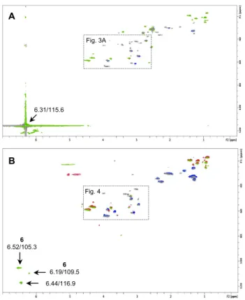
Characteristic signals of 4-(13C)-MI at 6.31 (1H)/115.6 (13C) ppm, and 5-(13C)-MI at 8.47 (1H)/145.6
(13C) ppm, were still present after 24 h of incubation as shown on Fig. 2A in the case of 4-(13C)-MI. On
the spectra obtained after 48 h of incubation no residual signal of MI was detected. This means that MI, still present after 24 h, was completely consumed, hydrolysed or detoxified after 48 h.
MI was found reactive in RHE with formation of several new signals. The region of the g-HSQC spectra shown is limited to the one where changes were observed after 24 h of incubation with 4-(13
C)-MI (Fig. 3A) or with 5-(13C)-MI (Fig. 3B). No evolution was observed after 48 h incubation. In the case
of MI 13C-substituted at position 4, new signals (grey) were of CH
position 5, new signals (green) were of CH nature. Thus, well-defined CH2 signals appeared at 2.56
(1H)/46.8 (13C) ppm, 2.71 (1H)/41.0 (13C) ppm, 2.87 (1H)/43.0 (13C) ppm, 3.18 (1H)/47.5 (13C) ppm and in
the region 2.60-2.80 (1H)/30.9-37.2 (13C) ppm (Fig. 3A) while new CH signals appeared at 3.34
(1H)/33.3 (13C) ppm, 4.27 (1H)/60.1 (13C) ppm and 4.27 (1H)/80.1 (13C) ppm (Fig. 3B). Based on the
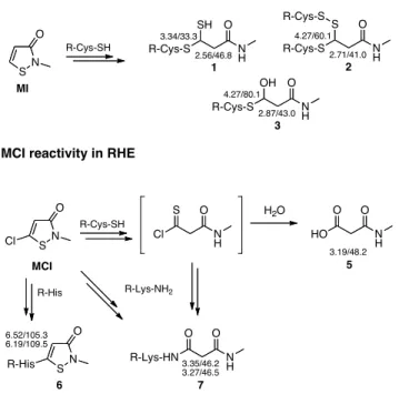
reported reaction chemistry of MI towards model nucleophiles, N-acetyl amino acids and model peptides (23, 27, 28), chemical shifts were characteristic of reactions with the nucleophilic thiol group of cysteine residues leading to the formation, through a complex chemistry, of adducts 1-3 (Fig. 5). Structures were identified by combining NMR data obtained from reaction of 4-(13C)-MI and 5-(13
C)-MI. The C-4 chemical shift (δ) value of 46.8 ppm, correlated by HSQC data to protons at 2.56 ppm, and the C-5 δ at 33.3 ppm correlated to a proton at 3.34 ppm, were characteristic of 1 bearing a cysteine unit and a thiol chemical group bound at C-5. As described for MI in solution with an excess of amino acid, in a situation similar to that of RHE, a new disulfide bond can be formed through oxidation of the C-5 thiol chemical group and further reaction with a new unit of cysteine, giving 2 with distinctive δ values at 41.0 (13C)/2.71 (1H) ppm for CH
2-4 and at 60.1 (13C)/4.27 (1H) ppm for CH-5
(28). We also observed adduct 3 for which the C-5 δ at 80.1 ppm, correlated to a proton at 4.27 ppm, was characteristic of a hemithioacetal structure. For adducts 1-3 the theoretical δ values for the methylene group of the lateral chain of linked cysteine would be around 2.60-3.00 (1H)/30.0-35.0 (13C)
ppm. As indicated above, in this region we detected new CH2 signals at 2.60-2.80 (1H)/30.9-37.2 (13C)
ppm, this being therefore also in favour of 1-3 formation. In parallel, another new CH2 signal was
observed in the experiments with 4-(13C)-MI, with δ values 3.18 (1H)/47.5 (13C) ppm.The δ at 3.18/47.5
ppm was characteristic of a methylene group bearing a carboxylic acid and an amide chemical function. Most likely, water present in RHE could compete with thiol groups and proceed to MI hydrolysis. The hydrolysis product 4 (non observed in the spectra) appeared to be oxidized in situ, most probably through an aldehyde dehydrogenase, to form acid 5 (Fig. 6).
Studies with MCI
RHE were treated with 4-(13C)-MCI followed by 24 h and 48 h of incubation. Thesignal of 4-(13C)-MCI
at 6.44 (1H)/116.9 (13C) ppm was still present after 24 h of incubation as shown on Fig. 2B. As for MI,
after 48 h of incubation no residual signal of MCI was detected.
MCI was reactive in RHE but with a different chemistry taking place and involving several amino acids. First of all, two groups of new signals at 6.52 (1H)/105.3 (13C) ppm and 6.19 (1H)/109.5 (13C) ppm
(Fig. 2B) may correspond to 6-like adducts on histidine (Fig. 5). The δ are indeed very similar to those we previously reported for MCI adducts on histidine residues of human serum albumin (35). Also, new signals appeared in the same region of the g-HSQC spectra we showed for MI (Fig. 3A versus Fig. 4).
No evolution was observed after 48 h incubation. Well-defined signals (grey) were of CH2 nature at
3.35 (1H)/46.2 (13C) ppm, 3.27 (1H)/46.5 (13C) ppm and 3.19 (1H)/48.2 (13C) ppm. Based on the reported
reaction chemistry of MCI (23, 27, 28, 35), these δ values were characteristic of 7-like adducts and of carboxylic acid 5 (Fig. 5). MCI was thus able to also react with the amino group of lysine residues. The C-4 δ values of 46.2 and 46.5 ppm, correlated to protons at 3.35 and 3.27 ppm respectively, are both representative of adduct 7 possessing an amide chemical function obtained by reaction with the amino group of the lysine lateral chain. A reactivity of MCI towards thiol groups cannot however be excluded as 5 is most probably formed via hydrolysis of a thioacyl chloride derivative that we have shown, in previous studies in solution, results from reaction of MCI with cysteine thiol units (Fig. 5) (28).
Discussion
MI is today responsible of a growing epidemic in skin contact allergy reactions. Active ingredient with MCI of the biocide Kathon® CG (MCI/MI 3:1), contact allergy to this mixture was mainly attributed to
the chlorinated derivative MCI which is considered a stronger sensitizer than MI. When the use of MI alone was introduced in industrial products in the early 2000s, and in 2005 in cosmetics at a maximum concentration of 100 ppm, there was an increase in its use that produced a dramatic rise of contact allergy cases to MI and to the MCI/MI mixture. At this time, in relation with this issue, questions raise again on the potential cross-reactivity between MI and MCI. Although challenge and patch test studies have been undertaken with both pure compounds and MCI/MI mixture (5, 21), the answer to this question is still not clear.
Cross-reactivity is observed when an individual, initially sensitized to an allergen A reacts to a second allergen B, different from A and to which he has not been previously exposed. The first compound is considered as the primary sensitizer while the second, eliciting a reaction, is a secondary allergen. However, the term cross-reactivity is often misused and should be restricted to the well defined-cases that can be called true allergens (36). In many cases, the identification of cross-allergic responses is difficult and may be confused with co-sensitization. At the molecular level, main factors controlling antigen recognition are the nature of the chemical reactive group, its pattern of reactivity towards nucleophilic amino acids and the molecular shape and spatial geometry of the formed adducts (22). Thus even if the chemical group is very important, serving to define what are commonly called group allergies, the volume and shape of the activated T-cell receptors are essential. Cross-reacting molecules must have similar size and spatial geometry to be recognized by the same T-cell receptor. MI and MCI appear as structurally very similar chemicals and cross-reactivity could thus be postulated. However, their chemical reactive sites are somewhat different due to the chlorine substitution at position 5 of MCI inducing a different chemistry. The aim of this work was to bring, from a chemistry viewpoint, a new insight perspective on the possibility of MI and MCI to cross-react.
For many years, reactions of MI and MCI towards protein nucleophilic amino acids have been studied in solution, buffer or semi-organic media (23, 27, 28). In all studies, MI was found to react exclusively with thiol -SH groups. This was further confirmed with the synthetic peptides used in the Direct Peptide Reactivity Assay (DPRA) for screening contact allergens (28, 37). Peptides containing cysteine (Pep-Cys) or lysine (Pep-Lys) as nucleophilic amino acids, confirmed the highly specific reactivity of MI towards cysteine as MI depleted exclusively Pep-Cys (98 % depletion) but was completely inert towards Pep-Lys (38). On the other hand, MCI, apart from reacting with thiol groups was found to react also with the imidazole lateral chain of histidine and the amino -NH2 group of lysine
(28, 35), and this through different reaction mechanisms to those described for MI. Reactions were described to be apparently faster with thiols than with amines. It was thus reasonably logical to observe depletion of both DPRA peptides, Pep-Cys (96% depletion) and Pep-Lys (35 % depletion), at different
rates (38). So far such investigations carried out in buffer or semi-organic solutions were the main source of information on how chemical sensitizers were behaving in the presence of nucleophiles and the only base for prediction of potential cross-reactions. However, the epidermis is a 3D tissue where new issues, especially regarding bioavailability and potential metabolic transformations, are to be considered.
Since the 1980s, RHE models based on keratinocytes cultures have been developed for the assessment of skin penetration, the evaluation of skin irritancy of chemicals, and for predicting epidermal responses to irritants and skin sensitizers (39-41). In this study we used the SkinEthic™ RHE model showing high similarity with in vivo human epidermis morphologically and in metabolic activity (42), and the HRMAS NMR technique (43) allowing, in association with carbon substituted molecules, to follow for the first time the in situ chemical behaviour of skin sensitizers (24). Heterogeneous samples (i. e. RHE) do not typically produce high quality NMR spectra, but the association of sample spinning and fast dynamics of some of the sample constituents allows the access to highly resolved spectra. By spinning the sample at the magic angle θm 54.74° with respect to the
direction of the magnetic field, the normally line broadening effects due to dipolar interactions and susceptibility differences within the RHE sample are removed resulting in high resolution quality spectra (44).
In here, by incubating the RHE with 4-(13C)-MI/5-(13C)-MI and 4-(13C)-MCI, we observed that
penetration, bioavailability and reactions were quite fast as all adducts/products are present in less than 24 h. The spectra obtained after 48 h of incubation were strictly the same except that MI and MCI were then completely consumed. On the one hand, MI was found to react in RHE exclusively with cysteine thiol residues confirming our previous studies in solution. Reaction with thiol groups starts by a nucleophilic attack on the electrophilic sulphur atom of MI leading to an opening of the ring and formation of an adduct where a cysteine residue is linked to the molecule through a disulfide bond. A new molecule of cysteine can then react with the disulfide bond and form an electrophilic thioxopropanamide intermediate, that can further react with cysteine thiol groups to form the observed adducts 1-3 (Fig. 5). On the other hand, MCI was found to react in RHE with histidine and lysine residues, in good agreement with model studies in solution and also with human serum albumin (28, 35). Reaction of MCI with cysteine residues will lead to a highly electrophilic thioacyl chloride derivative that can then react with lysine to give 7 or water to give 5 (Fig. 5). The reaction between MCI and lysine can therefore either derive from a direct addition-elimination at the C-5 electrophilic carbon atom of MCI followed by an opening of the cycle or by a direct reaction of the lysine side chain with the intermediate thioacyl chloride derivative. The imidazole ring of histidine can also react through a direct addition-elimination at the C-5 electrophilic carbon atom of MCI affording 6 that is not subjected to ring opening.
Thus, final adducts that were identified in RHE were different for MI and MCI. A different chemoselectivity (cysteine versus lysine and histidine) was evidenced. The spatial volumes of these potential antigenic adducts were also different. Taking into consideration these elements MI/MCI cross-reactivity would not be expected from the chemical point of view. However, looking at the overall chemical reactions produced to form 1-3 and 6-7, it seemed to be difficult to conclude on this undoubtedly. Putting aside the fact that MCI reacted with lysine and histidine by direct addition-elimination at the C-5 electrophilic carbon atom, which was not the case of MI, initially MI and MCI can react with cysteine. Similar first intermediates are then obtained where a cysteine unit is linked to the molecule through a S-S disulphide bond. They only differ in the presence of chlorine atom in the MCI product (28). We do not know if these intermediates could be at the origin of similar antigenic determinants, supporting then cross-reactivity. Besides, further reaction of another cysteine unit on the S-S disulphide bond produced a thioxopropanamide intermediate derived from MI, and a thioacyl chloride derivative from MCI. We know both intermediates had dissimilar associated reactions and afforded, as said above, the different adducts we observed in RHE with different chemoselectivity. Yet, a by-product of this reaction is a modified protein where two cysteine units have been converted to a cystine unit. Supposing that proteins involved in MI and MCI reactivity were the same, these modified proteins would be the same from MI an MCI and we do not know neither if they could lead to similar antigenic determinants in which case cross-reactivity would be expected.
The formation in RHE of the carboxylic acid 5 was evidenced for both MI and MCI. This clearly illustrates the further value of using RHE compared to classical peptide/protein in solution for reaction chemistry studies. Indeed, RHE have been demonstrated to have a metabolic activity similar to that of
ex vivo human skin (45). In the case of MI, acid 5 is most probably the metabolic oxidation outcome of
MI hydrolysis product 4, possibly through the action of an aldehyde dehydrogenase (Fig. 6). This N-methyl malonamic acid 5 was reported as one of the main metabolites found in the urine of rats orally exposed to MI (46) but can obviously also be formed in the epidermis. This confirms that RHE is metabolically active and that our approach allows observing the metabolic behaviour of haptens in addition to their chemical reactions. Interestingly, acid 5 is also formed by a direct chemical hydrolysis of MCI in RHE as already reported in solution studies (23, 27, 28). This metabolite 5 is the only common chemical formed in RHE after exposure to either MI or MCI but its involvement in the sensitization to MI and to MCI is not expected (47).
A very complex chemistry came out from these studies in RHE. Multi-step reactions have been highlighted, leading to the formation of various potential antigenic adducts. These were not the result of a simple reaction between one molecule of MI or MCI and one single cysteine amino acid or one single residue such as lysine or histidine, but are involving several residues with the formation of multiple adducts. As highlighted on Fig. 5 and Fig. 6, the chemistry of MI is different from that of MCI that reacts on proteins also with lysine and histidine residues to form final adducts of very different
structures (35). The knowledge of these adducts will now allow performing modelling studies to evaluate potential statistical association based on molecular structures. Despite the similar structure of these two biocides it is thus still difficult to draw a conclusion on the risk of cross-reactivity from the chemical point of view, although a common hydrolysis/metabolic non-sensitizing product 5 is formed.
Acknowledgements
References
1 Schnuch A, Lessmann H, Geier J, Uter W. Contact allergy to preservatives. Analysis of IVDK data 1996-2009. Br J Dermatol 2011: 164: 1316–1325.
2 Thyssen J P, Engkilde K, Lundov M D, Carlsen B C, Menné T, Johansen J D. Temporal trends of preservative allergy in Denmark (1985-2008). Contact Dermatitis 2010: 62: 272–273.
3 Svedman C, Andersen K E, Brandão F M, Bruynzeel D P, Diepgen T L, Frosch P J, Rustemeyer T, Giménez Arnau A, Gonçalo M, Goossens A, Johansen J D, Lahti A, Menné T, Seidenari S, Tosti A, Wahlberg J E, White I R, Wilkinson J D, Mowitz M, Bruze M. Follow-up of the monitored levels of preservatives sensitivity in Europe. Overview of the years 2001-2008. Contact Dermatitis 2012: 67: 312–314.
4 Bruze M, Dahlquist I, Fregert S, Gruvberger B, Persson K. Contact allergy to the active ingredients of Kathon® CG. Contact Dermatitis 1987: 16: 183–188.
5 Bruze M, Fregert S, Gruvberger B, Persson K. Contact allergy to the active ingredients of Kathon®
CG in the guinea pig. Acta Dermato-Venereol 1987: 67: 315–320.
6 Burnett C L, Bergfeld W F, Belsito D V, Klaassen C D, Marks J G Jr, Shank R C, Slaga T J, Snyder P W, Alan Andersen F. Final report of the safety assessment of methylisothiazolinone. Int J
Toxicol 2010: 29: 187S–213S.
7 Gonçalo M, Goossens A. Whilst Rome burns: the epidemic of contact allergy to methylisothiazolinone. Contact Dermatitis 2013: 68: 257–258.
8 Lundov M D, Opstrup M S, Johansen J D. Methylisothiazolinone contact allergy – a growing epidemic. Contact Dermatitis 2013: 69: 271–275.
9 Thyssen J P, Sederberg-Olsen N, Thomsen J F, Menné T. Contact dermatitis from methylisothiazolinone in a paint factory. Contact Dermatitis 2006: 54: 322–324.
10 García-Gavín J, Vansina S, Kerre S, Naert A, Goossens A. Methylisothiazolinone, an emerging allergen in cosmetics? Contact Dermatitis 2010: 63: 96–101.
11 Lundov M D, Thyssen J P, Zachariae C, Johansen J D. Prevalence and cause of methylisothiazolinone contact allergy. Contact Dermatitis 2010: 63: 164–167.
12 Aerts O, Cattaert N, Lambert J, Goossens A. Airborne and systemic dermatitis, mimicking atopic dermatitis, caused by methylisothiazolinone in a young child. Contact Dermatitis 2013: 68: 250– 251.
13 Lundov M D, Zachariae C, Menné T, Johansen J D. Airborne exposure to preservative methylisothiazolinone causes severe allergic reactions. BMJ 2012: 345: e8221.
14 Geier J, Lessmann H, Schnuch A, Uter W. Recent increase in allergic reactions to methylchloroisothiazolinone/methylisothiazolinone: is methylisothiazolinone the culprit? Contact
15 Urwin R, Wilkinson M. Methylchloroisothiazolinone and methylisothiazolinone contact allergy: a new “epidemic”. Contact Dermatitis 2013: 68: 253–255.
16 Basketter D A, Gilmour N J, Wright Z M, Walters T, Boman A, Lidén C. Biocides: characterization of the allergenic hazard of methylisothiazolinone. J Toxicol Cutan Ocul Toxicol 2003: 22: 187–199.
17 Cosmetic Directive 2005/42/EC. Off J Eur Union 2005: L158: 17–19.
18 Bruze M, Engfeldt M, Gonçalo M, Goossens A. Recommendation to include methylisothiazolinone in the European baseline patch test series – on behalf of the European Society of Contact Dermatitis and the European Environmental and Contact Dermatitis Research Group. Contact Dermatitis 2013: 69: 263–270.
19 European Commission, Scientific Committee on Consumer Safety. Opinion on Methylisothiazolinone (P94) Submission II (Sensitisation only), 2013. Available at: http://
http://ec.europa.eu/health/scientific_committees/consumer_safety/opinions/index_en.htm.
20 Rustemeyer T, De Groot J, Von Blomberg B M E, Bruynzeel D P, Frosch P J, Scheper RJ. Assessment of contact allergen cross-reactivity by retesting. Exp Dermatol 2002: 11: 257–265. 21 Isaksson M, Gruvberger B, Bruze M. Patch testing with serial dilutions of various isothiazolinones
in patients hypersensitive to methylchloroisothiazolinone/methylisothiazolinone. Contact
Dermatitis 2014: 70: 270–275.
22 Lepoittevin J-P. Molecular aspects in allergic and irritant contact dermatitis. In : Contact Dermatitis, 5th edition (Johansen J D, Frosch P J, Lepoittevin J-P Eds): Berlin, Springer-Verlag, 2011: 91–110.
23 Alvarez-Sánchez R, Basketter D, Pease C, Lepoittevin J-P. Studies of chemical selectivity of hapten, reactivity and skin sensitization potency. 3. Synthesis and studies on the reactivity towards model nucleophiles of the 13C-labeled skin sensitizers, 5-chloro-2-methylisothiazol-3-one (MCI)
and 2-methylisothiazol-3-one (MI). Chem Res Toxicol 2003: 16: 627–636.
24 Elbayed K, Berl V, Debeuckelaere C, Moussallieh F-M, Piotto M, Namer I-J, Lepoittevin J-P. HR-MAS NMR spectroscopy of reconstructed human epidermis: potential for the in situ investigation of the chemical interactions between skin allergens and nucleophilic amino acids. Chem Res
Toxicol 2013: 26: 136–145.
25 Piotto M, Elbayed K, Wieruszeski J M, Lippens G. Practical aspects of shimming a high resolution magic angle spinning probe. J Magn Reson 2005: 173: 84–89.
26 Davis A L, Keeler J, Laue E D, Moskau D. Experiments for recording pure–adsorption heteronuclear correlation spectra using pulse field gradients. J Magn Reson 1992: 98: 207–216. 27 Alvarez-Sánchez R, Basketter D, Pease C, Lepoittevin J-P. Covalent binding of the 13C-labeled skin
sensitizers 5-chloro-2-methylisothiazol-3-one (MCI) and 2-methylisothiazol-3-one (MI) to a model peptide and glutathione. Bioorg Med Chem Lett 2004: 14: 365–368.
28 Mutschler J, Giménez-Arnau E, Foertsch L, Gerberick G F, Lepoittevin J-P. Mechanistic assessment of peptide reactivity assay to predict skin allergens with Kathon® CG isothiazolinones. Toxicol in Vitro 2009: 23: 439–446.
29 Eilstein J, Giménez-Arnau E, Duché D, Rousset F, Lepoittevin J-P. Synthesis and reactivity towards nucleophilic amino acids of 2,5-[13C]-dimethyl-para-benzoquinonediimine. Chem Res Toxicol 2006: 19: 1248–1256.
30 Fleischel O, Giménez-Arnau E, Lepoittevin J-P. NMR studies on covalent modification of amino acids thiol and amino residues by monofunctional aryl 13C-isocyanates, models of skin and respiratory sensitizers. Transformation of thiocarbamates into urea adducts. Chem Res Toxicol 2009: 22: 1106–1115.
31 Kireche M, Giménez-Arnau E, Lepoittevin J-P. Preservatives in cosmetics: reactivity of allergenic formaldehyde releasers towards amino acids through breakdown products other than formaldehyde. Contact Dermatitis 2010: 63: 192–202.
32 Lippens G, Bourdonneau M, Dhalluin C, Warrass R, Richert T, Seetharaman C, Boutillon C, Piotto M. Study of compounds attached to solid supports using high resolution magic angle spinning NMR. Curr Org Chem 1999: 3: 147–169.
33 Bathen T F, Sitter B, Sjøbakk T E, Tessem M B, Gribbestad I S. Magnetic resonance metabolomics of intact tissue: a biotechnological tool in cancer diagnostics and treatment evaluation. Cancer Res 2010: 70: 6692–6696.
34 Martínez-Bisbal M C, Martí-Bonmatí L, Piquer J, Revert A, Ferrer P, Llácer J L, Piotto M, Assemat O, Celda B. 1H and 13C HR-MAS spectroscopy of intact biopsy samples ex vivo and in vivo 1H MRS study of human high grade gliomas. NMR Biomed 2004: 17: 191–205.
35 Alvarez-Sánchez R, Divkovic M, Basketter D, Pease C, Panico M, Dell A, Morris H, Lepoittevin J-P. Effect of glutathione on the covalent binding of the 13C-labeled skin sensitizer
5-chloro-2-methylisothiazol-3-one to human serum albumin: identification of adducts by nuclear magnetic resonance, matrix-assisted laser desorption/ionization mass spectrometry, and nanoelectrospray tandem mass spectrometry. Chem Res Toxicol 2004: 17: 1280–1288.
36 Benezra C, Maibach H. True cross-sensitization, false cross-sensitization and otherwise. Contact
Dermatitis 1984: 11: 65–69.
37 Gerberick G F, Vassallo J D, Bailey R E, Chaney J G, Morrall S W, Lepoittevin J-P. Development of a peptide reactivity assay for screening contact allergens. Toxicol Sci 2004: 81: 332–343.
38 Gerberick G F, Vassallo J D, Foertsch L M, Price B B, Chaney J G, Lepoittevin J-P. Quantification of chemical peptide reactivity for screening contact allergens: a classification tree model approach.
39 Gysler A, Kleuser B, Sippl W, Lange K, Korting H C, Höltje H-D, Schäfer-Korting M. Skin penetration and metabolism of topical glucocorticoids in reconstructed epidermis and in excised human skin. Pharm Res 1999: 16: 1386–1391.
40 Tornier C, Rosdy M, Maibach H I. In vitro skin irritation testing on reconstituted human epidermis: reproducibility for 50 chemicals tested with two protocols. Toxicol in Vitro 2006: 20: 401–416.
41 Alépée N, Tornier C, Robert C, Amsellem C, Roux M-H, Doucet O, Pachot J, Méloni M, Brugerolle de Fraissinette A. A catch up validation study on reconstructed human epidermis (SkinEthic RHE) for full replacement of the Draize skin irritation test. Toxicol in Vitro 2010: 24: 257–266.
42 Ponec M. Reconstructed human epidermis in vitro: an alternative to animal testing. ATLA 1995:
23: 97–110.
43 Piotto M, Moussallieh F-M, Dillmann B, Imperiale A, Neuville A, Brigand C, Bellocq J P, Elbayed K, Namer I J. Metabolic characterization of primary human colorectal cancers using high resolution magic angle spinning H-1 magnetic resonance spectroscopy. Metabolomics 2009: 5: 292–301.
44 Alam T M, Jenkins J E. HR-MAS NMR spectroscopy in material science. In: Advanced Aspects of Spectroscopy (Farrukh M A Ed). InTech publisher, 2012: 279–306.
45 Eilstein J, Léreaux G, Budimir N, Hussler G, Wilkinson S, Duché D. Comparison of xenobiotic metabolizing enzyme activities in ex vivo human skin and reconstructed human skin models from SkinEthic. Arch Toxicol 2014: 88: 1681–1694.
46 Burnett C, Bergfeld W F, Belsito D V, Klaassen C D, Marks J G, Shank R C, Slaga T J, Snyder P W, Andersen F A. Final report of the safety assessment of methylisothiazolinone. Int J Toxicol 2010: 29S: 187–213.
47 Bruze M, Gruvberger B. Patch testing with degradation products of kathon CG. Contact Dermatitis 1989: 21: 124.
Figure Legends
Fig.1. Chemical structures of 4-(13C)-MI , 5-(13C)-MI and 4-(13C)-MCI (* indicate the 13C-substituted
position).
Fig.2. (A) Full g-HSQC spectrum of RHE treated with 4-(13C)-MI after 24 h, superimposed to native
RHE. Residual signal of 4-(13C)-MI at 6.31 (1H)/115.6 (13C) ppm is visible. The spectral region were
interesting reactions were observed is focused in Fig. 3A. (B) Full g-HSQC spectrum of RHE treated with 4-(13C)-MCI after 24 h, superimposed to native RHE. Residual signal of 4-(13C)-MCI at 6.44
(1H)/116.9 (13C) ppm is visible. Adducts 6-like are indicated. The spectral region were more reactions
were observed is focused in Fig. 4.
Fig.3. HRMAS 1H-13C g-HSQC spectra for MI. (A) RHE treated with 4-(13C)-MI after 24 h,
superimposed to native RHE. (B) RHE treated with 5-(13C)-MI after 24 h, superimposed to native RHE.
Colour of signals: Treated RHE: green CH/CH3, grey CH2; Native RHE: red CH/CH3, blue CH2.
Fig.4. HRMAS 1H-13C g-HSQC spectra of RHE treated with 4-(13C)-MCI after 24 h, superimposed to
native RHE. Colour of signals: Treated RHE: green CH/CH3, grey CH2; Native RHE: red CH/CH3, blue
CH2.
Fig.5. Reactions of MI and MCI and adducts characterised. Characteristic 1H and 13C NMR chemical
shifts for 13C-substituted carbon atoms are indicated.
Figure 1 S N O 1 2 3 4 5 * S N O 1 2 3 4 5* 4-(13C)-MI 5-(13C)-MI S N O 1 2 3 4 5 * 4-(13C)-MCI Cl
Figure 2 6.31/115.6 Fig. 3A A 6.44/116.9 6 6.19/109.5 6 6.52/105.3 Fig. 4 B
Figure 3 ! 1 3.34/33.3 3 4.27/80.1 3 2.87/43.0 5 3.18/47.5 2 2.71/41.0 2 4.27/60.1 1 2.56/46.8 A B
Figure 4 ! 7 3.35/46.2 5 3.19/48.2 7 3.27/46.5
Figure 5 SN O R-Cys-SH N H O SH R-Cys-S 1 2.56/46.8 3.34/33.3 N H O S R-Cys-S 2 2.71/41.0 4.27/60.1 R-Cys-S N H O OH R-Cys-S 3 2.87/43.0 4.27/80.1 MI SN O R-Cys-SH N H O S Cl MCI Cl NH O O HO H2O 3.19/48.2 5 R-Lys-NH2 R-His SN O R-His 6.52/105.3 6.19/109.5 6 N H O O R-Lys-HN 7 3.35/46.2 3.27/46.5 MI reactivity in RHE
Figure 6 SN O S N O Cl MI MCI R-Cys-SH 1, 2, 3 H2O H N H O O 4 AIDH RHE HO N H O O 5 H2O 6, 7 R-Lys-NH2 R-His