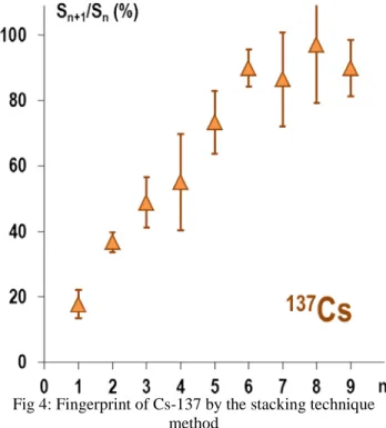
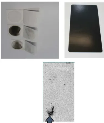
Digital Autoradiography technique an efficient tool for sampling procedure
4
0
0
Texte intégral
Figure

Documents relatifs
is assumed to be periodic, regardless of the specific trajectory (i.e. Owing to the 1/ε-term in front of the vector field G, the solution of the transport equation evolves in
Consigne : Pose et effectue
Consigne : Pose et effectue
Consigne : Pose et effectue
Consigne : Pose et effectue
Consigne : Pose et effectue
[r]
We also present an improved dichroic atomic vapor laser lock (DAVLL) method for frequency stabilization based on a nanocell with a λ/2
