HAL Id: hal-01801588
https://hal.sorbonne-universite.fr/hal-01801588
Submitted on 28 May 2018
HAL is a multi-disciplinary open access
archive for the deposit and dissemination of
sci-entific research documents, whether they are
pub-lished or not. The documents may come from
teaching and research institutions in France or
abroad, or from public or private research centers.
L’archive ouverte pluridisciplinaire HAL, est
destinée au dépôt et à la diffusion de documents
scientifiques de niveau recherche, publiés ou non,
émanant des établissements d’enseignement et de
recherche français ou étrangers, des laboratoires
publics ou privés.
Distributed under a Creative Commons Attribution| 4.0 International License
Short-term temporal memory in idiopathic and
Parkin-associated Parkinson’s disease
Bertrand Degos, Ilhame Ameqrane, Sophie Rivaud-Pechoux, Pierre Pouget,
Marcus Missal
To cite this version:
Bertrand Degos, Ilhame Ameqrane, Sophie Rivaud-Pechoux, Pierre Pouget, Marcus Missal.
Short-term temporal memory in idiopathic and Parkin-associated Parkinson’s disease. Scientific Reports,
Nature Publishing Group, 2018, 8, pp.87637. �10.1038/s41598-018-25751-8�. �hal-01801588�
Short-term temporal memory in
idiopathic and Parkin-associated
Parkinson’s disease
Bertrand Degos
1,4,5, Ilhame Ameqrane
2,3, Sophie Rivaud-Péchoux
2, Pierre Pouget
2&
Marcus Missal
3In a rapidly changing environment, we often know when to do something before we have to do it. This preparation in the temporal domain is based on a ‘perception’ of elapsed time and short-term memory of previous stimulation in a similar context. These functions could be perturbed in Parkinson’s disease. Therefore, we investigated their role in eye movement preparation in sporadic Parkinson’s disease and in a very infrequent variant affecting the Parkin gene. We used a simple oculomotor task where subjects had to orient to a visual target and movement latency was measured. We found that in spite of an increased average reaction time, the influence of elapsed time on movement preparation was similar in controls and the two groups of PD patients. However, short-term temporal memory of previous stimulation was severely affected in sporadic PD patients either ON or OFF dopaminergic therapy. We conclude that the two different contributions to temporal preparation could be dissociated. Moreover, a short-term temporal memory deficit might underlie temporal cognition deficits previously observed in PD.
Parkinson’s disease (PD) is a neurodegenerative disease characterized by motor (resting tremor, rigidity, bradyki-nesia and postural instability) and non-motor symptoms1. Particularly, cognitive decline is frequently observed
early in the evolution of the pathology, causing rapid impairment of functions like memory2–7, attention8 and
executive functions9. Cognitive decline has important consequences on the quality of life of patients and often
precedes dementia10. It has been shown repeatedly that temporal cognition is also affected in PD11–13. Temporal
cognition refers to a large set of functions among which the ability to estimate and reproduce durations in differ-ent sensory modalities. It is also essdiffer-ential before movemdiffer-ent preparation14,15. Temporal preparation refers to this
ability to plan a motor response in expectation of an upcoming trigger16,17. For instance, temporal preparation
occurs while waiting for a traffic light to turn green. This predictive capacity is based on stored memories of previously experienced similar situations containing temporal cues. In an experimental environment, temporal preparation is studied using a warning stimulus before the presentation of the target of an upcoming movement18.
The warning stimulus can be a sound or any stimulus allowing the subject to prepare to an upcoming imperative stimulus, e.g. a visual target or a response button. The period between the warning and the imperative stimuli is often referred to as the foreperiod (FP)19,20. When the duration of the FP is randomly selected from a uniform
distribution with the same probability, the reaction time of the motor responses decreases with elapsed time. This ‘FP effect’ has been observed in all species where it has been investigated and in several different sensorimotor domains. In the oculomotor domain, if a target appears in the periphery of the visual field after a variable FP, the latency of saccadic eye movements progressively decrease with increasing FP duration21. This effect could be
explained by a perception of an increased likelihood that the target will appear as time elapses during the FP22,23.
However, the FP effect could also be attributed to the influence of the previous FP duration on current temporal preparation24. Indeed, the behavior of humans and animals in response to a sensory stimulation is often partly
determined by the history of previous similar stimulation held in short-term memory, even if this influence goes unnoticed by the subject and is implicit.
1Neurology department, Parkinson’s disease expert centre, Salpêtriêre Hospital, AP-HP, Paris, France. 2Sorbonne
Universités, UPMC Univ Paris 06, Inserm U1127, CNRS UMR 7225, UM 75, ICM, F-75013, Paris, France. 3Institute
of Neuroscience (IONS), Cognition and Systems (COSY), Université catholique de Louvain, Avenue Mounier 53 bte B1.53.04 1200, Brussels, Belgium. 4Neurology Unit, Avicenne University Hospital, AP-HP, Bobigny, France. 5Present
address: Center for Interdisciplinary Research in Biology, Collège de France, UMR CNRS 7241/INSERM 1050, Labex Memolife, Paris, France. Correspondence and requests for materials should be addressed to M.M. (email: marcus. missal@uclouvain.be)
Received: 9 November 2017 Accepted: 27 April 2018 Published: xx xx xxxx
www.nature.com/scientificreports/
The aim of the present study was to investigate temporal preparation and short-term memory in PD patients ON or OFF antiparkinsonian medication. We also compared idiopathic PD (referred to as ‘iPD’) with a genetic variant of the disease (referred to as ‘Parkin’). Indeed, there is a small population of PD patients that develops a form of the disease early in life due to mutations of the Parkin gene that causes autosomal recessive juve-nile parkinsonism25. Parkin are thought to express mostly motor symptoms and less cognitive deficits26. To our
knowledge, there is no study that addressed temporal preparation and memory in Parkin patients. Therefore, we compared these two variants of the disease in order to better estimate the influence of the heterogeneity of PD on temporal preparation and short-term memory.
Methods
Participants.
Seventeen healthy control subjects (2 females, 15 males; 12% females) and twenty nine Parkinson’s disease (PD) patients took part to the study. Amongst the 29 PD patients, 19 presented the idio-pathic form of the disease (referred to as ‘iPD’ patients, amongst which 3 females; 16% females) and 10 had a genetic variant of PD (referred to as ‘Parkin’ patients; amongst which 2 females; 20% females). Although more females were present in the Parkin group of patients, there is no available experimental evidence or theoretical considerations suggesting that temporal preparation and short-term memory could be influenced by sex. Parkin patients were compound heterozygote/homozygote carriers of Parkin mutations. Sample size was determined by the availability of Parkin patients visiting the Neurology Department at the Salpêtrière Hospital in Paris, France. Ten patients could be recruited, an exceptionally large sample. Indeed, Parkin patients constitute only 10–20% of the population of early onset PD patients that itself constitutes only 15% of all PD patients27. Sample size wastherefore intrinsically reduced for Parkin patients. The sample size of iPD patients was adapted to this limitation of sample size in Parkin patients. However, in each subjects, we collected approximately two hundred saccadic eye movements. All patients fulfilled the UKPDSBB criteria28. Subjects had normal or corrected to normal vision.
All patients were taking medication to control their symptoms. All patients were taking levodopa and/or a dopa-minergic agonist. The equivalent L-dopa daily dose was computed using the procedure described in Tomlinson
et al.29. Patients of the present study were assessed using the Hoehn and Yahr scale30 and the motor part of the
Unified Parkinson’s disease Rating Scale31 (UPDRS part III; see Table 1 for details). The Starkstein scale was used
to measure apathy32. The Montreal Cognitive Assessment (MoCA) was used to assess dementia33–35. Subjects with
a MoCA score of 25 or below were excluded.
In order to test the influence of L-Dopa treatment, iPD patients were attributed to one of two groups. In the first group, patients were ON dopaminergic treatment during the first visit and testing (n = 9). Before the second visit, these patients withdrew from their current PD medications for the purpose of participating in this research project. The OFF state was practically defined as a 12-hour withdrawal of dopaminergic medication. In the sec-ond group of iPD patients (n = 10), the sequence of states was reversed: OFF during the first visit and then ON L-Dopa during the second visit. Parkin patients (n = 10) were always tested ON L-Dopa during the first visit and then OFF L-Dopa during the second visit. Given that Parkin patients are very infrequent and sometimes live at long distances, it was not possible to constitute a second group of these patients to test the OFF - ON transition. Amongst the 17 control subjects, 10 were tested twice as iPD patients, and 7 were tested only once. For all sub-jects, the two visits were separated by 7 to maximum 10 days and lasted approximately 1 hour each in all subjects.
Ethical approval and informed consent.
Experiments were approved by the local ethics committee (CPP Ile de France Paris VI, n° 212-A01056-37). Methods were carried out in accordance with relevant guidelines and regulations. Written informed consent was obtained from all participants. The study referred to as ‘PEDUPARK’ has been registered with clinicaltrials.gov (NCT02126475). All participants were informed about the purpose of the study and procedures before they gave their informed consent.Experimental paradigm.
Subjects were facing a LCD screen which presented stimuli at a refresh rate of 140 Hz. An EyeLink 1000 infrared eye tracking system (SR Research, Mississauga, Ontario) was used to record eyeCont
n = 17 iPDn = 19 Parkinn = 10 P-value t-testiPD/Parkin
Age (years) 57.4 ± 2.0 59.0 ± 1.7 42.6 ± 3.9 <0.001** Age onset (years) na 54.3 ± 1.9 23.3 ± 3.0 <0.001** Duration (years) na 4.6 ± 0.7 18.8 ± 2.8 <0.001** Hoehn and Yahr (/5) na 1.8 ± 0.1 1.6 ± 0.3 0.522 Starkstein ON (/42) 7.9 ± 0.8 11.4 ± 1.1 11.4 ± 2.1 0.992 Starkstein OFF (/42) na 11.4 ± 1.3# 12.1 ± 2.4# 0.783
UPDRS III ON (/108) na 13.6 ± 1.6 9.0 ± 1.7 0.081 UPDRS III OFF (/108) na 22.3 ± 2.2 20.2 ± 3.4 0.603 MoCA (/30) 28.8 ± 0.2 28.3 ± 0.6 28.5 ± 0.5 0.844 L-Dopa equ (mg) na 511 ± 56 443.5 ± 104.0 0.539
Table 1. Demographics and clinical measures. Cont: control subjects; iPD: idiopathic Parkinson’s disease; L-Dopa equ (mg): Levodopa equivalence in milligrams; na = not applicable. #For the Starkstein score OFF,
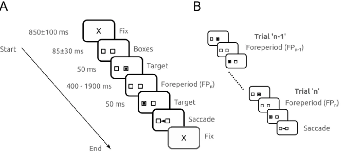
movements at 1 KHz. Stimulus display and oculomotor data collection were synchronized on a frame-by-frame basis. Saccades were detected offline in MATLAB (MathWorks, Natic, MA) with a velocity threshold of 30 deg/sec. Figure 1A depicts the time line of stimuli presentation on the screen facing the subject. Each trial started with an initial fixation period of a small cross (0.7 deg) appearing on the screen for a random duration (850 ± 100 ms; see ‘X’ on Fig. 1A). At the end of this period, two empty square ‘boxes’ appeared on the screen (1.4 × 1.4 deg), one in the center of the screen and one 9 deg eccentric, randomly to the right or to the left. Afterwards, a square target was flashed in the central box for 50 ms. Extinction of the initial target indicated to subjects the beginning of the foreperiod (FP). Subjects were required to hold on fixation of the central box until another target was briefly presented for 50 ms in the eccentric box. The FP duration could take one of 4 different values with the same probability: 400 ms, 900 ms, 1400 ms and 1900 ms. Subjects were required to wait until stimulus appearance in the eccentric box to make a saccade (black arrowhead on Fig. 1A). Saccadic latency (reaction time) was defined as the time elapsed between the appearance of the eccentric target and movement onset. Figure 1B shows a schematic diagram to illustrate the influence of short-term memory of the forepriod during the preceding trial (FPn−1) on
saccadic latency during trial’n’. Each sequence from the initial to the final fixation period will be referred to as a ‘trial’. Figure 1B shows two trials in succession. The foreperiod during the current trial (trial ‘n’) will be referred to as ‘FPn’. The foreperiod during the previous trial (trial ‘n−1’) will be referred to as ‘FPn−1’.
Statistical analyses.
The significance threshold α for all analysis was 0.01. All analyses were realized using analysis of variance (ANOVA) unless specified otherwise. Analyses were performed using SPSS 23 (SPSS Inc., Chicago, IL). Mixed-design ANOVA’s and multiple regression analysis were used. In all analyses, subject identity was used as a random factor to take into account the influence of uncontrolled variability observed between sub-jects. Results will be presented as mean ± standard error of the mean unless specified.Data availability.
The datasets generated during and/or analysed during the current study are available from the corresponding author on reasonable request.Results
Demographics and clinical characteristics.
The mean age of iPD patients was 59.0 ± 1.7 years (n = 19) with a mean duration of the disease of 4.7 ± 0.7 years. The mean age of control subjects was 57.4 ± 1.7 years (n = 17). The mean age of Parkin patients was 43.0 ± 3.9 years (n = 10) with a mean duration of the disease of 18.8 ± 2.8 years. Parkin patients were statistically younger (F[1, 27] = 20.74; P < 0.001) and had a longer duration of the disease (F[1, 27] = 40.28; P < 0.001). However, Hoehn and Yahr stages were similar in both groups (F[1, 27] = 0.42; P = 0.52; NS) as well as the UPDRS III score ON L-Dopa treatment (F[1, 27] = 6.80; P = 0.015; NS; Table 1). The mean daily levodopa equivalent dose was similar in iPD (511 ± 56 mg, n = 19) and Parkin patients (444 ± 104 mg; F[1, 27] = 0.39; P = 0.54; NS; see Table 1). The mean age of iPD and control subjects was not sta-tistically different (F[1, 34] = 5.03; P = 0.483; NS).Saccades in iPD and Parkin patients.
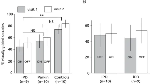
Given that an oculomotor form of hypokinesia could be observed in patients, we firstly compared the percentage of executed visually-guided saccades in patients and controls (see Fig. 2). At least two blocks of 100 trials were presented to all subjects (TT = 200 trials total) and a total of 100 to200 saccades (TS) were recorded per subject. The percentage of executed saccades was computed as [(TS/TT)*100].
To compare the percentage of saccades executed in the different groups, a repeated-measures ANOVA with sub-ject group as between subsub-jects factor and visit as within subsub-ject factor was performed. In this test, we used the ON-OFF group of iPD patients, in order to compare it with Parkin patients who were tested only with this same sequence of treatments (iPD ON-OFF group, n = 9; Parkin, n = 10; controls, n = 10; see Fig. 2). We found that the percentage of saccades significantly increased between visits (F[1] = 20.807; P < 0.001) and that subject group had Figure 1. (A) Schematic description of the experimental paradigm. Subjects were facing a computer screen where the sequence of stimuli was presented. Fix: appearance of the fixation cross at the beginning of a trial. See Methods for details. (B) Schematic diagram illustrating the influence of short-term memory of the forepriod during the preceding trial (FPn−1) on saccadic latency during trial ‘n’ (FPn).
www.nature.com/scientificreports/
a significant influence on the percentage of saccades (F[2] = 6.423; P = 0.005). However, there was no significant interaction between factors (F[2] = 0.018; P = 0.983). A post-hoc Bonferroni test suggests that on average the percentage of saccades was higher in controls than in iPD patients (P = 0.005) but not different between controls and Parkin patients (P = 0.073) or between iPD and Parkin patients (P = 0.757).
In iPD patients, we also compared the percentage of saccades between the two groups who experienced a dif-ferent order of treatments (ON-OFF versus OFF-ON). In iPD patients, treatment order had no significant influ-ence (F[1] = 0.001; P = 0.977). The percentage of visually-guided saccades did not significantly increase between visits (F[1] = 0.527; P = 0.473) and there was no significant interaction between factors (F[1, 3] = 0.234; P = 0.631; see Fig. 2B).
In summary, statistically less visually-guided saccades were observed in iPD patients than in controls. Parkin patients apparently made less-visually guided saccades than controls but this difference did not reach statisti-cal significance. In iPD patients, dopaminergic treatment did not significantly alter the percentage of saccades observed that tended to increase with repetition (without reaching statistical significance).
Temporal preparation in Parkinson’s disease.
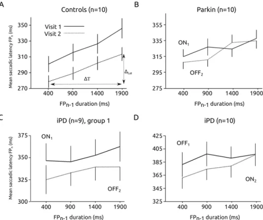
The foreperiod effect is a behavioral measure of temporal preparation that could be affected in PD. Figure 3 shows the relationship between saccadic latency and foreperiod (FP) duration in the 4 groups of subjects (controls, Parkin, iPD ON1 – OFF2 and OFF1 – ON2). Figure 3A showsthat, in control subjects, there was a significant influence of FP duration on the latency of visually guided sac-cades (ANOVA with FP duration as fixed factor and subject as random factor; F[3, 27.363] = 27.663; p < 0.001; n1 = 1508 saccades; n2 = 1702 saccades; n = 10 subjects). As expected, saccadic latencies decreased with
increas-ing FP duration (the FP effect). Although latencies tended to decrease on average between visit 1 and visit 2, this effect did not reach statistical significance (F[1, 9.037] = 7.304; p = 0.024; NS). Moreover, there was no significant interaction between FP duration and visit (F[3, 28.415] = 0.446; p = 0.722). In Parkin patients (see Fig. 3B), FP duration also had a significant effect on movement latency (F[3, 30.807] = 47.120; p < 0.001) but dopaminergic treatment had no effect (F[1, 9.242] = 0.550; p = 0.477; nON = 2272 saccades, nOFF = 2544). No interaction between
FP duration and treatment was found (F[3, 31.197] = 0.947; p = 0.430; n = 10 patients). In iPD patients, we tested the two different transitions between states in two different groups of subjects (ON1-OFF2 transition, see Fig. 3C;
OFF1-ON2 transition, see Fig. 3D). In the ON1-OFF2 group (n = 9), FP duration (F[3, 29.950] = 9.382; p < 0.001;
nON = 853 saccades, nOFF = 1021) and treatment (F[1, 11.033] = 13.094; p = 0.004) had a significant effect on
laten-cies but no significant interaction between FP duration and treatment was found (F[3, 30.982] = 1.056; p = 0.382). Saccadic latencies were longer during visit 1 ON L-Dopa but the FP effect remained unaltered. In the OFF1-ON2
group of iPD patients (n = 10), FP duration had a significant effect on latencies (F[3, 33.080] = 21.060; p < 0.001; nOFF = 932 saccades, nON = 1021) but not dopaminergic treatment (F[1, 11.786] = 0.277; p = 0.608). Here also, no
significant interaction between FP duration and treatment was found (F[3, 36.503] = 0.554; p = 0.649).
These results show that the FP effect was similar whether subjects were ON or OFF L-Dopa (absence of FP duration x dopaminergic treatment interaction). A significant influence of dopaminergic treatment on average saccadic latencies was present in the ON1-OFF2 iPD group, but there was no significant influence of the treatment
on the FP effect itself. Therefore, we conclude that the FP effect did not depend on dopaminergic medication. Alteration of mean reaction time could be dissociated from the FP effect.
Figure 2. Saccadic hypokinesia in PD patients. (A) Percentage of visually-guided saccades towards the eccentric visual target in the different groups of subjects. Note that PD patients performed rather poorly compared with healthy controls. Data from subjects tested twice (visit 1 & visit 2). (B) Percentage of visually-guided saccades in the two groups of iPD patients. **Indicates a statistically significant differences (P < 0.01).
Influence of the short-term temporal memory.
It has been shown that the FP effect could depend heavily on the previously experienced FP duration24. This influence could be characterized as a sequence ortem-poral short-term memory effect. Indeed, the content of a short-term temtem-poral storage (duration of FPn−1) could
be influencing saccadic latency during the current foreperiod (FPn). Given that short-term memory is essential
for executive functions, it could be a reliable indicator of cognitive decline in PD. Figure 4A shows the influ-ence of FPn−1 duration on average saccadic latency during FPn in controls. Saccadic latency tended to regularly
increase with increasing FPn−1 duration during both visits, and a significant effect of FPn−1 on saccadic latency
was found (mixed-design ANOVA with FPn−1 as fixed factor and subject as random factor: F[3, 28.344] = 28.199;
P < 0.001; n = 3210 saccades total). However, there was no significant influence of visit (F[1, 9.037] = 8.403; P = 0.018) and no significant interaction between fixed factors (F[3, 28.195] = 0.873; P = 0.467). A similar trend was found in Parkin patients. Indeed, on average saccadic latency increased with increasing FPn−1 duration (F[3,
33.918] = 11.646; P < 0.001; n = 4816 saccades) but there was no significant influence of visit (F[1, 9.241] = 0.248; P = 0.630) and no significant interaction between fixed factors (F[3, 34.476] = 1.577; P = 0.213).
The influence of short-term temporal memory was statistically similar in controls and Parkin patients. However, the situation was different in iPD patients. Figure 4C,D show the relationship between FPn−1
dura-tion and saccadic latency during FPn in iPD patients (group 1: ON1 – OFF2, Fig. 4C; group 2: OFF1 – ON2,
Fig. 4D). In group 1, saccadic latency did not increase with increasing FPn−1 duration (F[3, 33.049] = 1.180;
P = 0.332; n = 1874 saccades). There was a significant influence of visit (F[1, 10.484] = 13.196; P = 0.004) but no significant interaction between fixed factors (F[3, 49.654] = 0.315; P = 0.815). These results suggest an absence of influence of short-term memory and a small test-retest effect on average saccadic latencies. In group 2, there was no significant effect of FPn−1 on saccadic latency (F[3, 44.759] = 3.041; P = 0.039; n = 1953 saccades), no
significant influence of visit (F[1, 12.358] = 0.629; P = 0.443) and no significant interaction between fixed factors (F[3, 37.090] = 0.337; P = 0.798). To better capture this effect, we computed for each subject the average slope of the relationship between saccadic latency during trial’n’ and FPn−1 duration (see Fig. 4A): Slope = ΔLAT/ΔT
with: ΔLAT = [(average latency of saccades for FPn−1 = 1900 ms)-(average latency of saccades for FPn−1 = 400 ms)];
ΔT = 1900–400 = 1500 ms. Figure 5 shows the average value of the slope for each group. If there were a modu-lation of saccadic latency with short-term temporal memory, the average value of the slope should be different from zero. In iPD patients, the slope was more variable than in Parkin patients or controls. We applied a one sam-ple two-tailed t-test with a test-value of zero for each group. In iPD patients, there was no significant difference Figure 3. The foreperiod effect. Saccadic latency decreased as a function of increasing delay duration in controls (A), Parkin (B) and iPD patients (C, ON-OFF group; D, OFF-ON group). Although saccadic latency could be affected by treatment, the foreperiod effect itself (the decreasing saccadic latency with increasing foreperiod duration) was not affected by dopaminergic medication. Data from subjects tested twice (visit 1 & visit 2).
www.nature.com/scientificreports/
between average slope values and zero in either the OFF1 – ON2 or the ON1 – OFF 2 group (P > 0.01). However,
slopes were significantly different from zero in Parkin patients and controls (see legend of Fig. 5 for detailed statis-tics). We found no correlation between the average slope values for each subject and demographics (age, duration of the disease), L-Dopa equivalent or UPDRS III score (Pearson correlation, two-tailed test, P > 0.01).
In order to determine whether on average there was a different linear relationship between FPn−1 and
sacca-dic latency between iPD and Parkin patients with respect to controls, a Mixed-model multiple linear regression analysis was performed with subjects as random factor, group as fixed factor and FPn−1 as co-variate. This analysis
should give a significant interaction term (moderator effect) if there is a different linear relationship between var-iables in iPD and Parkin patients with respect to controls. Figure 6A shows the relationships between FPn−1 and
saccadic latency for the 2 groups of patients tested ON1 – OFF 2 (Parkin and iPD) and controls. It can be seen that
saccadic latency increased rapidly with FPn−1 duration in controls but that this relationship could have a shallower
slope in patients. For Parkin patients the slope of the relationship was apparently intermediate between the other Figure 4. Influence of the duration of previous foreperiod (FPn−1) on saccadic latency during the current
foreperiod (FPn) in the different groups of subjects. The influence of the previous foreperiod was significant only
in controls (A) and Parkin patients (B) but not in iPD patients (C, ON1-OFF2 group; D, OFF1-ON2). See text for
statistics.
Figure 5. Short-term temporal memory deficit in PD patients. Average slopes in the different groups. Statistics: iPD: OFF1 (t = 0.12; df = 9; P = 0.25) – ON2 (t = 2.27; df = 9; P = 0.05); iPD: ON1 (t = 0.29; df = 9;
P = 0.78) – OFF2 (t = 0.1; df = 8; P = 0.92); Parkin: ON1 (t = 12.79; df = 9; P < 0.001) – OFF2 (t = 6.58; df = 9;
P < 0.001); controls: visit 1 (t = 8.10; df = 9; P < 0.001) – visit 2 (t = 6.24; df = 9; P < 0.001). Data from subjects tested twice (visit 1 & visit 2).
two groups. If there is a moderator effect in the mixed-model multiple linear regression analysis, then it could be hypothesized that the influence of short-term temporal memory on saccadic latency is different between groups of subjects taking controls as reference. We did not find a significant main effect of patient group (F[2, 9894] = 1.836; P < 0.160) but a significant main effect of FPn−1 duration (F[1, 24.804] = 82.359; P < 0.001). However, we found
that the moderator effect was not significant (F[2, 22.207] = 2.286; P = 0.125). Therefore, based on this data set, we could not conclude that there is a different linear relationship between FPn−1 duration and saccadic latency
between groups. This conclusion could be due to the small sample of subjects. In order to lift this limitation, we pooled iPD subjects of the ON1-OFF2 and OFF1-ON2 groups together. This pooling is legitimated by the
observa-tion that dopaminergic treatment had no significant influence on short-term memory in iPD patients (see above). iPD patients were either ON1 or OFF1. This could not be done for Parkin patients. Indeed, there is not enough
Parkin patients in France to constitute two groups. Moreover, given that age is a major factor of general decline
that could occur rapidly, we collected additional data on 7 male healthy subjects in order to create cohorts of equal sizes that have less than a year of age difference between controls and iPD patients (controls: 57.9+/−2.0 years, n = 15; iPD patients: 57.5+/−1.9 years, n = 15; ~3000 saccades in each group). These additional control subjects were tested only once (visit 1 only). We found no significant difference in age between groups (F[1] = 0.022; P = 0.883). Here also, the average slopes of the FPn−1/saccade latency relationship were significantly different from
zero in controls (t[14] = 4.825; P < 0.001) but not in iPD patients (t[14] = 0.280; P = 0.783). Moreover, we carried out a similar Mixed-model multiple regression analysis with these two groups of subjects. In this case, we found that FPn−1 duration (F[1, 28.040] = 10.052; P = 0.004) had a significant effect but not subject group (controls or
iPD; F[1, 3347] = 3.732; P = 0.053). However, a significant interaction (moderator effect) was found between independent variables (F[1, 28.040] = 8.447; P = 0.007; see Fig. 6B). This result suggests that the linear FPn−1/
saccade latency relationship was indeed different between iPD patients and controls.
Discussion
We found that in all subjects and patients groups, temporal preparation (the foreperiod effect) was preserved. Given the motor deficits observed in PD, several authors have tested the FP effect in these patients36–38. All these
previous studies confirmed that the FP effect was observed (but see [38] for a dissociation between reflexive and voluntary behaviors). Accordingly, in the present study, in both groups of PD patients, latencies steadily decreased as time elapsed during the delay period. Observation of a preserved variable FP effect in PD patients is not sur-prising. Indeed, this effect is very robust in several neurological diseases, and does not depend on an explicit con-scious manipulation of temporal information that is affected in PD12. Stated differently, the implicit monitoring
of elapsed time is functional in PD patients. Given that the duration of the previous FP (FPn−1) has been shown to
influence movement latency and the FP effect in healthy subjects, we analyzed the influence of FPn−1 on saccadic
latency. Within-group analyses revealed that memory of the previous FP duration affected movement latency during the current trial’n’ in controls and Parkin patients but not in iPD patients. The slope analysis confirmed this observation. A Mixed-model multiple linear regression analysis revealed that the posited linear relationship between FPn−1 and saccadic latency was not statistically different between groups. This absence of effect might be
related to the small sample size dictated by the small population of Parkin patients available in France. However, comparison between controls and iPD subjects matched for a difference of age of less than a year and inde-pendently of dopaminergic status showed that memory of previous FP played a significantly different role btween these two groups. Most cognitive processes rely on working memory that could be defined as the ability to main-tain and manipulate meaningful information during a short time period for usage in complex tasks like language, learning and reasoning39. Several studies suggest an impairment of working memory in PD patients40. Indeed,
the pathophysiological process of PD might affect working memory. Short-term memory could be described as the input buffer to working memory. If working memory is not properly fed with temporal information, then all time-dependent cognitive abilities might be affected. The ability to keep track of the history of previous foreper-iods (FPn−1) is essential in order to build an internal representation of the likelihood of future foreperiods (FPn,
FP , etc…). Memory is useful if it helps to anticipate or predict future events. If prediction based on short-term Figure 6. (A) Between group comparison of the relationship between previous FP duration and saccadic latency. (B) Same analysis on an additional data set with controls and iPD patients differing by <1 year of age.
www.nature.com/scientificreports/
memory does not operate properly, iPD patients might be in a constant state of unpreparedness when the target appears, generating longer latency responses. It could be argued that a loss of cognitive flexibility could explain observed effects. Cognitive flexibility could be defined as the capacity to weight different alternatives when faced with a complex problem. It involves also the capacity to shift attention from one rule valid in a given context to a new rule. Idiopathic PD patients tend to perseverate if rules to execute a given task are unexpectedly changed, showing an obvious lack of cognitive flexibility41–46. Therefore, one could argue that reduced cognitive flexibility
is sufficient to explain the perturbed sequence effect or reduced short-term memory observed in iPD patients in the present study. For at least two reasons we believe this hypothesis could be rejected. Firstly, it should be emphasized that cognitive flexibility is always tested in tasks where information must be explicitly manipulated in working memory in order to achieve a particular goal. This is not the case here. Indeed, no instruction about trial sequence was required from subjects. They were asked to ‘make a saccade to the eccentric target when it appears in the periphery’. Secondly, in the present experimental paradigm, if loss of cognitive flexibility were a valid expla-nation, it should manifest itself by perseverance based on previous information and therefore a stronger influence of FPn−1. Another alternative explanation to reduced short-term temporal memory is that bradyphrenia or
brady-kinesia could explain observed effects. Bradyphrenia is a general slowing of cognitive processes1 and bradykinesia
is a slowing down of movements generally observed in iPD47. Could these two commonly observed phenomena
in iPD explain observation of the present study? Firstly, a strong influence of the current FP (the foreperiod effect) was found in our patients in spite of an increased average saccadic latency (oculomotor hypo/bradykinesia). Therefore, implicit temporal processing during the current trial was not affected. However, this information was not subsequently used in the planning of the next saccadic eye movement. Secondly, cognitive abilities evaluated by the MoCA test did not differ between controls and iPD patients. Therefore, we hypothesize that a reduced or more variable influence of FPn−1 as presented here results of an impairment of temporal short-term memory (or
sequence effect) and not of a general slowing of motor and cognitive processes that should be detected with the MoCA test.
In conclusion, the oculomotor variant of the variable foreperiod paradigm used in the present study con-firmed that temporal preparation could depend of both an implicit ‘perception’ of elapsed time and short-term memory. The influence of elapsed time during the current foreperiod was not affected by PD or dopaminergic status even if saccadic latency tended to be longer in PD patients. A within-group analysis showed that the influ-ence of the previous foreperiod (FPn−1) was present in controls and Parkin patients, but not in iPD patients. In
iPD patients, a robust foreperiod effect could be observed in the absence of a short-term memory effect. The two different contribution to the foreperiod effect could be dissociated. However, we could not determine if there was a significantly different linear relationship between FPn−1 and saccadic latency in iPD and Parkin patients with
respect to controls.
References
1. Kehagia, A. A., Barker, R. A. & Robbins, T. W. Neuropsychological and clinical heterogeneity of cognitive impairment and dementia in patients with Parkinson’s disease. Lancet Neurol. 9, 1200–1213 (2010).
2. Dubois, B. & Pillon, B. Cognitive deficits in Parkinson’s disease. J Neurol. 244, 2–8 (1997).
3. Cooper, J. A., Sagar, H. J., Jordan, N., Harvey, N. S. & Sullivan, E. V. Cognitive impairment in early, untreated Parkinson’s disease and its relationship to motor disability. Brain. 114, 1095–2122 (1991).
4. Owen, A. M. Cognitive dysfunction in Parkinson’s disease: the role of frontostriatal circuitry. Neuroscientist. 10, 525–37 (2004). 5. Meireles, J. & Massano, J. Cognitive impairment and dementia in Parkinson’s disease: clinical features, diagnosis, and management.
Front. Neurol. 3, 88, https://doi.org/10.3389/fneur.2012.00088c (2012).
6. Kudlicka, A., Clare, L. & Hindle, J. V. Executive functions in Parkinson’s disease: systematic review and meta-analysis. Mov. Disord.
26, 2305–15, https://doi.org/10.1002/mds.23868 (2011).
7. Morris, R. G. et al. Planning and spatial working memory in Parkinson’s disease. J. Neurol. Neurosurg. Psychiatry. 51, 757–66 (1998). 8. Ballard, C. G. et al. Fluctuations in attention: PD dementia vs DLB with parkinsonism. Neurology 59, 1714–1720 (2002). 9. Levy, G. et al. Memory and executive function impairment predict dementia in Parkinson’s disease. Mov. Disord 17, 1221–26 (2002). 10. Levy, G. et al. The association of incident dementia with mortality in PD. Neurology 59, 1708–13 (2002).
11. Buhusi, C. V. & Meck, W. H. What makes us tick? Functional and neural mechanisms of interval timing. Nat. Rev. Neurosci. 6, 755–65 (2005).
12. Malapani, C., Deweer, B. & Gibbon, J. Separating storage from retrieval dysfunction of temporal memory in Parkinson’s disease. J.
Cogn. Neurosci. 14, 311–22 (2002).
13. Pastor, M. A., Artieda, J., Jahanshahi, M. & Obeso, J. A. Time estimation and reproduction is abnormal in Parkinson’s disease. Brain.
115, 211–25.70 (1992).
14. Nobre, A., Correa, A. & Coull, J. The hazards of time. Curr. Opin. Neurobiol. 17, 465–70 (2007).
15. Coull, J. T. Neural substrates of mounting temporal expectation. PLoS Biol. Aug; 7(8), e1000166, https://doi.org/10.1371/journal. pbio.1000166. Epub2009 Aug 4.
16. Vallesi, A. et al. The neural basis of temporal preparation: Insights from brain tumor patients. Neuropsychologia. 45, 2755–63 (2007). 17. Los, S. A., Kruijne, W., Meeter, M. Outlines of a multiple trace theory of temporal preparation. Front Psychol. 5, 1058. https://doi.
org/10.3389/fpsyg.2014.01058. ECollection (2014).
18. Woodrow, H. The measurement of attention. Psychological Monographs. 5, 1–158 (1914).
19. Niemi, P. & Näätänen, R. Foreperiod and simple reaction time. Psychological Bulletin 89, 133–62 (1981).
20. Los, S. Foreperiod and sequential effects: theory and data. In Coull and Nobre (Eds), Attention and Time. Oxford (2010). 21. Ameqrane, I., Pouget, P., Wattiez, N., Carpenter, R. H. & Missal, M. Implicit and explicit timing in oculomotor control. PLoS One.
9(4), e93958, https://doi.org/10.1371/journal.pone.0093958 (2014).
22. Janssen, P. & Shadlen, M. N. A representation of the hazard rate of elapsed time in macaque area LIP. Nat. Neurosci. 8, 234–41 (2005). 23. Oswal, A., Ogden, M. & Carpenter, R. H. The time course of stimulus expectation in a saccadic decision task. J. Neurophysiol. 97,
2722–30 (2007).
24. Vallesi, A., Shallice, T. & Walsh, V. Role of the prefrontal cortex in the foreperiod effect: TMS evidence for dual mechanisms in temporal preparation. Cereb. Cortex. 17, 466–74 (2007).
25. Gasser, T. Genetics of Parkinson’s disease. Curr. Op. Neurol. 18, 363–369 (2005).
26. Alcalay, R. N. et al. Cognitive and motor function in long-duration PARKIN-associated Parkinson disease. JAMA Neurol. 71, 62–7 (2014).
27. Klein, C. & Schlossmacher, M. G. The genetics of Parkinson disease: Implications for neurological care. Nat Clin Pract Neurol. 2, 136–46 (2006).
28. Hughes, A. J., Daniel, S. E., Kilford, L. & Lees, A. J. Accuracy of clinical diagnosis of idiopathic Parkinson’s disease: a clinico-pathological study of 100 cases. J. Neurol. Neurosurg. Psychiatry. 55, 181–4 (1992).
29. Tomlinson, C. L. et al. Systematic review of levodopa dose equivalency reporting in Parkinson’s disease. Mov. Disord. 25, 2649–53 (2010).
30. Hoehn, M. M. & Yahr, M. D. Parkinsonism: onset, progression and mortality. Neurology 17, 427–442 (1967).
31. Goetz, C. G. et al. Movement Disorder Society-sponsored revision of the Unified Parkinson’s Disease Rating Scale (MDS-UPDRS): scale presentation and clinimetric testing results. Mov. Disord. 23, 2129–2170 (2008).
32. Starkstein, S. E. et al. The syndromal validity and nosological position of apathy in Parkinson’s disease. Mov. Disord. 24, 1211–6 (2009).
33. Gill, D. J., Freshman, A., Blender, J. A. & Ravina, B. The Montreal cognitive assessment as a screening tool for cognitive impairment in Parkinson’s disease. Mov. Disord. 23, 1043–6 (2008).
34. Hoops, S. et al. Validity of the MoCA and MMSE in the detection of MCI and dementia in Parkinson disease. Neurology. 73, 1738–45 (2009).
35. Kulisevsky, J. & Pagonabarraga, J. Cognitive impairment in Parkinson’s disease: tools for diagnosis and assessment. Mov. Disord. 24, 1103–10 (2009).
36. Jahanshahi, M., Brown, R. G. & Marsden, C. D. Simple and choice reaction time and the use of advanced information for motor preparation in Parkinson’s disease. Brain 115, 539–564 (1992).
37. Jordan, N., Sagar, H. J. & Cooper, J. A. Cognitive component of reaction time in Parkinson’s disease. Journal of Neurology,
Neurosurgery, and Psychiatry. 55, 658–664 (1992).
38. Jurkowski, A. J., Stepp, E. & Hackley, S. A. Variable foreperiod deficits in Parkinson’s disease: dissociation across reflexive and voluntary behavior. Brain Cogn. 58, 49–61 (2005).
39. Baddeley, A. Working memory. Science. 255, 556–9 (1992).
40. Owen, A. M., Iddon, J. L., Hodges, J. R., Summers, B. A. & Robbins, T. W. Spatial and non-spatial working memory at different stages of Parkinson’s disease. Neuropsychologia. 35, 519–32 (1997).
41. Cools, R., Barker, R. A., Sahakian, B. J. & Robbins, T. W. Enhanced or impaired cognitive function in Parkinson’s disease as a function of dopaminergic medication and task demands. Cerebral cortex (New York, NY: 1991) 11, 1136–1143 (2001).
42. Cools, R., Barker, R. A., Sahakian, B. J. & Robbins, T. W. Mechanisms of cognitive set flexibility in Parkinson’s disease. Brain. 124, 2503–2512 (2001).
43. Cools, R., Clark, L. & Robbins, T. W. Differential responses in human striatum and prefrontal cortex to changes in object and rule relevance. J Neurosci 24, 1129–1135 (2004).
44. Cools, R., Ivry, R. B. & D’Esposito, M. The human striatum is necessary for responding to changes in stimulus relevance. J Cogn
Neurosci 18, 1973–1983 (2006).
45. Cools, R. Dopaminergic modulation of cognitive function-implications for L-DOPA treatment in Parkinson’s disease. Neurosci
Biobehav Rev. 30, 1–23 (2006).
46. Svenningsson, P., Westman, E., Ballard, C. & Aarsland, D. Cognitive impairment in patients with Parkinson’s disease: diagnosis, bio-markers and treatment. Lancet Neurol. 11, 697–707 (2012).
47. Braak, H. et al. Staging of brain pathology related to sporadic Parkinson’s disease. Neurobiol Aging. 24, 197–211 (2003).
Author Contributions
Bertrand Degos, study concept and design, analysis and interpretation of data, drafting or revising the manuscript for intellectual content, study supervision. Ilhame Ameqrane, acquisition of data. Sophie Rivaud-Péchoux, analysis and interpretation of data, critical revision of manuscript for intellectual content, acquisition of data. Pierre Pouget, study concept and design, analysis and interpretation of data, critical revision of manuscript for intellectual content, study supervision. Marcus Missal, study concept and design, analysis and interpretation of data, study supervision, drafting or revising the manuscript for intellectual content.
Additional Information
Competing Interests: Bertrand Degos Employment: APHP (assistance Publique des Hôpitaux de Paris); Grants: INSERM (COSSEC); France Parkinson; MERZ-pharma. Other: none. Ilhame Ameqrane Employment: INSERM. Pierre Pouget Boards: Scientific Reports. Employment: CNRS; Grants: ANR Carnot, Bettencourt Schuller, FRC. Sophie Rivaud-Péchoux Employment: INSERM. Marcus Missal Employment: Université catholique de Louvain. Grants: FRSM, FRS-FNRS, ARC; Other: none.
Publisher's note: Springer Nature remains neutral with regard to jurisdictional claims in published maps and institutional affiliations.
Open Access This article is licensed under a Creative Commons Attribution 4.0 International License, which permits use, sharing, adaptation, distribution and reproduction in any medium or format, as long as you give appropriate credit to the original author(s) and the source, provide a link to the Cre-ative Commons license, and indicate if changes were made. The images or other third party material in this article are included in the article’s Creative Commons license, unless indicated otherwise in a credit line to the material. If material is not included in the article’s Creative Commons license and your intended use is not per-mitted by statutory regulation or exceeds the perper-mitted use, you will need to obtain permission directly from the copyright holder. To view a copy of this license, visit http://creativecommons.org/licenses/by/4.0/.


