HAL Id: hal-01706143
https://hal.archives-ouvertes.fr/hal-01706143
Submitted on 10 Feb 2018
HAL is a multi-disciplinary open access
archive for the deposit and dissemination of
sci-entific research documents, whether they are
pub-lished or not. The documents may come from
teaching and research institutions in France or
abroad, or from public or private research centers.
L’archive ouverte pluridisciplinaire HAL, est
destinée au dépôt et à la diffusion de documents
scientifiques de niveau recherche, publiés ou non,
émanant des établissements d’enseignement et de
recherche français ou étrangers, des laboratoires
publics ou privés.
Interventional planning and assistance for ascending
aorta dissections
Christophe Lohou, Pawel Lubniewski, Nawel Fetnaci, Bruno Miguel, Laurent
Sarry
To cite this version:
Christophe Lohou, Pawel Lubniewski, Nawel Fetnaci, Bruno Miguel, Laurent Sarry. Interventional
planning and assistance for ascending aorta dissections. Colloque Recherche en Imagerie et
Technolo-gies pour la Santé - RITS 2013, Apr 2013, Bordeaux, France. �hal-01706143�
Interventional planning and assistance for ascending aorta dissections
Christophe Lohou
1, Pawe l Lubniewski
1,2
, Nawel Fetnaci
1, Bruno Miguel
1,3, Laurent Sarry
11. Clermont Universit´e, Universit´e d’Auvergne, ISIT-UMR 6284 UdA-CNRS, BP 10448, F-63000 Clermont-Ferrand (France) 2. Cardinal Stefan Wyszy´nski University, Faculty of Mathematics and Natural Sciences, Warsaw (Poland)
3. CHU Gabriel Montpied, Service de Chirurgie Cardio-Vasculaire, Pˆole Cardiologie, 58 rue Montalembert, BP69, F-63003 Clermont-Ferrand (France)
Abstract — In this paper, we present our actual works concerning interventional planning and assistance for ascending aorta dissections. The first part is about aortic dissection re-trieval in Computed Tomography Angiography (CTA) images. It is based on fast marching seg-mentation, mathematical morphology and digi-tal topology frameworks.
The second part is about the registration of the outer aortic wall onto X-ray fluoroscopic an-giographic images. It is based on either Digital Reconstructed Radiographs (DRRs) or para-metric model projection, then on image trans-formation descriptors (ITDs) computing. Our motivation is first to give 3D views of dis-secting aorta in peroperative step to assess the dissection, then to augment the current angio-graphic view by registering aortic dissection features during the intraoperative step to as-sist clinicians.
Keywords
aortic dissections, segmentation, 3D/2D registration, augmented reality
I. INTRODUCTION
Aortic dissections are caused by one or more aortal tissue perforation(s) because of tissue weakness and blood pressure. That consists of one or several intimal tear(s) (Fig. 1 (left)) and a clear separation between layers of the aortic media, resulting in two separate blood flow channels : the true lumen (the normal bed of blood in the aorta) and the false lumen (a chan-nel entirely within the media which appears during an aortic dissection). The layer of detached aorta tissues between lumens is called intimal flap.
To deal with aortic dissections, physicians require preoperative 3D Computed Tomography Angiography (CTA) images (Fig. 1 (right)), then intraoperative 2D X-ray (here fluoroscopic, by a monoplane C-arm sys-tem) angiographic images (Fig. 3).
Our works consist in segmenting 3D CTA images to retrieve aortic dissection features, and to register them to X-ray images.
Figure1 – (left) (1), (2) and (3) respectively intima, media and adventitia layers ; (a) an intimal tear, (b) the false lumen ; (c) the true lumen. (right) 2D grays-cale Computed Tomography Angiography slice (T : true lumen, F : false lumen).
II. SEGMENTATION
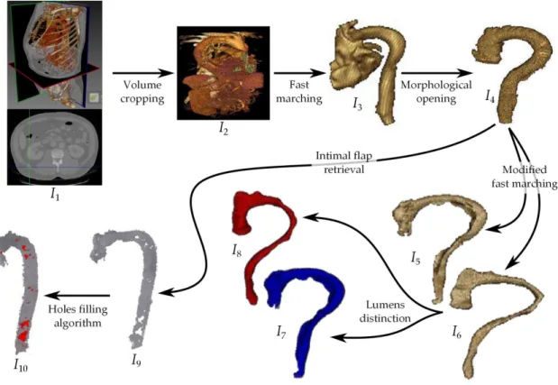
In this section, we summarize our different works rela-ted to aortic dissection features extraction. They are schematized in Fig. 2.
Preprocessing. Let us consider an initial CT aortic dissection image : I1. We perform a crop to
accele-rate image processing : I2.
Lumens retrieval. Fast marching method (FM in the following) is an efficient implementation of level set deformable models. We initialize FM by defining a single voxel inside a lumen. Is it possible to limit the number of iterations of the evolution process to avoid the propagation to other arteries (collateral arteries) but the 3D propagation front reaches the heart : I3. By using a morphological opening, we
may cut that part of heart : we obtain the image of the two interconnected lumens I4.
By modifying the speed function of FM, and using an intermediate labelling image (of points in the neighborhood of the intimal flap [dark grayscale]), we succeed in separating the two lumens[2] : I5 and
I6.
Lumens distinction. By using the fact that grays-cale value of blood is higher inside the true lumen, a quick operator (mean intensity inside each lumen) allowed us to distinguish between the true lumen and the false lumen, obtaining thus the false lumen image I7 and the true lumen image I8.
se-I
1I
2I
3I
4I
5I
7I
8I
9I
10 Volume cropping Fast marching Morphological openingFigure 2 – Aortic dissection segmentation : I7false lumen, I8true lumen, I9 intimal flap, I10 entry tears in red.
quel of n morphological dilations followed by a se-quel of n morphological erosions) gives the image of the intimal flap : I9.
Intimal tears materializing. The first challenge of our works was to obtain the entry tears from a CT. Because they do not correspond to physical tissues or organs, such a segmentation cannot be direct. In-timal tears may be considered as 3D holes in the intimal flap. Therefore, we used a holes filling algo-rithm proposed in Digital Topology framework[1] : the surfaces filling holes in I9 are shown in red in
I10; they correspond to the location of intimal tears.
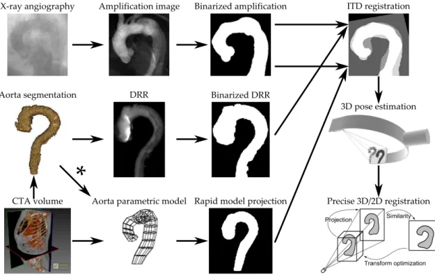
III. REGISTRATION
The main goal of our registration framework is to pro-vide an alignment of pre- and intra-operational medi-cal images (see Fig. 3).
Source image for registration. See the top row of Fig. 3. The reference image is a 2D X-ray fluoro-scopic angiography sequence, acquired directly in the interventional room. In order to augment the aorta visibility, a filtering technique has been ap-plied on the whole set of angiographic frames, to yield a single 2D image called Amplification image. Binaring the previous image, we obtain the Bina-rized amplification, which is the fixed input for the Registration algorithm.
Target image for registration. The Segmented
aorta (issued from a CTA 3D image) plays the role of the (moving) target to establish the 3D/2D correspondence.
Now, we give three ways to compute a 2D model of that target :
– One way [center row of Fig. 3] is to compute a Digitally Reconstructed Radiograph (DRR) from the aorta interior ; the projection parameters are computed in the registration process.
– Another alternative solution [bottom row of Fig. 3] is to define an aorta parametric model (by inter-active construction[3]) and to generate its Rapid model projection.
– The parametric model reconstruction issued from the segmentation image could also be considered (* on Fig. 3).
Both Binarized DRR and Model projection images are the alternative moving inputs for the registra-tion algorithm.
Initial 3D pose computing. A novel ITD regis-tration technique of rapid 3D/2D alignment is applied[4]. It computes directly the spatial corres-pondence between projections, using image transfor-mation descriptors (ITDs), such as moments, area ... Then, the volume 3D pose estimation is achieved. Accurate registration. The last stage of the pipe-line is a Precise 3D/2D registration technique, well initialized by the previously described algorithm. We will consider one of the iconic methods with
mul-3D pose estimation
Precise 3D/2D registration X-ray angiography Amplification image Binarized amplification
Aorta segmentation
Aorta parametric model Rapid model projection
DRR Binarized DRR
CTA volume
ITD registration
*
Figure3 – 3D/2D registration framework.
timodal similarity measure (correlation or entropy based).
(a)
(b)
(c)
(d)
Figure 4 – Virtual reality : (a) 3D view of the two lumens, (b) virtual angioscopy. Augmented reality (c) on CTA-images, (d) on X-ray images.
IV. CONCLUSION
In this paper, we presented our actual works concer-ning aortic dissection features extraction from a CTA image, and the registration of a segmented outer aor-tic wall on a X-ray fluoroscopic image. We have distin-guished the two lumens (Fig. 4 (a)), augmented CTA slices with these data (Fig. 4 (b)), proposed a virtual angioscopy (Fig. 4 (c)).
Our future works consist in registering the whole set of features extraction (intimal tears, intimal flap, true lumen, false lumen) on fluoroscopic angiographic
images ; a sketch is given in Fig. 4 (d). REFERENCES
[1] C. Lohou and B. Miguel. Detection of the aortic intimal tears by using 3D digital topology. SPIE Electronic Imaging 2011.
[2] N. Fetnaci, P. Lubniewski, B. Miguel and C. Lo-hou. 3D segmentation of the true and false lumens on CT aortic dissection images. SPIE Electronic Imaging 2013.
[3] P. Lubniewski, B. Miguel, V. Sauvage and C. Lo-hou. Interactive 3D segmentation by tubular enve-lope model for the aorta treatment. SPIE Electro-nic Imaging 2011.
[4] P. Lubniewski, L. Sarry, B. Miguel and C. Lohou. 3D/2D image registration by image transforma-tion descriptors (ITDs) for thoracic aorta imaging. SPIE Electronic Imaging 2013.