HAL Id: inserm-00413491
https://www.hal.inserm.fr/inserm-00413491
Submitted on 23 Sep 2010HAL is a multi-disciplinary open access
archive for the deposit and dissemination of sci-entific research documents, whether they are pub-lished or not. The documents may come from teaching and research institutions in France or abroad, or from public or private research centers.
L’archive ouverte pluridisciplinaire HAL, est destinée au dépôt et à la diffusion de documents scientifiques de niveau recherche, publiés ou non, émanant des établissements d’enseignement et de recherche français ou étrangers, des laboratoires publics ou privés.
Retinoic acid receptors exhibit cell-autonomous
functions in cranial neural crest cells.
Valérie Dupé, Isabelle Pellerin
To cite this version:
Valérie Dupé, Isabelle Pellerin. Retinoic acid receptors exhibit cell-autonomous functions in cranial neural crest cells.. Developmental Dynamics, Wiley, 2009, 238 (10), pp.2701-11. �10.1002/dvdy.22087�. �inserm-00413491�
Retinoic Acid Receptors exhibit Cell-autonomous Functions
in Cranial Neural Crest Cells
Valérie Dupé and Isabelle Pellerin
Institut de Génétique et Développement, CNRS UMR6061, Université de Rennes 1, IFR140 GFAS, Faculté de Médecine, 2, avenue du Professeur Léon Bernard, Rennes 35043 Cedex, France.
C
orrespondence (valerie.dupe@univ-rennes1.fr)Abbreviations: BA, branchial arch; E, embryonic day; NCC, neural crest cells; RA, retinoic acid; ALDH1A, retinaldehyde dehydrogenase; RAR, retinoic acid receptor; RXR, rexinoid receptor, VAD, vitamin A-deficient.
Key words: retinoic acid; branchial arch; craniofacial development; apoptosis; conditional mutagenesis; mouse.
ABSTRACT
Previous work has emphasized the crucial role of retinoic acid (RA) in the ontogenesis of the vast majority of mesenchymal structures derived from the neural crest cells (NCC), which migrate through, or populate, the frontonasal process and branchial arches. Using somatic mutagenesis in the mouse, we have selectively ablated two or three retinoic acid receptors (i.e., RAR!/RAR", RAR!/RAR# and RAR!/RAR"/RAR#) in NCC. By rigorously analysing these mutant mice, we found that survival and migration of NCC is normal until gestational day 10.5, suggesting that RAR-dependent signalling is not intrinsically required for the early steps of NCC development. However, ablation of Rara and Rarg genes in NCC yields an agenesis of the median portion of the face, demonstrating that RAR! and RAR" act cell-autonomously in post-migratory NCC to control the development of structures derived from the frontonasal process. In contrast, ablation of the three Rar genes in NCC leads to less severe defects of the branchial arches derived structures compared to Rar compound null mutants. Therefore, RARs exert a function in the NCC as well as in a separated cell population. This work demonstrates that RARs use distinct mechanisms to pattern cranial NCC.
INTRODUCTION
The morphogenesis of the craniofacial region results from complex developmental processes involving the migration of neural crest cells (NCC) and their subsequent differentiation through interactions with endoderm and ectoderm (Veitch et al., 1999; Trainor and Krumlauf, 2001). Cranial NCC arise from fore-, mid- and hindbrain in distinct migratory streams. Fore- and midbrain NCC give rise to the frontonasal skeleton, whereas pre-otic hindbrain populations colonize the first two branchial arches (BA), where they differentiate to form skeletal elements of the jaw, middle ear and tongue. Cardiac NCC participate in the development of aortic arch arteries, as well as thymus, thyroid and parathyroid glands (Le Douarin et al., 1993; Santagati and Rijli, 2003). Since patterning and morphogenesis of these cranio-facial structures require a finely orchestrated series of tissue interactions (Graham and Smith, 2001; Trainor and Krumlauf, 2001), perturbation of different processes may yield similar developmental abnormalities. For instance, inactivation of genes expressed in either NCC, endoderm or ectoderm, results in similar cardiovascular defects, as remodelling of BA arteries into the definitive arterial vessels and heart outflow tract requires coordinated communication between NCC, endoderm and ectoderm (Depew et al., 1999; Lindsay et al., 2001; Abu-Issa et al., 2002; Stalmans et al., 2003).
Malformations of cranial NCC-derived structures are generated upon impairment of retinoic acid (RA) signalling in various vertebrate species (Lohnes et al., 1994; Mark et al., 1998; Schneider et al., 2001; Jiang et al., 2002a; Jiang et al., 2002b; Kopinke et al., 2006; Halilagic et al., 2007; Vieux-Rochas et al., 2007). The effects of RA on embryonic development are mediated by heterodimers between RA receptor (RAR) isotypes (RAR!, " and #) that are widely expressed in the embryo (Mollard et al., 2000; Dollé, 2009) and rexinoid receptors (RXRs) (Chambon, 1996; Mark et al., 2009). The requirement of RARs for pattern formation, differentiation, proliferation and apoptosis during organogenesis has been investigated through generation and analysis of Rar-null mouse models (Mark et al., 2009). These studies have shown that Rara/g-null mutants display malformations in a large variety of structures derived from cranial and cardiac NCC (Lohnes et al., 1994; Mendelsohn et al., 1994; Ghyselinck et al., 1997). In particular, structures derived from the frontonasal process are agenic in these mutants, resulting in a median facial cleft. In Rara/g-null and Rara/b-null mutants, BA2 or BA3 to BA6 are hypoplastic and the laryngeal cartilages are malformed (Mendelsohn et al., 1994; Dupé et al., 1999b; Wendling et al., 2001). The aortico-pulmonary
septum and derivatives of the 3rd
, 4th
, and 6th
aortic arches are also consistently altered in these mutants, leading to persistent truncus arteriosus and to a variety of abnormal arterial patterns (Mendelsohn et al., 1994; Jiang et al., 2002a). In addition, various forms of parathyroid or thymic ectopia or agenesis are observed (Mendelsohn et al., 1994; Ghyselinck et al., 1997). Altogether, these observations led to the proposal that cranial NCC giving rise to mesenchymal derivatives are major targets of RA action (Mark et al., 1998).
While previous work has demonstrated that RA is produced by the ventral epithelium of the frontonasal process and then targets the cranial NCC (Schneider et al., 2001), the question whether RARs have their functions in both cranial NCC and craniofacial ectoderm is still unclear. In addition, various data favoured the involvement of interactions between the arch endoderm and the NCC in the patterning of BAs (Graham et al., 2005). In experimental approaches where RA signalling is impaired, hypoplasia of BA3-4 is always correlated with changes in expression of genes that are crucial for the normal patterning of BAs, notably Hoxa1 and Hoxb1 in NCC and endoderm, as well as Pax1 and Pax9 in endoderm (Peters et al., 1998; Dupé et al., 1999b; Wendling et al., 2000; Vermot et al., 2003; Mark et al., 2004). These data strongly suggest that RA exerts at least part of its action in the endoderm. Furthermore, a previous study using specific inactivation of RXR! in the NCC suggests that the persistent truncus arteriosus phenotype would be non cell-autonomous for the NCC (Jiang et al., 2002a). However, the actual involvement of each of the RARs in NCCs versus other craniofacial tissues has not been deciphered. The mouse knockout models that have been analyzed so far did not discriminate between direct effects of RARs in a given NCC population and indirect effects mediated by RARs in neighbouring cell populations. In the present work, we have investigated the phenotypes of somatic mutants lacking RAR!, RAR" and/or RAR#, selectively in NCC and demonstrated that RARs exerts a dual function during craniofacial development. We show that within the frontonasal mass, RAR! and RAR# act cell-autonomously in NCC to determine the morphogenesis of NCC-derived elements. In contrast, RARs appear mostly dispensable in cranial NCC giving rise to BA2-6 derived tissues, suggesting that they predominantly exert their role in another embryonic cell population such as the BA endoderm.
METHODS
Mice. All mice, with a mixed C57BL/6–129/Sv (50%:50%) genetic background, were housed
in an animal facility licensed by the French Ministry of Agriculture (agreement N°B67–218– 5), in compliance with the European legislation on care and use of laboratory animals. Heterozygous mice were mated overnight, and animals with a vaginal plug at noon of the next day were considered as embryonic day (E) 0.5. The generation and genotyping of loxP-flanked Rara (Rara+/L2
), Rarb (Rarb+/L2
) and Rarg (Rarg+/L2
) have been described (Chapellier et al., 2002a; Chapellier et al., 2002b; Chapellier et al., 2002c). The Wnt1-Cre mouse, which carries a transgene containing a Cre cassette under the control of the Wnt1 promoter, and the R26R reporter mouse, which expresses E. coli ß-galactosidase upon Cre-mediated excision of a loxP-flanked intervening DNA sequence, were obtained from A. McMahon and P. Soriano, respectively (Danielian et al., 1998; Soriano, 1999). The Wnt1-Cre, Rara+/L2
, Rarb+/L2
and/or Rarg+/L2
parental lines were intercrossed to generate mutant embryos and fetuses lacking RAR! and RAR" or RAR! and RAR# in NCC (hereafter designated as Rara/b
and Rara/gNCC-/-, respectively) and lacking all three RARs in NCC (hereafter designated as
Rara/b/g
mice). Littermate embryos without the Wnt1-Cre transgene were used as controls (hereafter designated as WT). All mutants were obtained at a Mendelian ratio at birth.
Phenotyping of embryos and fetuses. For histology, samples were fixed in Bouin’s fluid for
5 days, embedded in paraffin, serially sectioned and stained with Mallory’s trichrome (Mark et al., 1993). Skeletons were stained with alcian blue and alizarin red. For "–galactosidase activity, detection was performed as described (Rossant et al., 1991). In situ RNA hybridization was carried out as described (Matt et al., 2003). The digoxigenin–labeled antisense riboprobes were synthesized using cDNA templates (references upon request). Terminal transferase–mediated dUTP-Nick-End-Labeling (TUNEL) was performed using the Apoptag® kit (Chemicon International) on 7µm-thick serial frontal sections from paraffin-embedded embryos.
Cell proliferation was assessed on 7µm-thick serial histological sections using the anti-phosphohistone H3 antibody (Sigma). The number of positive cell nuclei was determined by counting more than 2000 frontonasal cells on each section at the rate of 20 sections by embryo.
RESULTS
Ablation of the three RARs in NCC does not alter their migration
We first tested whether inactivation of all three RARs in the neural crest lineage (i.e., in Rara/b/g
mutants) altered migration of NCC from the neurectoderm into the frontonasal process and BAs. For this purpose, we used the reporter line (R26R-LacZ), which is expressed in the progeny of the NCC that have undergone Cre-mediated recombination (Jiang et al., 2000). At E9.5, both mutant and WT embryos possessed 4 BAs, three of which (BA1 to BA3) have externally visible bulges (Fig. 1a,b). Importantly, the pattern of ß-galactosidase activity, identifying NCC in the frontonasal process and BAs, was identical in E9.5 Rara/b/g
NCC-/-mutants and in control Wnt1-cre/R26R embryos (Fig.1a-f). At E9.5, normal BAs consisted of a core of paraxial mesoderm centred by an aortic arch artery, surrounded by NCC and covered externally with ectoderm and internally with endoderm (Fig. 1e). This BA organization was present in Rara/b/g
embryos, with histologically normal endoderm and ectoderm (n=2; Fig. 1e-f). To further analyse the migration and patterning of NCC in mutant embryos, we performed whole mount in situ hybridizations using two markers of migrating NCC, Crabp1 and Dlx5 (Dencker et al., 1990; Depew et al., 1999). We found a normal distribution of Crabp1 and Dlx5 in E9.5 Rara/b/g
mutants (Fig. 1g-j). Lastly, to confirm that segmentation of the BAs occurred correctly in Rara/b/g
mutants, we analysed the distribution of Pax1 transcript, which is expressed in the pharyngeal pouch endoderm (Deutsch et al., 1988). As shown in figure 1, Pax1 was normally expressed in E9.5 Rara/b/g
mutants (n=2; Fig. 1k-l). Altogether, these data clearly indicate that ablation of all RARs in NCC affect neither their specification and migration, nor the formation of the BAs.
Ablation of the three RARs in NCC does not alter the development of cranial nerve ganglia
To assess whether removing all three RARs in NCC affected the development of cranial nerves, we compared the axonal paths using antineurofilament immunostaining in E10.5 WT and Rara/b/gNCC-/-
mutants. Additionally, we analysed the expression of Sox10, a marker of the neurogenic NCC (Kuhlbrodt et al., 1998). We detected identical patterns of neurofilament distribution and of Sox10 expression in WT and Rara/b/gNCC-/- embryos at E10.5 (n=2; Fig.
1m-p). These data indicate that ablation of RARs in NCC does not alter the fate of their neural derivatives.
RAR! and RAR# function in cranial NCC is necessary for the formation of most of the frontonasal skeletal elements
At E9.5, Rara/b/gNCC-/-
mutants were morphologically normal (Fig. 1a-b). However, at birth, Rara/b/gNCC-/-
mutant mice displayed a truncated face and a mid-facial cleft (n=6; Fig. 2c,d), similar to that described in Rara/g-null mutants although exencephaly was not observed (Lohnes et al., 1994). Skeletal analysis of E18.5 Rara/b/g
fetuses revealed agenesis or malformations of NCC-derived craniofacial bones and cartilages (Noden, 1988; Le Douarin et al., 1993; Chai and Maxson, 2006; Gross and Hanken, 2008; McBratney-Owen et al., 2008; Yoshida et al., 2008). Large portions of frontal bones were missing; the premaxilla, palatal process of premaxilla and nasal cartilage were rudimentary; the upper incisors were absent, the presphenoid bone hardly distinguishable, and the basisphenoid bone was deformed in its anterior part. In contrast, the more posterior structures derived from paraxial mesoderm, i.e. the different parts of the occipital (basi-, exo-, supraoccipital) bone were normal (Table 1; Fig. 2e-i). However, the parietal bone also derived from the mesoderm was reduced (Fig. 2i). The interparietal bone that has a dual origin: its medial portion being crest-derived, and its lateral portion mesoderm-derived (Jiang et al., 2002b), was normal in Rara/b/g
fetuses (Fig. 2i). Rara/b/g
mutants also exhibited a supernumerary skeletal element previously detected in Rara/g-null mutants, separating the trigeminal ganglion from the brain. This element corresponds to a reminiscent of the pila antotica, a structure that was lost during evolution from reptiles to mammals (Table 1, data not shown) (Lohnes et al., 1994; Ghyselinck et al., 1997; Mark et al., 1998). Analysis of Rara/g
fetuses revealed an extensive loss of NCC-derived craniofacial skeletal elements undistinguishable from that seen in Rara/b/g
mutants. Contrasting with the multiple frontonasal skeletal deficiencies seen in Rara/g
and Rara/b/g
mutants, alteration of a single skeletal element was observed in Rarg
mutants, corresponding to a severe hypoplasia of the presphenoid bone (fig2j-k). Rara/b
mutants did not show abnormalities in the frontonasal region (Table 1). Altogether, these observations indicate that RAR! and RAR# act cell-autonomously within the frontonasal mass NCC-derived mesenchyme to direct the morphogenesis of skeletal elements, whereas RAR" is not involved in this process.
All derivatives of the first and second BA are present in Rara/b/gNCC-/- mutants
In Rara/b/gNCC-/- mutants, the dentary (mandibular) bone, Meckel’s cartilage and malleus, all deriving from the mandibular portion of the BA1, were normal (Table 1 and Fig. 2e-i). Conversely, the maxillary bones, as well as the alisphenoid, squamosal, incus, tympanic bone and gonial bone, were present although misshapen. The maxillary, the alisphenoid and the squamosal bone were reduced (Table 1 and Fig. 2e-i). The short process of the incus (SI) was conspicuously larger than in WT, a defect also observed in all RaraNCC-/-
mutants (Fig. 2l). The tympanic ring was shortened and the gonial bone distorted. This atypical gonial bone was also observed in Rara/b
mutants; it was the only skeletal malformation associated to the inactivation of both Rara and Rarb in NCCs.
A pterygoquadrate element (i.e., a skeletal element connecting the alisphenoid bone and the incus) was never observed in Rara/b/gNCC-/- mutants (Table 1), whereas it was found in Rara/g-null and Rara/b-null mutants (Lohnes et al., 1994; Ghyselinck et al., 1997). The cartilaginous derivatives of the 2nd branchial arch, i.e. the stapes, the styloid and the lesser horn of the hyoid bone (Noden, 1988; Le Douarin et al., 1993; Santagati and Rijli, 2003) were normal in Rara/b/gNCC-/- mutants (Table 1). The otic capsule, which has a dual embryological origin from ectoderm and NCC (Torres and Giraldez, 1998; O'Gorman, 2005) was also normal. Agenesis of the pinna (the externally visible portion of the outer ear) was never observed in Rara/b/g
mutants (Fig. 2a-c) although it was found in Rara/g-null mutants (Lohnes et al., 1994). Thus, the mild craniofacial skeletal defects of the 1st BA maxillary derivatives previously seen in Rara/g-null mutants were also observed in Rara/b/gNCC-/- mutants. However, the severe craniofacial defects of the 1st BA mandibular derivatives and of the 2nd BA derivatives observed in Rara/g-null mutants were not recapitulated in
Rara/b/gNCC-/- mutants (Table 1). Altogether, these data indicate that RARs are not involved in NCC for the initial specification of the 1st
and 2nd
BA derivatives, but rather play a role later in the morphogenesis of some skeletal elements of the 1st
and 2nd
BA derivatives.
Ablation of RARs in NCC yields relatively few defects in tissues derived from the post-otic BA
We next investigated the status of the great cephalic arteries (i.e., arch of the aorta, subclavian and common carotid arteries), the aorticopulmonary septum, the thymus and the parathyroid glands, as their normal development is supported by the cardiac NCC and they were found to
be severely altered and/or deficient in Rara/b- and Rara/g-null mutant mice (Lohnes et al., 1994; Mendelsohn et al., 1994; Ghyselinck et al., 1997). About half of the Rara/b/g
NCC-/-mutants (3 out of 7) displayed a patterning defect of the great arteries consisting of an abnormally positioned aortic arch on the right side of the midline and associated with a persistent truncus arteriosus (PTA) and an interventricular septal defect (Fig. 3a, b and Table 2). Along the same lines, one third of the Rara/g
mutants, exhibited a retroesophagal subclavian artery (2 out of 6; Table 2). In contrast, Rara/b
mutants never exhibited defects of the great arteries (n=6). Notably, Rara/b/g
NCC-/-, Rara/g
and Rara/b
mutants never displayed developmental defects of the thymus and thyroid derived from the posterior branchial arch arteries (Table 2). However, the parathyroid glands were always found more rostrally than expected, i.e. never associated with thyroid gland, in Rara/b/gNCC-/-
mutants at E18.5 (n=3; Fig. 2j-l). One can also note that, despite a normal shape of the hyoid bone, a fusion between the hyoid greater horn and the rostral horn of the thyroid cartilage was consistently observed in Rara/gNCC-/-
and Rara/b/gNCC-/-
mutants (Fig. 3c-d). Altogether, these data indicate that RAR! and RAR#, but not RAR", have a function in NCC for the proper formation of the post-otic BA derivatives.
Ablation of RAR! and RAR# alters the apoptotic pattern in the frontonasal area
In E10.5 Rara/b/gNCC-/-
mutants, the nasolateral and nasomedial processes were present on either side of the olfactory pit. However, at E11.5, these processes are spaced more laterally than in the WT situation (compare double-arrows in Fig. 4). The abnormally wide spacing became more pronounced later during development and result in defective merging of the nasomedial processes in the midline. These observations suggest that ablation of RARs in NCC impacted the medial expansion of the nasal processes, possibly through regulation of cell proliferation or apoptosis. Cell proliferation was assessed at E10.5 and E11.5 using an anti-phosphohistone H3 (PHH3) antibody (a marker of the M-phase of the cell cycle). In the frontonasal mesenchyme of E10.5 WT, the number of PPH3-positive cells was 132+/-15 for 2000 counting cells (n=4) versus 144+/-17 (n=2) in Rara/b/gNCC-/-
mutants. At E11.5 Fig. 5a,e), the number of PPH3-positive cells in the frontonasal region of the WT (n= 4) was 160+/-22 versus 154+/-25 in Rara/b/gNCC-/-
mutants (n=3). Therefore cell proliferation in the frontonasal region was not significantly affected by ablation of RARs in the NCC.
The frontonasal region of E10.5 WT embryos displayed, similarly to E11.5 embryos, a cluster of TUNEL-positive cells in the dorsal region of the nasal process (arrowheads in Fig. 5b,d).
Apoptosis was assessed at E10.5 and E11.5 on serial histological sections. At E10.5, out of 30 sections through the entire frontonasal region the apoptotic cluster was consistently observed in WT and Rara/b/gNCC-/-
mutants (n=3; Fig. 5b,f). Therefore, at E10,5, no significant change was observed for the distribution of apoptotic cells in the frontonasal region of Rara/b/gNCC-/-
mutants. In E11,5 WT embryos, the large cluster of TUNEL-positive cells located dorsally to the nasal pits was detected on at least 35 consecutives serial sections (n=7; arrowheads in Fig. 5d). In Rara/b/gNCC-/-
mutants (n=2; Fig. 5h) and Rara/gNCC-/-
mutants (n=2), this cluster was not detected. Only a few scattered apoptotic cells were observed (arrows in Fig. 5h). Altogether, these data indicate that apoptosis in the frontonasal mesenchyme is controlled by RAR! and RAR#. At E11.5, we also identified on WT embryos a single cluster of apoptotic cells located on the midline (n=7; brackets in Fig. 5c), which was replaced by two smaller apoptotic clusters on each side of the midline in Rara/b/g
(n=2) and Rara/g
mutants (n=2; Fig. 5g). This difference can be accounted for by the abnormal wide space between the 2 nasomedial processes in Rara/b/g
mutants.
DISCUSSION
Although, the crucial role of RARs in NCC-derived craniofacial structures has been established for years (Lohnes et al., 1994; Ghyselinck et al., 1997; Mark et al., 2009), evidence that this role could be cell autonomous was still lacking. The difficulty of understanding the functions of RARs in NCC originated from several problems. Firstly, conventional germline RAR compound null mutants display a broad spectrum of abnormalities affecting a variety of organs constituted by cells of multiple origins. Secondly, RARs exhibit widespread and rapidly changing expression patterns in the developing embryo (Dollé, 2009), and RA is a secreted molecule which can act in a paracrine manner (Matt et al., 2005; Duester, 2008; Matt et al., 2008; Niederreither and Dollé, 2008). To overcome these difficulties and to gain insights into the functions of RA, it was necessary to produce and analyse mice in which RARs were specifically ablated in NCC.
RAR! and RAR# exert cell-autonomous function in frontonasal NCC
The present work shows that somatic inactivation of Rara and Rarg in NCC (i.e., Rara/g
NCC-/-mutants) phenocopies almost all the abnormalities displayed by the corresponding germline null mutants (i.e., Rara/g-null mutants) in the frontonasal skeletal elements, namely the
frontal, nasal, premaxillary, presphenoid, and basisphenoid bones, as well as in the upper incisors (Lohnes et al., 1994). This demonstrates that RAR! and RAR# are cell-autonomously implicated in the development of the NCC-derived frontonasal skeletal elements. Besides, as Rarg
mutants display severely reduced presphenoid bones, RAR# is predominantly involved in the formation of this bone.
Additionally, the supraoccipital and interparietal bone appear normal in Rara/g
mutant mice, while they are severely affected in Rara/g-null mutants. These observations suggest that RAR! and RAR# also exert a function in the mesodermal mesenchyme, as the supraoccipital bone and the lateral portion of the interparietal derive from the mesoderm (Chai and Maxson, 2006). Interestingly, the mesoderm-derived parietal bone is reduced in Rara/g
mutants, suggesting that it may be the result of a defect in the underlying NCC-derived meninges (Jiang et al., 2002b).
It is noteworthy that the otic capsule is normal in Rara/g
mutant mice, while severely malformed in Rara/g-null mutants (Lohnes et al., 1994). As this structure is derived both from mesoderm and NCC (Torres and Giraldez, 1998; O'Gorman, 2005) these observations suggest that the mesoderm can construct a normal otic capsule without involvement of RARs in the NCC.
The additional ablation of Rarb (yielding Rara/b/g
mutants) does not increase the severity or penetrance of the craniofacial skeletal defects, but induces severe ocular malformations (Matt et al., 2008 for a full description). This is accounted for by the functional redundancy between Rarb and Rarg in the periocular mesenchyme (Matt et al., 2005; Matt et al., 2008), rather than in the bone anlagen (present study). Thus, our data show that RAR! and RAR# are the functional receptors transducing the RA signal in frontonasal NCC.
RAR! and RAR# functions are fulfilled after the cessation of NCC migration
We show that NCC lacking all RARs are able to migrate into the frontonasal process and BAs, according to normal paths, as assessed from the distribution of Crabp1 transcripts and from the NCC-specific ß-galactosidase activity in Rara/b/gNCC-/-
mutants. Therefore, RARs are dispensable in NCC for their migration and survival until E10.5. However at E11.5, the nasal processes develop too widely apart and fail to fuse, eventually causing a wide midline cleft by E18.5. To understand at a cellular level the basis of the aberrant anatomy of E11.5 mutants, we analyse cellular processes underlying growth of the nasal prominences by measuring
proliferation and cell death. We could not detect any significant differences in proliferation. However, the widespread cell death normally occurring in E11.5 WT frontonasal mesenchyme is absent in Rara/b/gNCC-/-
and in Rara/gNCC-/-
mutants, indicating that RAR! and RAR# mediate, without any contribution from RAR", apoptosis during frontonasal mesenchyme maturation.
A failure of RA-induced physiological cell death in the frontonasal processes may be one of the mechanisms to account for the abnormal lateral position of the nasal processes. As the crucial merging of the facial primordial processes depends on their proper outgrowth, a change in the critical mass is likely to compromise this process. We cannot exclude the possibility that other mechanisms may also participate to this phenotype, however this impaired apoptosis constitutes an additional example illustrating a RAR-dependent cell-death process acting to sculpt the embryo. Analogous situations have been observed during heart, eye and limb and development (Ghyselinck et al., 1998; Dupé et al., 1999a; Matt et al., 2005; Rodriguez-Guzman et al., 2007).
RA activating RAR! and RAR# in frontonasal NCC is likely from (neuro)ectodermal origin
A local source of RA in the chick rostral head initiates a regulatory cascade that coordinates frontonasal process morphogenesis (Schneider et al., 2001). Similarly, in mice, expression pattern analyses have indicated that the RA-synthesizing enzyme ALDH1A2 is transiently expressed in the prospective forebrain neuroectoderm (Wagner et al., 2000; Smith et al., 2001), while ALDH1A3 is expressed later in the facial ectoderm (Mic et al., 2000) and ALDH1A1 is only expressed in the eye (McCaffery et al., 1991). In addition, craniofacial defects similar to those displayed by Rara/gNCC-/-
mutants have been described in Aldh1a2/Aldh1a3 compound null mutants (Halilagic et al., 2006). These data, together with the present results, suggest that RA synthesized by ALDH1A2 then by ALDH1A3 in (neuro)ectodermal tissues may control the morphogenesis of the frontonasal process through diffusing towards the neighbouring RAR!- and RAR#-expressing NCC-derived mesenchyme. In this context, RA would act in a paracrine manner to pattern the skeletogenic frontonasal NCC. Interestingly, a similar situation has been observed in the developing ocular region: RA is synthesized by the retinal pigmented epithelium, the retina and the corneal ectoderm [all derived from the (neuro)ectoderm], and it orchestrates eye morphogenesis through diffusing to the NCC-derived periocular mesenchyme where it activates RXR/RAR heterodimers.
Surprisingly, during these two processes RARs would have no autonomous function in the ectodermal tissues (Matt et al., 2005; Matt et al., 2008).
RARs are not required in NCC for the formation of branchial arches
The BA2 to BA4 are hypoplasic in Rara/g- and Rara/b-null mutants (Lohnes et al., 1994; Ghyselinck et al., 1997; Dupé et al., 1999b; Wendling et al., 2001), indicating that formation of these structures is RAR-dependent. The hypoplasia or absence of posterior BA in embryos deficient for RA signalling is correlated with changes in Hoxa1, Hoxb1, Pax1 and Pax9 gene expression in the endoderm (Dupé et al., 1999b; Wendling et al., 2000; Niederreither et al., 2003; Vermot et al., 2003). Since Hoxa1 and Hoxb1 are direct RA-target genes (Dupé et al., 1997; Huang et al., 1998), and considering that BA endoderm is a primary target tissue of RA-induced teratogenesis (Matt et al., 2003; Vieux-Rochas et al., 2007), a model in which RARs in the endoderm regulate the formation of the pharyngeal pouches has been proposed (Mark et al., 2004; Vieux-Rochas et al., 2007).
As Pax1 is severely down-regulated in the endoderm of Rara/g- and Rara/b-null mutants, we have tested its expression in Rara/b/g
and observed a normal expression pattern. In addition, by observing histological section of E9.5 embryos, we show that the formation of BA2 to BA4 is essentially not affected in Rara/b/gNCC-/-
embryos. Therefore, these two data unambiguously demonstrate that NCC do not represent the main target tissue of RARs during BA formation. Along the same lines, we have previously shown in Rara/b-null mutants that RAR! and RAR" are together essential for the organization of the NCC-derived post-otic cranial nerve and of their distal ganglia (Dupé et al., 1999b; Wendling et al., 2001). Surprisingly, cranial nerves of Rara/b/gNCC-/- mutants are not affected, thereby indicating that
RAR do not fulfil cell-autonomous functions in the patterning of neurogenic NCC.
RARs are largely dispensable in NCC for the early morphogenesis of BA-derived structures
In Rara/g-null mutants, the formation of BA1 is unaffected (Wendling et al., 2001). Accordingly, BA1-derivatives are all identifiable although some of them are misshapen. We previously proposed that this can be accounted for by the severe malformations of the neighbouring (i) mid-facial skeletal elements (as to defects of the maxillary, squamosal and alisphenoid bone) and (ii) otic capsule (as to defects of the malleus, meckel, incus cartilages). In addition, more anterior structures derived from the mandibular portion of BA1 (i.e.,
dentary bone, mandibular molars and incisors) are unaffected (Lohnes et al., 1994; Ghyselinck et al., 1997). In Rara/b-null mutants, the cranium is essentially normal (Lohnes et al., 1994; Ghyselinck et al., 1997).
The present study shows that in Rara/b/gNCC-/-
mutants, maxillary BA1-derived structures including the maxillary, alisphenoid and squamosal bones are well recognizable, but generally reduced, similar to Rara/g-null mutants. The presence of these misshapen bones may illustrate a RAR requirement in NCCs during later developmental process such as the modelling of skeletal elements. Indeed, the enlargement of the short process of the incus displayed also by RaraNCC-/-
mutants demonstrates that RARs are important in NCC for sculpting the morphology of skeletal element.
In contrast, in Rara/b/gNCC-/-
mutants, mandibular BA1-derived structures including Meckel’s cartilage and malleus appear normal. Thus, this observation suggests that the severely malformed malleus and Meckel cartilages previously described in Rara/g-null mutants may be explained by the severe malformation of the otic capsule. In contrast, the gonial and tympanic bones are still distorted in Rara/gNCC-/- mutants, it suggests that RAR! and RAR#
have a function in modelling these two bones. Since we observe discrete cranial skeletal irregularities, we propose that early morphogenesis of BA1-derived structures does not rely on RARs in NCC. Nevertheless, RARs in NCC are important later for sculpting the morphology of the skeletal components of the head. It may represent an important mechanism for driving morphological changes in facial architecture during evolution. Such a mechanism is reinforced by experiments implicating elevation of RA that also lead to skull and face bone defects (Vieux-Rochas et al., 2007; Maclean et al., 2009). In both Rara/g- and Rara/b-null mutants, the alisphenoid and incus, which are derived from the maxillary process of BA1, are fused through a supernumerary piece of cartilage to form a pterygoquadrate element that has been lost during mammalian evolution (Mark et al., 1998). Intriguingly, the pterygoquadrate is never observed in Rara/b/gNCC-/-
mutants, suggesting that RARs are required in a tissue (or a cell-type) distinct from NCC to allow involution of this atavistic structure of the upper jaw (Mark et al., 1998).
In contrast to BA1, the initial formation of BA2 is impaired in Rara/g-null mutants (Wendling et al., 2001), and BA2-derived elements (i.e., the stapes, hyoid lesser horn, styloid bone and pinna) are consequently severely malformed (Lohnes et al., 1994). We show in the present study that BA2 is properly formed in Rara/b/g
are affected. Thus, RAR-depleted NCC can differentiate normally into cartilage and their initial patterning is unaltered. Altogether these findings demonstrate that RARs are dispensable in NCC for the formation of BA2-derived structures, suggesting thereby that they predominantly exert their role in a cell population distinct from NCC.
Rara/b–null and Rara/g–null mutants display the complete set of defects that can be generated in the chick by surgical ablation of post–otic NCC normally migrating in BA3-BA4, namely aberrant patterning of great arteries, together with persistent truncus arteriosus (PTA) and interventricular septal defects, and thymus and parathyroid gland ageneses or ectopia (Hutson and Kirby, 2003). Post-otic mesenchymal NCC also contribute to the body of the hyoid bone and the thyroid cartilages (Kontges and Lumsden, 1996; Santagati and Rijli, 2003). Interestingly, the defects in the patterning of great arteries, which are severe and consistently observed in Rara/b- and Rara/g-null mutant (Mendelsohn et al., 1994; Ghyselinck et al., 1997), occur with a low penetrance in Rara/b/g
fetuses. Furthermore, BA4 appears to be the most affected BA, since the defects in great arteries observed (i.e. the right aortic arch and the retroesophagal subclavian artery), result from remodelling of the 4th
aortic arch. In addition, all the neck glands (i.e., thymus, thyroid and parathayroids) are present in Rara/b/g
fetuses (Ghyselinck et al., 1997). Finally, contrary to the situation in Rara/b-null mutants, the shapes of the hyoid bone and of the laryngeal cartilages are normal in Rara/b/g
fetuses, with the notable exception of a fusion between the greater horn of the hyoid bone and the rostral horn of the thyroid cartilage, which are also BA4-derived structures.
The present work thus indicates that RARs have a cell-autonomous function in NCC during the development of a few given post-otic BA-derived structures, mainly those derived from the BA4. However, the overall study clearly shows that RAR-dependent formation of most of the post-otic BA-derived structures does not rely on the presence of RARs in NCC. A previous study, based on pharmacological study (Matt et al., 2003), favoured a model in which the main function of RARs in BA2 to BA6 was taking place in the pharyngeal endoderm; the present genetic study clearly demonstrates its physiological relevance.
We are indebted to Professor Chambon, Professor Mark and Doctor Ghyselinck, who made this work feasible through providing funds and unlimited access to mouse mutants, materials, technical facilities and for reading of the manuscript. We also thank B. Weber and B. Féret for technical assistance. We are indebted to B. Osborne, P. Dollé and J. Gilthorpe for helpful comments on the manuscript. We thank Professor McMahon (Harvard University, Cambridge, MA) and Professor Soriano (Fred Hutchinson Cancer Research Center, Seattle, WA) for providing the Wnt1-Cre transgenic and Rosa26 reporter mouse lines, respectively. This work was supported by the Centre National de la Recherche Scientifique (CNRS), the Institut National de la Santé et de la Recherche Médicale (INSERM), Rennes Métropole and the Conseil Régional de Bretagne.
REFERENCES
Abu-Issa R, Smyth G, Smoak I, Yamamura K, Meyers EN. 2002. Fgf8 is required for pharyngeal arch and cardiovascular development in the mouse. Development 129:4613-4625.
Chai Y, Maxson RE, Jr. 2006. Recent advances in craniofacial morphogenesis. Dev Dyn 235:2353-2375.
Chambon P. 1996. A decade of molecular biology of retinoic acid receptors. Faseb J 10:940-954.
Chapellier B, Mark M, Bastien J, Dierich A, LeMeur M, Chambon P, Ghyselinck NB. 2002a. A conditional floxed (loxP-flanked) allele for the retinoic acid receptor beta (RARbeta) gene. Genesis 32:91-94.
Chapellier B, Mark M, Garnier JM, Dierich A, Chambon P, Ghyselinck NB. 2002b. A conditional floxed (loxP-flanked) allele for the retinoic acid receptor gamma (RARgamma) gene. Genesis 32:95-98.
Chapellier B, Mark M, Garnier JM, LeMeur M, Chambon P, Ghyselinck NB. 2002c. A conditional floxed (loxP-flanked) allele for the retinoic acid receptor alpha (RARalpha) gene. Genesis 32:87-90.
Danielian PS, Muccino D, Rowitch DH, Michael SK, McMahon AP. 1998. Modification of gene activity in mouse embryos in utero by a tamoxifen-inducible form of Cre recombinase. Curr Biol 8:1323-1326.
Dencker L, Annerwall E, Busch C, Eriksson U. 1990. Localization of specific retinoid-binding sites and expression of cellular retinoic-acid-retinoid-binding protein (CRABP) in the early mouse embryo. Development 110:343-352.
Depew MJ, Liu JK, Long JE, Presley R, Meneses JJ, Pedersen RA, Rubenstein JL. 1999. Dlx5 regulates regional development of the branchial arches and sensory capsules. Development 126:3831-3846.
Deutsch U, Dressler GR, Gruss P. 1988. Pax 1, a member of a paired box homologous murine gene family, is expressed in segmented structures during development. Cell 53:617-625.
Dollé P. 2009. Developmental expression of retinoic receptors. Nuclear Receptor Signaling in press.
Duester G. 2008. Retinoic acid synthesis and signaling during early organogenesis. Cell 134:921-931.
Dupé V, Davenne M, Brocard J, Dollé P, Mark M, Dierich A, Chambon P, Rijli FM. 1997. In vivo functional analysis of the Hoxa-1 3' retinoic acid response element (3'RARE). Development 124:399-410.
Dupé V, Ghyselinck NB, Thomazy V, Nagy L, Davies PJ, Chambon P, Mark M. 1999a. Essential roles of retinoic acid signaling in interdigital apoptosis and control of BMP-7 expression in mouse autopods. Dev Biol 208:30-43.
Dupé V, Ghyselinck NB, Wendling O, Chambon P, Mark M. 1999b. Key roles of retinoic acid receptors alpha and beta in the patterning of the caudal hindbrain, pharyngeal arches and otocyst in the mouse. Development 126:5051-5059.
Ghyselinck NB, Dupé V, Dierich A, Messaddeq N, Garnier JM, Rochette-Egly C, Chambon P, Mark M. 1997. Role of the retinoic acid receptor beta (RARbeta) during mouse development. Int J Dev Biol 41:425-447.
Ghyselinck NB, Wendling O, Messaddeq N, Dierich A, Lampron C, Décimo D, Viville S, Chambon P, Mark M. 1998. Contribution of retinoic acid receptor beta isoforms to the formation of the conotruncal septum of the embryonic heart. Dev Biol 198:303-318. Graham A, Okabe M, Quinlan R. 2005. The role of the endoderm in the development and
evolution of the pharyngeal arches. J Anat 207:479-487.
Graham A, Smith A. 2001. Patterning the pharyngeal arches. Bioessays 23:54-61.
Gross JB, Hanken J. 2008. Review of fate-mapping studies of osteogenic cranial neural crest in vertebrates. Dev Biol 317:389-400.
Halilagic A, Ribes V, Ghyselinck NB, Zile MH, Dollé P, Studer M. 2006. Retinoids control anterior and dorsal properties in the developing forebrain. Dev Biol.
Halilagic A, Ribes V, Ghyselinck NB, Zile MH, Dollé P, Studer M. 2007. Retinoids control anterior and dorsal properties in the developing forebrain. Dev Biol 303:362-375. Huang D, Chen SW, Langston AW, Gudas LJ. 1998. A conserved retinoic acid responsive
element in the murine Hoxb-1 gene is required for expression in the developing gut. Development 125:3235-3246.
Hutson MR, Kirby ML. 2003. Neural crest and cardiovascular development: a 20-year perspective. Birth Defects Res C Embryo Today 69:2-13.
Jiang X, Choudhary B, Merki E, Chien KR, Maxson RE, Sucov HM. 2002a. Normal fate and altered function of the cardiac neural crest cell lineage in retinoic acid receptor mutant embryos. Mech Dev 117:115-122.
Jiang X, Iseki S, Maxson RE, Sucov HM, Morriss-Kay GM. 2002b. Tissue origins and interactions in the mammalian skull vault. Dev Biol 241:106-116.
Jiang X, Rowitch DH, Soriano P, McMahon AP, Sucov HM. 2000. Fate of the mammalian cardiac neural crest. Development 127:1607-1616.
Kontges G, Lumsden A. 1996. Rhombencephalic neural crest segmentation is preserved throughout craniofacial ontogeny. Development 122:3229-3242.
Kopinke D, Sasine J, Swift J, Stephens WZ, Piotrowski T. 2006. Retinoic acid is required for endodermal pouch morphogenesis and not for pharyngeal endoderm specification. Dev Dyn 235:2695-2709.
Kuhlbrodt K, Herbarth B, Sock E, Hermans-Borgmeyer I, Wegner M. 1998. Sox10, a novel transcriptional modulator in glial cells. J Neurosci 18:237-250.
Le Douarin NM, Ziller C, Couly GF. 1993. Patterning of neural crest derivatives in the avian embryo: in vivo and in vitro studies. Dev Biol 159:24-49.
Lindsay EA, Vitelli F, Su H, Morishima M, Huynh T, Pramparo T, Jurecic V, Ogunrinu G, Sutherland HF, Scambler PJ, Bradley A, Baldini A. 2001. Tbx1 haploinsufficieny in the DiGeorge syndrome region causes aortic arch defects in mice. Nature 410:97-101.
Lohnes D, Mark M, Mendelsohn C, Dollé P, Dierich A, Gorry P, Gansmuller A, Chambon P. 1994. Function of the retinoic acid receptors (RARs) during development (I). Craniofacial and skeletal abnormalities in RAR double mutants. Development 120:2723-2748.
Maclean G, Dollé P, Petkovich M. 2009. Genetic disruption of CYP26B1 severely affects development of neural crest derived head structures, but does not compromise hindbrain patterning. Dev Dyn 238:732-745.
Mark M, Ghyselinck NB, Chambon P. 2004. Retinoic acid signalling in the development of branchial arches. Curr Opin Genet Dev 14:591-598.
Mark M, Ghyselinck NB, Chambon P. 2009. Function of retinoic acid receptors during embryonic development. Nuclear Receptor Signaling in press.
Mark M, Ghyselinck NB, Kastner P, Dupé V, Wendling O, Krezel W, Mascrez B, Chambon P. 1998. Mesectoderm is a major target of retinoic acid action. Eur J Oral Sci 106 Suppl 1:24-31.
Mark M, Lufkin T, Vonesch JL, Ruberte E, Olivo JC, Dollé P, Gorry P, Lumsden A, Chambon P. 1993. Two rhombomeres are altered in Hoxa-1 mutant mice. Development 119:319-338.
Matt N, Dupé V, Garnier JM, Dennefeld C, Chambon P, Mark M, Ghyselinck NB. 2005. Retinoic acid-dependent eye morphogenesis is orchestrated by neural crest cells. Development 132:4789-4800.
Matt N, Ghyselinck NB, Pellerin I, Dupé V. 2008. Impairing retinoic acid signalling in the neural crest cells is sufficient to alter entire eye morphogenesis. Dev Biol 320:140-148.
Matt N, Ghyselinck NB, Wendling O, Chambon P, Mark M. 2003. Retinoic acid-induced developmental defects are mediated by RARbeta/RXR heterodimers in the pharyngeal endoderm. Development 130:2083-2093.
McBratney-Owen B, Iseki S, Bamforth SD, Olsen BR, Morriss-Kay GM. 2008. Development and tissue origins of the mammalian cranial base. Dev Biol 322:121-132.
McCaffery P, Tempst P, Lara G, Drager UC. 1991. Aldehyde dehydrogenase is a positional marker in the retina. Development 112:693-702.
Mendelsohn C, Lohnes D, Décimo D, Lufkin T, LeMeur M, Chambon P, Mark M. 1994. Function of the retinoic acid receptors (RARs) during development (II). Multiple abnormalities at various stages of organogenesis in RAR double mutants. Development 120:2749-2771.
Mic FA, Molotkov A, Fan X, Cuenca AE, Duester G. 2000. RALDH3, a retinaldehyde dehydrogenase that generates retinoic acid, is expressed in the ventral retina, otic vesicle and olfactory pit during mouse development. Mech Dev 97:227-230.
Mollard R, Viville S, Ward SJ, Décimo D, Chambon P, Dollé P. 2000. Tissue-specific expression of retinoic acid receptor isoform transcripts in the mouse embryo. Mech Dev 94:223-232.
Niederreither K, Dollé P. 2008. Retinoic acid in development: towards an integrated view. Nat Rev Genet 9:541-553.
Niederreither K, Vermot J, Le Roux I, Schuhbaur B, Chambon P, Dollé P. 2003. The regional pattern of retinoic acid synthesis by RALDH2 is essential for the development of posterior pharyngeal arches and the enteric nervous system. Development 130:2525-2534.
Noden DM. 1988. Interactions and fates of avian craniofacial mesenchyme. Development 103 Suppl:121-140.
O'Gorman S. 2005. Second branchial arch lineages of the middle ear of wild-type and Hoxa2 mutant mice. Dev Dyn 234:124-131.
Peters H, Neubuser A, Kratochwil K, Balling R. 1998. Pax9-deficient mice lack pharyngeal pouch derivatives and teeth and exhibit craniofacial and limb abnormalities. Genes Dev 12:2735-2747.
Rodriguez-Guzman M, Montero JA, Santesteban E, Ganan Y, Macias D, Hurle JM. 2007. Tendon-muscle crosstalk controls muscle bellies morphogenesis, which is mediated by cell death and retinoic acid signaling. Dev Biol 302:267-280.
Rossant J, Zirngibl R, Cado D, Shago M, Giguere V. 1991. Expression of a retinoic acid response element-hsplacZ transgene defines specific domains of transcriptional activity during mouse embryogenesis. Genes Dev 5:1333-1344.
Santagati F, Rijli FM. 2003. Cranial neural crest and the building of the vertebrate head. Nat Rev Neurosci 4:806-818.
Schneider RA, Hu D, Rubenstein JL, Maden M, Helms JA. 2001. Local retinoid signaling coordinates forebrain and facial morphogenesis by maintaining FGF8 and SHH. Development 128:2755-2767.
Smith D, Wagner E, Koul O, McCaffery P, Drager UC. 2001. Retinoic acid synthesis for the developing telencephalon. Cereb Cortex 11:894-905.
Soriano P. 1999. Generalized lacZ expression with the ROSA26 Cre reporter strain. Nat Genet 21:70-71.
Stalmans I, Lambrechts D, De Smet F, Jansen S, Wang J, Maity S, Kneer P, von der Ohe M, Swillen A, Maes C, Gewillig M, Molin DG, Hellings P, Boetel T, Haardt M, Compernolle V, Dewerchin M, Plaisance S, Vlietinck R, Emanuel B, Gittenberger-de Groot AC, Scambler P, Morrow B, Driscol DA, Moons L, Esguerra CV, Carmeliet G, Behn-Krappa A, Devriendt K, Collen D, Conway SJ, Carmeliet P. 2003. VEGF: a modifier of the del22q11 (DiGeorge) syndrome? Nat Med 9:173-182.
Torres M, Giraldez F. 1998. The development of the vertebrate inner ear. Mech Dev 71:5-21. Trainor PA, Krumlauf R. 2001. Hox genes, neural crest cells and branchial arch patterning.
Curr Opin Cell Biol 13:698-705.
Veitch E, Begbie J, Schilling TF, Smith MM, Graham A. 1999. Pharyngeal arch patterning in the absence of neural crest. Curr Biol 9:1481-1484.
Vermot J, Niederreither K, Garnier JM, Chambon P, Dollé P. 2003. Decreased embryonic retinoic acid synthesis results in a DiGeorge syndrome phenotype in newborn mice. Proc Natl Acad Sci U S A 100:1763-1768.
Vieux-Rochas M, Coen L, Sato T, Kurihara Y, Gitton Y, Barbieri O, Le Blay K, Merlo G, Ekker M, Kurihara H, Janvier P, Levi G. 2007. Molecular dynamics of retinoic acid-induced craniofacial malformations: implications for the origin of gnathostome jaws. PLoS ONE 2:e510.
Wagner E, McCaffery P, Drager UC. 2000. Retinoic acid in the formation of the dorsoventral retina and its central projections. Dev Biol 222:460-470.
Wendling O, Dennefeld C, Chambon P, Mark M. 2000. Retinoid signaling is essential for patterning the endoderm of the third and fourth pharyngeal arches. Development 127:1553-1562.
Wendling O, Ghyselinck NB, Chambon P, Mark M. 2001. Roles of retinoic acid receptors in early embryonic morphogenesis and hindbrain patterning. Development 128:2031-2038.
Yoshida T, Vivatbutsiri P, Morriss-Kay G, Saga Y, Iseki S. 2008. Cell lineage in mammalian craniofacial mesenchyme. Mech Dev 125:797-808.
FIGURE LEGENDS
Fig. 1: Comparison of neural crest cells distribution in E9.5 wild type (WT) and
Rara/b/g
mutant embryos. (a-f) Distribution of ß-galactosidase activity in mice
harbouring both R26R and Wnt1-Cre transgenes. The "-galactosidase staining in WT and Rara/b/g
is undistinguishable in (a,b) whole embryo preparations and (c-f) histological sections. (g-n) In situ hybridization analyses: (g,h) Crabp1 and (i,j) Dlx5 expressions are normal in branchial arches of Rara/b/g
mutants, while (k,l) expression of Pax1 demonstrates that the branchial arches are normally segmented. (m-p) Analysis of cranial nerves using: (m,n) Sox10 as a molecular marker and (o,p) an anti-neurofilament antibody demonstrate a normal pattern of the cranial nerves and associated ganglia in Rara/b/g
NCC-/-mutants. Abbreviations. A1-A3: aortic arches 1-3; B1-B6: branchial arches 1-6; EN: BA endoderm; EC: BA ectoderm; F: frontonasal mesenchyme; FB: forebrain; H: heart; M: mesoderm; NP: nasal process; P1-P3: pharyngeal pouches 1-3; G5: trigeminal ganglion; G7/8: facial-acoustic ganglion; N9: 9th
cranial nerve; N10: 10th
cranial nerve.
Fig. 2: Craniofacial defects in Rara/b/g
mutants. (a-d) Heads of (a) WT, (b) Rara/g NCC-
and (c,d) Rara/b/g
E18.5 fetuses. White arrowheads (in a-b) point to the eye and the asterisk indicates the absence of externally visible eye structures (Matt et al., 2008). Black arrowheads (in b-d) point to the median facial cleft. (e-l) Skeletal preparations of heads from E18.5 WT, Rara/b/gNCC-/-, RargNCC-/- and RaraNCC-/- mutants (genotypes indicated in panels).
(e,f) Lateral view; (g,h) Ventral view. In (f) the asterisk indicates the hypoplasic frontonasal skeleton. In (i), comparative view of dissected bones. Interparietal and dentary bones are normal. Mutants have smaller parietal, frontal and squamosal bones. Tympanic and gonial bones are distorted. In (k) the asterisk indicates the severely reduced presphenoid bone of the Rarg
mutant. In (l) note the lengthening of the caudal process (CI) of the Rara
NCC-/-mutant incus. Abbreviations: AL: alisphenoid bone; BO: basioccipital bone; BS: basisphenoid bone; CI: caudal process of the incus; D: dentary (mandibular) bone; E: exoccipital bone; EE: external ear; F: frontal bone; G: gonial; I: interparietal bone; N: nasal bone; O: otic capsule; P: parietal bone; PS: presphenoid bone; PX: premaxillary bone; S: supraoccipital bone; SI: stapedial process of the incus; SQ: squamosal bone; TB: tympanic bone; X: maxillary bone.
Fig. 3: Cardiovascular and glandular defects in Rara/b/g
mutants. Histological
sections of the heart outflow tract region of E14.5 fetuses show separated aortic (AO) and pulmonary (PT) trunks in (a) WT, whereas (b) Rara/b/g
mutants exhibit an unseptated outflow tract (i.e., a persistent truncus arteriosus; PTA). (c-e) Histological sections through the thyroid gland region of (c) WT and (d-e) Rara/b/g
E18.5 fetuses, showing (d) a lack of parathyroid glands within the thyroid glands in the mutant and (e) their ventral ectopia. (k) The black asterisk indicates an ectopic cartilage connecting the body of the hyoid bone to the thyroid cartilage. Abbreviations: AO: aorta; H: body of the hyoid bone; LH: lesser horn of the hyoid bone; PT: pulmonary trunk; PTA: persistent truncus arteriosus; PTh: parathyroid glands; T: thyroid cartilage; Th: thyroid gland.
Fig. 4: E11.5 Rara/g
and Rara/b/g
mutants present a median facial cleft. (a,b)
Heads of E11.5 (a) WT and (b) Rara/g
embryos. Double-headed black arrows point to the distance between the nasal placodes. FB: forebrain; NP: nasal process.
Fig. 5: Ablation of RARs in neural crest cells decreases apoptosis in the frontonasal mass. (a,e) Distribution of proliferating cells (as assessed by phosphohistone H3
immunodetection) in E11.5 (a) WT and (e) Rara/b/g
embryos. (b-d, f-h) Distribution of apoptotic cells (as assessed by TUNEL assays) at E10.5 (b,f) and E11.5 (c,d,g,h). Brackets (in c and g) indicate clusters of apoptotic cells in the midline region. Black arrowheads (in b,d) indicate clusters of apoptotic cells located in the dorsal frontonasal area of WT embryos. Abbreviations. F: frontonasal mesenchyme; FB: forebrain; GE: ganglionic eminence; H: heart; M: maxillary primordium; MD: mandibular primordium; N: nasal cavity; NP: nasal process.
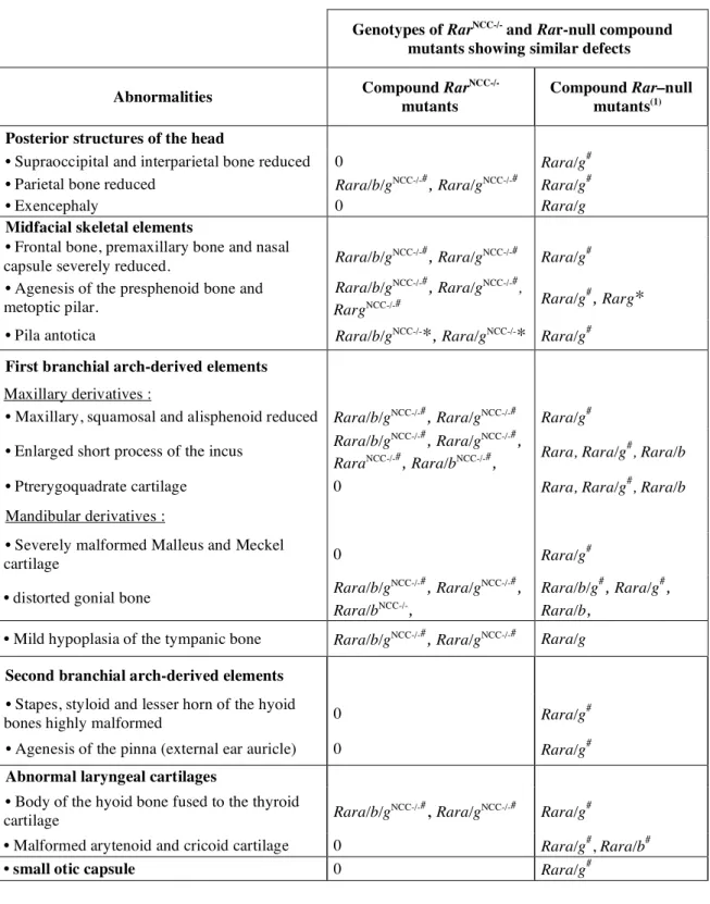
Table 1: Malformations of cranial skeletal elements in Rar
mutants compared to the
Rar-null compound mutants observed at E18.5. Legend. *: abnormality present in a majority of the mutants; #: abnormality fully penetrant; (1) Lohnes et al., 1994 and Ghyselinck et al., 1997.
Genotypes of RarNCC-/- and Rar-null compound
mutants showing similar defects Abnormalities Compound Rar
NCC-/-mutants
Compound Rar–null mutants(1)
Posterior structures of the head
• Supraoccipital and interparietal bone reduced 0 Rara/g#
• Parietal bone reduced Rara/b/gNCC-/-#, Rara/gNCC-/-# Rara/g#
• Exencephaly 0 Rara/g
Midfacial skeletal elements
• Frontal bone, premaxillary bone and nasal
capsule severely reduced. Rara/b/g
NCC-/-#, Rara/gNCC-/-# Rara/g#
• Agenesis of the presphenoid bone and metoptic pilar.
Rara/b/gNCC-/-#, Rara/gNCC-/-#,
RargNCC-/-# Rara/g
#,
Rarg*
• Pila antotica Rara/b/gNCC-/-*, Rara/gNCC-/-* Rara/g# First branchial arch-derived elements
Maxillary derivatives :
• Maxillary, squamosal and alisphenoid reduced Rara/b/gNCC-/-#, Rara/gNCC-/-# Rara/g#
• Enlarged short process of the incus Rara/b/g
NCC-/-#
, Rara/gNCC-/-#
,
RaraNCC-/-#, Rara/bNCC-/-#, Rara, Rara/g
#
, Rara/b
• Ptrerygoquadrate cartilage 0 Rara, Rara/g#, Rara/b
Mandibular derivatives :
• Severely malformed Malleus and Meckel
cartilage 0 Rara/g
#
• distorted gonial bone Rara/b/g
NCC-/-# , Rara/gNCC-/-# , Rara/bNCC-/-, Rara/b/g#, Rara/g#, Rara/b,
• Mild hypoplasia of the tympanic bone Rara/b/gNCC-/-#, Rara/gNCC-/-# Rara/g Second branchial arch-derived elements
• Stapes, styloid and lesser horn of the hyoid
bones highly malformed 0 Rara/g
#
• Agenesis of the pinna (external ear auricle) 0 Rara/g#
Abnormal laryngeal cartilages
• Body of the hyoid bone fused to the thyroid
cartilage Rara/b/g
NCC-/-#
, Rara/gNCC-/-#
Rara/g#
• Malformed arytenoid and cricoid cartilage 0 Rara/g#, Rara/b#
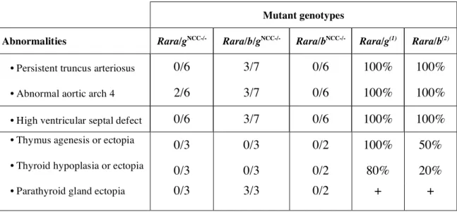
Table 2: Cardiovascular and glandular abnormalities in Rar
mutants. Legend. + :
presence of ectopic parathyroid gland, percentage not determined. (1) Lohnes et al., 1994; (2) Ghyselinck et al., 1997. Cardiovascular defects were evaluated at E14.5 and E18.5 whereas glandular defects were evaluated at E18.5.
Mutant genotypes
Abnormalities Rara/gNCC-/- Rara/b/gNCC-/- Rara/bNCC-/- Rara/g(1) Rara/b(2)
• Persistent truncus arteriosus 0/6 3/7 0/6 100% 100%
• Abnormal aortic arch 4 2/6 3/7 0/6 100% 100%
• High ventricular septal defect 0/6 3/7 0/6 100% 100%
• Thymus agenesis or ectopia 0/3 0/3 0/2 100% 50%
• Thyroid hypoplasia or ectopia 0/3 0/3 0/2 80% 20%