HAL Id: inserm-01153067
https://www.hal.inserm.fr/inserm-01153067
Submitted on 19 May 2015
HAL is a multi-disciplinary open access
archive for the deposit and dissemination of
sci-entific research documents, whether they are
pub-lished or not. The documents may come from
teaching and research institutions in France or
abroad, or from public or private research centers.
L’archive ouverte pluridisciplinaire HAL, est
destinée au dépôt et à la diffusion de documents
scientifiques de niveau recherche, publiés ou non,
émanant des établissements d’enseignement et de
recherche français ou étrangers, des laboratoires
publics ou privés.
Mahmood, Charlotte Allan, Anya Topiwala, Vyara Valkanova, Eric Brunner,
Martin Shipley, et al.
To cite this version:
Nicola Filippini, Enikö Zsoldos, Rita Haapakoski, Claire Sexton, Abda Mahmood, et al.. Study
protocol: The Whitehall II imaging sub-study.. BMC Psychiatry, BioMed Central, 2013, 14, pp.159.
�10.1371/journal.pone.0003583�. �inserm-01153067�
S T U D Y P R O T O C O L
Open Access
Study protocol: the Whitehall II imaging sub-study
Nicola Filippini
1,2, Enik
ő Zsoldos
1, Rita Haapakoski
1, Claire E Sexton
1, Abda Mahmood
1, Charlotte L Allan
1,
Anya Topiwala
1, Vyara Valkanova
1, Eric J Brunner
3, Martin J Shipley
3, Edward Auerbach
4, Steen Moeller
4,
Kâmil U
ğurbil
4, Junqian Xu
4,5, Essa Yacoub
4, Jesper Andersson
2, Janine Bijsterbosch
2, Stuart Clare
2,
Ludovica Griffanti
2, Aaron T Hess
6, Mark Jenkinson
2, Karla L Miller
2, Gholamreza Salimi-Khorshidi
2,
Stamatios N Sotiropoulos
2, Natalie L Voets
2, Stephen M Smith
2, John R Geddes
1, Archana Singh-Manoux
3,7,
Clare E Mackay
1, Mika Kivimäki
3and Klaus P Ebmeier
1*Abstract
Background: The Whitehall II (WHII) study of British civil servants provides a unique source of longitudinal data to investigate key factors hypothesized to affect brain health and cognitive ageing. This paper introduces the multi-modal magnetic resonance imaging (MRI) protocol and cognitive assessment designed to investigate brain health in a random sample of 800 members of the WHII study.
Methods/design: A total of 6035 civil servants participated in the WHII Phase 11 clinical examination in 2012–2013. A random sample of these participants was included in a sub-study comprising an MRI brain scan, a detailed clinical and cognitive assessment, and collection of blood and buccal mucosal samples for the characterisation of immune function and associated measures. Data collection for this sub-study started in 2012 and will be completed by 2016. The participants, for whom social and health records have been collected since 1985, were between 60–85 years of age at the time the MRI study started. Here, we describe the pre-specified clinical and cognitive
assessment protocols, the state-of-the-art MRI sequences and latest pipelines for analyses of this sub-study. Discussion: The integration of cutting-edge MRI techniques, clinical and cognitive tests in combination with retrospective data on social, behavioural and biological variables during the preceding 25 years from a well-established longitudinal epidemiological study (WHII cohort) will provide a unique opportunity to examine brain structure and function in relation to age-related diseases and the modifiable and non-modifiable factors affecting resilience against and vulnerability to adverse brain changes.
Keywords: Epidemiology, Magnetic resonance imaging, Diffusion tensor imaging, White matter, Functional MRI, Connectome, Resting state brain networks, Neuropsychology, Dementia, Affective disorders
Background
Over the next few decades, increases in life expectancy will result in fundamental changes to the population struc-ture. Associated with this demographic change, health and social care services will need to cope with a greater preva-lence of mental and neurological disorders. Clinical de-pression and cognitive decline have a combined estimated prevalence of 7-20% in the population over 65 years [1-4]. Furthermore, according to some estimates, the number of people with neurodegenerative disorders will quadruple in
the next 20 years causing a significant increase in the cost of care [5]. To extend the productive period in citizens’ lives and to reduce costs of care in late life a greater know-ledge of prevention and treatment of these common con-ditions is needed. This will not be possible without a better understanding of the causal mechanisms of disease and, equally importantly, the factors associated with resili-ence to age-related dysfunction [6].
The Whitehall II (WHII) study of 10,308 British civil servants provides a remarkable source of longitudinal data to explore factors hypothesized to affect brain health and cognitive ageing (Table 1). The cohort was established in 1985 at University College London (UCL) with the aim of advancing knowledge of the causal chain through which
* Correspondence:[email protected]
1
Department of Psychiatry, University of Oxford, Warneford Hospital, Oxford OX3 7JX, UK
Full list of author information is available at the end of the article
© 2014 Filippini et al.; licensee BioMed Central Ltd. This is an Open Access article distributed under the terms of the Creative Commons Attribution License (http://creativecommons.org/licenses/by/4.0), which permits unrestricted use, distribution, and reproduction in any medium, provided the original work is properly credited. The Creative Commons Public Domain Dedication waiver (http://creativecommons.org/publicdomain/zero/1.0/) applies to the data made available in this article, unless otherwise stated.
Filippiniet al. BMC Psychiatry 2014, 14:159 http://www.biomedcentral.com/1471-244X/14/159
social circumstances influence health [7]. By September 2011, the study had acquired 25 years of rich social, behav-ioural and biological data enabling its transformation into a unique study of ageing. Of the original 10,308 non-industrial civil servants recruited in Phase 1, 6035 par-ticipated in the ‘Phase 11’ assessment in 2012–13. As participants in this study continue into older adult life, the research now focuses on life course factors affecting health and personal functioning at older ages.
We randomly selected 800 WHII Phase 11 participants to take part in the WHII imaging sub-study, which in-cludes a detailed clinical and cognitive assessment, meas-urement of immune parameters and a magnetic resonance imaging (MRI) scan. MRI scans provide a non-invasive window into the living brain, giving unique access to un-derstanding normal and pathological processes that affect brain structure and function. Incorporating state-of-the-art imaging techniques and cognitive measures with the WHII’s longitudinal dataset of social, behavioural and bio-logical variables, represents a unique opportunity to study the ageing process and to directly link 25-year exposure history to old-age cognition and a variety of measures of ‘brain health’. Analysis of immunological variables and linkage of these studies with behavioural and imaging data enables a more comprehensive investigation of the patho-physiological processes of dysfunction and cognitive im-pairment in later life.
In this paper, we provide a description of the study’s or-ganisation and funding structure, its participants’ inclusion/ exclusion criteria and of the cognitive, imaging and blood specimen protocols employed in the study. For the imaging protocol, careful consideration has been given to harnessing the most recent technical developments, whilst maintaining clinical relevance. A preliminary description of the tech-niques to be used to pre-process and examine MRI-related
measures will also be presented, and results of a direct comparison between a recently developed and a more standard MRI acquisition approach for investigating brain functional organization will also be shown. We provide an overview of our original hypotheses at the time of applica-tion for funding.
Methods/design
Study organization and funding
The sub-study is funded by the Lifelong Health and Well-being Phase-3 programme grant “Predicting MRI abnor-malities with longitudinal data of the Whitehall II sub-study” (MRC-G1001354; Ebmeier KP (PI), Geddes JR, Kivimäki M, Mackay CE, Singh-Manoux A, Smith SM), as well as the HDH Wills 1965 (English Charity Register: 1117747; Ebmeier KP (PI)), and the Gordon Edward Small Charitable (Scottish Charity Register: SC008962; Ebmeier KP (PI)) Trusts. Collection of blood and buccal mucosal samples for a characterisation of immune function and as-sociated measures is funded by the UK Medical Research Council programme grant K013351 (“Adult determinants of late life depression, cognitive decline and physical func-tioning - The Whitehall II Ageing Study”, Kivimäki M (PI), Singh-Manoux A, Brunner E, Batty GD, Kumari M, Ebmeier KP, Hingorani A) and the ESRC professional fel-lowship scheme to Kivimäki.
Ethical approval was granted generically for the“Protocol for non-invasive magnetic resonance investigations in healthy volunteers” (MSD/IDREC/2010/P17.2) by the Uni-versity of Oxford Central UniUni-versity / Medical Science Div-ision Interdisciplinary Research Ethics Committee(CUREC/ MSD-IDREC), who also approved the specific protocol: “Predicting MRI abnormalities with longitudinal data of the Whitehall II sub-study” (MSD-IDREC-C1-2011-71). The Health Research Authority NRES Committee
Table 1 Phases of Whitehall II with available measures
Phase 1 1985-88 Phase 3 91-93 Phase 5 97-99 Phase 7 03-04 Phase 9 07-09 Phase 11 12-13 Ages [years] 35-55 39-64 45-69 50-74 55-79 60-85 Participants [n] 10308 8637 7830 6967 6755 6035
Social circumstances & behaviour, smoking, alcohol, exercise, sleep diet × × × × × × Biological measures: blood pressure, BMI, lipids, glucose, insulin, stored blood (−80C) × × × × × ×
2-h oral glucose tolerance test – × × × × ×
Inflammatory markers – × × × × –
Autonomic (HRV) – – × × × ×
Genetic material – – – × × ×
Psychosocial factors: work, social support,− participation, care provision × × × × × × Health outcomes: CHD, stroke, diabetes, cancer, mortality, medications × × × × × ×
Function: physical, social & mental × × × × × ×
Cognitive tests, physical/lung function tests – – × × × ×
South Central – Oxford B approved the Study: “The Whitehall II Immune Function Sub-study” (REC refer-ence: 13/SC/0072, IRAS project ID: 120516).
The study follows the Medical Research Council (MRC) Policy on data sharing, i.e. images and other data will be available for analysis by other groups after completion of the study, as is the case with the Whitehall II study (see http://www.ucl.ac.uk/whitehallII/data-sharing [8,9].
Participants’ recruitment and cognitive protocol description Participants
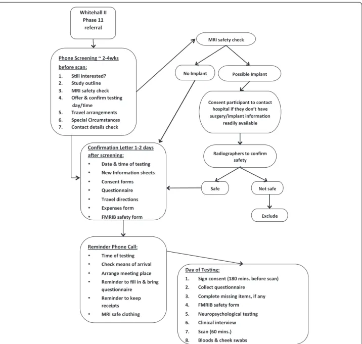
In order to make the sample as representative as possible of the cohort at baseline, a random sample of 800 WHII Phase 11 participants willing and able to give informed consent have been invited to attend the imaging sub-study at the Oxford Centre for Functional MRI of the Brain (FMRIB). To achieve a sufficient number of participants with depression, we added 30 participants with depressive symptoms based on previous WHII clinical examinations. We excluded participants with contraindications to MRI scanning (including but not limited to a history of claus-trophobia, certain metallic implants and metallic injury to the eye) or who were unable to travel to Oxford without assistance. A schematic flow-chart describing the different stages of the study is provided in Figure 1.
Clinical and cognitive assessment
Each participant recruited for the WHII imaging sub-study undergoes a detailed clinical and cognitive assess-ment lasting up to two hours.
The clinical assessment consists of a (A) self-administered questionnaire, a (B) semi-structured clin-ical interview and (C) cognitive assessment (median = 56 minutes, interquartile range: 51–61 minutes).
Self-administered questionnaire
General Health Questionnaire-30 (GHQ-30; [10]): The GHQ-30 is a 30-item self-administered screening ques-tionnaire for the detection of psychiatric illness that ac-companies ill-health, in non-psychiatric clinical and community settings (routinely applied from scan 200).
Mood Disorder Questionnaire (MDQ; [11]): The MDQ is a brief self-report questionnaire for the assessment of life time history of bipolar disorders, based on the DSM-IV.
Centre for Epidemiological Studies Depression Scale (CES-D; [12]): The CES-D is short self-report scale that measures major depressive symptomatology in the general population.
State and Trait Anxiety Inventory (STAI; [13]): The STAI measures both S (state)- and T (trait)-Anxiety in clinical and research settings [14]. It is a self-administered questionnaire that consists of twenty statements assessing how the individual feels at the moment (S-Anxiety) and
twenty assessing how they generally feel (administered to n = 15 before routinely applied from scan 200).
CHAMPS Physical Activity Questionnaire for Older Adults[15]: The CHAMPS is a self-administered physical activity questionnaire for older persons. Participants re-port the weekly frequency and duration of various physical activities, typically undertaken by older adults, allowing calculation of metabolic equivalent of task (MET) and cal-oric expenditure values per week.
Locus for Causality Exercise Questionnaire (LCE;[16]): The LCE is a 3-item self-administered scale that assesses how much an individual feels that they choose to exercise (internal perceived locus of causality) rather than have to exercise for some reason (external perceived locus of caus-ality). It is thought that individuals are more likely to en-gage in physical exercise when the perceived locus of causality is internal [17].
Pittsburgh Sleep Quality Index (PSQI; [18]): The PSQI is a self-rated questionnaire made up of seven component scores that assess sleep quality and disturbance over a one month period in clinical and research settings.
Jenkins Sleep Questionnaire (JSQ;[19]): The JSQ is a 4-item self-rated questionnaire for the assessment of sleep disturbances over a month period.
Life-Orientation Revised (LOT-R; [20]): The LOT-R was devised to measure individual optimism for future events in the general population.
Life Events [21,22]): A modified version of the List of Threatening Experiences questionnaire (LTE-Q) is used, in which participants are asked about seven types of stressful life events. Participants are asked to remember if any of the events happened to them in the past, and when they happened.
MacArthur stress reactivity questionnaire [23]: It is a nine-item self-rated questionnaire in which the participant is required to rate nine statements on a 5-point scale, regarding to how they handle their emotions in stressful situations.
Penn State Worry Questionnaire Ultra-brief Version (PSWQ;[24]): The PSWQ ultra-brief is the 3-item version of the widely used self-report questionnaire for patho-logical worry, the 16-item long PSWQ. The 3-items capture pathological worry as defined by the DSM-IV; perceived uncontrollability, multiple domains and high frequency of worry. The PSWQ was introduced into the assessment after scan 200.
Handedness ([25]): It is a self-administered questionnaire that assesses which is the participant’s preferred hand to complete a list of twelve tasks, as well as left-handedness in the family.
Participants also provide information on medical history (detailing hospitalizations, longstanding illnesses, diseases or medical conditions), alcohol and nicotine intake, and general information, such as age and education. Their blood pressure is measured twice in a sitting position, after Filippiniet al. BMC Psychiatry 2014, 14:159 Page 3 of 16 http://www.biomedcentral.com/1471-244X/14/159
the cognitive protocol (OMRON HEM-907; OMRON Healthcare UK Ltd., Milton Keynes).
Semi-structured clinical interview
Structured Clinical Interview for DSM-IV-TR Axis I Dis-orders [26]: The SCID-I is a semi-structured interview for diagnosing current and past DSM-IV Axis I disorders and is administered by a trained graduate psychologist or psychiatrist.
Further structured assessments
Further tests were performed when the clinical history and SCID data of the participant suggested a diagnosis and
indicated that a more detailed assessment was required (the number of each test carried out so far is listed in brackets):
Hamilton Depression Scale (HAMD;[27]): The HAMD is a 17-item severity scale administered to individuals di-agnosed with ‘affective disorder of depressive type’. It has been devised to quantify the intensity of the depres-sive symptoms of the patient, based on the necessary in-formation elicited by the interviewer. This scale was administered to n = 16 participants currently symptom-atic on the SCID-I [26].
Young Mania Rating Scale (YMRS;[28]): The YMRS is an 11-item rating scale for the assessment of manic symptoms based on the subjective report of the patient’s Figure 1 Flow-chart summarising the stages of the WHII imaging sub-study.
experience over the past forty-eight hours. It follows a rating style of symptom severity similar to that of the HAMD [27] and is administered to participants cur-rently symptomatic on the SCID-I [26] (n = 2).
Yale-Brown Obsessive Compulsive Scale (Y-BOCS; [29,30]): The Y-BOCS is a clinician-rated ten-item scale of the severity of symptoms of obsessive-compulsive disorder, with separate subtotals for obsessions and compulsions. This scale is applied to participants currently symptomatic on the SCID-I [26] (n = 0).
CAGE Questionnaire [31]: Four questions make up this questionnaire to detect dependence on alcohol. They request information on whether the individual needs to “Cut down” their drinking, feels Annoyed by criticism of their drinking, feels Guilty about their alcohol use, and whether they use alcohol first thing in the morning as an ‘Eye-opener’. This scale is administered to participants currently symptomatic on the SCID-I [26] (n = 6).
Brief Psychiatric Rating Scale (BPRS; [32]): The BPRS is an 18-24-point rating scale for the assessment of psychotic symptoms, and is used in both clinical and re-search settings. This scale is administered to participants currently symptomatic on the SCID-I [26] (n = 0).
Cognitive assessment
Cognitive test battery administered to all participants: Montreal Cognitive Assessment (MoCA;[33]): The MoCA is a 30-point cognitive screening test assessing multiple cognitive domains: a) visuo-spatial abilities (4 points), assessed using a three-dimensional cube-drawing (1 point) and a clock-drawing task (3 points); b) short-term memory recall task (5 points), which involves learning 5 nouns and recalling them approximately 5 minutes afterwards; c) ex-ecutive function (3 points), which include an alternation task (1 point) and a verbal abstraction task (2 points); d) at-tention, orientation and working memory (6 points), which are evaluated using a forward- and backward-digit task (2 points), a sustained attention task (1 point), and a serial subtraction task (3 points); e) language (6 points), which is measured using a three-item naming task (3 points), the repetition of two syntactically complex sentences (2 points) and a phonemic fluency task (1 point); and f) orientation to time and space (6 points). Participants receive an additional (1 point) if their education level is≤ 12 years. Since the MoCA assesses multiple cognitive domains, it is a useful cognitive screening tool for several neurological diseases, such as Parkinson’s disease, vascular cognitive impairment, Huntington’s disease, multiple sclerosis, and other condi-tions, such as traumatic brain injury, depression and schizophrenia [33,34].
Trail Making Test (TMT) versions A and B ([35,36]: The TMT is a visual attention and task-switching test consisting of two parts in which the subject is instructed to connect a set of twenty-five consecutive dots (A:
numbers and B: numbers and letters) on a sheet of paper as fast as possible while still maintaining accuracy. It provides information about visual search speed, speed of processing, mental flexibility, as well as executive func-tioning [36]. It is sensitive to the detection of cognitive impairment including Alzheimer’s disease [37].
Rey Complex Figure Test and Recognition Trial (RCFT; [38,39]): The RCF involves copying and then recalling a complex geometric diagram at increasing time intervals [40]. Different cognitive abilities are needed for a correct performance, including visuo-spatial abilities, memory, attention, planning, and working memory. It is used to investigate the effects of brain injury and to test the presence of neurodegenerative conditions [41].
Verbal fluency test (adapted from the Addenbrooke’s Cognitive Examination Revised (ACE-III)[42]): The verbal fluency test requires participants to say as many words as possible from a category (animals) in a specified time (60 seconds). It is used to investigate the presence of cog-nitive impairment, neurodegenerative and psychiatric dis-orders [43].
Hopkins Verbal Learning Test-Revised (HVLT-R; [44]): The HVLT-R test provides a measure of verbal learning and memory ability [45,46]. The participant is required to learn a list of twelve words over the course of three trials, and recall and recognise them at increasing time intervals. It is widely used to test the presence of amnes-tic disorders [47,48].
Boston Naming Test (BNT-60; [49]): The BNT-60 is a 60-item test graded in difficulty used to measure semantic memory ability and requires naming of a series of images shown to the participant [50]. It is used in individuals with aphasia or any language disturbance caused by neurological insults, such as stroke or neurodegenerative disorders [51].
Digit Span (DS) and Coding (DC) tests from the Wechsler Adult Intelligence Scale - Fourth Edition (WAIS-IV; [52]): The DS test is used to investigate short-term memory abilities. It includes recall of a lengthening list of digits forwards, backwards, and rearranged in ascend-ing sequence (DSF, DSB, DSS) [53,54]. In the DC test participants have to write the appropriate novel symbol for each number within a given time.
Test of Premorbid Functioning (TOPF;[55]): The TOPF consists of a list of seventy written words, which must be read aloud and is marked according to pronunciation. The TOPF is used to estimate an individual's level of in-tellectual functioning before the onset of injury or ill-ness. Premorbid IQ can be calculated from the raw score, adjusted for sex and years of education.
Dots and letters (adapted from the Addenbrooke’s Cog-nitive Examination III;[42]): The participant is asked to count four sets of dots without pointing to them and identify four partially drawn letters. These tasks assess perceptual abilities.
Filippiniet al. BMC Psychiatry 2014, 14:159 Page 5 of 16 http://www.biomedcentral.com/1471-244X/14/159
CLOX [56]: The CLOX is a clock drawing task; in the first part the participant is given a set of instructions to draw a clock and in the second part the examiner draws a clock face, which the participant then has to copy. The CLOX was designed to assess executive impairment and non-executive failure, and is used to discriminate demen-tia sub-groups [56].
Cambridge Neuropsychological Test Automated Battery Reaction Time touchscreen version (CANTAB RTI; CAN-TABeclipse 5.0; Cambridge Cognition Ltd. www.camcog. com): The CANTAB RTI is a computerised (touchscreen) latency task that measures latency and movement time without having to control for tremor. The task is divided into a simple and 5-choice reaction time stage. During the task the participant must react as soon as a yellow dot ap-pears; moving their finger on the screen from a pre-defined location to the location of the yellow dot. In the simple stage the yellow dot always appears in the same lo-cation, and in the five-choice stage in one out of five po-tential locations. The CANTAB RTI is often used to assess visuo-spatial and visuo-motor coordination abilities [57], motor speed [58], and understand sustained attention and reaction time [59].
Purdue Pegboard Test [60,61]: The Purdue Pegboard measures two types of dexterity; gross movement of the fingers, hands and arms, and fine fingertip dexterity. The participant places pins into a row of holes using right, left and both hands (gross movement) and assembles a set of structures from pins, collars and washers using both hands (fine dexterity) as fast as they can, within a given time. The Purdue Pegboard test was devised for employee selec-tion for industrial jobs but is also used in clinical settings. Impaired peg placement was found among patients with Parkinson’s disease [62,63]), Huntington’s disease [62] and schizophrenia [64]. The Purdue Pegboard can also aid lateralization of function [65]. Healthy older people with-out neuropsychiatric or other disease who showed MRI white matter hyperintensities (WMH) performed worse on the assembly subtest (fine dexterity) than those without WMH [66].
Imaging protocol description
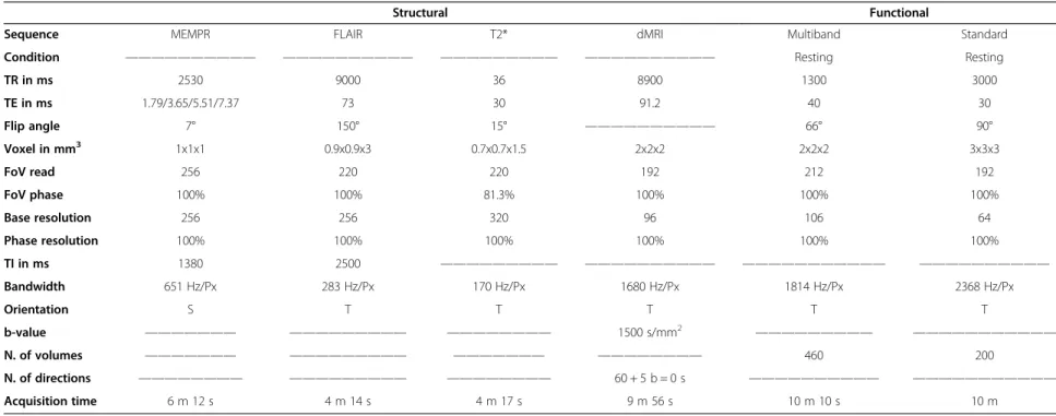
Scanning is carried out at the Oxford Centre for Func-tional MRI of the Brain (FMRIB) using a 3 T Siemens Magnetom Verio (Erlangen, Germany) Scanner with a 32-channel receive head coil. The neuroimaging protocol comprises both structural and functional sequences and lasts approximately 50 minutes. MRI sequences include: a) high-resolution T1-weighted, b) diffusion MRI (dMRI), c) resting-state functional MRI (rfMRI), d) Fluid Attenuated Inversion Recovery (FLAIR) and e) T2*. A full description of the MRI parameters adopted in our sequences is pro-vided in Table 2.
T1-weighted
This sequence is primarily used to study grey matter (GM) structural macroscopic tissue in both cortical and subcor-tical brain regions. GM reductions have been widely asso-ciated with impending disease and age-related cognitive dysfunction [67-69].
A Multi-Echo MPRAGE (MEMPR) with motion correc-tion, developed at the Massachusetts General Hospital (MGH, Boston), was employed [70,71]. This sequence has the advantage of combining the properties of the classical MPRAGE sequence, which has high contrast aiding cor-tical segmentation, with Multi-Echo FLASH, which im-proves segmentation of subcortical regions.
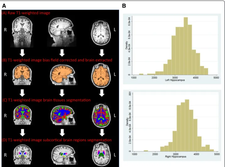
The pre-processing pipeline includes: a) re-orientating images to the standard (MNI) template, b) bias field cor-rection, c) registration to the MNI template using both linear (FLIRT) and non-linear (FNIRT) registration tools and d) brain extraction. Brain tissues are segmented using FMRIB's Automated Segmentation Tool (FAST) that allows extracting measures of total GM, WM and cerebrospinal fluid (CSF). FIRST (http://fsl.fmrib.ox.ac. uk/fsl/fslwiki/FIRST) [72], an automated model-based segmentation/registration tool, is applied to extract cortical grey matter structures. Brain tissues and sub-cortical regions are visually inspected to ensure an accurate segmentation (Figure 2A,B).
Diffusion MRI (dMRI)
Diffusion MRI exploits the principles of traditional MRI to measure the random motion of water molecules and subse-quently to 1) infer information about white matter (WM) microstructural properties and 2) delineate the gross axonal organisation of the brain [73]. WM is characterised by bun-dles of myelinated axons surrounded by myelin sheaths that are built up by layers of membranes. This restricts diffusion of free water molecules; i.e. the myelin layers and the axonal membrane cause a lower restriction along than across the axon and thus a higher anisotropy.
A number of strategies were used to minimise distor-tions caused by, for example, magnetic susceptibility, eddy-currents, and subject-motion. We employed mono-polar diffusion encoding gradients with parallel imaging (GRAPPA) to minimise echo time, which increases the signal to noise ratio (SNR), at the cost of a small increase in eddy-current distortion. We used a recently developed dMRI correction strategy that takes advantage of the com-plementary information from pairs of diffusion images ac-quired with reversed phase-encoding (PE) directions to correct for susceptibility-induced distortions [74]. A single non-diffusion weighted (b-value = 0 s/mm2) volume with reversed PE was combined with the non-reversed dMRI data to estimate an off-resonance field, which is then ap-plied to correct susceptibility distortions [75].
Table 2 MRI sequences and parameters used in the study; S and T define Sagittal and Transversal orientation, respectively
Structural Functional
Sequence MEMPR FLAIR T2* dMRI Multiband Standard
Condition —————————— —————————— ————————— —————————— Resting Resting
TR in ms 2530 9000 36 8900 1300 3000 TE in ms 1.79/3.65/5.51/7.37 73 30 91.2 40 30 Flip angle 7° 150° 15° —————————— 66° 90° Voxel in mm3 1x1x1 0.9x0.9x3 0.7x0.7x1.5 2x2x2 2x2x2 3x3x3 FoV read 256 220 220 192 212 192 FoV phase 100% 100% 81.3% 100% 100% 100% Base resolution 256 256 320 96 106 64 Phase resolution 100% 100% 100% 100% 100% 100% TI in ms 1380 2500 ————————— —————————— ——————————— —————————— Bandwidth 651 Hz/Px 283 Hz/Px 170 Hz/Px 1680 Hz/Px 1814 Hz/Px 2368 Hz/Px Orientation S T T T T T b-value ——————— ————————— ———————— 1500 s/mm2 ————————— ——————————— N. of volumes ——————— ————————— ——————— ———————— 460 200 N. of directions ———————— ————————— ———————— 60 + 5 b = 0 s —————————— ——————————— Acquisition time 6 m 12 s 4 m 14 s 4 m 17 s 9 m 56 s 10 m 10 s 10 m Filippini et al. BMC Psychiatr y 2014, 14 :159 Page 7 o f 1 6 http://ww w.biomedce ntral.com/1 471-244X/14 /159
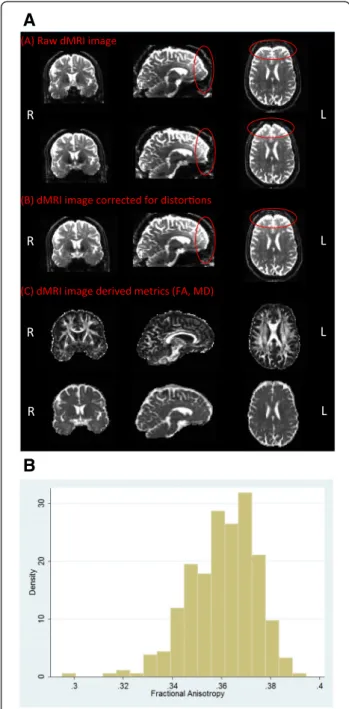
The Pre-processing pipeline uses a recently developed approach that simultaneously considers and corrects for susceptibility-induced distortions, eddy-currents and head motion (based on methods developed and applied to the Human Connectome Project (HCP) diffusion MRI data [74]). Briefly, a generative model approach is used to esti-mate all types of distortion and a single resampling step with spline interpolation is used to correct for all of them simultaneously. Fractional anisotropy (FA), mean diffusiv-ity (MD), axial diffusivdiffusiv-ity (AD) and radial diffusivdiffusiv-ity (RD) maps are generated using DTIFit, part of FMRIB’s Diffu-sion Toolbox (http://fsl.fmrib.ox.ac.uk/fsl/fdt), that fits a diffusion tensor model at each voxel [76,77] (Figure 3, b). Subsequently, crossing fibre orientations can be estimated, and probabilistic tractography can be performed to recon-struct white matter bundles and assess recon-structural connect-ivity [78].
Resting-state functional MRI (rfMRI)
rfMRI is used to investigate resting state networks (RSNs), which comprise brain regions sharing a common time-course of spontaneous fluctuations, and are thought to represent functionally-critical neuronal networks that reflect properties of functional brain organisation [79]. RSNs have been consistently observed across subjects, sessions and functional brain imaging modalities (fMRI, PET, EEG), and their presence has also been reported in studies when participants were asleep, and in anaesthe-tized monkeys and rats [79-85]. Although potentially harder to interpret than task-based fMRI, the rfMRI ap-proach has the considerable advantage of providing an assay of brain function without requiring subjects to undertake a specific task, particularly in cases where a subject may be less able to understand and/or respond to complex instructions.
Figure 2 T1-weighted imaging– analysis pipeline and initial volumetric results. A. Schematic figure representing the pipeline set up to analyse T1-weighted images. The (A) raw T1-weighted image is initially (B) pre-processed (bias field corrected, neck cropped and brain extracted). Subsequently, (C) brain tissues are segmented into gray matter (GM, red), white matter (WM, blue) and cerebrospinal fluid (CSF, green). Finally, (D) subcortical brain regions are segmented. R and L define the right and the left hemisphere, respectively. B. Histogram of hippocampal sizes in the first 300 participants. Volume measures in 10-3ml.
We compared a recently developed Multiband MRI sequence [86,87] with‘standard’ EPI. Multiband provides a considerable improvement in temporal (Multiband: 1.3 sec-onds vs. Standard EPI: 3 secsec-onds) and spatial (Multiband: 2 mm isotropic vs. Standard EPI: 3 mm isotropic)
resolution, which allows: a) better definition of the spatial maps, b) wider frequency range exploration in time-series analyses and c) more detailed network analyses. To ensure that the new multiband sequence was robust in our older population, we acquired both sequences (standard and multiband) on a subset of participants (N = 76). Results of this comparison will be presented in the next section. In all cases subjects were instructed to lie in dimmed light with their eyes open, blink normally, but not to fall asleep.
rfMRI data pre-processing (motion correction, brain ex-traction, high-pass temporal filtering with a cut-off of 100 s, and field-map correction is carried out using MELODIC (Multivariate Exploratory Linear Optimized Decomposition into Independent Components, part of FSL http://fsl.fmrib. ox.ac.uk/fsl/fslwiki/melodic/) [88,89]. In order to reduce the presence of spatially and/or temporally-related artefacts a data-cleaning approach is applied. Single-subject independ-ent componindepend-ent analysis (ICA) is followed by an automatic component classification with FMRIB's ICA-based X-noiseifier (FIX) to identify and regress out the “signal” of the artefactual components reflecting non-neuronal fluctu-ations [90,91]. The pre-processed and“cleaned” functional data are registered to the individual's structural scan and standard space images using FNIRT, then optimized using boundary-based-registration approach [92], and finally spatially smoothed using an isotropic Gaussian kernel of 6 mm full width at half maximum (FWHM).
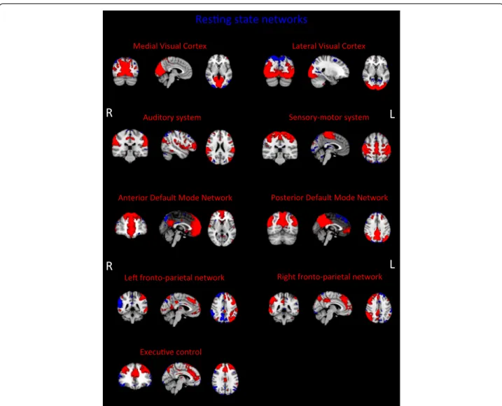
This project represents the first major application of the Multiband sequence in older adults: therefore, as part of the protocol development, the following were tested: a) whether it was possible to identify the previously reported set of “canonical” RSNs with the Multiband sequence [81,88]; b) whether the Multiband sequence was effectively associated with a“better” signal relative to a Standard EPI sequence. For the first analysis MELODIC was used across the first 50 consecutive participants and was able to detect a set of RSNs that matched the“canonical” RSNs (Figure 4). For the second analysis a direct comparison was made be-tween Multiband and Standard EPI sequences that had both been acquired on a subset (N =76) of participants. In order to be able to compare the two sequences with respect to spatial detail, spatial smoothing was not applied in this analysis. Additionally, an independent set of RSNs’ spatial maps, derived from data acquired for the Human Connec-tome Project (HCP), was used as a common template to extract time series and spatial maps from Multiband and Standard EPI sequence data using the dual regression ap-proach [93,94]. HCP templates are of higher resolution and therefore contain more information compared with the standard template maps. Group maps were then obtained performing a one-sample t-test on the subjects’ spatial maps (output of the second stage of dual regression) for each component, calculating the corresponding z map and ap-plying a mixture model correction to ensure comparable
A
B
Figure 3 Diffusion MRI - analysis pipeline and initial results for fractional anisotropy. A. Schematic figure representing the raw data (b=0) for both anterior-to-posterior and posterior-to-anterior encoding directions (A) and after application of the two reversed phase encoding (B). Red circles show before and after correction for distortions. Diffusion MRI (dMRI)-derived metrics for fractional anisotropy (FA) and mean diffusivity (MD) are also shown (C). R and L define the right and the left hemisphere, respectively. B. Histogram of mean fractional anisotropy in the first 300 participants.
Filippiniet al. BMC Psychiatry 2014, 14:159 Page 9 of 16 http://www.biomedcentral.com/1471-244X/14/159
null distributions [95]. Figure 5 shows the quantile-quantile plots (Q-Q plots) of the group maps against the normal distribution. The group maps’ z-statistic distribu-tion was expected to follow a normal distribudistribu-tion around zero (random noise– the null part of the spatial maps), while the tails should deviate from it according to the RSNs’ identified signal. Therefore our results suggest that Multiband sequence allows stronger RSN signal detection (higher z-values) with respect to Standard EPI sequence.
Fluid attenuated inversion recovery (FLAIR)
This sequence is commonly used in clinical practice, for example to characterise periventricular lesions adjacent to the sulci, WM hyperintensities and WM lesions. Age-related increase in the number of hyperintensities and/or periventricular lesions has been widely reported [96,97]. Moreover, it has been shown that WM changes are associ-ated with subtle structural and functional brain changes
[98], and with decline in essential cognitive abilities in healthy elderly people [99]. Clinical ratings of FLAIR are performed by trained neuroscientists based on visual rat-ing scores. WM lesions and/or hyperintensities are visually identified on FLAIR images by three independent raters who make assessments blind to demographic details. All axial slices of each subject are visually inspected. Peri-ventricular and deep WM hyperintensities are rated separ-ately using a 4-point ordinal scale from 0–3; the sum of these two ratings lead to the total Fazekas score (an inte-ger from 0–6) [100] (Figures 6A and B).
T2* (T2 star)
The T2* sequence allows the identification of cerebral microbleeds, which reflect small deposits of the iron-storing protein hemosiderin in the brain and may be a sign of cerebral small-vessel disease [101,102]. Microbleeds can be found all over the brain and have been shown to be Figure 4 Resting state networks (RSNs) derived from Multiband data. Images represent group maps of 50 subjects. All RSN maps are thresholded at Z = 3. R and L define respectively the right and the left hemisphere. Maps reported here are not smoothed.
associated with neurological dysfunction and both clin-ical and cognitive impairment [103,104]. Their under-lying mechanism is still under investigation but the deleterious effect, probably due to inflammatory effects, has been proved to affect neuron functionality and/or cortical cerebral activity [105]. All axial slices of T2*
images are visually explored performed by trained neu-roscientists and microbleeds identified (Figure 6C).
Blood specimen protocol for the characterization of immune function
Following the MRI examination, blood samples (3 × 8 ml) and two buccal mucosal epithelial samples are taken from each participant. Blood samples are drawn using Vacutai-ner CPT tubes (Becton Dickinson) in a single venepunc-ture, inverted 8 times to mix anticoagulant additive with blood and processed within two hours of collection. Buc-cal mucosal epithelial cells are collected by nylon swabs (microbiome sample) and by twirling the brush against the epithelium and shaking the brush in RNAlater solution for 15 sec to remove the cells (RNA sample). CPT tubes are centrifuged in 1600 g for 30 min, allowing the separation of serum from white blood cells and from red blood cells. After the centrifugation, serum samples and per-ipheral blood mononuclear cells (PBMCs), i.e. T cells, B cells and NK cells are collected. PBMC cells are washed with phosphate-buffered saline (PBS), counted under microscope and cryopreserved using Cell Freezing Medium (5% DMSO/11%HSA in PBS). All samples are stored in−80°C for later analysis.
In order to investigate immune function in sample mater-ial, PBMC cells are stimulated in vitro withαCD3/αCD28 mAb and with TLR2/1 ligand Pam3CSK4 (Pam3Cys) and
TLR4 ligand LPS in RPMI at 37°C in 5% CO2.Cells and
su-pernatants are collected after 6 and 24 h of stimulation and stored for later analysis. RNA samples are investigated using RT-PCR or transcriptomics, while serum samples and cell supernatants are analysed with ELISA, Luminex or Figure 5 Q-Q plots of group maps’ z values. Continuous lines
represent Q-Q plots for Multiband (green), Standard EPI (red) and Human Connectome Project template (blue). Dashed-dotted lines join the first and third quartiles of each distribution, to help evaluate the linearity of the data.
Figure 6 FLAIR and T2* images. (A) periventricular hyperintensities (PWH) and (B) deep white matter (DWM) lesions on FLAIR. (C) T2* image showing microbleed. R and L define the right and the left hemisphere, respectively.
Filippiniet al. BMC Psychiatry 2014, 14:159 Page 11 of 16 http://www.biomedcentral.com/1471-244X/14/159
proteomics. Also, genomics/epigenomics and metabolomics techniques as well as advanced bioinformatics tools will be employed in the sample analysis.
Hypotheses
We will examine the following overarching hypotheses in the sub-cohort recruited from WH II Phase 11:
(1) Early risk factors for cognitive dysfunction and late onset depression (such as cardio-vascular risk, see (i.) to (v.) below) will be associated with cerebral atrophy, re-duced perfusion and impaired white matter integrity. Resulting impaired cognitive function (as measured by tests of episodic memory and executive function, see below) will be associated with specific brain volumes mea-sured by advanced methods such as voxel based analysis and the automatic identification of specific structures, such as hippocampus, and also with reduced white matter integrity, quantified both globally [106], and homing in on fibres of interest (including measures of structural con-nectivity) [78];
(2) Resilience, measured in the sub-study directly [107] and by the absence of depression and cognitive impair-ment particularly in the presence of higher early risk (see above), is associated with high white matter integrity [108], and increased coherence of frontal regions in resting state networks (which provides a measure of functional connectivity) [109] or (compensatory) increased frontal BOLD signal in a memory encoding task [93]. The most important risk factors will be: (i) Antecedent vascular and metabolic risk trajectories and morbidity (adverse major blood lipids/apolipoproteins, hyperglycaemia and diabetes, adiposity, high blood pressure, smoking, chronic inflam-mation (C-reactive protein, interleukin 6), ECG abnormal-ities, angina, myocardial infarction, stroke; Framingham cardiovascular risk scores); (ii) low antecedent levels of physical and mental activity (measured by questionnaire [110-112]); (iii) baseline cognitive performance levels and up to 15-year gradients of memory and executive function decrement; (iv) history of depressed mood (3 to 8 repeat measurements using the CESD [12] and the GHQ [10], (v) genotype (APOE4 plus >48 k single nucleotide polymor-phisms relevant to cardiovascular, metabolic and inflam-matory syndromes [113]). The primary clinical hypotheses which we will address are: a) The quality of frontal com-pensatory activity will be affected by vascular risk, hypertension, including absence of protective factors, such as physical fitness, cardio-vascular and cerebrovas-cular prophylaxis (aspirin, statins, antihypertensives), by mental activity (“use it or lose it”), and by frontal lobe atrophy (as observed in treatment resistant depression); b) Time-trajectories derived from the WHII data set, e.g. of cognitive function or depression scores, and vascular risks-factor trajectories, will account for more of the diver-sity of outcome than singular measurements during any
one of the previous follow-phases. This may allow imputa-tions about the natural history of brain changes, but from clinician’s point of view, the least labour and time-intensive predictive test will be the most attractive. An important clinical task would, therefore, be to determine the mini-mum data set to predict outcome. c) Overall resilience, as supported by the absence of current and past affective or cognitive symptoms and good performance [78], can be modelled longitudinally from observations antecedent to medial temporal atrophy and frontal compensatory activity and structural integrity (high FA in DTI scan) as described in (a) and (b). In addition, we will address the following specific hypotheses which are of independent interest but will also support the primary findings: (d) Global cognitive performance depends on both hippocampal and frontal in-tegrity/connectivity and (compensatory) frontal activity measured relatively higher frontal coherence within execu-tive resting state networks; (e) Clinical impairment (out-come) appears if functional frontal compensation does not keep up with the degree of hippocampal and frontal atro-phy observed; (f) First onset of depression after the age of 65 compared with no depression ever (outcome), matched for age, sex, education, and potential causal factors, such as vascular risk, is associated with reduced frontal/executive network structural and functional connectivity; (g) Clinical impairment may present as impaired cognition and/or as major depressive syndrome depending on the frontal net-works affected; (h) Greater hippocampal atrophy will be observed with increasing age and APOEε4 alleles; the ab-sence of antidepressant medication; and among those with a family history of dementia.
Discussion
The proposed programme of work will lead to the de-velopment and validation of methods and measures to integrate biological, physical, psychological, and socio-economic markers or indicators of health and wellbeing in later life, combining the Whitehall II repeat data from 1985 onwards and the psychiatric assessment in Phase 11 (2012–13), with detailed structural and func-tional imaging data collected in 2012–2016.
Our focus will be on understanding the development and impact of age-related conditions such as depression and cognitive decline, their implications for employment and work in later life, and specifically the neural mecha-nisms of compensation for cognitive decline and resilience to age-related stress by identifying the mechanisms of neural scaffolding, and the factors and mechanisms associ-ated with successful ageing in the face of brain changes and risk factors. This hopefully will generate ways of pro-tecting against age-related cognitive decline - an approach that is urgently needed, given the current limited progress in specifically preventing dementia.
The programme requires the close collaboration between the epidemiological and clinical team at UCL, the psychi-atric and neuropsychological teams in the Oxford Depart-ment of Psychiatry and the expertise and resources of the neuroimaging centre at FMRIB. The research programme not only crosses boundaries within the MRC, but also in-volves engineering and physics components by virtue of its complex MRI acquisition and analysis protocol. The Whitehall II data-base includes extensive socio-economic data, which will allow testing of relevant hypotheses that cross over to risk factors employed in medical epidemi-ology, and cover the entire adult life course from early adulthood (age 35) to old age. The programme is relevant to biological mechanisms within the neurosciences, as the imaging methods employed will require further interpret-ation and possible adjunct projects to investigate the bio-logical mechanism responsible for MRI abnormalities. There are of course other large-scale projects, including the UK Biobank and the Connectome Project. One aspect of specific added value in the Whitehall MRI substudy will be the availability of a fine-grained chronological clinical and life-style record with very detailed cognitive assess-ment and comprehensive MRI data covering structural and functional brain connectivity.
Abbreviations
3 T:3 Tesla (magnetic field strength); ACE-III: Addenbrooke’s cognitive examination 3rd revision; AD: Axial diffusivity; APOE: Apolipoprotein E gene/ allele; B cells: B lymphocytes play a role in the humoral immunity of the adaptive immune system; BD: Becton Dickinson (company); BNT-60: Boston naming (60-item) test; BOLD: MRI blood oxygen level dependent; BPRS: Brief psychiatric rating scale; CANTAB RTI: Cambridge neuropsychological test automated battery reaction time; CES-D: Centre for epidemiological studies depression scale; CHAMPS: Community Healthy Activities Model Program for Seniors (Physical Activity Questionnaire for Older Adults); CHD: Coronary heart disease; CO2: Carbon dioxide; CPT: Cell preparation tube; CSF: Cerebrospinal fluid; CUREC: Central University Research Ethics Committee; DC: Digit coding test; DMSO: Dimethyl sulfoxide; DS: Digit span test; DSB: Digit span backwards; DSF: Digit span forwards; DSM-IV: Diagnostic and Statistical Manual 4th edition; DSS: Digit span rearranged in ascending sequence; DTI: MRI diffusion tensor imaging; dMRI: Diffusion weighted MRI; EEG: Electroencephalography; ELISA: Enzyme-linked immunosorbent assay; EPI: MRI echo planar imaging; FAST: FMRIB’s automated segmentation tool; FAST: Fractional anisotropy; FIRST: FMRIB’s model-based segmentation/registration tool; FIX: FMRIB’s ICA-based X-noiseifier; FLAIR: MRI fluid attenuated inversion recovery; FLASH: MRI fast low angle shot; FLIRT: FMRIB’s linear image registration tool; FMRIB Centre: Functional Magnetic Resonance Imaging of the Brain Centre (Oxford); FNIRT: FMRIB’s nonlinear image registration tool; FSL: FMRIB software library; FWHM: Full width at half maximum (spatial resolution); GHQ-30: General health questionnaire-30; GM: Brain grey matter; HAMD: Hamilton depression scale; HCP: Human connectome project; HRV: Heart rate variability; HSA: Human serum albumin; HVLT-R: Hopkins verbal learning test-revised; ICA: Independent component analysis; JSQ: Jenkins sleep questionnaire; LCE: Locus for causality exercise questionnaire; LOT-R: Life-orientation revised; LPS: Lipopolysaccharide (Endotoxin); LTE-Q: List of threatening experiences questionnaire;
mAb: Monoclonal antibody; MD: Mean diffusivity; MDQ: Mood disorder questionnaire; MEMPR: Multi-echo MPRAGE; MGH: Massachusetts general hospital; MoCA: Montreal cognitive assessment; MPRAGE: MRI magnetization-prepared 180 degrees radio-frequency pulses and rapid gradient-echo; MRC: Medical Research Council (UK); MRI: Magnetic resonance imaging; MSD-IDREC: Medical Science Division Interdisciplinary Research Ethics Committee; NK cells: Natural killer cells (or NK cells) are a type of cytotoxic lymphocyte critical to the innate immune system; OCMR: Oxford Centre for Clinical Magnetic Resonance; Pam3Cys:
(S)-(2,3-bis(palmitoyloxy)-(2RS)-propyl)-N-palmitoyl-(R)-Cys-(S)-Ser(S)-Lys4-OH
trihydrochloride (synthetic lipopeptide, TLR2/1 agonist); PBMC: Peripheral blood mononuclear cells; PBS: Phosphate buffered saline; PE: Phase-encoding; PET: Positron emission tomography; PI: Principal investigator; PSQI: Pittsburgh sleep quality index; PSWQ: Penn state worry questionnaire ultra-brief version; Q-Q plots: Quantile-quantile plots; RCFT: Rey complex figure test; RD: Radial diffusivity; REC: Research Ethics Committee; RNA: Ribonucleic acid; RPMI: Roswell Park Memorial Institute; rfMRI: Resting-state functional MRI; RSN: Resting State Network; RT-PCR: Reverse transcription polymerase chain reaction; SCID-1: Structured clinical interview for DSM-IV-TR axis I disorders; SNR: Signal to noise ratio; STAI: State and trait anxiety inventory; T cells: T lymphocytes play a central role in cell-mediated immunity; T1: MRI spin–lattice relaxation time; T2*: MRI measure of the loss of coherence in an ensemble of spins that includes all interactions (including static dephasing); TLR: Toll-like receptor; TMT: Trail making test; TOPF: Test of premorbid functioning; UCL: University College London; WAIS-IV: Wechsler Adult Intelligence Scale - fourth edition; WHII: The Whitehall II study; WHII Phase 11: Whitehall Two Phase 11 (2011–2012); WM: Brain white matter; Y-BOCS: Yale-Brown obsessive compulsive scale; YMRS: Young mania rating scale.
Competing interests
Financial competing interests: NF, EZs, RH, CES, AM, CLA, AT, VV, EJB, MJS, EA, SM, KU, JX, EY, JA, JB, SC, LG, ATH, MJ, KLM, SNS, NLV, SMS, JRG, AS-M, CEM, MK declare no financial competing interest. GR-K is director of R&D in AIG, with potential financial competing interests. KPE has received consultation fees from Eli Lilly in connection with Amyvid™. Non-financial competing interests: NF, EZs, RH, CES, AM, CLA, AT, VV, EJB, MJS, EA, SM, KU, JX, EY, JA, JB, SC, LG, ATH, MJ, KLM, GS-K, SNS, NLV, SMS, JRG, AS-M, CEM, MK, and KPE declare no non-financial competing interests.
Authors’ contributions
NF wrote part of the first draft, planned the imaging protocol and performed some of the analyses. EZs wrote part of the first draft, planned the imaging, clinical and neuropsychological protocol, as well as the data management, and performed some of the analyses. RH wrote part of the first draft, planned the immunology protocol and performed some of the analyses. CES planned the data management, as well as the imaging, clinical and neuropsychological protocol, and critically read the manuscript. AM contributed to the data management, as well as some of the clinical and neuropsychological protocol, and critically read the manuscript. CLA contributed to planning the study and critically read the manuscript. AT and VV reviewed the clinical and neuropsychological protocol, and critically read the manuscript. EJB and MJS contributed to the data analysis plan and critically read the manuscript. EA, SM, KU, JX and EY led the development of multiband image acquisition sequences, and critically read the manuscript. JB, KLM, LG, NLV, RSK and SC set up the multiband sequence development and analysis pipeline and critically read the manuscript. JA, MJ and SNS assisted with the Diffusion MRI acquisition protocol, set up the data pre-processing pipeline and critically read the manuscript. ATH set up the structural sequence development and analysis pipeline and critically read the manuscript. SMS planned the study, contributed to the application for funding and critically revised the draft of this paper. JRG planned the study, contributed to the application for funding and critically revised the draft of this paper. ASM planned the study, contributed to the application for funding and critically revised the draft of this paper. CEM planned the study, contributed to the application for funding and critically revised drafts of this paper. MK planned the study, contributed to the application for funding and critically revised the draft of this paper. KPE (PI) planned the study, contributed to the application for funding, managed the study, and critically revised drafts of this paper. All Authors read and approved the final manuscript. Acknowledgements
Thank you to all participants and to the Whitehall staff at UCL, who so helpfully collaborated with us. We are grateful to all FMRIB staff, in particular radiographers Jon Campbell, Michael Sanders, Caroline Young and David Parker, IT staff David Flitney, Christopher Gallagher, Duncan Mortimer and Matthew Webster, FMRIB administrative staff Sue Field and Marilyn Goulding, and last but not least my PA Amanda Pipkin, for help with coordinating the appointments. We would like to thank Martin Turner and his colleagues for advising on incidental findings and taking over clinical responsibility for such participants. We are grateful for provision of the multiband pulse sequence and reconstruction algorithms to the Center for Magnetic Resonance Research, University of Minnesota, USA. We are grateful to Siemens Healthcare for the provision of the DTI and MEMPR sequences. The DTI Filippiniet al. BMC Psychiatry 2014, 14:159 Page 13 of 16 http://www.biomedcentral.com/1471-244X/14/159
sequence was a Works-in-Progress package for advanced EPI diffusion imaging, developed by Thorsten Feiweier, Siemens AG, Healthcare Sector, Erlangen, Germany. The Multi-Echo MPRAGE sequence is a Works-in-Progress package, developed by Siemens in collaboration with the Athinoula A. Martinos, Center for Biomedical Imaging, Massachusetts General Hospital. Work on this study was mainly funded by the“Lifelong Health and Wellbeing” Programme Grant:“Predicting MRI abnormalities with longitudinal data of the Whitehall II Substudy” (UK Medical Research Council: G1001354). Collection of blood and buccal mucosal samples for a characterisation of immune function and associated measures is supported by the UK Medical Research Council grant K013351 and the ESRC professional fellowship scheme to Kivimäki. NF and AM are funded by the HDH Wills 1965 Charitable Trust (Nr: 1117747). CLA and AT are supported by Oxford University Clinical Academic Graduate School. NLV is funded by the Medical Research Council, RH by the Economic and Social Research Council (ES/J023299/1). KU receives funding from NIH (NIH U54MH091657, NIH P41 EB015894). LG was funded by Ricerca Corrente 2012–2013 (Italian Ministry of Health). KLM is funded by the Wellcome Trust. SNS is funded by the WU-Minn Human Connectome Project (1U54MH091657-01) from the 16 NIH Institutes and Centers that support the NIH Blueprint for Neuroscience Research; ASM receives research support from the US National Institutes of Health (R01AG013196, R01AG034454). Some researchers (CEM, NLV) are supported by the National Institute for Health Research (NIHR) Oxford Biomedical Research Centre based at Oxford University Hospitals NHS Trust and University of Oxford (The views expressed are those of the author(s) and not necessarily those of the NHS, the NIHR or the Department of Health).
Author details
1
Department of Psychiatry, University of Oxford, Warneford Hospital, Oxford OX3 7JX, UK.2Nuffield Department of Clinical Neurosciences, University of Oxford, Oxford, UK.3Department of Epidemiology & Public Health, University College London, London, UK.4Center for Magnetic Resonance Research (CMRR), University of Minnesota, Minneapolis, MN, USA.5Icahn School of Medicine at Mount Sinai, New York, NY, USA.6Centre for Clinical Magnetic Resonance Research, University of Oxford, Oxford, UK.7Centre for Research in Epidemiology and Population Health, Hôpital Paul Brousse, INSERM, U1018, 94807 Villejuif, Cedex, France.
Received: 16 May 2014 Accepted: 21 May 2014 Published: 30 May 2014
References
1. Berr C, Wancata J, Ritchie K: Prevalence of dementia in the elderly in Europe. Eur Neuropsychopharmacol 2005, 15(4):463–471.
2. Blazer DG: Depression in late life: review and commentary. J Gerontol A Biol Sci Med Sci 2003, 58(3):249–265.
3. Huang CQ, Wang ZR, Li YH, Xie YZ, Liu QX: Cognitive function and risk for depression in old age: a meta-analysis of published literature. Int Psychogeriatr 2011, 23(4):516–525.
4. Steffens DC, Fisher GG, Langa KM, Potter GG, Plassman BL: Prevalence of depression among older Americans: the Aging, Demographics and Memory Study. Int Psychogeriatr 2009, 21(5):879–888.
5. Brookmeyer R, Johnson E, Ziegler-Graham K, Arrighi HM: Forecasting the glo-bal burden of Alzheimer's disease. Alzheimers Dement 2007, 3(3):186–191. 6. Spijker J, MacInnes J: Population ageing: the timebomb that isn't? BMJ
2013, 347:f6598.
7. Marmot M, Brunner E: Cohort Profile: the Whitehall II study. Int J Epidemiol 2005, 34(2):251–256.
8. Medical Research Council (UK): MRC ethics series Good research practice:− Principles and guidelines. In London: Medical Research Council (UK); 2012. URL: http://www.mrc.ac.uk/documents/pdf/good-research-practice-principles-and-guidelines/.
9. Medical Research Council (UK): MRC Guidance on data sharing requirements for population and patient studies. In London: Medical Research Council (UK); 2011. URL: http://www.mrc.ac.uk/news-events/publications/mrc-policy-and-guidance-on-sharing-of-research-data-from-population-and-patient-studies/. 10. Goldberg D, Williams P: A user’s guide to the general health questionnaire.
London: GL Assessment Limited; 2006.
11. Hirschfeld RM: The Mood Disorder Questionnaire: A Simple, Patient-Rated Screening Instrument for Bipolar Disorder. Prim Care Companion J Clin Psychiatry 2002, 4(1):9–11.
12. Radloff LS: The CES-D Scale: A Self-Report Depression Scale for Research in the General Population. Applied Psychological Measurement 1977, 1:385–401. 13. Spielberger CD: State-Trait Anxiety Inventory: A comprehensive bibliography.
Palo Alto, CA: Consulting Psychologists Press; 1983.
14. Spielberger CD: Theory and research on anxiety. In Anxiety and behavior. Edited by Spielberger CD. New York: Academic; 1966.
15. Stewart AL, Mills KM, King AC, Haskell WL, Gillis D, Ritter PL: CHAMPS physical activity questionnaire for older adults: outcomes for interventions. Med Sci Sports Exerc 2001, 33(7):1126–1141.
16. Deci EL, Ryan RM: Intrinsic Motivation and Self-Determination in Human Be-havior. New York: Plenum; 1985.
17. Markland D: Self-determination moderates the effects of perceived competence on intrinsic motivation in an exercise setting. J Sport Exerc Psychol 1999, 21(4):351–361.
18. Buysse DJ, Reynolds CF 3rd, Monk TH, Berman SR, Kupfer DJ: The Pittsburgh Sleep Quality Index: a new instrument for psychiatric practice and research. Psychiatry Res 1989, 28(2):193–213.
19. Jenkins CD, Stanton BA, Niemcryk SJ, Rose RM: A scale for the estimation of sleep problems in clinical research. J Clin Epidemiol 1988, 41(4):313–321. 20. Scheier MF, Carver CS, Bridges MW: Distinguishing Optimism from
Neuroticism (and Trait Anxiety, Self-Mastery, and Self-Esteem) - a Reevalua-tion of the Life OrientaReevalua-tion Test. J Pers Soc Psychol 1994, 67(6):1063–1078. 21. Brugha T, Bebbington P, Tennant C, Hurry J: The List of Threatening
Experiences: a subset of 12 life event categories with considerable long-term contextual threat. Psychol Med 1985, 15(1):189–194.
22. Brugha TS, Cragg D: The List of Threatening Experiences: the reliability and validity of a brief life events questionnaire. Acta Psychiatr Scand 1990, 82(1):77–81.
23. Taylor SE, Seeman TE: Psychosocial resources and the SES-health relation-ship. Ann N Y Acad Sci 1999, 896:210–225.
24. Berle D, Starcevic V, Moses K, Hannan A, Milicevic D, Sammut P: Preliminary Validation of an Ultra-brief Version of the Penn State Worry Question-naire. Clin Psychol Psychother 2011, 18(4):339–346.
25. Briggs GG, Nebes RD: Patterns of hand preference in a student population. Cortex 1975, 11(3):230–238.
26. First MB, Gibbon M, Spitzer RL, Williams JBW: User's Guide for the Structured Clinical Interview for DSM-IV-TR Axis I Disorders - Research Version - (SCID-I for DSM-IV-TR, November 2002 Revision. New York: Biometric Research Department, New York State Psychiatric Intitute; 2002.
27. Hamilton M: A Rating Scale for Depression. J Neurol Neurosurg Psychiatry 1960, 23(1):56–62.
28. Young RC, Biggs JT, Ziegler VE, Meyer DA: A rating scale for mania: reliability, validity and sensitivity. Br J Psychiatry 1978, 133:429–435. 29. Goodman WK, Price LH, Rasmussen SA, Mazure C, Fleischmann RL, Hill CL,
Heninger GR, Charney DS: The Yale-Brown Obsessive Compulsive Scale: I. Development, use, and reliability. Arch Gen Psychiatry 1989, 46(11):1006–1011. 30. Goodman WK, Price LH, Rasmussen SA, Mazure C, Delgado P, Heninger GR,
Charney DS: The Yale-Brown Obsessive Compulsive Scale: II. Validity. Arch Gen Psychiatry 1989, 46(11):1012–1016.
31. Ewing JA: Detecting alcoholism: The CAGE questionnaire. JAMA 1984, 252(14):1905–1907.
32. Overall JE, Gorham DR: The Brief Psychiatric Rating-Scale. Psychol Rep 1962, 10(3):799–812.
33. Nasreddine ZS, Phillips NA, Bedirian V, Charbonneau S, Whitehead V, Collin I, Cummings JL, Chertkow H: The Montreal Cognitive Assessment, MoCA: a brief screening tool for mild cognitive impairment. J Am Geriatr Soc 2005, 53(4):695–699.
34. Smith T, Gildeh N, Holmes C: The Montreal Cognitive Assessment: validity and utility in a memory clinic setting. Can J Psychiatry 2007, 52(5):329–332. 35. Lezak MD, Howieson DB, Loring DW: Neuropsychological Assessment. 4th
edition. New York: Oxford University Press; 2004.
36. Gaudino EA, Geisler MW, Squires NK: Construct validity in the Trail Making Test: what makes Part B harder? J Clin Exp Neuropsychol 1995, 17(4):529–535. 37. Tombaugh TN: Trail Making Test A and B: normative data stratified by
age and education. Arch Clin Neuropsychol 2004, 19(2):203–214. 38. Meyers JE, Meyers KR: Rey complex figure test and recognition trial:
Professional manual. Lutz, FL: Psychological Assessment Resources, Inc; 1995. 39. Meyers J, Meyers K: The Rey Complex Figure and the Recognition Trial under
four different administration procedures. Clin Neuropsychol 1995, 9:65–67. 40. Liberman J, Stewart W, Seines O, Gordon B: Rater agreement for the
41. Cherrier MM, Mendez MF, Dave M, Perryman KM: Performance on the Rey-Osterrieth Complex Figure Test in Alzheimer disease and vascular de-mentia. Neuropsychiatry Neuropsychol Behav Neurol 1999, 12(2):95–101. 42. Hsieh S, Schubert S, Hoon C, Mioshi E, Hodges JR: Validation of the
Addenbrooke's Cognitive Examination III in frontotemporal dementia and Alzheimer's disease. Dement Geriatr Cogn Disord 2013, 36(3–4):242–250. 43. Mioshi E, Dawson K, Mitchell J, Arnold R, Hodges JR: The Addenbrooke's
Cognitive Examination Revised (ACE-R): a brief cognitive test battery for dementia screening. Int J Geriatr Psychiatry 2006, 21(11):1078–1085. 44. Brandt J: The Hopkins verbal learning test: Development of a new memory
test with six equivalent forms. Clin Neuropsychol 1991, 5(2):125–142. 45. Shapiro AM, Benedict RH, Schretlen D, Brandt J: Construct and concurrent
validity of the Hopkins Verbal Learning Test-revised. Clin Neuropsychol 1999, 13(3):348–358.
46. Woods SP, Scott JC, Conover E, Marcotte TD, Heaton RK, Grant I, HIVNRC Group: Test-retest reliability of component process variables within the Hopkins Verbal Learning Test-Revised. Assessment 2005, 12(1):96–100. 47. Lacritz LH, Cullum CM, Weiner MF, Rosenberg RN: Comparison of the
hopkins verbal learning test-revised to the California verbal learning test in Alzheimer's disease. Appl Neuropsychol 2001, 8(3):180–184.
48. O'Neil-Pirozzi TM, Goldstein R, Strangman GE, Glenn MB: Test-re-test reliability of the Hopkins Verbal Learning Test-Revised in individuals with traumatic brain injury. Brain Inj 2012, 26(12):1425–1430.
49. Kaplan E, Goodglass H, Weintraub S: Boston Naming Test. 2nd edition. Philadelphia, PA: Lippincott Williams & Wilkins; 2001.
50. LaBarge E, Edwards D, Knesevich JW: Performance of normal elderly on the Boston Naming Test. Brain Lang 1986, 27(2):380–384.
51. Knesevich JW, LaBarge E, Edwards D: Predictive value of the Boston Naming Test in mild senile dementia of the Alzheimer type. Psychiatry Res 1986, 19(2):155–161.
52. Wechsler D: Wechsler Adult Intelligence Scale - Fourth Edition (WAIS-IV). Bloomington, MN: Pearson Education Inc.; 2008.
53. Keiser TW: Schizotype and the Wechsler Digit Span Test. J Clin Psychol 1975, 31(2):303–306.
54. Leung JL, Lee GT, Lam YH, Chan RC, Wu JY: The use of the Digit Span Test in screening for cognitive impairment in acute medical inpatients. Int Psychogeriatr 2011, 23(10):1569–1574.
55. Wechsler D: Test of Premorbid Functioning. UK Version (TOPF UK). Bloomington, MN: Pearson Inc.; 2011.
56. Royall DR, Cordes JA, Polk M: CLOX: an executive clock drawing task. J Neurol Neurosurg Psychiatry 1998, 64(5):588–594.
57. Stip E, Sepehry AA, Prouteau A, Briand C, Nicole L, Lalonde P, Lesage A: Cognitive discernible factors between schizophrenia and schizoaffective disorder. Brain Cogn 2005, 59(3):292–295.
58. Majer M, Welberg LA, Capuron L, Miller AH, Pagnoni G, Reeves WC: Neuropsychological performance in persons with chronic fatigue syndrome: results from a population-based study. Psychosom Med 2008, 70(7):829–836.
59. Gau SS, Huang WL: Rapid visual information processing as a cognitive endophenotype of attention deficit hyperactivity disorder. Psychol Med 2014, 44(2):435–446.
60. Lafayette Instrument Company: Instructions and normative data for Model 32020, Purdue Pegboard. Lafayette, IN: Lafayette Instrument Company; 1985. 61. Tiffin J, Asher EI: The Purdue Pegboard: Norms and studies of reliability
and validity. J Appl Psychol 1948, 32:234–247.
62. Brown RG, Jahanshahi M, Marsden CD: The execution of bimanual movements in patients with Parkinson's, Huntington's and cerebellar disease. J Neurol Neurosurg Psychiatry 1993, 56(3):295–297.
63. Pernat K, Kritikos A, Phillips JG, Bradshaw JL, Iansek R, Kempster P, Bradshaw JA: The association between clinical and quantitative indexes of Parkinsonian symptomatology. Neuropsychiatry Neuropsychol Behav Neurol 1996, 9(4):234–241.
64. Flyckt L, Sydow O, Bjerkenstedt L, Edman G, Rydin E, Wiesel FA: Neurological signs and psychomotor performance in patients with schizophrenia, their relatives and healthy controls. Psychiatry Res 1999, 86(2):113–129. 65. Rapin I, Tourk L, Costa LD: Evaluation of the Purdue Pegboard as
screening test for brain damage. Dev Med Child Neurol 1966, 8:45–54. 66. Schmidt R, Fazekas F, Offenbacher H, Dusek T, Zach E, Reinhart B, Grieshofer P,
Freidl W, Eber B, Schumacher M, Koch M, Lechner H: Neuropsychologic correlates of MRI white matter hyperintensities: a study of 150 normal volunteers. Neurology 1993, 43(12):2490–2494.
67. Raz N, Gunning-Dixon FM, Head D, Dupuis JH, Acker JD: Neuroanatomical correlates of cognitive aging: evidence from structural magnetic reson-ance imaging. Neuropsychology 1998, 12(1):95–114.
68. Raz N, Lindenberger U, Rodrigue KM, Kennedy KM, Head D, Williamson A, Dahle C, Gerstorf D, Acker JD: Regional brain changes in aging healthy adults: general trends, individual differences and modifiers. Cereb Cortex 2005, 15(11):1676–1689.
69. Raz N, Rodrigue KM, Head D, Kennedy KM, Acker JD: Differential aging of the medial temporal lobe: a study of a five-year change. Neurology 2004, 62(3):433–438.
70. Tisdall MD, Hess AT, Reuter M, Meintjes EM, Fischl B, van der Kouwe AJ: Volumetric navigators for prospective motion correction and selective reacquisition in neuroanatomical MRI. Magn Reson Med 2012, 68(2):389–399. 71. van der Kouwe AJ, Benner T, Salat DH, Fischl B: Brain morphometry with
multiecho MPRAGE. Neuroimage 2008, 40(2):559–569.
72. Patenaude B, Smith SM, Kennedy DN, Jenkinson M: A Bayesian model of shape and appearance for subcortical brain segmentation. Neuroimage 2011, 56(3):907–922.
73. Mori S, Zhang J: Principles of diffusion tensor imaging and its applications to basic neuroscience research. Neuron 2006, 51(5):527–539. 74. Sotiropoulos SN, Jbabdi S, Xu J, Andersson JL, Moeller S, Auerbach EJ,
Glasser MF, Hernandez M, Sapiro G, Jenkinson M, Feinberg DA, Yacoub E, Lenglet C, Van Essen DC, Ugurbil K, Behrens TE, for the WU-Minn HCP Consortium: Advances in diffusion MRI acquisition and processing in the Human Connectome Project. Neuroimage 2013, 80:125–43.
75. Andersson JL, Skare S, Ashburner J: How to correct susceptibility distortions in spin-echo echo-planar images: application to diffusion tensor imaging. Neuroimage 2003, 20(2):870–888.
76. Basser PJ, Mattiello J, LeBihan D: Estimation of the effective self-diffusion tensor from the NMR spin echo. J Magn Reson B 1994, 103(3):247–254. 77. Pierpaoli C, Jezzard P, Basser PJ, Barnett A, Di Chiro G: Diffusion tensor MR
imaging of the human brain. Radiology 1996, 201(3):637–648. 78. Behrens TE, Berg HJ, Jbabdi S, Rushworth MF, Woolrich MW: Probabilistic
diffusion tractography with multiple fibre orientations: What can we gain? Neuroimage 2007, 34(1):144–155.
79. Raichle ME, MacLeod AM, Snyder AZ, Powers WJ, Gusnard DA, Shulman GL: A default mode of brain function. Proc Natl Acad Sci U S A 2001, 98(2):676–682. 80. Chen S, Ross TJ, Zhan W, Myers CS, Chuang KS, Heishman SJ, Stein EA, Yang Y: Group independent component analysis reveals consistent resting-state networks across multiple sessions. Brain Res 2008, 1239:141–151. 81. Damoiseaux JS, Rombouts SA, Barkhof F, Scheltens P, Stam CJ, Smith SM,
Beckmann CF: Consistent resting-state networks across healthy subjects. Proc Natl Acad Sci U S A 2006, 103(37):13848–13853.
82. Fukunaga M, Horovitz SG, de Zwart JA, van Gelderen P, Balkin TJ, Braun AR, Duyn JH: Metabolic origin of BOLD signal fluctuations in the absence of stimuli. J Cereb Blood Flow Metab 2008, 28(7):1377–1387.
83. Goldman RI, Stern JM, Engel J Jr, Cohen MS: Simultaneous EEG and fMRI of the alpha rhythm. Neuroreport 2002, 13(18):2487–2492.
84. Kannurpatti SS, Biswal BB, Kim YR, Rosen BR: Spatio-temporal
characteristics of low-frequency BOLD signal fluctuations in isoflurane-anesthetized rat brain. Neuroimage 2008, 40(4):1738–1747.
85. Vincent JL, Patel GH, Fox MD, Snyder AZ, Baker JT, Van Essen DC, Zempel JM, Snyder LH, Corbetta M, Raichle ME: Intrinsic functional architecture in the anaesthetized monkey brain. Nature 2007, 447(7140):83–86.
86. Feinberg DA, Moeller S, Smith SM, Auerbach E, Ramanna S, Gunther M, Glasser MF, Miller KL, Ugurbil K, Yacoub E: Multiplexed echo planar imaging for sub-second whole brain FMRI and fast diffusion imaging. PloS One 2010, 5(12):e15710.
87. Moeller S, Yacoub E, Olman CA, Auerbach E, Strupp J, Harel N, Ugurbil K: Multiband multislice GE-EPI at 7 tesla, with 16-fold acceleration using partial parallel imaging with application to high spatial and temporal whole-brain fMRI. Magn Reson Med 2010, 63(5):1144–1153.
88. Beckmann CF, DeLuca M, Devlin JT, Smith SM: Investigations into resting-state connectivity using independent component analysis. Philos Trans R Soc Lond B Biol Sci 2005, 360(1457):1001–1013.
89. Jenkinson M: Fast, automated, N-dimensional phase-unwrapping algo-rithm. Magn Reson Med 2003, 49(1):193–197.
90. Salimi-Khorshidi G, Douaud G, Beckmann CF, Glasser MF, Griffanti L, Smith SM: Automatic denoising of functional MRI data: combining independent component analysis and hierarchical fusion of classifiers. Neuroimage 2014, 90:449–468.
Filippiniet al. BMC Psychiatry 2014, 14:159 Page 15 of 16 http://www.biomedcentral.com/1471-244X/14/159