HAL Id: hal-01931345
https://hal.uca.fr/hal-01931345
Submitted on 22 Nov 2018
HAL is a multi-disciplinary open access
archive for the deposit and dissemination of
sci-entific research documents, whether they are
pub-lished or not. The documents may come from
teaching and research institutions in France or
abroad, or from public or private research centers.
L’archive ouverte pluridisciplinaire HAL, est
destinée au dépôt et à la diffusion de documents
scientifiques de niveau recherche, publiés ou non,
émanant des établissements d’enseignement et de
recherche français ou étrangers, des laboratoires
publics ou privés.
Cyclomodulins in Urosepsis Strains of Escherichia coli
Didier Dubois, J. Delmas, A. Cady, F. Robin, Adeline Sivignon, E. Oswald, R.
Bonnet
To cite this version:
Didier Dubois, J. Delmas, A. Cady, F. Robin, Adeline Sivignon, et al.. Cyclomodulins in Urosepsis
Strains of Escherichia coli. Journal of Clinical Microbiology, American Society for Microbiology, 2010,
48 (6), pp.2122 - 2129. �10.1128/JCM.02365-09�. �hal-01931345�
Published Ahead of Print 7 April 2010.
10.1128/JCM.02365-09.
2010, 48(6):2122. DOI:
J. Clin. Microbiol.
Adeline Sivignon, Eric Oswald and Richard Bonnet
Damien Dubois, Julien Delmas, Anne Cady, Frédéric Robin,
Escherichia coli
Cyclomodulins in Urosepsis Strains of
http://jcm.asm.org/content/48/6/2122
Updated information and services can be found at:
These include:
REFERENCES
http://jcm.asm.org/content/48/6/2122#ref-list-1
at:
This article cites 70 articles, 46 of which can be accessed free
CONTENT ALERTS
more»
articles cite this article),
Receive: RSS Feeds, eTOCs, free email alerts (when new
http://journals.asm.org/site/misc/reprints.xhtml
Information about commercial reprint orders:
http://journals.asm.org/site/subscriptions/
To subscribe to to another ASM Journal go to:
on January 8, 2014 by I-20DAW
http://jcm.asm.org/
Downloaded from
on January 8, 2014 by I-20DAW
http://jcm.asm.org/
Downloaded from
JOURNAL OF
CLINICAL
MICROBIOLOGY, June 2010, p. 2122–2129
Vol. 48, No. 6
0095-1137/10/$12.00
doi:10.1128/JCM.02365-09
Copyright © 2010, American Society for Microbiology. All Rights Reserved.
Cyclomodulins in Urosepsis Strains of Escherichia coli
䌤
Damien Dubois,
1,2Julien Delmas,
1,2Anne Cady,
3Fre
´de
´ric Robin,
1,2Adeline Sivignon,
2Eric Oswald,
4,5,6,7and Richard Bonnet
1,2*
CHU Clermont-Ferrand, Centre de Biologie, Laboratoire de Bacte
´riologie, Clermont-Ferrand F-63003,
1Clermont Universite
´,
Universite
´ d’Auvergne, Evolution des bacte
´ries pathoge
`nes et susceptibilite
´ de l’ho
ˆte, JE 2526 usc INRA 2018, Clermont-Ferrand F-63001,
2CHU Rennes, Ho
ˆpital Pontchaillou, Po
ˆle des Micro-organismes, Service de Bacte
´riologie, Virologie et Hygie
`ne Hospitalie
`re,
Rennes F-35033,
3Universite
´ de Toulouse, UPS, Faculte
´ de Me
´decine, Toulouse F-31400,
4Laboratoire de Bacte
´riologie-Hygie
`ne,
CHU de Toulouse, Institut Fe
´de
´ratif de Biologie, Toulouse F-31059,
5INRA, UMR 1225, Toulouse F-31076,
6and
Universite
´ de Toulouse, ENVT, UMR 1225, Toulouse F-31076,
7France
Received 3 December 2009/Returned for modification 15 January 2010/Accepted 29 March 2010
Determinants of urosepsis in Escherichia coli remain incompletely defined. Cyclomodulins (CMs) are a
growing functional family of toxins that hijack the eukaryotic cell cycle. Four cyclomodulin types are actually
known in E. coli: cytotoxic necrotizing factors (CNFs), cycle-inhibiting factor (Cif), cytolethal distending toxins
(CDTs), and the pks-encoded toxin. In the present study, the distribution of CM-encoding genes and the
functionality of these toxins were investigated in 197 E. coli strains isolated from patients with
community-acquired urosepsis (n
ⴝ 146) and from uninfected subjects (n ⴝ 51). This distribution was analyzed in relation
to the phylogenetic background, clinical origin, and antibiotic resistance of the strains. It emerged from this
study that strains harboring the pks island and the cnf1 gene (i) were strongly associated with the B2
phylogroup (P, <0.001), (ii) frequently harbored both toxin-encoded genes in phylogroup B2 (33%), and (iii)
were predictive of a urosepsis origin (P, <0.001 to 0.005). However, the prevalences of the pks island among
phylogroup B2 strains, in contrast to those of the cnf1 gene, were not significantly different between fecal and
urosepsis groups, suggesting that the pks island is more important for the colonization process and the cnf1
gene for virulence. pks- or cnf1-harboring strains were significantly associated with susceptibility to antibiotics
(amoxicillin, cotrimoxazole, and quinolones [P, <0.001 to 0.043]). Otherwise, only 6% and 1% of all strains
harbored the cdtB and cif genes, respectively, with no particular distribution by phylogenetic background,
antimicrobial susceptibility, or clinical origin.
The bacterial species Escherichia coli comprises a wide
di-versity of strains belonging to the commensal intestinal flora of
humans and warm-blooded animals. Among these strains,
sev-eral pathogenic variants cause intestinal or extraintestinal
in-fections in humans and animals (33). Population genetic
stud-ies based on multilocus enzyme electrophoresis and various
DNA markers (10, 20, 44) classify the E. coli strains into four
major phylogenetic groups (A, B1, B2, and D). The groups are
diversely associated with certain ecological niches and
propen-sities to cause disease.
Extraintestinal pathogenic E. coli (ExPEC) strains are
fac-ultative pathogens that are not yet fully described. They are
reported to belong mainly to phylogroups B2 and D, and they
possess high numbers of virulence genes that belong to a
flex-ible gene pool (43, 53). Among ExPEC strains, uropathogenic
E. coli (UPEC) strains take advantage of host behavior and
susceptibility by employing virulence factors that facilitate
bac-terial growth and persistence in the urinary tract (5, 28–30).
Important virulence mechanisms are adhesion, invasion,
sub-version of host defenses, and direct interference with host
cellular functions via secreted effectors (33, 69).
These effectors include the cyclomodulins (CMs), a
func-tional class of toxins that hijack the cell cycle, a fundamental
host cell process (48). In the species E. coli, four kinds of CMs
have been identified: the rho GTPase-targeting toxins CNF-1
to CNF-3 (cytotoxic necrotizing factors) (34), the
cycle-inhib-iting factor (Cif) (38), and two kinds of genotoxins, cytolethal
distending toxins (CDTs) I to V (19) and the recently
discov-ered colibactin (41). Colibactin is probably a hybrid
polyketide-nonribosomal peptide toxin, whose activity is encoded by the
genomic pks island (41). CDTs, Cif, and colibactin block
mi-tosis, whereas CNFs promote DNA replication without
cyto-kinesis. CM production can therefore be detected by the
anal-ysis of the cytopathic effects induced (41, 50).
E. coli CMs are encoded by mobile elements (genomic
is-lands, plasmids, and bacteriophages) that belong to the flexible
gene pool of E. coli (22). The aim of the present study was to
compare the prevalences of CMs in E. coli strains that differed
in their clinical origins (community-acquired urosepsis or
fe-ces), phylogroups, and susceptibilities to antimicrobial agents.
MATERIALS AND METHODS
Recruitment of patients and strains.One hundred forty-six E. coli strains were collected from blood cultures of 146 adults with community-acquired urosepsis between May 2006 and July 2008 in two university hospitals in France (Clermont-Ferrand [n⫽ 86] and Rennes [n ⫽ 60]). Community-acquired urosepsis was defined as the association of a urinary tract infection (⬎104
leukocytes/ml and ⬎105CFU/ml) with bacteremia due to the same E. coli strain in patients who had
not previously been admitted to hospital and whose hospital stay did not exceed 48 h. Patients with histories of urine disorders (previous hospitalization in a urology or nephrology unit, urethral instrumentation, or nephrostomy) were excluded. Of the 146 isolates, 123 came from samples taken in emergency wards, 5 in intensive care units, and 18 in other medical units. The median age of
* Corresponding author. Mailing address: Laboratoire de Bacte
´ri-ologie, 28 place H. Dunant, 63001 Clermont-Ferrand, France. Phone:
33 4 73 75 49 20. Fax: 33 4 73 75 49 22. E-mail: rbonnet@chu
-clermontferrand.fr.
䌤
Published ahead of print on 7 April 2010.
2122
on January 8, 2014 by I-20DAW
http://jcm.asm.org/
patients with urosepsis was 76 years (range, 20 to 96 years). There were 38 males (26%) (median age, 71 years; range, 22 to 93 years) and 108 females (74%) (median age, 76 years; range, 20 to 96 years).
Fecal isolates (n ⫽ 51) were collected by anal swabs from adults in the community who had no evidence of acute infection or other disorders of the gastrointestinal tract and who had not received antibiotics in the preceding month. The healthy individuals had no health care association within the past 6 months. Healthy individuals who had had urinary tract infections in the previous 12 months and patients with benign or malignant gastrointestinal disease were excluded. The median age of the 51 patients was 46.5 years (range, 23 to 62 years). There were 18 males (35%) (median age, 30.5 years; range, 24 to 59 years) and 33 females (65%) (median age, 50 years; range, 23 to 62 years). One arbitrarily selected E. coli colony per sample was analyzed. Previous data show that there is an 86% probability that an arbitrarily selected fecal E. coli colony represents the quantitatively predominant clone in the sample (35).
The E. coli strains were identified with the automated Vitek II system
(bioMe´rieux). The experimental guidelines of the authors’ institutions were followed in the conduct of clinical research. Epidemiological and laboratory results for each episode were recorded anonymously in a computer database in accordance with French law. E. coli archetypal reference strains and the E.
coli K-12 strain DH5␣, which are used as positive and negative controls, respectively, for genotypic and phenotypic analyses, are given in Table 1. All strains were stored on 15% glycerol-supplemented Luria-Bertani (LB) me-dium at⫺80°C.
DNA extraction.Template DNA was extracted by the boiling-water method. Briefly, three to five bacterial colonies from a freshly grown culture were sus-pended in 150l sterile distilled water and were incubated for 15 min at 95°C. After chilling on ice, bacterial debris was removed by centrifugation at 15,000⫻
g for 5 min at 4°C. The supernatant was transferred to a fresh microcentrifuge
tube and was stored at⫺20°C until required.
PCR phylogenetic grouping and fingerprint profiling.E. coli strains were
classified according to the Escherichia coli Reference Collection (ECOR) system (23) into phylogenetic groups A, B1, B2, and D by using a multiplex PCR technique (10). The uidA gene was used as an internal control of amplification, and the anonymous gene svg was detected to distinguish the B21ribotyping/ST29
multilocus sequence type (MLST) isolates from the B2 strains (3). Three sub-types of phylogroup B2 strains were differentiated by this technique and were designated B21, Tsp-negative B2, and Tsp-positive B2. A DNA positive control
was performed with strain RS218, which harbors all the genes targeted by the multiplex PCR. In order to investigate the clonal relationships among the pks-and cnf1-positive strains, fingerprint profiles had been generated for each isolate using the enterobacterial repetitive intergenic consensus (ERIC)-PCR scheme reported by Adamus-Bialek et al. (1).
Detection of CM-producing genes by dot blot DNA hybridization. CM-encod-ing genes were detected by dot blot DNA hybridization experiments. The probes were obtained by PCR with previously published primers (Table 2) by using the PCR DIG probe synthesis kit (Roche Molecular Biochemicals, France) accord-ing to the manufacturer’s instructions. Two-microgram DNA samples were fixed onto positively charged nylon membranes by UV illumination for 20 min. Hy-bridization was performed using the Roche labeling and detection kit (Roche Molecular Biochemicals) as indicated by the manufacturer. Each spot was checked with a 16S rRNA gene probe. The pks island, which contains the
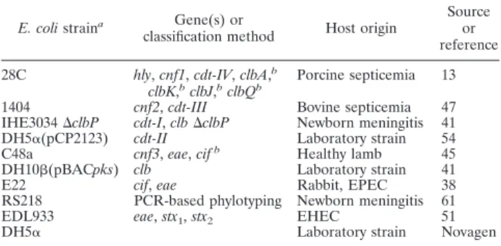
TABLE 1. Archetypal E. coli control strains used in this study
E. coli straina Gene(s) or
classification method Host origin
Source or reference 28C hly, cnf1, cdt-IV, clbA,b
clbK,bclbJ,bclbQb
Porcine septicemia 13 1404 cnf2, cdt-III Bovine septicemia 47 IHE3034⌬clbP cdt-I, clb⌬clbP Newborn meningitis 41 DH5␣(pCP2123) cdt-II Laboratory strain 54 C48a cnf3, eae, cifb Healthy lamb 45
DH10(pBACpks) clb Laboratory strain 41 E22 cif, eae Rabbit, EPEC 38 RS218 PCR-based phylotyping Newborn meningitis 61 EDL933 eae, stx1, stx2 EHEC 51
DH5␣ Laboratory strain Novagen
a
Strain C48a was kindly provided by José Antonio Orden, and strain RS218 was kindly provided by Philippe Bidet and Edouard Bingen.
b
The clbA, clbK, clbJ, clbQ, and cif genes were detected during this study.
TABLE 2. Primers used in this study
Primer Oligonucleotide sequence (5⬘–3⬘) Tmof PCR(°C) Specificity
PCR product size (bp)
Source or reference
91E
TCAAA(G,T)GAATTGACGGGGGC
54
16S rRNA gene
473
58
13B
GCCCGGGAACGTATTAC
18
pksORF9-10.1KJ
ATTCGATAGCGTCACCCAAC
58
clbK-clbJ
2,119
41
pksORF9-10.2KJ
TAAGCGTCTGGAATGCAGTG
IHAPJPN42
CAGATACACAGATACCATTCA
55
clbA
1,002
27
IHAPJPN46
CTAGATTATCCGTGGCGATTC
IHAPJPN55
TTATCCTGTTAGCTTTCGTTC
55
clbQ
821
27
IHAPJPN56
CTTGTATAGTTACACAACTATTTC
CNF-1s
GGGGGAAGTACAGAAGAATTA
48
cnf1
1,112
64
CNF-1as
TTGCCGTCCACTCTCACCAGT
CNF-2s
TATCATACGGCAGGAGGAAGCACC
48
cnf2
1,241
66
CNF-2as
GTCACAATAGACAATAATTTTCCG
CNF3-3D
TAACGTAATTAGCAAAGA
48
cnf3
757
45
CNF-3as
GTCTTCATTACTTACAGT
This study
CDT-s1
GAAAGTAAATGGAATATAAATGTCCG
60
cdtB-II, cdtB-III, cdtB-V
467
64
CDT-as1
AAATCACCAAGAATCATCCAGTTA
CDT-IIas
aTTTGTGTTGCCGCCGCTGGTGAAA
62
cdtB-II
556
64
CDT-IIIas
aTTTGTGTCGGTGCAGCAGGGAAAA
62
cdtB-III, cdtB-V
555
64
CDT-s2
GAAAATAAATGGAACACACATGTCCG
56
cdtB-I, cdtB-IV
467
64
CDT-as2
AAATCTCCTGCAATCATCCAGTTA
CDT-Is
CAATAGTCGCCCACAGGA
56
cdtB-I
411
64
CDT-Ias
ATAATCAAGAACACCACCAC
CDT-IVs
CCTGATGGTTCAGGAGGCTGGTTC
56
cdtB-IV
350
64
CDT-IVas
TTGCTCCAGAATCTATACCT
P105
GTCAACGAACATTAGATTAT
49
cdtC-V
748
25
c2767r
ATGGTCATGCTTTGTTATAT
cif-int-s
AACAGATGGCAACAGACTGG
55
cif
383
38
cif-int-as
AGTCAATGCTTTATGCGTCAT
a
Used with primer CDT-s1.
VOL. 48, 2010
CYCLOMODULINS IN UROSEPSIS STRAINS OF ESCHERICHIA COLI
2123
on January 8, 2014 by I-20DAW
http://jcm.asm.org/
colibactin-producing gene cluster clb, was screened with a probe overlapping the
clbK and clbJ genes. The cnf genes were detected with a mixture of probes
specific to cnf1, cnf2, and cnf3. The cdtB genes were detected by two hybridiza-tion experiments with the cdtB-II–cdtB-III and cdtB-I–cdtB-IV probe mixtures. The cif gene was detected using an internal specific probe. The sensitivities and specificities of the probes were checked on each membrane by spotting DNA extracts of all CM control strains.
Identification of the CM-producing genes and other virulence factors by PCR assays.Positive hybridizations with a CM probe were subjected to confirmatory PCR assays using the primers given in Table 2. The reaction mixture contained 50 ng DNA sample, 0.2 mM each deoxynucleoside triphosphate (dNTP), 0.4M each primer, 3 mM MgCl2, and 1.0 U RedGoldStar DNA polymerase
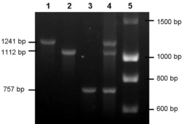
(Euro-gentec, France) in the corresponding reaction buffer. Primers located in the 5⬘ and 3⬘ regions of the pks island (the clbA and clbQ genes) were used to confirm the full presence of the colibactin-producing island. A multiplex PCR was used to distinguish the cnf1, cnf2, and cnf3 genes. Figure 1 shows the standardization of the cnf multiplex PCR. cdtB-I, cdtB-II, cdtB-III/cdtB-V, and cdtB-IV were differentiated by simplex PCR. The cdt-III and cdt-V operons were differentiated by cdtC gene sequencing (Cogenics, Meylan, France) (Table 2) (4). Isolates harboring cdt or cif were further tested by PCR for the additional virulence factor genes stx1, stx2, and eae, as previously described (8).
Phenotypic tests.The cytopathic effects of CNF and CDTs were investigated in all strains, and those of colibactin and Cif were investigated in nonhemolytic strains, as previously described (41, 50). Briefly, the effects of CDT and CNF were detected with a cell-lysate-interacting test. After a 48-h culture at 37°C with shaking in Luria-Bertani broth medium, bacterial cells were sonicated and were sterile filtered separately using 0.22-m-pore-size filters. HeLa cells were treated with the sterile sonicated lysates until analysis. The effects of colibactin and Cif were detected using a cell-bacterium-interacting test, which was based on the interaction between HeLa cells and bacteria. Overnight Luria-Bertani broth cultures of E. coli were diluted in interaction medium, and HeLa cell cultures were infected at multiplicities of infection (numbers of bacteria per cell at the onset of infection) of 100 and 200. Cells were washed 3 to 6 times 4 h after inoculation and were incubated in cell culture medium with 200g/ml gentami-cin until analysis. After 72 h of incubation at 37°C under a 5% CO2atmosphere,
the medium was removed by several washes of the HeLa cell monolayers. The morphological changes characteristic of CDT, CNF, colibactin, and Cif were observed after staining with Giemsa stain, as described elsewhere (41).
The detection of alpha-hemolysin was performed for all strains studied by overnight growth at 37°C on Columbia sheep blood (5%) agar (Oxoid, Dardilly, France). Antibiotic susceptibility tests were performed by the disc diffusion method (Bio-Rad), using CA-SFM interpretative criteria (7). E. coli 25922 (ATCC) was used as the reference strain.
Statistical analysis.Statistical analysis was performed using the Fisher exact and chi-square tests. (For multiple-group comparisons, an initial chi-square test for heterogeneity was done, and only if this yielded a P value of⬍0.05 were the individual pairwise comparisons tested.) The threshold for statistical significance was a P value of⬍0.05.
No statistical difference in the distribution of phylogenetic groups, CM geno-types, or antimicrobial resistance phenotypes was observed between strain
groups of the same clinical origin from different geographic sources (except for resistance to extended-spectrum cephalosporins [C3G], where the P value was 0.041). The strains were therefore grouped together and were analyzed according to their uroseptic or fecal origin.
RESULTS AND DISCUSSION
E. coli phylogroups and clinical origin.
E. coli strains
iso-lated from the fecal and urosepsis groups differed significantly
in the prevalences of phylogenetic groups A, B1, and B2 but
not in that of phylogroup D (Table 3). Phylogroup B2 was
predominant in urosepsis strains (54%), followed by
phylo-groups D (23%), A (18%), and B1 (5%), as previously
ob-served (5, 28–30). Fecal isolates belonged most frequently to
phylogenetic group A (37%), followed by phylogroups B1 and
B2, with identical prevalences (24%). These results are
differ-ent from those of other studies, which found B2 strains
pre-dominant in fecal samples of healthy subjects, notably in
in-dustrialized countries (14, 16, 17, 29, 42, 71). This difference
might be explained by the impact of geographic/climatic
con-ditions, dietary factors, and/or the use of antibiotics or host
genetic factors on the commensal flora (14, 16, 62). Hence,
phylogroup A, and to a lesser extent phylogroup B1, was
sig-nificantly more prevalent among fecal strains than among
uro-sepsis strains (37% versus 18% [P, 0.011] and 24% versus 5%
[P,
⬍0.001], respectively). In contrast, phylogroup B2, known
to encompass the most virulent ExPEC strains (15), was
sig-nificantly more predominant in urosepsis strains than in fecal
strains (54% versus 24% [P,
⬍0.001]).
Distribution of CM-encoding genes according to
phyloge-netic background.
Table 4 shows a clear heterogeneity in the
FIG. 1. Standardization of cnf gene typing by multiplex PCR.
Lanes: 1, strain 1404 (cnf2); 2, strain 28C (cnf1); 3, strain C48a (cnf3);
4, mixture of equal quantities of 1404, 28c, and C48a DNAs (cnf1, cnf2,
cnf3); 5, DNA ladder (Eurogentec).
TABLE 3. Distribution of phylogenetic groups among 197
E. coli isolates recovered from patients with urosepsis
and from the feces of healthy individuals
Phylogeneticgroup
No. (%) of E. coli isolates
Pa Fecal (n⫽ 51) Urosepsis (n⫽ 146)
A
19 (37)
27 (18)
(0.011)
B1
12 (24)
7 (5)
(
⬍0.001)
B2
12 (24)
79 (54)
⬍0.001
D
8 (16)
33 (23)
aP values (by Fisher’s exact test) are shown where P is⬍0.05. P values given
in parentheses indicate negative associations between the prevalence of the urosepsis strains and the particular phylogroup.
TABLE 4. Phylogenetic distribution of cyclomodulin genes and Hly
among 197 E. coli isolates recovered from patients with urosepsis
and from the feces of healthy individuals
Virulencefactor
No. (%) of E. coli isolates
Pa Total (n⫽ 197) Group A (n⫽ 46) Group B1 (n⫽ 19) Group B2 (n⫽ 91) Group D (n⫽ 41) pks 53 (27) 0 0 53 (58) 0 ⬍0.001 cnf1-Hly 36 (18) 1 (2) 1 (5) 34 (37) 0 ⬍0.001 Hly alone 15 (8) 5 (11) 0 8 (9) 2 (5) cnf1-Hly, pks 30 (15) 0 0 30 (33) 0 ⬍0.001 cdtB-I 5 (2) 1 (2) 0 4 (4) 0 cdtB-IV 6 (3) 0 0 5 (5) 1 (2) cdtB-V 1 (1) 0 1 (5) 0 0 cif 2 (1) 0 2 (11) 0 0 a
P values (by Fisher’s exact test) are shown where P is⬍0.05 and are for
comparisons of group B2 versus all other groups combined.
2124
DUBOIS ET AL.
J. CLIN. MICROBIOL.
on January 8, 2014 by I-20DAW
http://jcm.asm.org/
prevalences of CM genes, of which the most frequent were the
pks island (27%) and cnf1 (18%), and in that of
alpha-hemo-lysin expression (26%), three traits that were strongly
associ-ated with phylogroup B2.
All pks-harboring strains belonged to phylogroup B2, as
previously observed (27, 41, 56). This association was
deep-seated (P,
⬍0.001 for B2 versus A, B1, and D combined or
separately). Fifty-eight percent of B2 strains possessed this
trait, which was observed in all B2 subtypes according to
phy-logenetic grouping by PCR (3 among 12 B2
1strains, 5 among
7 Tsp-negative B2 strains, and 45 among 75 Tsp-positive B2
strains).
Multiplex PCR-based cnf typing revealed only cnf1 genes.
No cnf2 or cnf3 genes, which were first observed in E. coli
strains isolated from animals (12, 45), were detected in our
human strains, a finding consistent with the absence or very
weak prevalence of these genes observed in previous studies (9,
12, 32, 37, 55, 63, 64). These results suggest that cnf2- and
cnf3-positive strains are almost entirely absent in humans and
thus are probably involved only in animal diseases.
cnf1-har-boring strains were highly associated with phylogroup B2,
in-cluding 37% of E. coli B2 strains, but were barely present in the
other phylogroups (P,
⬍0.001 for B2 versus other groups), as
previously observed (5, 26, 29, 30). Moreover, 33% of B2
strains harbored both the pks island and the cnf1 gene (P,
⬍0.001 for B2 versus other groups), as previously observed
(27).
All cnf1-positive strains exhibited the alpha-hemolytic
phe-notype. This association was probably due to the presence of
the pathogenicity island (PAI) II
J96-like domain, in which the
cnf1 gene is located just downstream of the hlyCABD operon,
which encodes the alpha-hemolytic phenotype (6, 34). We
ob-served two isolates from one patient, one hemolytic
cnf1-pos-itive and one nonhemolytic cnf1-negative isolate, sharing the
same randomly amplified polymorphic DNA (RAPD) 1283
typing and antimicrobial susceptibility profiling, suggesting the
spontaneous loss of a PAI II
J96-like domain in the second
isolate, as a result of the instability of the PAI (39). Only the
hemolytic cnf1-positive isolate was retained for statistical
anal-ysis. Only 8% of strains exhibited an alpha-hemolytic
pheno-type without the cnf1 gene. These strains, which accounted for
29% of the alpha-hemolytic strains, had no particular
phylo-genetic distribution pattern. They probably possessed another
PAI containing the hly operon, similar to the PAI I
CFT073-like
domain (21).
cdtB genes were observed in 6% of strains, with only one
cdtB subtype per strain. Although 75% of cdtB-positive strains
belonged to phylogroup B2, no significant phylogenetic
distri-bution pattern clearly emerged, even among cdtB subtypes. In
our study, cdtB-I (n
⫽ 5) and cdtB-IV (n ⫽ 6) types were
overrepresented compared to the cdtB-V (n
⫽ 1) type. In
contrast, no cdt-II- or cdt-III-positive strains were found. Of
the cdt genes, cdt-I and cdt-IV are the most closely
homolo-gous, and both genes, framed with lambdoid prophage genes
(65), might have been acquired from a common ancestor by
phage transduction. cdt genes did not possess any particular
association with the pks island or the cnf1 gene (see Table 6).
Since cdt genes have been extensively investigated in Shiga
toxin-producing E. coli (STEC) strains, cdt-harboring strains
were screened for the eae and stx genes (4, 25, 46, 49). No eae
or stx genes were detected in our cdt-encoding strains.
Two cif-harboring strains were observed. They belonged to
phylogroup B1 and harbored no other CM-encoding gene. Cif
is an effector protein of the type 3 secretion system (T3SS)
encoded by the locus of enterocyte effacement pathogenicity
island, and previous observations of the cif gene were restricted
to enteropathogenic E. coli (EPEC) and enterohemorrhagic
E. coli (EHEC) strains (36, 38). The two cif-positive strains of
this study were confirmed as EPEC strains by detection of the
eae gene but not of stx genes.
Distribution of CM-encoding genes according to clinical
origin.
Overall, the urosepsis strains had a significantly higher
prevalence of the pks island than the fecal strains (32% versus
12%; P, 0.005) (Table 5). The pks island could therefore be
involved in uropathogenesis; however, the prevalences of the
pks island among phylogroup B2 strains were not significantly
different between the fecal and urosepsis groups. The urosepsis
strains were also significantly more likely to harbor Hly
(P,
⬍0.001) and cnf1 associated with Hly (P, ⬍0.001) than the
fecal strains. Likewise, the B2 urosepsis strains harbored cnf1
associated with Hly significantly more frequently than their B2
fecal counterparts (P, 0.028).
The weak prevalence of cdtB genes in urosepsis strains (4%)
has been documented elsewhere (28, 29, 31). These
observa-tions suggest that CDTs are not major virulence factors for
urosepsis. cdt genes in E. coli urosepsis strains were
repre-TABLE 5. Distribution of cyclomodulin genes and Hly among 197 E. coli isolates according to phylogenetic group and clinical origin
CM gene(s) No. (%) of all isolates (n⫽ 197) P value for all isolatesa No. (%) of isolates P value for group B2a No. (%) of group D isolates (n⫽ 41) Group A (n⫽ 46) Group B1 (n⫽ 19) Group B2 (n⫽ 91)
Fecal (n⫽ 51) Urosepsis (n⫽ 146) Fecal (n⫽ 19) Urosepsis (n⫽ 27) Fecal (n⫽ 12) Urosepsis (n⫽ 7) Fecal (n⫽ 12) Urosepsis (n⫽ 79) Fecal (n⫽ 8) Urosepsis (n⫽ 33)
pks
6 (12)
47 (32)
0.005
0
0
0
0
6 (50)
47 (59)
0
0
cnf1-Hly
1 (2)
35 (24)
⬍0.001
0
1 (4)
0
1 (14)
1 (8)
33 (42)
0.028
0
0
Hly alone
3 (6)
12 (8)
2 (11)
3 (11)
0
0
1 (8)
7 (9)
0
2 (6)
cnf1-Hly, pks
1 (2)
29 (20)
0.001
0
0
0
0
1 (8)
29 (37)
0
0
cdtB-I
2 (4)
3 (2)
1 (5)
0
0
0
1 (8)
3 (4)
0
0
cdtB-IV
3 (6)
3 (2)
0
0
0
0
2 (17)
3 (4)
1 (12)
0
cdtB-V
1 (2)
0
0
0
1 (8)
0
0
0
0
0
cif
0
2 (1)
0
0
0
2 (29)
0
0
0
0
aP values (by Fisher’s exact test) are shown where P is⬍0.05.
VOL. 48, 2010
CYCLOMODULINS IN UROSEPSIS STRAINS OF ESCHERICHIA COLI
2125
on January 8, 2014 by I-20DAW
http://jcm.asm.org/
sented exclusively by the cdtB-I and cdtB-IV types. cdtB genes
were more prevalent in the fecal isolates than in the urosepsis
isolates (12% versus 4%), but not to the level of significance.
This is in contrast with other studies in which cdtB prevalence
in fecal strains ranged from 0.9% to 5%, with similar
preva-lences in urosepsis strains (29, 30, 32, 64). This difference may
be explained in part by the exhaustive research into cdt
sub-types that was performed in our study. In several studies, CDTs
were detected in EPEC or STEC/EHEC strains isolated from
patients with diarrhea (2, 4, 46, 49). Our CDT-producing
strains were not isolated during bouts of enteric disease and
did not possess eae or stx genes. A case-control study of
CDT-producing E. coli showed no association between CDT-positive
E. coli and diarrhea (2). Our cdtB-V-positive fecal strain did
not possess stx and eae genes, while the cdtB-V gene has been
reported only in STEC collections (4, 46). Finally, the two
cif-positive, eae-positive urosepsis strains suggest the potential
involvement of EPEC as an opportunistic organism in
extraint-estinal infections. The patients were both female and
⬎80
years old, two risk factors for urinary sepsis.
Distribution of phylogroup-CM gene profiles according to
clinical origin.
CM gene profiles and phylogroups were
coana-lyzed to determine whether combinations of CMs and
phylo-groups can also differentiate between the two clinical phylo-groups of
strains (Table 6). Phylogroup B1 and A strains with no
CM-encoding genes were significantly more prevalent in feces than
in the blood cultures of patients with urosepsis (P, 0.003 and
0.018, respectively), whereas the association of the CM profile
pks cnf1 with phylogroup B2 strains was more widespread in
urosepsis strains (P, 0.003). Overall, both the pks island and the
cnf1 gene, whether associated with another CM or not, were
highly predictive of a urosepsis origin (20% versus 2%;
P, 0.001) (Table 5). Three fingerprint profiles (encompassing
17, 10, and 3 strains) were obtained from the 30 pks-, cnf1-, and
group B2-positive strains, suggesting that these urosepsis
strains can belong to a distinct genetic background.
During urosepsis, colibactin and CNF1 may induce
pro-found changes in cellular signaling pathways. Colibactin
in-duces DNA double-strand breaks (41). CNF1 modulates a high
number of cellular functions by hijacking rho GTPases (34),
particularly attenuates polymorphonuclear leukocyte functions
(11), and induces a severe and controlled inflammatory
re-sponse (40, 57, 59, 60). By affecting the immune rere-sponse,
CNF1 could lengthen the brief time window between the
es-tablishment of bacteriuria and the activation of a host defense
during urosepsis, consequently enhancing UPEC survival and
allowing invasion of the parenchyma and bacteremia. CNF1
and probably colibactin may also favor host colonization, since
their encoding genes have been found together in group B2
strains responsible for asymptomatic bacteriuria (70). In this
study, the difference in the prevalences of the pks island and
the cnf1 gene in B2 fecal strains suggest that colibactin may act
mainly as a colonization factor and CNF-1 as a virulence
factor (Table 5). Further experimental investigations are
needed to shed light on the pathogenic and/or colonizer
roles of these CMs.
Phenotypic detection of CMs.
Interpretation of the four
CM-related phenotypes in eukaryotic cells was straightforward
in all but two strains, which harbored the pks island and the cdt
gene and for which the phenotypes are similar in the
cell-bacterium-interacting test. Thus, we were not able to affirm
that the pks islands of these two strains were functional.
Nev-ertheless, all cdt- and cnf-positive strains produced a cytopathic
effect, and a cytopathic effect induced by the pks island was
observed in all but three strains. This result suggests
nonfunc-tional pks islands. However, we cannot rule out the possibility
that other genes, potentially involved in the secretion of
coli-bactin or in the synthesis of precursors used by pks
island-encoded enzymes, were altered. It is noteworthy that two of
these nonfunctional pks islands did not belong to ExPEC
strains and accounted for 33% of pks-positive fecal isolates.
Only one of the two cif genes produced a cytopathic effect.
Loukiadis et al. reported that 66% of cif-positive E. coli strains
did not induce a Cif-related phenotype in eukaryotic cells due
to frameshift mutations or an insertion sequence in the cif
gene (36). However, a nonfunctional T3SS may also explain
the absence of a cytopathic effect. In addition, all strains
that induced cytopathic effects harbored CM-encoding
genes, suggesting that the genomic techniques used had
reliable sensitivity.
Antibiotic susceptibilities of urosepsis strains according to
CM-encoding genes and phylogenetic background.
The
prev-alence of antibiotic resistance among urosepsis strains was as
follows: 59% were resistant to amoxicillin, 5% to
extended-spectrum cephalosporins (C3G), 25% to cotrimoxazole, 18%
to nalidixic acid and norfloxacin, 14% to ciprofloxacin, 2% to
gentamicin, and 0% to amikacin. Table 7 shows the
preva-lences of CM-encoding genes and phylogenetic groups
accord-ing to antibiotic susceptibility status. Quinolone susceptibility
was associated with phylogenetic group B2 (59% of susceptible
versus 33% of resistant isolates; P, 0.019) and the pks island
TABLE 6. Distribution of phylogenetic group-cyclomodulin gene
profiles among 197 E. coli isolates recovered from patients with
urosepsis and from the feces of healthy individuals
Phylogeneticgroup CM gene profile
No. (%) of isolates Pa Fecal (n⫽ 51) Urosepsis (n⫽ 146)
B1
None
11 (21.6)
4 (2.7)
(0.003)
cdtB-V
1 (2)
0
cif
0
2 (1.4)
cnf1
0
1 (0.7)
A
None
18 (35.3)
26 (17.8) (0.018)
cnf1
0
1 (0.7)
cdtB-I
1 (2)
0
B2
None
4 (7.8)
26 (17.8)
cnf1
0
4 (2.7)
pks
4 (7.8)
17 (11.6)
pks, cnf1
1 (2)
26 (17.8)
0.003
pks, cnf1, cdtB-I/cdtB-IV
b0
3 (2)
pks, cdtB-I
1 (2)
1 (0.7)
cdtB-I/cdtB-IV
b2 (4)
2 (1.4)
D
None
7 (13.7)
33 (22.6)
cdtB-IV
1 (2)
0
aP values (by Fisher’s exact test) are shown where P is⬍0.05. P values given
in parentheses indicate negative associations between the prevalence of the urosepsis strains and the particular phylogenetic group-cyclomodulin gene pro-file.
bBecause of their small numbers, high level of homology, and close
epidemi-ology, cdt-I and cdt-IV were aggregated.
2126
DUBOIS ET AL.
J. CLIN. MICROBIOL.
on January 8, 2014 by I-20DAW
http://jcm.asm.org/
(37% versus 11%; P, 0.011), whereas quinolone resistance was
associated with group A (41% of resistant versus 13% of
sus-ceptible isolates; P, 0.002). Likewise, amoxicillin susceptibility
was associated with group B2 (78% versus 37%; P,
⬍0.001),
the combination of the alpha-hemolytic phenotype and the
cnf1 gene (35% versus 16%; P, 0.011), and the pks island (48%
versus 21%; P,
⬍0.001), whereas amoxicillin resistance was
associated with groups A and D (28% versus 5% [P,
⬍0.001]
and 29% versus 13% [P, 0.028], respectively). Cotrimoxazole
susceptibility was also associated with group B2 (60% versus
36%; P, 0.02), the alpha-hemolytic phenotype (36% versus
17%; P, 0.024), the combination of the alpha-hemolytic
phe-notype and the cnf1 gene (28% versus 11%; P, 0.043), and the
pks island (41% versus 6%; P,
⬍0.001). C3G susceptibility was
associated with group B2 (57% versus 0%; P, 0.003), whereas
C3G resistance (six strains with overexpression of
chromo-some-mediated cephalosporinases and one with an
extended-spectrum
-lactamase, CTX-M-14) was associated with group
A (86% versus 15%; P,
⬍0.001).
These results suggest that resistance to quinolones is
asso-ciated with the less virulent phylogenetic groups and with a
weak prevalence of virulence factors, as previously observed
(24, 26, 52, 67, 68). We obtained similar results for resistance
against cotrimoxazole, amoxicillin, and C3G. The acquisition
of antibiotic resistance by horizontal gene transfers or
muta-tions may therefore require a particular genetic background, as
observed for virulence factors (15). Some studies have
sug-gested that the low-virulence group A isolates are more
ex-posed to antibiotic selection pressure within the intestinal tract
(24, 26), which indicates the possible importance of
environ-mental factors.
Conclusion.
The distribution of CM-encoding genes,
includ-ing the recently described pks genomic island, and the
func-tionality of these toxins were investigated in E. coli strains in
relation to their phylogenetic background, clinical origin, and
antibiotic resistance. One finding to emerge from the present
study was the frequent association of the pks island with the
cnf1 gene and the alpha-hemolytic phenotype, and their
pres-ence in amoxicillin-, cotrimoxazole-, and quinolone-susceptible
E. coli strains of the B2 phylogenetic background isolated from
patients with urosepsis. The widespread diffusion of the pks
island and the cnf1 gene in E. coli help to distinguish ExPEC
from commensal strains and reinforce the idea that these genes
of the E. coli flexible gene pool are involved in pathogenicity
and/or in the ability to survive in new ecological niches, such as
the human urinary tract.
ACKNOWLEDGMENTS
This work was supported by le Ministe
`re Franc
¸ais de l’Education
Nationale, de la Recherche et de la Technologie (grant JE2526) and by
l’Institut National de la Recherche Agronomique (grant USC-2018).
We thank Rolande Perroux and Marle
`ne Jan for helpful technical
assistance.
REFERENCES
1. Adamus-Bialek, W., A. Wojtasik, M. Majchrzak, M. Sosnowski, and P. Parniewski.2009. (CGG)4-based PCR as a novel tool for discrimination of uropathogenic Escherichia coli strains: comparison with enterobacterial re-petitive intergenic consensus-PCR. J. Clin. Microbiol. 47:3937–3944. 2. Albert, M. J., S. M. Faruque, A. S. Faruque, K. A. Bettelheim, P. K. Neogi,
N. A. Bhuiyan, and J. B. Kaper.1996. Controlled study of cytolethal dis-tending toxin-producing Escherichia coli infections in Bangladeshi children. J. Clin. Microbiol. 34:717–719.
3. Bidet, P., A. Metais, F. Mahjoub-Messai, L. Durand, M. Dehem, Y. Aujard, E. Bingen, X. Nassif, and S. Bonacorsi.2007. Detection and identification by PCR of a highly virulent phylogenetic subgroup among extraintestinal patho-genic Escherichia coli B2 strains. Appl. Environ. Microbiol. 73:2373–2377. 4. Bielaszewska, M., M. Fell, L. Greune, R. Prager, A. Fruth, H. Tschape, M. A.
Schmidt, and H. Karch.2004. Characterization of cytolethal distending toxin genes and expression in Shiga toxin-producing Escherichia coli strains of non-O157 serogroups. Infect. Immun. 72:1812–1816.
5. Bingen-Bidois, M., O. Clermont, S. Bonacorsi, M. Terki, N. Brahimi, C. Loukil, D. Barraud, and E. Bingen.2002. Phylogenetic analysis and preva-lence of urosepsis strains of Escherichia coli bearing pathogenicity island-like domains. Infect. Immun. 70:3216–3226.
6. Blum, G., V. Falbo, A. Caprioli, and J. Hacker. 1995. Gene clusters encoding the cytotoxic necrotizing factor type 1, Prs-fimbriae and alpha-hemolysin form the pathogenicity island II of the uropathogenic Escherichia coli strain J96. FEMS Microbiol. Lett. 126:189–195.
7. CA-SFM. 2007. Comite´ de l’Antibiogramme de la Socie´te´ Franc¸aise de Microbiologie, communique´ 2007. CA-SFM, Paris, France.
8. Chassagne, L., N. Pradel, F. Robin, V. Livrelli, R. Bonnet, and J. Delmas.
TABLE 7. Distribution of antibiotic susceptibility according to cyclomodulin genes/Hly and phylogenetic background among 143 E. coli
isolates recovered from patients with urosepsis
aVariable Quinolones–fluoroquinolonesb Amoxicillin C3Gc Gentamicin Cotrimoxazole No. (%) of isolates Pd No. (%) of isolates Pd No. (%) of isolates Pd
No. (%) of isolates No. (%) of isolates
Pd R (n⫽ 27) (n⫽ 119)S (n⫽ 86)R (n⫽ 60)S (nR⫽ 7) (n⫽ 139)S (nR⫽ 3) (n⫽ 143)S (n⫽ 36)R (n⫽ 110)S Phylogenetic groups A 11 (41) 16 (13) (0.002) 24 (28) 3 (5) (⬍0.001) 6 (86) 21 (15) (⬍0.001) 2 (67) 25 (17) 9 (25) 18 (16) B1 2 (7) 5 (4) 5 (6) 2 (3) 1 (14) 6 (4) 1 (33) 6 (4) 3 (8) 4 (4) B2 9 (33) 70 (59) 0.019 32 (37) 47 (78) ⬍0.001 0 (0) 79 (57) 0.003 0 (0) 79 (55) 13 (36) 66 (60) 0.020 D 5 (19) 28 (24) 25 (29) 8 (13) (0.028) 0 (0) 33 (24) 0 (0) 33 (23) 11 (31) 22 (20) CMs pks 3 (11) 44 (37) 0.011 18 (21) 29 (48) ⬍0.001 0 47 (34) 0 47 (33) 2 (6) 45 (41) ⬍0.001 cnf1-Hly 7 (26) 28 (24) 14 (16) 21 (35) 0.011 2 (29) 33 (24) 0 35 (24) 4 (11) 31 (28) 0.043 Hly alone 1 (4) 11 (9) 8 (9) 4 (7) 0 12 (9) 0 12 (8) 2 (6) 10 (9) cdtB-I 0 3 (3) 1 (1) 2 (3) 0 3 (2) 0 3 (2) 0 3 (3) cdtB-IV 0 3 (3) 1 (1) 2 (3) 0 3 (2) 0 3 (2) 0 3 (3) cif 0 2 (2) 1 (1) 1 (2) 0 2 (1) 0 2 (1) 0 2 (2) a R, resistant; S, susceptible. b
All resistant isolates were resistant both to quinolone (nalidixic acid) and to fluoroquinolones (either norfloxacin alone or norfloxacin and ciprofloxacin).
c
C3G, extended-spectrum cephalosporins.
d
P values (by Fisher’s exact test) are shown where P is⬍0.05. P values shown in parentheses indicate negative associations between the prevalence of the susceptible
strains and the particular trait (phylogenetic group or CM).
VOL. 48, 2010
CYCLOMODULINS IN UROSEPSIS STRAINS OF ESCHERICHIA COLI
2127
on January 8, 2014 by I-20DAW
http://jcm.asm.org/
2009. Detection of stx1, stx2, and eae genes of enterohemorrhagic Escherichia
coli using SYBR Green in a real-time polymerase chain reaction. Diagn.
Microbiol. Infect. Dis. 64:98–101.
9. Clark, C. G., S. T. Johnson, R. H. Easy, J. L. Campbell, and F. G. Rodgers. 2002. PCR for detection of cdt-III and the relative frequencies of cytolethal distending toxin variant-producing Escherichia coli isolates from humans and cattle. J. Clin. Microbiol. 40:2671–2674.
10. Clermont, O., S. Bonacorsi, and E. Bingen. 2000. Rapid and simple deter-mination of the Escherichia coli phylogenetic group. Appl. Environ. Micro-biol. 66:4555–4558.
11. Davis, J. M., H. M. Carvalho, S. B. Rasmussen, and A. D. O’Brien. 2006. Cytotoxic necrotizing factor type 1 delivered by outer membrane vesicles of uropathogenic Escherichia coli attenuates polymorphonuclear leukocyte an-timicrobial activity and chemotaxis. Infect. Immun. 74:4401–4408. 12. De Rycke, J., A. Milon, and E. Oswald. 1999. Necrotoxic Escherichia coli
(NTEC): two emerging categories of human and animal pathogens. Vet. Res. 30:221–233.
13. Dozois, C. M., S. Clement, C. Desautels, E. Oswald, and J. M. Fairbrother. 1997. Expression of P, S, and F1C adhesins by cytotoxic necrotizing factor 1-producing Escherichia coli from septicemic and diarrheic pigs. FEMS Mi-crobiol. Lett. 152:307–312.
14. Duriez, P., O. Clermont, S. Bonacorsi, E. Bingen, A. Chaventre, J. Elion, B. Picard, and E. Denamur.2001. Commensal Escherichia coli isolates are phylogenetically distributed among geographically distinct human popula-tions. Microbiology 147:1671–1676.
15. Escobar-Pa´ramo, P., O. Clermont, A. B. Blanc-Potard, H. Bui, C. Le Bou-guenec, and E. Denamur.2004. A specific genetic background is required for acquisition and expression of virulence factors in Escherichia coli. Mol. Biol. Evol. 21:1085–1094.
16. Escobar-Pa´ramo, P., K. Grenet, A. Le Menac’h, L. Rode, E. Salgado, C. Amorin, S. Gouriou, B. Picard, M. C. Rahimy, A. Andremont, E. Denamur, and R. Ruimy.2004. Large-scale population structure of human commensal
Escherichia coli isolates. Appl. Environ. Microbiol. 70:5698–5700.
17. Escobar-Pa´ramo, P., A. Le Menac’h, T. Le Gall, C. Amorin, S. Gouriou, B. Picard, D. Skurnik, and E. Denamur.2006. Identification of forces shaping the commensal Escherichia coli genetic structure by comparing animal and human isolates. Environ. Microbiol. 8:1975–1984.
18. Gauduchon, V., L. Chalabreysse, J. Etienne, M. Celard, Y. Benito, H. Lepidi, F. Thivolet-Bejui, and F. Vandenesch.2003. Molecular diagnosis of infective endocarditis by PCR amplification and direct sequencing of DNA from valve tissue. J. Clin. Microbiol. 41:763–766.
19. Ge, Z., D. B. Schauer, and J. G. Fox. 2008. In vivo virulence properties of bacterial cytolethal-distending toxin. Cell. Microbiol. 10:1599–1607. 20. Gordon, D. M., O. Clermont, H. Tolley, and E. Denamur. 2008. Assigning
Escherichia coli strains to phylogenetic groups: multi-locus sequence typing
versus the PCR triplex method. Environ. Microbiol. 10:2484–2496. 21. Guyer, D. M., J. S. Kao, and H. L. Mobley. 1998. Genomic analysis of a
pathogenicity island in uropathogenic Escherichia coli CFT073: distribution of homologous sequences among isolates from patients with pyelonephritis, cystitis, and catheter-associated bacteriuria and from fecal samples. Infect. Immun. 66:4411–4417.
22. Hacker, J., U. Hentschel, and U. Dobrindt. 2003. Prokaryotic chromosomes and disease. Science 301:790–793.
23. Herzer, P. J., S. Inouye, M. Inouye, and T. S. Whittam. 1990. Phylogenetic distribution of branched RNA-linked multicopy single-stranded DNA among natural isolates of Escherichia coli. J. Bacteriol. 172:6175–6181. 24. Houdouin, V., S. Bonacorsi, P. Bidet, M. Bingen-Bidois, D. Barraud, and E.
Bingen.2006. Phylogenetic background and carriage of pathogenicity island-like domains in relation to antibiotic resistance profiles among Escherichia
coli urosepsis isolates. J. Antimicrob. Chemother. 58:748–751.
25. Janka, A., M. Bielaszewska, U. Dobrindt, L. Greune, M. A. Schmidt, and H. Karch.2003. Cytolethal distending toxin gene cluster in enterohemorrhagic
Escherichia coli O157:H⫺ and O157:H7: characterization and evolutionary considerations. Infect. Immun. 71:3634–3638.
26. Jaure´guy, F., E. Carbonnelle, S. Bonacorsi, C. Clec’h, P. Casassus, E. Bin-gen, B. Picard, X. Nassif, and O. Lortholary. 2007. Host and bacterial determinants of initial severity and outcome of Escherichia coli sepsis. Clin. Microbiol. Infect. 13:854–862.
27. Johnson, J. R., B. Johnston, M. A. Kuskowski, J. P. Nougayrede, and E. Oswald.2008. Molecular epidemiology and phylogenetic distribution of the
Escherichia coli pks genomic island. J. Clin. Microbiol. 46:3906–3911.
28. Johnson, J. R., M. A. Kuskowski, A. Gajewski, S. Soto, J. P. Horcajada, M. T. Jimenez de Anta, and J. Vila. 2005. Extended virulence genotypes and phylogenetic background of Escherichia coli isolates from patients with cys-titis, pyelonephritis, or prostatitis. J. Infect. Dis. 191:46–50.
29. Johnson, J. R., K. Owens, A. Gajewski, and M. A. Kuskowski. 2005. Bacterial characteristics in relation to clinical source of Escherichia coli isolates from women with acute cystitis or pyelonephritis and uninfected women. J. Clin. Microbiol. 43:6064–6072.
30. Johnson, J. R., F. Scheutz, P. Ulleryd, M. A. Kuskowski, T. T. O’Bryan, and T. Sandberg.2005. Host-pathogen relationships among Escherichia coli
iso-lates recovered from men with febrile urinary tract infection. Clin. Infect. Dis. 40:813–822.
31. Johnson, J. R., and A. L. Stell. 2000. Extended virulence genotypes of
Escherichia coli strains from patients with urosepsis in relation to phylogeny
and host compromise. J. Infect. Dis. 181:261–272.
32. Kadhum, H. J., D. Finlay, M. T. Rowe, I. G. Wilson, and H. J. Ball. 2008. Occurrence and characteristics of cytotoxic necrotizing factors, cytolethal distending toxins and other virulence factors in Escherichia coli from human blood and faecal samples. Epidemiol. Infect. 136:752–760.
33. Kaper, J. B., J. P. Nataro, and H. L. Mobley. 2004. Pathogenic Escherichia
coli. Nat. Rev. Microbiol. 2:123–140.
34. Lemonnier, M., L. Landraud, and E. Lemichez. 2007. Rho GTPase-activat-ing bacterial toxins: from bacterial virulence regulation to eukaryotic cell biology. FEMS Microbiol. Rev. 31:515–534.
35. Lidin-Janson, G., B. Kaijser, K. Lincoln, S. Olling, and H. Wedel. 1978. The homogeneity of the faecal coliform flora of normal school-girls, character-ized by serological and biochemical properties. Med. Microbiol. Immunol. 164:247–253.
36. Loukiadis, E., R. Nobe, S. Herold, C. Tramuta, Y. Ogura, T. Ooka, S. Morabito, M. Kerouredan, H. Brugere, H. Schmidt, T. Hayashi, and E. Oswald.2008. Distribution, functional expression, and genetic organization of Cif, a phage-encoded type III-secreted effector from enteropathogenic and enterohemorrhagic Escherichia coli. J. Bacteriol. 190:275–285. 37. Mainil, J. G., E. Jacquemin, and E. Oswald. 2003. Prevalence and identity of
cdt-related sequences in necrotoxigenic Escherichia coli. Vet. Microbiol. 94:
159–165.
38. Marche`s, O., T. N. Ledger, M. Boury, M. Ohara, X. Tu, F. Goffaux, J. Mainil, I. Rosenshine, M. Sugai, J. De Rycke, and E. Oswald.2003. Enteropatho-genic and enterohaemorrhagic Escherichia coli deliver a novel effector called Cif, which blocks cell cycle G2/M transition. Mol. Microbiol. 50:1553–1567.
39. Middendorf, B., B. Hochhut, K. Leipold, U. Dobrindt, G. Blum-Oehler, and J. Hacker.2004. Instability of pathogenicity islands in uropathogenic
Esch-erichia coli 536. J. Bacteriol. 186:3086–3096.
40. Munro, P., G. Flatau, A. Doye, L. Boyer, O. Oregioni, J. L. Mege, L. Land-raud, and E. Lemichez.2004. Activation and proteasomal degradation of rho GTPases by cytotoxic necrotizing factor-1 elicit a controlled inflammatory response. J. Biol. Chem. 279:35849–35857.
41. Nougayre`de, J. P., S. Homburg, F. Taieb, M. Boury, E. Brzuszkiewicz, G. Gottschalk, C. Buchrieser, J. Hacker, U. Dobrindt, and E. Oswald.2006.
Escherichia coli induces DNA double-strand breaks in eukaryotic cells.
Sci-ence 313:848–851.
42. Nowrouzian, F. L., I. Adlerberth, and A. E. Wold. 2006. Enhanced persis-tence in the colonic microbiota of Escherichia coli strains belonging to phy-logenetic group B2: role of virulence factors and adherence to colonic cells. Microbes Infect. 8:834–840.
43. Nowrouzian, F. L., A. E. Wold, and I. Adlerberth. 2005. Escherichia coli strains belonging to phylogenetic group B2 have superior capacity to persist in the intestinal microflora of infants. J. Infect. Dis. 191:1078–1083. 44. Ochman, H., and R. K. Selander. 1984. Standard reference strains of
Esch-erichia coli from natural populations. J. Bacteriol. 157:690–693.
45. Orden, J. A., G. Dominguez-Bernal, S. Martinez-Pulgarin, M. Blanco, J. E. Blanco, A. Mora, J. Blanco, and R. de la Fuente.2007. Necrotoxigenic
Escherichia coli from sheep and goats produce a new type of cytotoxic
necrotizing factor (CNF3) associated with the eae and ehxA genes. Int. Microbiol. 10:47–55.
46. Orth, D., K. Grif, M. P. Dierich, and R. Wurzner. 2006. Cytolethal distending toxins in Shiga toxin-producing Escherichia coli: alleles, serotype distribution and biological effects. J. Med. Microbiol. 55:1487–1492.
47. Oswald, E., J. de Rycke, P. Lintermans, K. van Muylem, J. Mainil, G. Daube, and P. Pohl.1991. Virulence factors associated with cytotoxic necrotizing factor type two in bovine diarrheic and septicemic strains of Escherichia coli. J. Clin. Microbiol. 29:2522–2527.
48. Oswald, E., J. P. Nougayre`de, F. Taieb, and M. Sugai.2005. Bacterial toxins that modulate host cell-cycle progression. Curr. Opin. Microbiol. 8:83–91. 49. Pandey, M., A. Khan, S. C. Das, B. Sarkar, S. Kahali, S. Chakraborty, S.
Chattopadhyay, S. Yamasaki, Y. Takeda, G. B. Nair, and T. Ramamurthy. 2003. Association of cytolethal distending toxin locus cdtB with enteropatho-genic Escherichia coli isolated from patients with acute diarrhea in Calcutta, India. J. Clin. Microbiol. 41:5277–5281.
50. Pe´re`s, S. Y., O. Marches, F. Daigle, J. P. Nougayre`de, F. Herault, C. Tasca, J. De Rycke, and E. Oswald.1997. A new cytolethal distending toxin (CDT) from Escherichia coli producing CNF2 blocks HeLa cell division in G2/M
phase. Mol. Microbiol. 24:1095–1107.
51. Perna, N. T., G. Plunkett III, V. Burland, B. Mau, J. D. Glasner, D. J. Rose, G. F. Mayhew, P. S. Evans, J. Gregor, H. A. Kirkpatrick, G. Posfai, J. Hackett, S. Klink, A. Boutin, Y. Shao, L. Miller, E. J. Grotbeck, N. W. Davis, A. Lim, E. T. Dimalanta, K. D. Potamousis, J. Apodaca, T. S. Ananthara-man, J. Lin, G. Yen, D. C. Schwartz, R. A. Welch, and F. R. Blattner.2001. Genome sequence of enterohaemorrhagic Escherichia coli O157:H7. Nature 409:529–533.
52. Piatti, G., A. Mannini, M. Balistreri, and A. M. Schito. 2008. Virulence
2128
DUBOIS ET AL.
J. CLIN. MICROBIOL.
on January 8, 2014 by I-20DAW
http://jcm.asm.org/
factors in urinary Escherichia coli strains: phylogenetic background and quin-olone and fluoroquinquin-olone resistance. J. Clin. Microbiol. 46:480–487. 53. Picard, B., J. S. Garcia, S. Gouriou, P. Duriez, N. Brahimi, E. Bingen, J.
Elion, and E. Denamur.1999. The link between phylogeny and virulence in
Escherichia coli extraintestinal infection. Infect. Immun. 67:546–553.
54. Pickett, C. L., D. L. Cottle, E. C. Pesci, and G. Bikah. 1994. Cloning, sequencing, and expression of the Escherichia coli cytolethal distending toxin genes. Infect. Immun. 62:1046–1051.
55. Pickett, C. L., R. B. Lee, A. Eyigor, B. Elitzur, E. M. Fox, and N. A. Strockbine.2004. Patterns of variations in Escherichia coli strains that pro-duce cytolethal distending toxin. Infect. Immun. 72:684–690.
56. Putze, J., C. Hennequin, J. P. Nougayre`de, W. Zhang, S. Homburg, H. Karch, M. A. Bringer, C. Fayolle, E. Carniel, W. Rabsch, T. A. Oelschlaeger, E. Oswald, C. Forestier, J. Hacker, and U. Dobrindt.2009. Genetic structure and distribution of the colibactin genomic island among Enterobacteriaceae. Infect. Immun. 77:4696–4703.
57. Real, J. M., P. Munro, C. Buisson-Touati, E. Lemichez, P. Boquet, and L. Landraud.2007. Specificity of immunomodulator secretion in urinary sam-ples in response to infection by alpha-hemolysin and CNF1 bearing uro-pathogenic Escherichia coli. Cytokine 37:22–25.
58. Relman, D. A. 1993. The identification of uncultured microbial pathogens. J. Infect. Dis. 168:1–8.
59. Rippere-Lampe, K. E., M. Lang, H. Ceri, M. Olson, H. A. Lockman, and A. D. O’Brien.2001. Cytotoxic necrotizing factor type 1-positive Escherichia
coli causes increased inflammation and tissue damage to the prostate in a rat
prostatitis model. Infect. Immun. 69:6515–6519.
60. Rippere-Lampe, K. E., A. D. O’Brien, R. Conran, and H. A. Lockman. 2001. Mutation of the gene encoding cytotoxic necrotizing factor type 1 (cnf1)
attenuates the virulence of uropathogenic Escherichia coli. Infect. Immun. 69:3954–3964.
61. Silver, R. P., W. Aaronson, A. Sutton, and R. Schneerson. 1980. Comparative analysis of plasmids and some metabolic characteristics of Escherichia coli K1 from diseased and healthy individuals. Infect. Immun. 29:200–206. 62. Skurnik, D., D. Bonnet, C. Bernede-Bauduin, R. Michel, C. Guette, J. M.
Becker, C. Balaire, F. Chau, J. Mohler, V. Jarlier, J. P. Boutin, B. Moreau, D. Guillemot, E. Denamur, A. Andremont, and R. Ruimy.2008. Character-istics of human intestinal Escherichia coli with changing environments. En-viron. Microbiol. 10:2132–2137.
63. Tavechio, A. T., L. R. Marques, C. M. Abe, and T. A. Gomes. 2004. Detection of cytotoxic necrotizing factor types 1 and 2 among fecal Escherichia coli isolates from Brazilian children with and without diarrhea. Mem. Inst. Oswaldo Cruz 99:81–83.
64. To´th, I., F. Herault, L. Beutin, and E. Oswald.2003. Production of cytolethal distending toxins by pathogenic Escherichia coli strains isolated from human and animal sources: establishment of the existence of a new cdt variant (type IV). J. Clin. Microbiol. 41:4285–4291.
65. To´th, I., J. P. Nougayre`de, U. Dobrindt, T. N. Ledger, M. Boury, S. Morabito, T. Fujiwara, M. Sugai, J. Hacker, and E. Oswald.2009. Cytolethal distending toxin type I and type IV genes are framed with lambdoid prophage genes in extraintestinal pathogenic Escherichia coli. Infect. Immun. 77:492–500. 66. Van Bost, S., E. Jacquemin, E. Oswald, and J. Mainil. 2003. Multiplex PCRs
for identification of necrotoxigenic Escherichia coli. J. Clin. Microbiol. 41: 4480–4482.
67. Velasco, M., J. P. Horcajada, J. Mensa, A. Moreno-Martinez, J. Vila, J. A. Martinez, J. Ruiz, M. Barranco, G. Roig, and E. Soriano.2001. Decreased invasive capacity of quinolone-resistant Escherichia coli in patients with uri-nary tract infections. Clin. Infect. Dis. 33:1682–1686.
68. Vila, J., K. Simon, J. Ruiz, J. P. Horcajada, M. Velasco, M. Barranco, A. Moreno, and J. Mensa.2002. Are quinolone-resistant uropathogenic
Esch-erichia coli less virulent? J. Infect. Dis. 186:1039–1042.
69. Wiles, T. J., R. R. Kulesus, and M. A. Mulvey. 2008. Origins and virulence mechanisms of uropathogenic Escherichia coli. Exp. Mol. Pathol. 85:11–19. 70. Zdziarski, J., C. Svanborg, B. Wullt, J. Hacker, and U. Dobrindt. 2008. Molecular basis of commensalism in the urinary tract: low virulence or virulence attenuation? Infect. Immun. 76:695–703.
71. Zhang, L., B. Foxman, and C. Marrs. 2002. Both urinary and rectal
Esche-richia coli isolates are dominated by strains of phylogenetic group B2. J. Clin.
Microbiol. 40:3951–3955.