Publisher’s version / Version de l'éditeur:
Journal of Microbiological Methods, 73, 2, pp. 199-202, 2008-02-14
READ THESE TERMS AND CONDITIONS CAREFULLY BEFORE USING THIS WEBSITE. https://nrc-publications.canada.ca/eng/copyright
Vous avez des questions? Nous pouvons vous aider. Pour communiquer directement avec un auteur, consultez la première page de la revue dans laquelle son article a été publié afin de trouver ses coordonnées. Si vous n’arrivez pas à les repérer, communiquez avec nous à PublicationsArchive-ArchivesPublications@nrc-cnrc.gc.ca.
Questions? Contact the NRC Publications Archive team at
PublicationsArchive-ArchivesPublications@nrc-cnrc.gc.ca. If you wish to email the authors directly, please see the first page of the publication for their contact information.
NRC Publications Archive
Archives des publications du CNRC
This publication could be one of several versions: author’s original, accepted manuscript or the publisher’s version. / La version de cette publication peut être l’une des suivantes : la version prépublication de l’auteur, la version acceptée du manuscrit ou la version de l’éditeur.
For the publisher’s version, please access the DOI link below./ Pour consulter la version de l’éditeur, utilisez le lien DOI ci-dessous.
https://doi.org/10.1016/j.mimet.2008.01.016
Access and use of this website and the material on it are subject to the Terms and Conditions set forth at
Development of a SYBR safe TM technique for the sensitive detection
of DNA in cesium chloride density gradients for stable isotope probing
assays
Martineau, Christine; Whyte, Lyle G.; Greer, Charles W.
https://publications-cnrc.canada.ca/fra/droits
L’accès à ce site Web et l’utilisation de son contenu sont assujettis aux conditions présentées dans le site LISEZ CES CONDITIONS ATTENTIVEMENT AVANT D’UTILISER CE SITE WEB.
NRC Publications Record / Notice d'Archives des publications de CNRC:
https://nrc-publications.canada.ca/eng/view/object/?id=badd1c5b-cc10-478e-9c09-46cc5f6de442 https://publications-cnrc.canada.ca/fra/voir/objet/?id=badd1c5b-cc10-478e-9c09-46cc5f6de442Note
Development of a SYBR safe™ technique for the sensitive detection of DNA
in cesium chloride density gradients for stable isotope probing assays
Christine Martineau
a,b, Lyle G. Whyte
b, Charles W. Greer
a,⁎
a
Biotechnology Research Institute, National Research Council of Canada, 6100 Royalmount Ave., Montreal, QC, Canada H4P 2R2
b
Department of Natural Resource Sciences, McGill University, Macdonald Campus, 21,111 Lakeshore, Ste-Anne-de-Bellevue, QC, Canada H9X 3V9
Received 5 December 2007; received in revised form 25 January 2008; accepted 25 January 2008 Available online 14 February 2008
Abstract
SYBR safe™, a fluorescent nucleic acid stain, was evaluated as a replacement for ethidium bromide (EtBr) in cesium chloride (CsCl) density gradients for DNA stable isotope probing (DNA-SIP) assays. The separation of12C- and13C-labelled DNA using SYBR safe™ gave similar results to those obtained using EtBr with pure cultures and environmental samples exposed to a13C-labelled substrate, while the detection limit of DNA was enhanced by the use of SYBR safe™ by at least 5 times. The results demonstrated that SYBR safe™ is a safe, sensitive and effective alternative to the use of ethidium bromide in CsCl density gradients for DNA-SIP assays.
Crown Copyright © 2008 Published by Elsevier B.V. All rights reserved.
Keywords: Stable isotope probing; Cesium chloride density gradient; SYBR safe™; Ethidium bromide
Stable isotope probing (SIP) of DNA is a technique that has become an important tool in microbial ecology (Radajewski et al., 2000). In this technique, compounds labelled with stable isotopes like 13C or 15N are provided as substrates for the growth of microorganisms in environmental samples. These microorgan-isms incorporate the stable isotopes into their cellular compounds, including phospholipid fatty acids (PLFA), RNA and DNA, which can be used as biomarkers to identify the bacterial population implicated in the metabolism of the substrate of interest.13C-labelled DNA is the biomarker that has been the most extensively used in SIP studies. The13C-labelled DNA can be separated from non-labelled12C-DNA by cesium chloride (CsCl) density gradient ultracentrifugation and screened through various phylogenetic and functional analyses. Using DNA stable isotope probing (DNA-SIP), the active microbial populations implicated in the degradation of various compounds have been identified (Friedrich, 2006).
One of the critical steps of the DNA-SIP technique is the CsCl density gradient ultracentrifugation. During this step, the 13
C-DNA is separated from the12C-DNA based on their respective buoyant densities. High concentrations of ethidium bromide (EtBr) are commonly added in the CsCl gradients to allow for the visualization of the DNA under UV light following the ultra-centrifugation. This technique has numerous disadvantages. EtBr, because of its tendency to intercalate between DNA bases, is known as a carcinogenic and mutagenic agent (Singer et al., 1999). Moreover, several studies have shown that UV exposure can damage DNA through photochemical reactions (Cariello et al., 1988; Emanuele et al., 2005) and subsequently compromise its use in downstream molecular biology analyses (Hartman, 1991; Gründemann and Schomig, 1996). Finally, EtBr wastes require safe disposal involving considerable environmental and monetary costs.
In the present study, we tested the use of SYBR safe™ (Invitrogen, Carlsbad, CA) as a replacement for EtBr in the CsCl density gradient ultracentrifugation step of SIP assays. SYBR safe™ is a fluorescent nucleic acid stain that has a lower muta-genic potential than EtBr and is classified as non-hazardous (http://probes.invitrogen.com/media/publications/494.pdf). Moreover, it was developed to be visualized optimally using a blue light transilluminator that does not damage DNA. The results obtained in this study also demonstrated that the use of
Journal of Microbiological Methods 73 (2008) 199 – 202
www.elsevier.com/locate/jmicmeth
⁎Corresponding author. Tel.: +1 514 496 6182; fax: +1 514 496 6265.
E-mail address:charles.greer@cnrc-nrc.gc.ca(C.W. Greer).
0167-7012/$ - see front matter. Crown Copyright © 2008 Published by Elsevier B.V. All rights reserved. doi:10.1016/j.mimet.2008.01.016
SYBR safe™ improves the detection limit of DNA in CsCl density gradients.
The first step of this study was to verify that a similar separation of the12C- and13C-DNA could be obtained using SYBR safe™ instead of EtBr in the CsCl density gradients.
12
C- and 13C-DNA was obtained through the cultivation of Methylosinus trichosporium on12C- or13C-methane. DNA was extracted following the protocol described by Pospiech and
Neumann (1995) and was quantified using the PicoGreen®
dsDNA quantitation assay (Invitrogen, Carlsbad, CA). Five micrograms of each12C- and13C-DNA from M. trichosporium was loaded into the ultracentrifugation tubes and duplicates were performed for each DNA stain. For the ultracentrifugation with EtBr, the tubes were prepared as described byRadajewski et al. (2004). For the ultracentrifugation with SYBR safe™, we determined that the tubes had to be prepared according to the following protocol. The volume of the DNA extract was adjusted to 700μL with TE (10:1) buffer. One gram of CsCl was dissolved in this solution and 25μL of 10 000× SYBR safe™ were added. The resulting mixture was loaded into a 13 × 51mm polyallomer Quick-Seal centrifuge tube (Beckman, Fullerton, CA, USA). The tubes were filled with a 1g mL− 1CsCl solution
and were heat-sealed. The final density of the solution was 1.72g mL− 1, as determined by refractometry (Reichert Abbe
Mark II Refractometer, Reichert, Depew, NY, USA). Tubes were ultracentrifuged (L8-70M ultracentrifuge, Beckman, Full-erton, CA, USA) at 265 000×g for 16h in a Vti80 rotor (Beck-man, Fullerton, CA, USA). Tubes containing SYBR safe™ were visualized using the Safe Imager™ blue light transillumi-nator (Invitrogen, Carlsbad, CA) while tubes containing EtBr were visualized using a long-wave UV hand light (Model UVL 56, UVP, Upland, CA, USA).
Typical results for the ultracentrifugation of 5μg of each12 C-and 13C-DNA from M. trichosporium with SYBR™ safe or EtBr are presented inFig. 1. Results were consistent between replicates (data not shown). Ultracentrifugation with SYBR safe™ resulted in the efficient separation of the two DNA bands (Fig. 1A). Band separation was similar to what was observed when the12C- and13C-DNA were ultracentrifuged with EtBr (Fig. 1B).
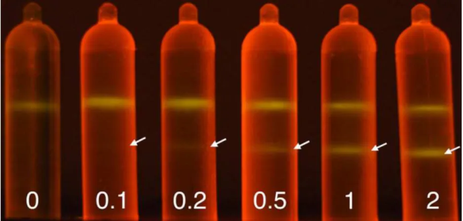
The detection limit of DNA in CsCl gradients prepared with SYBR safe™ as a dye was determined. Five ultracentrifuga-tion tubes were set up, ultracentrifuged and visualized as described previously for SYBR safe™, each one containing 2000ng of 12C-DNA and either 0, 100, 200, 500, 1000 or 2000ng of13C-DNA from M. trichosporium (Fig. 2). Using this approach, we found that a band containing 100ng of DNA was faint but detectable in the CsCl density gradient, while a 200ng band was easily visualized. The ultracentrifugation in CsCl density gradients of 100ng and 200ng of12C-DNA from M. trichosporium was repeated to confirm the value of the detection limit (data not shown).
The potential of using SYBR safe™ for the ultracentrifuga-tion in CsCl density gradients of DNA from environmental samples exposed to a13C-labelled substrate was also evaluated. Two microcosms containing 20g of an Arctic surface soil sample collected at Eureka (Ellesmere Island, Nunavut) were incubated at 4°C with 20mL of NMS medium until 5 or 40mL of13CH4were consumed. DNA was extracted from 10g of soil
from each of the two microcosms following the protocol described by Fortin et al. (2004), but without performing the PVPP purification step. Ultracentrifugation tubes containing the
Fig. 1. Comparison of the CsCl density gradient ultracentrifugation of 5 μg of
12
C- and13C-DNA from M. trichosporium with SYBR safe™ (A) or EtBr (B).
Fig. 2. CsCl density gradient ultracentrifugation with SYBR safe™ of 0, 0.1, 0.2, 0.5, 1.0 and 2.0 μg of13C-labelled DNA with a constant amount of 2.0 μg of12 C-DNA, both from M. trichosporium. Arrows indicate the position of the13C-DNA band.
DNA extracted from each microcosm were set up as described above, ultracentrifuged at 177 000×g for 40h and visualized with the Safe Imager™ blue light transilluminator.
CsCl density gradient ultracentrifugation of DNA from an environmental sample incubated under13C-methane using SYBR safe™ resulted in two bands corresponding to the12C-DNA and
13
C-DNA (Fig. 3). After the consumption of 5mL of 13 C-methane, the two DNA bands were clearly visible (Fig. 3A), while a longer incubation with up to 40mL of 13C-methane led to a stronger 13C-DNA band and a much fainter 12C-DNA band (Fig. 3B). SYBR safe™ was also used for the ultracentrifugation in CsCl density gradients of DNA from numerous other envi-ronmental samples exposed to13C-methane, with similar results. The results obtained in this study showed that SYBR safe™ is an effective replacement for EtBr in CsCl density gradients. The separation of the12C- and13C-DNA bands, which is crucial for SIP analysis, was achieved using SYBR safe™ as a dye in the CsCl density gradient, both for pure culture DNA and for DNA from an environmental sample previously exposed to a 13 C-labelled compound. Our results also demonstrated that the use of SYBR safe™ with the Safe Imager™ blue light transilluminator provided a sensitive detection of DNA in CsCl density gradients. While we were not able to detect less than 2μg of DNA with EtBr (data not shown) and detection limits between 0.5 and 2μg have been reported by others for EtBr (Cadisch et al., 2005; Neufeld et al., 2007), we were able to visualize a band containing amounts as low as 100ng of DNA using SYBR safe™. This increased sensitivity can play an important role in SIP analysis, where the labelling of DNA with13C can be challenging. The success of a SIP analysis resides not only in the labelling of enough DNA to be able to detect it, but also on short incubation times that limit the extent of cross-feeding. The use of a more sensitive dye provides the opportunity to detect lower amounts of DNA and, therefore, to reduce incubation times.
An interesting aspect of the use of SYBR safe™ in CsCl density gradients is that it is optimally visualized using the Safe Imager™, a blue light transilluminator that does not damage the DNA. The use of a blue light transilluminator can be of critical importance when the DNA retrieved from the gradient is to be
used in molecular techniques that require intact, undamaged DNA. For example, the13C-DNA retrieved from a CsCl density gradient ultracentrifugation tube can be used to create a meta-genomic library (Dumont et al., 2006). Such an approach, that has the potential of increasing the amount of information obtained through SIP studies, includes a cloning step that can be negatively affected by previous exposure of the DNA to UV (Hartman, 1991; Gründemann and Schomig, 1996).
DNA stable isotope probing is a powerful tool in microbial ecology and it has the potential to provide a great deal of information on microbial activity in environmental samples. However, the application of this technique can be limited by the ability to detect the labelled DNA. Here, we developed and tested a modification of the CsCl density gradient step of the SIP assay that permits a more sensitive detection of DNA. This modifica-tion, which consisted of replacing ethidium bromide by SYBR safe™ in the CsCl solution, is simple and, when combined with the use of the Safe Imager™ blue light transilluminator, provides several other advantages including increased safety for the user, much less potential damage to the resulting DNA, and does not generate hazardous waste material. Therefore, we suggest that SYBR safe™ is applicable to DNA-SIP and a significant im-provement over the currently employed EtBr-based technique. Acknowledgements
The authors would like to thank Diane Labbé for her technical support and David F. Juck for collecting the Arctic soil sample and taking the pictures. Logistical support from the Canadian Polar Continental Shelf Project (PCSP) is gratefully acknowledged. CM was supported by a NSERC postgraduate scholarship and by the Department of Indian and Northern Affairs — Northern Scientific Training Program.
References
Cadisch, G., Espana, M., Causey, R., Richter, M., Shaw, E., Morgan, J.A.W., et al., 2005. Technical considerations for the use of15N-DNA stable-isotope probing
for functional microbial activity in soils. Rapid Commun. Mass Spectrom. 19, 1424–1428.
Cariello, N.F., Keohavong, P., Sanderson, B.J.S., Thilly, W.G., 1988. DNA damage produced by ethidium bromide staining and exposure to ultraviolet light. Nucleic Acids Res. 16, 4157.
Dumont, M.G., Radajewski, S.M., Miguez, C.B., McDonald, I.R., Murrell, J.C., 2006. Identification of a complete methane monooxygenase operon from soil by combining stable isotope probing and metagenomic analysis. Environ. Microbiol. 8, 1240–1250.
Emanuele, E., Markovitsi, D., Millie, P., Zakrzewska, K., 2005. UV spectra and excitation delocalization in DNA: influence of the spectral width. Chem-PhysChem 6, 1387–1392.
Fortin, N., Beaumier, D., Lee, K., Greer, C.W., 2004. Soil washing improves the recovery of total community DNA from polluted and high organic content sediments. J. Microbiol. Methods 56, 181–191.
Friedrich, M.W., 2006. Stable-isotope probing of DNA: insights into the function of uncultivated microorganisms from isotopically labeled meta-genomes. Curr. Opin. Biotechnol. 17, 59–66.
Gründemann, D., Schomig, E., 1996. Protection of DNA during preparative agarose gel electrophoresis against damage induced by ultraviolet light. BioTechniques 21, 898–903.
Hartman, P.S., 1991. Transillumination can profoundly reduce transformation frequencies. BioTechniques 11, 747–748.
Fig. 3. CsCl density gradient ultracentrifugation with SYBR safe™ of DNA extracted from an environmental sample that had metabolized either 5 mL (A) or 40 mL (B) of13C-methane.
Neufeld, J.D., Vohra, J., Dumont, M.G., Lueders, T., Manefield, M., Friedrich, M.W., et al., 2007. DNA stable-isotope probing. Nat. Protoc. 2, 860–866. Pospiech, A., Neumann, B., 1995. A versatile quick-prep of genomic DNA from
Gram-positive bacteria. Trends Genet. 11, 217–218.
Radajewski, S., Ineson, P., Parekh, N.R., Murrell, J.C., 2000. Stable-isotope probing as a tool in microbial ecology. Nature 403, 646–649.
Radajewski, S., McDonald, I.R., Murrell, J.C., 2004. Stable isotope probing of nucleic acids to identify active microbial populations. In: Kowalchuk,
G.A., de Bruijn, F.J., Head, I.M., Akkermans, A.D., van Elsas, J.D. (Eds.), Molecular Microbial Ecology Manual. Kluwer Academic Publishers, Dordrecht, pp. 1661–1672.
Singer, V.L., Lawlor, T.E., Yue, S., 1999. Comparison of SYBR® Green I nucleic acid gel stain mutagenicity and ethidium bromide mutagenicity in the Salmonella/mammalian microsome reverse mutation assay (Ames test). Mutat. Res., Genet. Toxicol. Environ. Mutagen. 439, 37–47.

![[PDF] Cours avancé sur QTcreator : le Framework | Cours informatique](data:image/gif;base64,R0lGODlhAQABAIAAAP///wAAACH5BAEAAAAALAAAAAABAAEAAAICRAEAOw==)