A N OUTBREAK O F PSEUDOMONAS AERUGINOSA
P N E U M O N I A AND BLOODSTREAM INFECTION
ASSOCIATED W I T H INTERMITTENT O T I T I S EXTERNA
IN A HEALTHCARE W O R K E R
Anne Zawacki, BSN, MPH; Edward O'Rourke, MD; Gail Potter-Bynoe, BS, CIC; Ann Macone, BS; Stephan Harbarth, MD, MS; Donald Goldmann, MD
ARSTRACT
OBJECTIVES: To investigate an outbreak of
Pseudomonas aeruginosa pneumonia and bloodstream infection among four neonates, determine risk factors for infection, and implement preventive strategies.
DESIGN: Retrospective case finding; prospective
surveil-lance cultures of patients, personnel, and environmental sites; molecular typing by pulsed-field gel electrophoresis; and a matched case-control study.
PATIENTS AND SETTING: Neonates in the level-Ill
neonatal intensive care unit of a tertiary-care pediatric institution.
INTERVENTIONS: Cohorting of patients with positive
results for P. aeruginosa, work restrictions for staff with positive results, implementation of an alcohol-based hand product, review of infection control policies and procedures, and closure of the unit until completion of the investigation.
RESULTS: Seven (4%) of 190 environmental cultures and 5 (3%) of 178 cultures of individual healthcare workers' hands grew P. aeruginosa. All four outbreak isolates and one previous bloodstream isolate were genotypically identical, as were the P. aeruginosa isolates from the hands and external auditory canal of a healthcare worker with intermittent otitis externa. Four of 5 case-patients versus 5 of 15 matched control-patients had been cared for by this healthcare worker (P = .05). The healthcare worker was treated and no further cases occurred.
CONCLUSIONS: These findings suggest that a health-care worker with intermittent otitis externa may have caused this cluster of fatal P. aeruginosa infections, adding the external ear to the list of colonized body sites that may serve as a source of potentially pathogenic organisms {Infect Control Hosp Epidemiol 2004;25:1083-1089).
Pseudomonas aeruginosa is a well-known nosocomi-al pathogen but is not commonly associated with neonatnosocomi-al infection except in the outbreak setting.12 Outbreaks have been attributed to a variety of contaminated substances and objects that have direct or indirect contact with new-borns. Contaminated medications,34 respiratory equip-ment,5 hand lotions,6 laryngoscopes,7 blood gas analyz-ers,8 and a breast milk pump9 have all been implicated in common-source neonatal P. aeruginosa outbreaks. Indirect contact spread via healthcare workers' (HCWs) hands has also been cited as a possible mechanism for nosocomial transmission oiP. aeruginosa.1^12 Reported P. aeruginosa outbreaks have been linked to microbiologi-cally documented hand carriage of this organism by HCWs with onychomycosis,1213 long natural nails,10 and artificial nails.1012
Between July 13 and August 30,1997, four neonates in a neonatal intensive care unit (NICU) developed P. aeruginosa bloodstream infections (BSIs) and died within a day of the onset of symptoms. This article summarizes
the investigation of this outbreak that ultimately was linked to ear and hand carriage of P. aeruginosa by a sin-gle HCW who experienced intermittent untreated bouts of otitis externa.
M E T H O D S
Setting
The outbreak occurred in the 18-bed NICU of a tertiary-care pediatric hospital in Massachusetts. Approximately 500 neonates with complex medical and surgical conditions are admitted to this level-Ill unit each year. All patients are generally transferred from hospitals in the New England region.
Initial Outbreak Presentation
In July 1997, two patients developed nosocomial P. aeruginosa BSIs within 7 days of each other and died. In response, general infection control measures, including handwashing practices, were reviewed and reinforced with the NICU staff. An environmental inspection was
per-Dr. Zawacki, Ms. Potter-Bynoe, and per-Dr. Goldmann are from Children's Hospital; per-Dr. O'Rourke is from Harvard Medical School; and Ms. Macone is from Paratek Pharmaceuticals, Boston, Massachusetts. Dr. Harbarth is from Geneva University Hospitals, Geneva, Switzerland.
Address reprint requests to Donald Goldmann, MD, Division of Infectious Diseases, Children's Hospital Boston, 300 LongwoodAve., Boston, MA 02115.
Dr. Harbarth was supported by a research fellowship award from the Max-Kade Foundation, New York.
The authors thank Ann Stark, MD, Christine Psota, Joanna Krasinski, Anne Hansen, MD, Ann Colangelo, the Centers for Disease Control and Prevention, and the members of the Neonatal Intensive Care Unit and Microbiology Department for assistance with the investigation of this outbreak.
TABLE 1
CHARACTERISTICS OF THE FIVE NEONATES WTTH NOSOCOMIAL PSEUDOMONAS AERUGINOSA BACTEREMIA FROM FEBRUARY THROUGH AUGUST 1997 Case-Patient Admitting Diagnosis Gestational Age(wk) Birth Weight (g) Onset LOS Prior to
P. aeruginosa BSI (d) Death
P. aeruginosa Genotype 1 2 3 4 5 RDS RDS Metabolic disorder Esophageal atresia RDS 25 27 34 37 29 723 1,010 1,700 2,390 1,480 2/26/97 7/13/97 7/20/97 8/13/97 8/30/97 15 15 9 6 6 No Yes Yes Yes Yes A A A A A
RDS - respiratory distress syndrome; BSI - bloodstream infection; LOS - length of stay in the neonatal intensive care unit.
formed and samples for surveillance cultures for P. aeru-ginosa were obtained from the remaining patients on three separate occasions during the next 10 days. The two case-patients had been in adjacent beds and in close prox-imity to a long-term NICU patient known to have respira-tory colonization with P. aeruginosa. The surveillance cul-tures of all other patients were negative for P. aeruginosa. This led to the working hypothesis that the long-term, col-onized patient had served as a reservoir and that the first two case-patients had been infected by indirect contact via the hands of HCWs. The single colonized patient was transferred out of the NICU on July 21, thus removing the hypothesized source-patient. No additional patients were colonized as assessed through biweekly surveillance cul-tures. However, on August 13, a third fatal case of P. aeruginosa BSI occurred. The original hypothesis was rejected and a comprehensive epidemiologic investigation was initiated. Two weeks later, despite ongoing control efforts, a fourth fatal case occurred (Table 1).
Outbreak Investigation
The outbreak investigation included five concurrent areas of inquiry: (1) retrospective case finding and prospective screening cultures of NICU patients to identi-fy additional colonized patients and determine the scope of the outbreak; (2) assessment of infection control pro-cedures to prevent further transmission of P. aeruginosa;
(3) selected surveillance cultures of the environment and screening cultures of HCWs' hands in search of a possible common source of the outbreak; (4) molecular typing of available isolates to determine whether case-patients were infected or colonized with a single clone of P. aeruginosa; and (5) a matched case-control study to elucidate risk fac-tors for P. aeruginosa BSI.
Case Finding
Potential case-patients were initially defined as any NICU patient from July 1, 1994, through August 31, 1997, with P. aeruginosa colonization or infection at any body site. The computerized microbiology database and infection control surveillance records were reviewed. NICU mortali-ty records for the previous 3 years were reviewed to
identi-fy any cases with possible infectious etiology. The microbi-ology database and surveillance records were also used to identify additional cases of P. aeruginosa BSI in other areas of the hospital. After the outbreak genotype was identified, the preliminary case definition was revised and limited to patients with blood cultures positive for the P. aeruginosa outbreak strain from July 1,1994, through August 31,1997. Surveillance Cultures of Patients and the Environment
Patient surveillance cultures for colonization with P. aeruginosa were performed from mid-July through September 1997. Cultures were initially performed biweekly but were increased to triweekly after the occur-rence of the third case. Samples included stool or perirec-tal swabs for all NICU patients, and throat cultures were performed for patients who were not receiving ventilation and tracheal aspirate cultures for those who were.
A thorough review of the environment was per-formed including multiple inspections of the NICU and central respiratory equipment area. To identify a possible inanimate source,14 cultures were performed of moist areas (eg, sinks, faucets, and water samples), equipment
(eg, respirators and bedside monitors), and containers of hand lotions and soaps.
Samples for surveillance cultures were obtained using cotton-tipped swabs (Culturette Collection and Transport Systems, Becton Dickinson Microbiology Systems, Sparks, MD). Samples were immediately trans-ported to the microbiology laboratory and processed within 4 hours of arrival.
Surveillance Cultures of HCWs
All HCWs, including NICU staff as well as person-nel from additional support and consultative services car-ing for NICU patients, had a semiquantitative broth rinse hand culture performed on arrival for work and prior to performing hand hygiene, or on entering the unit.1516 Each HCW was screened once; most were screened two or three times during the course of the investigation. HCWs rubbed their hands with 50 mL of broth (10% tryp-ticase soy broth with asolectin and polyethylene glycol
sorbitan monopalmitate)17 in a sterile basin for 1 minute. Coded vials were stored at 4°C and plated within 8 hours of collection.
HCWs with hand cultures positive for P. aeruginosa were referred to the Occupational Health Service, where hand cultures were repeated, a physical examination of skin integrity was performed, and swabs of other body sites (nose, throat, ears, axillae, groin, toe webs, vagina, and rectum) were collected.
Microbiological Methods
Samples for surveillance cultures of patients and the environment and samples from body sites of HCWs with positive results for P. aeruginosa were inocu-lated onto MacConkey and cetrimide agar plates. Semiquantitative cultures were performed of the hand-washing samples by inoculating 0.1, 0.5, and 1.0 mL onto three MacConkey plates.
The media were incubated at 35° C and examined for growth at 24 and 48 hours. Non-lactose-fermenting, gram-negative rods were identified by oxidase and char-acteristic green pigment on cetrimide agar. Pigment neg-ative strains were identified by VITEK GNI+ card (bioMerieux, Hazelwood, MO).
Susceptibility testing was performed on all available P. aeruginosa isolates using the MicroScan Autoscan System (Dade Behring, Inc., Sacramento, CA).
Molecular Typing
All available clinical P. aeruginosa isolates from patients in the NICU from 1996 to 1997 (n = 6), all avail-able P. aeruginosa blood culture isolates from any hospital unit from July 1994 through August 1997 (n = 13), and all isolates from positive surveillance cultures (n = 16) were typed and characterized by pulsed-field gel electrophore-sis (PFGE) at the Centers for Disease Control and Prevention using previously validated methods.18
Epidemiologic Methods and Statistical Analysis A matched case-control study was performed including the five definitive case-patients with genotypi-cally identical P. aeruginosa bloodstream isolates. With the use of the 1997 NICU admission log and a random numbers table, three control-patients were selected for each case-patient and matched for birth weight and dura-tion of hospitalizadura-tion. A control-patient was defined as a neonate with a birth weight within 250 g of that of the case-patient, who was hospitalized in the NICU for at least as long as the time until the onset of P. aeruginosa infec-tion in the case-patient. All case-patients and control-patients were admitted between February 8 and August 23,1997. Seven of the 15 control-patients had overlapping NICU admissions, and 5 were hospitalized within 7 days of the matched case-patient. The other three control-patients were hospitalized within 1 to 3 months of the matched case-patient. Risk factors assessed included patient demographics, gestational age, admitting diagno-sis, underlying diseases, type and duration of ventilation,
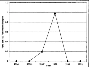
FIGURE 1 . Rate of Pseudomonas aeruginosa bloodstream infections among patients in the neonatal intensive care unit according to year. •Isolate not available for genotyping.
type of venous or arterial access, antimicrobial use, nutri-tion, medical interventions, and exposure to individual HCWs. Case-patients were considered exposed to a poten-tial risk factor if the exposure occurred prior to the onset of infection and matched control-patients were considered exposed to a potential risk factor if exposed during a com-parable number of ICU-days.
Data were collected retrospectively by review of patient charts and patient care assignment records using standardized forms and were transferred to Epi-Info soft-ware (version 6.04; Centers for Disease Control and Prevention, Atlanta, GA). Risk factors for P. aeruginosa BSI were determined by Mantel-Haenszel summary chi-square tests for matched set analysis and univariable con-ditional logistic regression analysis using STATA software (version 6.0; STATA Corp., College Station, TX).
R E S U L T S
Outbreak Description and Case Finding During a 6-week period in July and August 1997, four patients in the NICU died of P. aeruginosa infection. All four experienced P. aeruginosa pneumonia with sec-ondary BSI. Only two cases of P. aeruginosa BSI had occurred in the NICU during the 3 years prior to July 1997 (Fig. 1). Based on the lack of positive P. aeruginosa BSI prior to July 1996, the analysis was limited to July 1996 through September 1997.
The review of microbiological records from the lat-ter period revealed a total of 16 patients in the NICU with any culture positive for P. aeruginosa. Excluding the four case-patients, there were two neonates with BSI, two with eye infection, one with wound infection, and seven with respiratory or gastrointestinal colonization. The microbi-ology laboratory routinely saves only organisms isolated from sterile body fluids, so the isolates from the patients with eye, wound, respiratory, or gastrointestinal sources were not available for PFGE. However, the P. aeruginosa
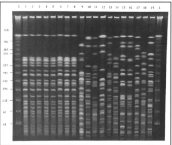
FIGURE 2. Results of pulsed-field gel electrophoresis of Pseudomonas aeruginosa strains isolated from neonates, healthcare workers (HCWs),
and environmental samples. Lane X = molecular-size ladder; lane 1 = blood from case-patient 4; lane 2 = blood from case-patient 3; lane 3 = blood from case-patient 2; lane 4 = blood from case-patient 1; lane 5 = blood from case-patient 5; lane 6 = handwash from HCW 2; lane 7 = ear swab from HCW 2; lane 8 = ventilator flow sensor; lane 9 = sink drain 1; lane 10 = faucet blades from sink 2; lane 11 = sink drain 3; lane 12 = faucet sink 4; lane 13 = handwash from HCW 5; lane 14 = handwash from HCW 3; lane 15 = handwash from HCW 1; lane 16 = sputum from patient initially hypothesized to be the source of the P. aeruginosa; lane 17 = rectal isolate from surveillance culture of neonatal intensive care unit (NICU) patient 1; lane 18 = rectal isolate from surveillance culture of NICU patient 2; and lane 19 = rectal isolate from surveillance culture of NICU patient 3.
isolate from the colonized, long-term patient hypothesized to be the reservoir for the first two cases of the outbreak was available and was submitted for PFGE.
The two additional cases of P. aeruginosa BSI that were identified by case finding occurred in July 1996 and February 1997. The patient from July 1996 underwent autopsy and had extensive necrotizing pneumonia with a blood culture positive for P. aeruginosa; however, this iso-late was not available for genotyping. The patient from February 1997 had a blood culture positive for P. aerugi-nosa without evidence of pneumonia and was included as a case-patient in our analysis because the isolate was tical to the outbreak genotype. No other cases were iden-tified by review of unit mortality records. Information regarding the five definitive case-patients is summarized in Table 1.
Microbiological Investigation of Patients and the Environment
The five available case isolates had the same antibi-otic susceptibility profile, typical of a community isolate (none was resistant to tobramycin, piperacillin, cef-tazadime, or imipenem). By PFGE, the five case isolates were identical (Fig. 2). The clinical isolate from the colonized patient originally thought to be the source of the outbreak (n = 1), the isolates obtained from patient sur-veillance cultures (n = 3), and the bloodstream isolates
from patients in other parts of the hospital (n = 13) were not related to the outbreak strain or to each other.
Seven (4%) of 190 environmental cultures were pos-itive for P. aeruginosa. Genotypically unrelated P. aerugi-nosa isolates were obtained from sink handles (n = 2) and sink drains (n = 4). A single culture sample from a wet ventilator flow sensor obtained during reprocessing, which had been cleaned but not disinfected, was positive for the outbreak genotype (Fig. 2).
Microbiological Investigation of HCWs
A total of 327 hand cultures were performed for 178 individual HCWs. Hand cultures of 5 (3%) of the 178 HCWs were repeatedly positive for P. aeruginosa, with colony counts ranging from 4 to more than 3,000 colonies per milliliter. Genotyping by PFGE was performed simul-taneously on all P. aeruginosa isolates of HCWs and the isolates from the five case-patients. The specimens from the hands of HCWs 1, 3, 4, and 5 were unrelated to the outbreak strain or to each other (Fig. 2). Samples for cul-tures collected from other body sites of HCWs 1, 3, 4, and 5 during the physical examination were negative for P. aeruginosa.
The initial hand culture from HCW 2 yielded 4 colonies per milliliter of P. aeruginosa. Three hand cul-tures performed during a 7-day period for HCW 2 yielded a range of 4 to 14 colonies per milliliter otP. aeruginosa. In addition, an external ear culture sample, obtained during the physical examination, yielded a moderate amount of/? aeruginosa with no clinical evidence of infection. Subsequent ear cultures were negative for P. aeruginosa. All case-patient isolates were identical to the P. aeruginosa isolates from the hand and external ear cultures of HCW 2 by PFGE (Fig. 2). Samples collected from other body sites of HCW 2 were negative for P. aeruginosa.
Epidemiologic Methods and Statistical Analysis The case-control study of five case-patients with P. aeruginosa BSI and 15 control-patients matched on birth weight and duration of NICU exposure revealed no signif-icant associations except a likely association with HCW 2. Four of 5 case-patients versus 5 of 15 matched control-patients had been cared for by HCW 2 (P = .05; Table 2). In addition, the mean days of exposure to ventilator flow sensors tended to be higher (P = .08) in case-patients than in control-patients (Table 2).
Infection Control Measures
Infection control measures included cohorting of staff and colonized or infected patients, reassignment of HCWs with positive results for P. aeruginosa (n = 5) to non-clinical activities, and implementation of contact pre-cautions for all patients infected or colonized with P. aeruginosa. Empiric antibiotic coverage of patients with sepsis of unknown origin was changed from ampicillin and gentamicin to piperacillin and tobramycin for optimal coverage of P. aeruginosa. In addition to a thorough review of patient care practices, handwashing compliance
TABLE 2
EPIDEMIOLOGIC CHARACTERISTICS OE
Characteristic
Mean birth weight, g (± SD)f Mean gestational age, wk (± SD) Exposure factors"
Surgery
Umbilical catheter Peripheral catheter Central venous catheter Arterial catheter Nasogastric tube Mechanical ventilation Mean days exposed to ventilator
flow sensors (± SD) Breastfeeding Chest tube
Ampicillin and gentamicin Cefazolin Vancomycin Clindamycin Exposure to HCW 2 PATIENTS W I T H AND Case-Patients (n = 5) 1,460 (± 290) 30.4 (± 2.2) 2 4 2 0 2 5 5 5.0 (± 1.6) 2 2 4 1 2 1 4
W I T H O U T PSEUDOMONAS AERUGINOSA BACTEREMIA* Control-Patients (n = 1,530 (± 31.5 ( 7 7 4 3 8 12 12 1.8 ( 10 2 15 1 3 2 5 15) 160) ±1.1) ±0.7) OR* <c95> 0.99 (0.98 to 1.01) 0.65 (0.31 to 1.36) 0.79 (0.11 to 5.43) 4.57 (0.34 to 60.86) 1.83 (0.20 to 16.49) Undefined 0.58 (0.07 to 4.88) Undefined Undefined 1.77 (0.93 to 3.37) 0.23 (0.1 to 2.68) 4.37 (0.37 to 51.24) 0.33 (<*> to 12.99) 3.0 (0.19 to 47.97) 3.56 (0.27 to 47.27) 1.73 (0.1 to 30.76) Undefined 1* >.2 >.2 >.2 >.2 >.2 >.2 >.2 >.2 >.2 .08 >.2 >.2 >.2 >.2 >.2 >.2 .05
OR = odds ratio; CI9S = 95% confidence interval; SD - standard deviation; HCW = healthcare worker. *Matched case-control study, univariable analysis.
This variable was used for the matching process.
*For continuous variables, the OR represents the odds of acquiring R aeruginosa bacteremia for each 1-unit (age or days exposed to a flow sensor) or 100-unit (weight) increment in the exposure vari-able of interest. An undefined OR is due to insufficient strata to calculate the effect estimate.
Calculated by Mantel-Haenszel summary chi-square tests. ''Exposure factors for cases prior to bacteremia due to R aeruginosa.
was observed and an alcohol-based handrub was intro-duced. Wall-mounted, previously opened soaps and lotions were removed and replaced with new, previously unopened stock. Personal hand lotions were removed from the unit. Pharmacy admixing and dosing practices were reviewed, but no changes were necessary. Discrepancies with recommended high-level disinfection policies for ventilator flow sensors were addressed. After the occurrence of the fourth case of fatal P. aeruginosa BSI, the NICU was closed to new admissions until com-pletion of the investigation.
No cases occurred following reassignment of the HCWs with positive results for P. aeruginosa to non-clini-cal duties. HCWs with positive results for P. aeruginosa performed a handwashing regimen of a 1-minute wash with a 2% chlorhexidine gluconate antimicrobial product hourly during waking hours for a 5-day period. HCWs 3, 4, and 5 had no evidence of nail infection and returned to clinical duties approximately 1 month after their initial positive handwashing culture, when it was determined that their isolates did not match the outbreak strain and repeat hand cultures were negative for P. aeruginosa. Eradication of P. aeruginosa was confirmed by two nega-tive hand cultures performed after completion of the handwashing regimen.
The P. aeruginosa isolate from HCW 1 was not relat-ed to the outbreak strain (Fig. 2). During the physical examination in Occupational Health Services, HCW 1 noted that treatment for onychomycosis had recently been completed as prescribed by a personal dermatolo-gist. HCW 1 was treated with a 14-day course of ciprofloxacin, performed the handwashing regimen and follow-up testing described above, and returned to clinical duties 7 weeks after the initial positive culture.
After identification of the probable source of the outbreak, the otitis externa of the implicated HCW (HCW 2) was treated with a 5-day course of combination steroid and antibiotic otic solution (hydrocortisone, neomycin sulfate, and polymyxin B). Clearance oiP aeruginosa from the hands was documented by three negative handwash-ing cultures performed durhandwash-ing a 1-month period, follow-ing completion of the previously described handwashfollow-ing regimen. To verify eradication of P. aeruginosa ear colo-nization, cultures of both ears were performed every 2 weeks for a 2-month period, followed by two additional sets 2 and 6 months later. HCW 2 resumed clinical duties 3 months after the initial positive handwashing culture was identified, when 3 negative hand cultures and 2 nega-tive ear cultures were performed after the completion of treatment.
DISCUSSION
The identification of a cluster of four patients with rapidly fatal P. aeruginosa infection prompted this out-break investigation. The third and fourth cases occurred despite comprehensive traditional outbreak control mea-sures, so the unit was closed for new admissions while the investigation continued. Using a combination of microbio-logical and epidemiologic methods, we identified a HCW with intermittent otitis externa as the most likely source for this cluster of infections. Although clinical evidence of otitis externa was absent during the initial physical exam-ination, the HCW was a swimmer and recalled bouts of intermittent otitis externa prior to and during the out-break. Although the mechanism of transmission cannot be definitively ascertained, it is plausible that P. aerugi-nosa could have been spread from ear to hand to patient. The outbreak terminated after the implicated HCW was removed from clinical duties, treated, and cleared of P. aeruginosa carriage. No further cases of P. aeruginosa infection or colonization with the implicated genotype have occurred.
An alternative hypothesis focused on contaminated respiratory care equipment as the potential source of infection. Although a problematic disinfection practice was identified, the appropriate high-level disinfection process was reimplemented 2 weeks prior to the occur-rence of the fourth case. In addition, of five flow sensors cultured during various stages of the discrepant disinfec-tion process, only one sensor was positive for the out-break strain and no cases occurred in two other ICUs that used the same ventilator sensors.
Neonatal infections due to P. aeruginosa include pneumonia, BSI, meningitis, conjunctivitis, and surgical-site, urinary tract, and gastrointestinal infection.1219 Although less common than some other causes of neona-tal infection,20 P. aeruginosa sepsis is particularly virulent in this patient population, with case-fatality rates exceed-ing 50%.2 Although sporadic cases do occur, most pub-lished reports have focused on outbreaks of P. aerugi-nosa}1 However, P. aeruginosa outbreaks have rarely been attributed to hand carriage of HCWs. Recently, two NICU outbreak investigations have associated nosocomial P. aeruginosa infection with hand carriage of HCWs.1012 Moolenaar et al.10 described an outbreak oiP. aeruginosa BSI and endotracheal tube colonization in a NICU and determined that exposure to two particular nurses was associated with acquiring P. aeruginosa. Interestingly, the genetic and environmental evidence found in this investi-gation suggested a possible role for long natural or artifi-cial fingernails in the colonization of the implicated HCWs' hands with P. aeruginosa. Foca et al.12 conducted an epidemiologic and molecular investigation of P. aerugi-nosa infection or colonization in their NICU. Cultures per-formed from environmental sources were negative, but cultures of the hands of 10 (6%) of 165 HCWs were posi-tive for P. aeruginosa. Significant risk factors for coloniza-tion included the use of artificial nails or nail wraps. However, only one HCW with Candida species
ony-chomycosis was persistently colonized with a P. aerugi-nosa strain that was identical to the predominant clone. All other P. aeruginosa isolates of HCWs were unrelated to the predominant clone or to other patient clones identi-fied.
In our study, a total of five HCWs were at least tran-siently colonized with P. aeruginosa strains, a finding pre-viously described.22 Because the initial outbreak control measures included reinforcement of good hand hygiene practices, our primary intention in requesting hand cul-tures of a variety of HCWs was to focus attention on the hands as a possible means of patient-to-patient indirect transmission. Although we knew from previous studies performed in our NICU that HCWs' hands may be colo-nized with gram-negative bacteria,23 we were surprised to find five HCWs with hand cultures positive for P. aerugi-nosa. Of these five HCWs, the implicated HCW (HCW 2) had the lowest colony count from the initial hand culture (4 colonies per milliliter), with a moderate amount of P. aeruginosa from the culture of the external ear, support-ing the theory of transient hand carriage with transmis-sion by indirect contact from the ear to hand to patient. The range of colony counts for the remaining four HCWs with positive results for P. aeruginosa was 930 to more than 3,000 colonies per milliliter. Of note, one HCW had previously received treatment for onychomycosis and was colonized with a non-outbreak strain. This particular strain differed by one band from the strain from the long-term, ventilator-dependent, colonized patient initially hypothesized to be the reservoir for the initial two cases inJuly(Fig.2).
Although culturing of HCWs is generally not per-formed in the early stages of an outbreak investigation, we elected to initiate hand cultures prior to the case-con-trol study. In our experience, hand cultures may have pro-vided a more rapid identification of a possible source of the outbreak, and reassignment of staff with positive results for P. aeruginosa to non-clinical duties may have avoided additional cases while the investigation contin-ued. Early HCW screening may also identify individuals with onychomycosis, dermatitis, or other conditions that might increase the risk of chronic hand colonization. However, it is not unusual for HCWs to have transient hand colonization with nosocomial pathogens, and aggressive culturing could lead to unnecessary exclusion of HCWs from clinical care and considerable anxiety.
Our investigation had limitations. First, case-patients may have been missed. Although the retrospec-tive review of the microbiological database from July 1996 through September 1997 revealed 16 patients with clinical cultures positive for P. aeruginosa, the laboratory routine-ly saved onroutine-ly isolates from sterile body sites. The blood-stream isolates were available, but isolates from 10 patients with P. aeruginosa from the eyes, wounds, and respiratory or gastrointestinal tracts were unavailable for molecular typing. In addition, the patient from July 1996 had a clinical course similar to those of the four case-patients who died, but the blood culture positive for P.
aeruginosa was of an autopsy specimen. Autopsy samples also were not routinely saved in the laboratory, so an addi-tional case-patient may have been missed.
Second, we must acknowledge that the small sam-ple size of the case-control study limited the precision of the matched set analysis, yielding large 95% confidence intervals. However, despite the limitations of such a small data set, the disappearance of the epidemic strain from the unit after removal and treatment of the implicated HCW strongly suggests that this HCW may have been the primary source of the epidemic.
Finally, although a significant association was demonstrated between the exposure of 4 of the 5 case-patients and 5 of the 15 control-case-patients to HCW 2 (P = .05), the relationship may have been attenuated by our definition of exposure. For our investigation, exposure was defined as being assigned the primary care of the patient, or participating in the initial transport of the patient to our facility. Although HCW 2 was not assigned the primary care or the transport of one of the five case-patients, HCW 2 had been assigned to a patient in the bed adjacent to the nonexposed case-patient. It is plausible that exposure may have occurred during cross-coverage, or when assistance with patient care was provided.
We investigated an outbreak of fatal neonatal pneu-monia and BSI caused by a single strain of P. aeruginosa. With the help of hand cultures and molecular and epi-demiologic analyses, we linked one HCW with intermit-tent otitis externa to the outbreak. Treatment and removal of the implicated HCW ended the outbreak. These find-ings demonstrate the hazards of infected or colonized HCWs in the NICU setting and the challenges related to rapid detection of asymptomatic carriage of potential pathogens. Detecting and eradicating HCWs' infection or colonization with potential pathogens may reduce the risk of life-threatening cross-infections.
R E F E R E N C E S
1. Gaynes RP, Edwards JR, Jarvis WR, et al. Nosocomial infections among neonates in high-risk nurseries in the United States: National Nosocomial Infections Surveillance System. Pediatrics 1996;98:357-361.
2. Leigh L, Stoll BJ, Rahman M, et al. Pseudomonas aeruginosa infection in very low birth weight infants: a case-control study. Pediatr Infect Dis
J 1995;14:367-371.
3. Baird RM, Shooter RA. Pseudomonas aeruginosa infections associated with use of contaminated medicaments. Br Med J 1976;2:349-350.
4. Archibald LK, Ramos M, Arduino MJ, et al. Enterobacter cloacae and
Pseudomonas aeruginosa polymicrobial bloodstream infections traced
to extrinsic contamination of a dextrose multidose vial. / Pediatr 1998;133:640-644.
5. Cobben NA, Drent M, Jonkers M, et al. Outbreak of severe
Pseudomonas aeruginosa respiratory infections due to contaminated
nebulizers. J Hosp Infect 1996;33:63-70.
6. Becks VE, Lorenzoni NM. Pseudomonas aeruginosa outbreak in a neonatal intensive care unit: a possible link to contaminated hand lotion. Am J Infect Control 1995;23:396-398.
7. Neal TJ, Hughes CR, Rothburn MM, et al. The neonatal laryngoscope as a potential source of cross-infection. / Hosp Infect 1995;30:315-317. 8. Garland SM, Mackay S, Tabrizi S, et al. Pseudomonas aeruginosa out-break associated with a contaminated blood-gas analyser in a neonatal intensive care unit. J Hosp Infect 1996;33:145-151.
9. Thom AR, Cole AP, Watrasiewicz K. Pseudomonas aeruginosa infection in a neonatal nursery, possibly transmitted by a breast-milk pump.
Lancet 1970;1:560-561.
10. Moolenaar RL, Crutcher JM, San Joaquin VH, et al. A prolonged out-break of Pseudomonas aeruginosa in a neonatal intensive care unit: did staff fingernails play a role in disease transmission? Infect Control Hosp
Epidemiol 2000;21:80-85.
11. Widmer AF, Wenzel RP, Trilla A, et al. Outbreak of Pseudomonas
aeru-ginosa infections in a surgical intensive care unit: probable
transmis-sion via hands of a health care worker. Clin Infect Dis 1993;16:372-376. 12. Foca M, Jakob K, Whittier S, et al. Endemic Pseudomonas aeruginosa
infection in a neonatal intensive care unit. N Engl J Med 2000;343:695-700.
13. Mermel LA, McKay M, Dempsey J, et al. Pseudomonas surgical-site infections linked to a healthcare worker with onychomycosis. Infect
Control Hosp Epidemiol 2003;24:749-752.
14. Grundmann H, Kropec A, Hartung D, et al. Pseudomonas aeruginosa in a neonatal intensive care unit: reservoirs and ecology of the nosoco-mial pathogen. J Infect Dis 1993;168:943-947.
15. Larson EL, Strom MS, Evans CA. Analysis of three variables in sam-pling solutions used to assay bacteria of hands: type of solution, use of antiseptic neutralizers, and solution temperature. / Clin Microbiol 1980;12:355-360.
16. Larson EL, Hughes CA, Pyrek JD, et al. Changes in bacterial flora associated with skin damage on hands of health care personnel. Am J
Infect Control 1998;26:513-521.
17. Isenberg HD. Epidemiologic and infection control microbiology. In: Gilchrist MJR, ed. Clinical Microbiology Procedures Handbook, vol. 2. Washington, DC: American Society for Microbiology; 1992. 18. Talon D, Cailleaux V, Thouverez M, et al. Discriminatory power and
usefulness of pulsed-field gel electrophoresis in epidemiological stud-ies of Pseudomonas aeruginosa. ] Hosp Infect 1996;32:135-145. 19. Richards MJ, Edwards JR, Culver DH, et al. Nosocomial infections in
pediatric intensive care units in the United States: National Nosocomial Infections Surveillance System. Pediatrics 1999;103:e39. 20. Holzel H, de Saxe M. Septicaemia in paediatric intensive-care patients
at the Hospital for Sick Children, Great Ormond Street. / Hosp Infect 1992;22:185-195.
21. Damjanovic V, van Saene HK Outbreaks of infection in neonatal inten-sive care units (NICUs). / Hosp Infect 1997;35:237-244.
22. Knittle MA, Eitzman DV, Baer H. Role of hand contamination of per-sonnel in the epidemiology of gram-negative nosocomial infections. /
Pediatr 1975;86:433-437.
23. Goldmann DA, Leclair J, Macone A. Bacterial colonization of neonates admitted to an intensive care environment. / Pediatr 1978;93:288-293.