HAL Id: hal-00529325
https://hal.archives-ouvertes.fr/hal-00529325
Submitted on 1 Jun 2020
HAL is a multi-disciplinary open access
archive for the deposit and dissemination of
sci-entific research documents, whether they are
pub-lished or not. The documents may come from
teaching and research institutions in France or
abroad, or from public or private research centers.
L’archive ouverte pluridisciplinaire HAL, est
destinée au dépôt et à la diffusion de documents
scientifiques de niveau recherche, publiés ou non,
émanant des établissements d’enseignement et de
recherche français ou étrangers, des laboratoires
publics ou privés.
Copyright
Extracellular DsbA-insensitive folding of Escherichia coli
heat-stable enterotoxin STa in vitro.
I. Batisson, M. Der Vartanian
To cite this version:
I. Batisson, M. Der Vartanian. Extracellular DsbA-insensitive folding of Escherichia coli heat-stable
enterotoxin STa in vitro.. Journal of Biological Chemistry, American Society for Biochemistry and
Molecular Biology, 2000, 275 (14), pp.10582-9. �hal-00529325�
Extracellular DsbA-insensitive Folding of Escherichia coli
Heat-stable Enterotoxin STa in Vitro*
(Received for publication, August 13, 1999, and in revised form, December 13, 1999) Isabelle Batisson and Maurice Der Vartanian‡
From the Laboratoire de Microbiologie, Institut National de la Recherche Agronomique, Centre de Recherches de Clermont-Ferrand-Theix, 63122 Saint-Gene`s-Champanelle, France
To study the folding of human Escherichia coli heat-stable enterotoxin STh, we used the major protein sub-unit of CS31A fimbriae (ClpG) as a marker of STh secre-tion and a provider of a signal peptide. We established that STh genetically fused to the N or C terminus of ClpG was able to mobilize ClpG to the culture superna-tant while still retaining full enterotoxicity. These fea-tures indicate that the STh activity was not altered by the chimeric structure and suggest that spatial confor-mation of STh in the fusion is close to that of the native toxin, thus permitting recognition and activation of the intestinal STh receptor in vivo. In contrast to other stud-ies, we showed that disulfide bond formation did not occur in the periplasm through the DsbA pathway and that there was no correlation between DsbA and secre-tion, folding, or activity. This discrepancy was not at-tributable to the chimeric nature of STh since there was no effect of dsbA or dsbB mutations on secretion and activity of recombinant STh from which ClpG had been deleted. Periplasmic and lysate fractions of dsbAⴙand dsbAⴚcells did not have any STh activity. In addition, the STh chimera was exclusively found in an inactive reduced form intracellularly and in an active oxidized form extracellularly, irrespective of the dsbA back-ground. Subsequently, a time course experiment in re-gard to the secretion of STh from both dsbAⴙand dsbAⴚ cells indicated that the enterotoxin activity (proper folding) in the extracellular milieu increased with time. Overall, these findings provide evidence that STa toxins can be cell-released in an unfolded state before being completely disulfide-bonded outside the cell.
Escherichia coli heat-stable enterotoxin STa (also referred to
as STI) is responsible for serious and sometimes fatal diarrheal disease in both humans and domestic animals (1). STa causes intestinal fluid secretion by binding to its cellular receptor, transmembrane guanylyl cyclase C (2), thus activating guany-lyl cyclase C and leading to an increase in intracellular mucosal cGMP. This increase in cGMP adversely affects electrolyte flux in the intestine; sodium absorption is inhibited, and chloride secretion is stimulated. These ion flux changes result in the secretory diarrhea characteristic of enterotoxigenic E. coli in-fection (3, 4). The receptor binding and enterotoxic properties of STa have been mapped to a highly conserved 13-amino acid
C-terminal core sequence essential for toxicity and the heat-stable nature of the toxin. Six cysteine residues are present within this domain, and the three intramolecular disulfide bonds formed between the cysteine residues are necessary for toxicity of the molecule (5). The two species of STa, STp and STh, from porcine (or bovine) and human strains of enterotoxi-genic E. coli, respectively, are typical extracellular peptides consisting of 18 (STp) or 19 (STh) amino acid residues (6, 7). STa is synthesized in the cytoplasm as a 72-amino acid precur-sor consisting of pre-, pro-, and mature STa regions (8, 9). The precursor is translocated across the inner membrane by the Sec-dependent export pathway (10); the 19-amino acid leader peptide is removed by signal peptidase; and the generated 53-amino acid pro-STa intermediate is delivered to the periplasm. The natural pre- and prosequences are dispensable for STa secretion (8). Mature STa without the prosequence may be able to gain access to the extracellular milieu upon its entry into the E. coli periplasm, once guided into this compartment by a heterologous periplasmic leader peptide (11). Conflicting observations have been reported on the mechanism of secretion of the toxin from the periplasm to the outside of the cell, making this mechanism poorly understood. Indeed, some au-thors found that the pro-STa region is cleaved in the periplas-mic space, where the thiol-disulfide oxidoreductase DsbA (14) directly catalyzes the formation of the disulfide bonds of STa, and that dsbA mutation causes a marked reduction in STa activity (12, 13). In contrast, other investigators hypothesized that pro-STa can exit to the extracellular milieu and that STa is disulfide-bonded outside the cell, making the role of DsbA in STa folding unclear (9, 15). Such disagreement may be ex-plained by the small size of the STa molecule and the escape velocity with which it is released into the extracellular milieu and thus by the difficulty in detecting and quantifying the intermediates in the different cellular compartments. In addi-tion, STa is poorly antigenic and not immunogenic and reacts unexpectedly in conventional protein treatments such as stain-ing, trichloroacetic acid precipitation, and electrophoresis (16), thus limiting progress in the elucidation of the STa secretion pathway. For these reasons, a number of attempts to develop genetic fusions between STa and heterologous carrier proteins have been made (11). Unfortunately, in most cases, the result-ing chimeras were periplasmic or membrane-associated, and no STa activity was detected in the extracellular milieu. In con-trast, in this work, we have shown that fusions containing various STa peptides at the N or C terminus of ClpG, the major protein subunit of E. coli CS31A fimbriae (17), result in the extracellular secretion of ClpG-STa hybrids with high entero-toxin activity. We used ClpG as a marker to facilitate STa detection in secretion and folding studies. We analyzed the effects of dsbA and dsbB mutations on the secretion and ente-rotoxicity of STa chimeras and determined the redox state of
STa, both inside and outside of dsbA⫹and dsbA⫺E. coli cells.
* This work was supported by the Conseil Re´gional Auvergne (Cler-mont-Ferrand, France) and the Institut National de la Recherche Agronomique (Paris, France). The costs of publication of this article were defrayed in part by the payment of page charges. This article must therefore be hereby marked “advertisement” in accordance with 18 U.S.C. Section 1734 solely to indicate this fact.
‡ To whom correspondence should be addressed. Tel.: 33-0473624243; Fax: 33-473624581; E-mail: dvartan@clermont.inra.fr.
© 2000 by The American Society for Biochemistry and Molecular Biology, Inc. Printed in U.S.A.
This paper is available on line at http://www.jbc.org
10582
at INRA Institut National de la Recherche Agronomique on June 18, 2018
http://www.jbc.org/
Disulfide bond formation during translocation of STa across the outer membrane was also examined.
EXPERIMENTAL PROCEDURES
Bacterial Strains, Plasmids, Oligonucleotides, and Media—The
bac-terial strains and plasmids used in this study are presented in Table I. Bacteria were grown at 37 °C in LB broth or LB agar with or without
the following antibiotics: ampicillin, 50 g/ml; chloramphenicol, 25
g/ml; kanamycin, 50 g/ml; and tetracycline, 12.5 g/ml.
clpG::STh Fusion Genes: Cloning Strategies, Constructions, and Fea-tures—We made a variety of constructs to increase our chances of
success in producing chimeric proteins with enterotoxigenic activity (see Fig. 1). Plasmid pHPCO838 was constructed from pDEV41155 (20) by in vitro site-directed mutagenesis (23) using mutagenic and selection
primers. The former primer (5
⬘-CGTGGCAGTAACTTATGTTAACTA-ATTGGCTTGA-3⬘) created a unique HpaI site and added a valine between the penultimate tyrosine and the last C-terminal residue (as-paragine) of ClpG. The latter primer
(5⬘-CGCAGGAAAGAAGATCT-GAGCAAAAGGCG-3⬘) mutated the single AflIII restriction site of the
pSK⫹plasmid vector into a single BglII site. Plasmid pSTN24 was
engineered from pHPCO838 as follows. A synthetic double-stranded DNA (the 5⬘ 3 3⬘ single coding strand was CGGCAGATCTGTACTGC- TGTGAACTTTGTTGTAATCCTGCCTGTACAGGATGTTACCCTGCA-GATCCTCATG) encoding the last 15 amino acids of mature STh and containing SphI-flanked ends was inserted in frame in the correct orientation into the SphI site of pHPCO838; the initial signal peptidase
cleavage sequence (⫺3AHA⫺12⫹1WT⫹2) was changed to an improved
processing site (⫺3AHA⫺12⫹1AD⫹2) based on the predictive accuracy
estimation of von Heijne (24). Plasmids pSTDG126 and pSPGST1, expressing the last 15 amino acids of mature STh and the entire 19-amino acid sequence of mature STh, respectively, were constructed after insertion of paired oligonucleotides (the two 5⬘ 3 3⬘ single coding strands were CGGCAGATCTGTACTGCTGTGAACTTTGTTGTAATC-CTGCCTGTACAGGATGTTACTAAGTT and CGAATTCTTCTAACTA- CTGCTGTGAACTTTGTTGTAATCCTGCCTGTACAGGATGTTACTA-AGTT, respectively) between the SphI and HpaI sites of pHPCO838. Plasmid pSTC22 was constructed by cloning a synthetic DNA duplex (the 5⬘ 3 3⬘ single coding strand was AACCCTGATAACCCCGGGAA-CTACTGCTGTGAACT TTGTTGTAATCCTGCCTGTACAGGATGTTA-CTAA) encoding the last 16 amino acids of mature STh and containing
HpaI and SmaI sites between the HpaI and XbaI sites of pHPCO838.
Plasmid pSTC17 was pSTC22 with the HpaI-SmaI fragment deleted, thus placing a glycine precisely at the ClpG-STh junction. Plasmid pProSTC28 was made by inserting a blunt-ended DNA fragment (the 5⬘ 3 3⬘ single coding strand was AACAAAAGTGGTCCTGAATCGATGA-ATTCTAGC) encoding amino acid residues 46 –53 (NKSGPESM) of the pro-STh peptide plus the first three residues (NSS) of mature STh between the HpaI and SmaI sites of pSTC22. We hypothesized that this natural linker might favor folding of the STh moiety in the hybrid in a more effective way. To obtain plasmid pEHProSTC28, the
MunI-Ecl136II fragment of pProSTC28 containing the clpG::STh fusion gene
was subcloned in the low-copy number plasmid pEH524 within the clp operon in place of the MunI-HpaI fragment carrying native clpG. This
same fusion gene was IPTG1-overexpressed by cloning the KpnI-BglII
fragment of pProSTC28 downstream of the Ptrcpromoter between the
KpnI and BamHI sites of pTrc99A, resulting in pTrcProSTC28. A
syn-thetic gene (the 5⬘ 3 3⬘ single coding strand was
CGGTTAACATGAAT-TCTGCAGTT) encoding the last 16 amino acids of mature STh and containing an HpaI site was placed in frame between the SphI and
HpaI sites of pSTC22, and the resulting construct was either HpaI/ HpaI-deleted, leading to pGMSTC71, or HpaI/SmaI-deleted, leading to
pGMSTC710. The same DNA fragment was also introduced between the SphI and HpaI sites of pProSTC28, and the resulting plasmid was
HpaI/HpaI-deleted, leading to pGMSTC13/19.
All other standard techniques were performed essentially as de-scribed previously (25). The oligonucleotides used in this study were supplied by Eurogentec (Belgium). Fusion genes were checked by se-quencing (26).
Production of Fusion Proteins—LB broth preculture (1 or 2 ml)
containing exponentially growing cells was poured onto LB agar plates, which were incubated overnight at 37 °C in a humid atmosphere with the agar surface facing up. Bacteria were carefully harvested by
scrap-ing from the agar surface. After centrifugation at 12,000⫻ g for 10 min,
the resulting supernatant was separated from the cell pellet and used as the solid culture supernatant fraction. Supernatants from LB broth cultures were obtained after centrifugation and used as the liquid culture supernatant fraction. The bacterial enumeration expressed in colony-forming units/ml was done by spreading out dilutions of PBS-suspended cells on MacConkey lactose agar medium containing the appropriate antibiotic.
Competitive ELISA—Competitive ELISA using a commercially
avail-able assay kit for E. coli STa (COLI ST EIA, Denka Seiken Co., Ltd., Tokyo, Japan) (27) was performed to detect and quantify fusion proteins in culture supernatants. The reactivity of the STa moiety of hybrids was determined using STa-monospecific horseradish peroxidase-conjugated antibodies and ELISA plates coated with synthetic STa peptide. After
the wells were washed once with the supplied buffer, 200l of serially
diluted samples and 10 l of conjugated monoclonal antibody were
added. After incubation for 90 min at room temperature, the contents were removed, and the wells were washed five times with buffer.
Enzyme substrate solution (100l), prepared by adding H2O2solution
to o-phenylenediamine, was added, and the plate was left in the dark at room temperature for 30 min. After the reaction was stopped by
addi-tion of 100l of 1.5NH2SO4, the absorbance at 490 nm was measured
using a Dynatech MR5000 microplate reader. A positive sample was identified by inhibition of binding of horseradish peroxidase-conjugated monoclonal antibodies to the well, as demonstrated by a decrease in
A490.The positive samples were defined as giving A490⬍ 0.2 (as this is
a competitive assay), and the negative samples were defined as giving
A490 ⱖ 1.2. The concentration of STa in culture supernatants was
determined by reference to a standard curve, which was constructed using known concentrations of purified native STp (Calbiochem)
rang-ing from 0 to 1.25g/ml.
Double Antibody Sandwich ELISA—Double antibody sandwich
ELISA was performed for determination of the amount of ClpG in supernatant samples. Microtiter plates (Immulon 2, Dynatech
Labora-tories Inc.) were coated by overnight incubation at 4 °C with 100l of
rabbit anti-ClpG antiserum (28) diluted (1:200) in 50 mMcarbonate
buffer (pH 9.6). After removal of the buffer, the plates were incubated overnight at 4 °C with blocking buffer (PBS, 2% dry milk, and 1% fetal calf serum) and washed twice with PBS and 0.05% Tween 20 and once with PBS. The supernatant samples were serially plated in 2-fold
dilutions (100l/well) in antibody buffer (PBS, 0.2% dry milk, and 0.5%
fetal calf serum) and incubated for 2 h at 37 °C. For the standard curve, known concentrations of purified ClpG in PBS were serially plated in 2-fold dilutions in antibody buffer. Purification of native ClpG has been described elsewhere (28). After washing twice with PBS and 0.05%
Tween 20 and once with PBS, 100 l of a 1:500 dilution of mouse
anti-ClpG antiserum were added to each well, and the plates were
incubated for 1.5 h at 37 °C. After washing, 100l of a 1:1000 dilution
of goat anti-mouse IgG were added, and the plates were incubated for
2 h at 37 °C. After washing, 100 l of 2 nM H2O2 and 10 mg of
2,2⬘-azinobis(3-ethylbenzthiazoline-6-sulfonic acid) diammonium salt
(Sigma) substrate in 100 ml of citrate/phosphate buffer (80 mMcitric
acid and 125 mMNa2HPO4) were added, and the plates were incubated
for 20 min in the dark at room temperature. The absorbance at 405 nm was read on a Dynatech MR5000 reader. An overnight supernatant of
E. coli DH5␣ (pDSPH524) served as a negative control. In terms of
molecular mass, ClpG represents 93% of the hybrid protein. Therefore,
1g of ClpG in the fusion protein mixture is equivalent to ⬃1.1 g of
ClpG-STh hybrid.
Nonreducing SDS-Polyacrylamide Gel Electrophoresis and Immuno-blotting—Samples containing the hybrids were mixed with an equal
volume of 2⫻ Laemmli buffer without reducing agent, applied without
boiling to a 12% SDS-polyacrylamide gel, and semidry-electrotrans-ferred onto 0.2-m nitrocellulose paper (Bio-Rad) as described by Tow-bin et al. (29). Blots were blocked and washed with 1% bovine serum albumin and 0.1% Tween 20 in Tris-buffered saline and incubated overnight with rabbit ClpG-specific antiserum or with a mixture of the STa-specific monoclonal antibodies 11C (30) and 20C1 (31). For probing with anti-ClpG antibodies, filters were developed with horseradish peroxidase-labeled goat anti-rabbit secondary antibodies and with
H2O2/␣-chloronaphthol as a substrate. For probing with STa
anti-bodies, a 1:1000 dilution of biotinylated goat anti-mouse IgG (Sigma) in Tris-buffered saline and 0.05% Tween 20 was used as a secondary antibody. After incubation for 1 h and washing, membranes were again incubated for 30 min with a 1:100 dilution of horseradish
peroxidase-conjugated streptavidin (250g/ml; Sigma) in Tris-buffered saline and
0.05% Tween 20 and developed with H2O2/␣-chloronaphthol.
Assay for Enterotoxin Activity—The enterotoxin activity of
superna-1The abbreviations used are: IPTG,
isopropyl--D
-thiogalactopyrano-side; PBS, phosphate-buffered saline; ELISA, enzyme-linked immu-nosorbent assay; G/C, gut weight/carcass weight ratio; DTT,
dithiothre-itol; AMS, 4-acetamido-4⬘-maleimidylstilbene-2,2⬘-disulfonic acid.
Folding of E. coli Heat-stable Enterotoxin STa
10583
at INRA Institut National de la Recherche Agronomique on June 18, 2018
http://www.jbc.org/
tant fractions was examined in the suckling mouse assay as described previously (32). Three-day-old Swiss OF1 suckling mice were separated from their mothers immediately before use and randomly divided into groups. A 0.1-ml aliquot of each sample was directly delivered to the stomachs of infant mice using flexible plastic tubing. Three hours later, the entire intestine from each mouse was removed and weighed, and the ratio of the gut weight to remaining carcass weight (G/C) was calculated. The mean G/C ratio and the S.E. of several separated assays
were determined for each mouse batch. A G/C ratio ofⱖ0.090
corre-sponded to the unambiguous accumulation of fluid in the gut lumen. One mouse unit was defined as the enterotoxin activity corresponding
to a minimum effective dose that gave a positive response (G/C ⱖ
0.090), and the enterotoxin titer was expressed as the highest dilution that gave 1 mouse unit of enterotoxin activity. The G/C value was plotted semilogarithmically versus dilution of the sample fraction, and the point at which the curve crossed the line equal to a G/C ratio of 0.090 was defined as the dilution that corresponds to 1 mouse unit of STa. In our study, the minimum effective dose of STa necessary to produce an activity of 1 mouse unit was 8 ng, as determined using a standard curve, which was developed following intragastric inoculation of suckling mice with 0.1 ml of sterile PBS containing amounts of pure STp ranging from 0.2 to 100 ng (data not shown).
Redox State of STh in the Fusion Protein—The redox state of fusion
proteins was assessed as described previously (33). To determine the redox state of intracellular fusion proteins, bacteria were cultured in LB
broth at 37 °C with or without 5 mMdithiothreitol (DTT). Cultures (25
ml) were grown to A550⬃ 0.5 and centrifuged. Cell pellets were
sus-pended in PBS and mixed with an equal volume of 10% trichloroacetic acid in PBS, followed by incubation on ice for 1 h. For determination of the redox state of extracellular fusion proteins, supernatants were
treated or not with 100 mMDTT for 10 min at 37 °C before
trichloro-acetic acid treatment as described above. In all cases, precipitates of trichloroacetic acid-denatured proteins were pelleted by centrifugation and washed with cold acetone to remove trace acid. The samples were centrifuged, and the acetone supernatants were discarded. The result-ing pellets were air-dried and dissolved in a solution (freshly made)
containing 50 mMTris-HCl (pH 8.0), 1% SDS, 1 mMEDTA, and 10 mM
4-acetamido-4⬘-maleimidylstilbene-2,2⬘-disulfonic acid (AMS; Molecu-lar Probes, Inc.). Proteins were separated by 12% SDS-polyacrylamide gel electrophoresis without using any reducing agent. Reduced and oxidized forms of proteins were visualized on Western blots as described above. AMS reacts specifically and irreversibly with sulfhydryl groups, conjugating a large 490-Da moiety to the free cysteines (34). This major change in molecular mass is used to clearly separate oxidized from reduced proteins on SDS-polyacrylamide gels (33). Indeed, unlike oxi-dized forms, reduced forms, which are sensitive to reaction with AMS, shift to a high molecular mass protein position.
RESULTS
DsbA and DsbB Are Not Required for Secretion and
Entero-toxicity of STh Chimeras—Plasmids carrying various
clpG::STh fusion genes (Fig. 1) were introduced into E. coli
strains JCB570 (dsbA⫹dsbB⫹), JCB571 (dsbA⫺), JCB819
(dsbB⫺), and JCB818 (dsbA⫺dsbB⫺) (Table I). The
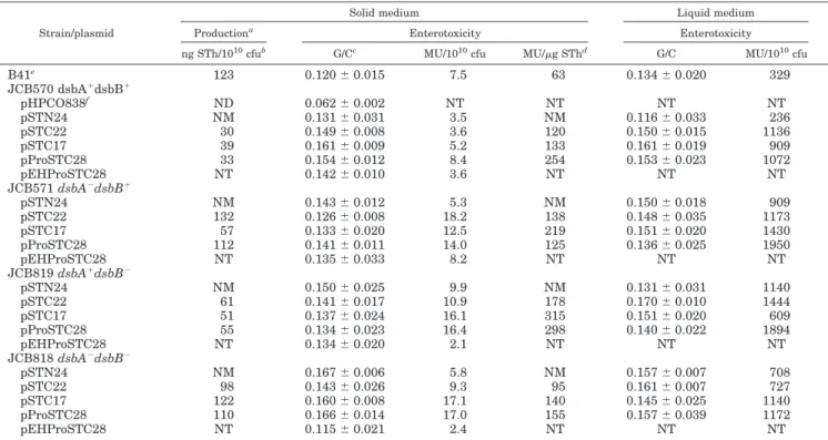
transfor-mants were grown overnight in LB broth or LB agar, and the supernatant fractions were analyzed for STh production by competitive ELISA and for STh activity by the suckling mouse assay. All STh chimeras were secreted with high biological
activity in solid and liquid media by both the dsb⫹wild-type
and dsb⫺ mutant cells (Table II). The percentage of positive
mice reached 100% for each of the recombinant plasmids tested in all mutant strains and in both solid and liquid media. Ac-tivity was 80 –170-fold higher in liquid medium than in solid
medium (data expressed in mouse units/1010 colony-forming
units in Table II), probably because of a better air oxygenation. In comparison to the wild-type strain, the production and
en-terotoxicity levels of the dsb⫺mutants were mostly improved.
Nevertheless, STh-specific activities (data expressed in mouse
units/g of STh in Table II) remained for the most part
un-changed, indicating a similar DsbA/DsbB-independent toxicity against these various fusion proteins. This is in contrast with other reports showing that the dsbA mutation caused a marked reduction in the secretion (13) and activity (12) of the native toxin. Therefore, questions remained regarding whether our results might be accounted for by the presence of ClpG or by a periplasmic leakage due to the high-copy number of the recom-binant plasmids.
First, we genetically deleted ClpG from ClpG-STh fusions
(Fig. 1). In all cases, the dsb⫹and dsb⫺strains secreted active
STh (Table III), indicating that STh free of ClpG reached the extracellular milieu simply upon its entry into the periplasm, once guided by the leader peptide of ClpG. However, the pro-duction and activity of STh were mostly slightly higher in the presence (Table II) than in the absence (Table III) of ClpG.
Second, we subcloned most of the clpG::STh fusion genes in a pSC101-related plasmid (Table I). Only results relative to the fusion protein carried by pProSTC28 (Fig. 1) are preferentially reported in the rest of our study, especially since all the ClpG-STh chimeras acted similarly. As shown in Table II, the ClpG-STh chimera expressed by the corresponding low-copy number re-combinant plasmid pEHProSTC28 was released as an active fusion protein whatever the dsb background. Therefore, the discrepancy (as discussed above) between our findings and those of Yamanaka et al. (12, 13) cannot merely be the result of the hybrid state of STh or a periplasmic leakage in our exper-iments, although pEHProSTC28 displayed a 1.7–7.8-fold re-duction in activity compared with pProSTC28 (Table II). Fur-thermore, none of the test periplasmic and cell lysate fractions
from dsb⫹and dsb⫺cells producing pProSTC28-encoded STh
FIG. 1. Structures of the fusion
pro-teins. In pSPGST1, STh is directly fused
to the leader peptide of ClpG (LPClpG), whereas pSTDG126, pGMSTC71, pGM-STC710, and pGMSTC13/19 are pSTN24, pSTC22, pSTC17, and pProSTC28 de-leted of clpG, respectively. The shaded
boxes represent the native mature ClpG
protein, and the lightface numbers above the boxes are the positions of the indi-cated amino acid residues relative to sig-nal peptide cleavage site ⫺1/⫹1 of the ClpG precursor sequence. Residues from the original STh (STa3) precursor (pre-pro-STa3) are shown in boldface, and ad-ditional residues are in lightface. The
boldface numbers refer to the positions of
the indicated amino acid residues relative to the prepro-STa3 precursor sequence.
Asterisks indicate stop codons (TAA) after
the STh or clpG gene.
at INRA Institut National de la Recherche Agronomique on June 18, 2018
http://www.jbc.org/
exhibited enterotoxicity (Table IV), strengthening the idea that active STh was absent in these cells.
Extracellular STh from dsb⫹ and dsb⫺ Cells Is Properly Folded, in Contrast to Intracellular STh—The three correct
intramolecular disulfide bonds of STa are needed for entero-toxicity (5). Therefore, the above findings strongly suggest that, in contrast to intracellular STh, extracellular STh from the
dsb⫹and dsb⫺strains was correctly folded in an active
disul-fide-bonded form. We verified this hypothesis by determining the redox state of STh outside (Fig. 2A) and inside (Fig. 2B) these bacteria.
Extracellular fusion protein from the JCB570 (dsbA⫹) and
JCB571 (dsbA⫺) strains appeared as two clearly
distinguish-able protein species (Fig. 2A,⫺AMS, ⫺DTT). These two species
do not represent singly reduced and oxidized forms since they tandemly migrated identically with or without AMS when not reduced by DTT. They consist of oxidized species since they were tandemly subjected to an upward change in position on immunoblots when treated with both AMS and DTT, but not with AMS alone. The shift of the band position signifies the chemical binding of AMS to sulfhydryl groups generated by the conversion of oxidized cysteines by DTT (see “Experimental Procedures”). The two oxidized species do not correspond to disulfide-bonded isoforms since they did not migrate as one band once reduced by DTT prior to AMS derivatization. Taken together with the finding that they reacted with STh-specific antibodies (not shown in Fig. 2A since they are indicated in Fig. 3c, samples marked Cox and O.N.), these results identify the upper and lower species as the folded full-length chimera and presumably a truncated disulfide-bonded fusion protein deriv-ative, respectively. Thus, STh in the fusion protein was totally
oxidized in the dsbA⫹and dsbA⫺supernatants.
To determine the redox state of intracellular STh, the
JCB570 and JCB818 (dsbA⫺dsbB⫺) strains carrying
pProSTC28 were directly cultured with or without DTT prior to AMS treatment. As shown in Fig. 2B, AMS-conjugated fusion proteins reduced or not with DTT had similar electrophoretic
profiles, in which they appeared as two distinguishable protein species. They represent reduced forms since they tandemly migrated identically at the “red” position on immunoblots when treated with only AMS or additionally with DTT and since they migrated more slowly than the DTT/AMS-untreated oxidized control. In addition, given that they did not react with antibod-ies to the ClpG portion as one band once treated with DTT, they do not consist of reduced isoforms, but most certainly of unoxi-dized precursor and mature forms. Moreover, no sample re-acted with STa-monospecific antibodies (data not shown), which are known to react less or not at all with reduced toxin (30), thus confirming that the STh chimera is unoxidized in JCB570 and JCB818 cells. Together with the fact that, in agreement with earlier studies (12, 13), no intracellular ente-rotoxin activity was found in the JCB570 strain (Table IV), these findings support the conclusion that disulfide formation does not happen in the periplasmic space through the DsbA pathway, in clear contrast to a previous report (12).
STh Is Secreted as an Unfolded Intermediate before Being Extracellularly Disulfide-bonded—The above data suggest that
STh from the JCB570 (dsbA⫹) and JCB571 (dsbA⫺) strains
folded after transit through the periplasm. To confirm this hypothesis, the redox state of STh was determined as soon as it translocated across the outer membrane during secretion by JCB570 and JCB571. For this purpose, we cloned the fusion
gene of pProSTC28 under the control of the IPTG-inducible Ptrc
promoter. The resulting plasmid, pTrcProSTC28 (Table I), was transferred into the JCB570 and JCB571 strains, and an IPTG-induced time course of folding (increased oxidized form) was performed (Fig. 3). A supernatant sample from the induced and uninduced cultures was taken at different time points and processed for determination of both the redox state and enter-otoxin activity of the STh chimera.
Approximately equal amounts of the oxidized form (resistant to AMS (Fig. 3b) and reacting with anti-STh antibodies (Fig. 3c)) and the reduced form (sensitive to AMS and lacking affin-ity for anti-STh antibodies) were found early in the culture
TABLE I
Strains and plasmids used in this study
Strain/plasmid Relevant genotype or phenotype Ref. or source
E. coli
B41 Bovine ETECaisolate (0101:K⫺:H⫺:K99,F41) producing STp 18
DH5␣ F⫺supE44,⌬(argF-lacZYA)U169 (⌬80d lacZ⌬M15), hsdR17 (rk⫺mk⫹), recA1,
endA1, gyrA96 (Nalr), thi-1, rel⌳1 Life Technologies, Inc.
JCB570 araD139⌬(araABC-leu) 7679 galU galK ⌬(lac) X74 rpsL thi phoR zih12⬋Tn10 14
JCB571 JCB570 dsbA⬋kan1 14
JCB819 JCB570 (malF-lacZ102) dsbB⬋kan1 19
JCB818 JCB570 (malF-lacZ102) dsbA⬋kan1 dsbB⬋kan1 Bardwell collection
Plasmid
pSK⫹ High-copy number cloning vector pBluescript SK⫹, pColE1 ori; ApR Stratagene
pDEV41155 pSK⫹carrying the clpG gene 20
pHPCO838 pDEV41155 with an additional GTT triplet just before the final codon AAC of clpG This study
pSTN24 pHPCO838 with a synthetic STh gene fused to the 5⬘-end of clpG This study
pSTC22 pHPCO838 with a synthetic STh gene fused to the 3⬘-end of clpG This study
pProSTC28 pHPCO838 with a synthetic STh gene fused to the 3⬘-end of clpG This study
pSTC17 pHPCO838 with a synthetic STh gene fused to the 3⬘-end of clpG This study
pSPGST1 pHPCO838 with clpG deleted and STh directly fused to the ClpG leader peptide This study
pSTDG126 pSTN24 with clpG deleted This study
pGMSTC71 pSTC22 with clpG deleted This study
pGMSTC710 pSTC17 with clpG deleted This study
pGMSTC13/19 pProSTC28 with clpG deleted This study
pHSG575 Low-copy number cloning vector, pSC101 ori; CmR 21
pEH524 pHSG575 carrying the CS31A fimbria-encoding clp operon 22
pDSPH524 pEH524 with clpG deleted 20
pEHSTN24 pEH524 in which clpG was replaced by the clpG⬋STh fusion gene from pSTN24 This study
pEHSTC22 pEH524 in which clpG was replaced by the clpG⬋STh fusion gene from pSTC22 This study
pEHProSTC28 pEH524 in which clpG was replaced by the clpG⬋STh fusion gene of pProSTC28 This study
pTrc99A Expression vector with IPTG-inducible Ptrcpromoter, lacI
9, rrnB; pColE1 ori; ApR Amersham Pharmacia Biotech
pTrcProSTC28 pTrc99A carrying the clpG⬋STh fusion gene of pProSTC28 This study
aETEC, enterotoxigenic E. coli; Ap, ampicillin; Cm, chloramphenicol.
Folding of E. coli Heat-stable Enterotoxin STa
10585
at INRA Institut National de la Recherche Agronomique on June 18, 2018
http://www.jbc.org/
supernatants of JCB571 (⬍15 min after induction). Appar-ently, the level of the reduced form decreased with time (Fig. 3b), whereas that of the oxidized form increased (Fig. 3c) so that a completely oxidized state was observed at the last time point (Fig. 3, b and c, O.N.). Likewise, both the enterotoxin activity (proper folding) (Fig. 3a) and concentration of hybrids in the supernatants increased with time (see legend to Fig. 3). Taken together, these observations support the idea that an increase in the oxidized form parallels a decrease in the re-duced form, although, in view of Fig. 3b, it is not clear whether
there is really more protein in the area of the oxidized form. Fig. 3b suggests that the quantity of the hybrid protein de-tected at 15 and 40 min was more than that at 65, 90, and 115 min, although its concentration in the supernatant increased with time. The most likely explanation is that variations in protein band thickness reflect variations in the yield of proteins recovered after trichloroacetic acid precipitation. The oxidized form first migrated as one band (Fig. 3c, 15 min and 40 min) and then as two bands reacting to anti-STa antibodies (Fig. 3c, 65–115 min), the upper appearing last. Emergence of the lower
TABLE II
Secretion and activity of extracellular STh chimeras
Strain/plasmid
Solid medium Liquid medium
Productiona Enterotoxicity Enterotoxicity
ng STh/1010cfub G/Cc MU/1010cfu MU/g SThd G/C MU/1010cfu
B41e 123 0.120⫾ 0.015 7.5 63 0.134⫾ 0.020 329 JCB570 dsbA⫹dsbB⫹ pHPCO838f ND 0.062⫾ 0.002 NT NT NT NT pSTN24 NM 0.131⫾ 0.031 3.5 NM 0.116⫾ 0.033 236 pSTC22 30 0.149⫾ 0.008 3.6 120 0.150⫾ 0.015 1136 pSTC17 39 0.161⫾ 0.009 5.2 133 0.161⫾ 0.019 909 pProSTC28 33 0.154⫾ 0.012 8.4 254 0.153⫾ 0.023 1072 pEHProSTC28 NT 0.142⫾ 0.010 3.6 NT NT NT JCB571 dsbA⫺dsbB⫹ pSTN24 NM 0.143⫾ 0.012 5.3 NM 0.150⫾ 0.018 909 pSTC22 132 0.126⫾ 0.008 18.2 138 0.148⫾ 0.035 1173 pSTC17 57 0.133⫾ 0.020 12.5 219 0.151⫾ 0.020 1430 pProSTC28 112 0.141⫾ 0.011 14.0 125 0.136⫾ 0.025 1950 pEHProSTC28 NT 0.135⫾ 0.033 8.2 NT NT NT JCB819 dsbA⫹dsbB⫺ pSTN24 NM 0.150⫾ 0.025 9.9 NM 0.131⫾ 0.031 1140 pSTC22 61 0.141⫾ 0.017 10.9 178 0.170⫾ 0.010 1444 pSTC17 51 0.137⫾ 0.024 16.1 315 0.151⫾ 0.020 609 pProSTC28 55 0.134⫾ 0.023 16.4 298 0.140⫾ 0.022 1894 pEHProSTC28 NT 0.134⫾ 0.020 2.1 NT NT NT JCB818 dsbA⫺dsbB⫺ pSTN24 NM 0.167⫾ 0.006 5.8 NM 0.157⫾ 0.007 708 pSTC22 98 0.143⫾ 0.026 9.3 95 0.161⫾ 0.007 727 pSTC17 122 0.160⫾ 0.008 17.1 140 0.145⫾ 0.025 1140 pProSTC28 110 0.166⫾ 0.014 17.0 155 0.157⫾ 0.039 1172 pEHProSTC28 NT 0.115⫾ 0.021 2.4 NT NT NT a
The amount of STh hybrids in supernatants was estimated by a competitive ELISA from a standard curve as described under “Experimental Procedures.”
bcfu, colony-forming units; MU, mouse units (the activity corresponding to a minimum effective dose that gives a G/C ratio of 0.090); ND, not
detectable in undiluted sample following ELISA; NT, not tested; NM, not measurable in ELISA because of a lack of affinity for anti-STa monoclonal antibodies.
cEach value represents the mean G/C ratio⫾ S.E. of a group of five suckling mice. Values ⱖ0.090 were considered positive.
dSpecific activity.
eStrain used as a positive reference control.
f
Plasmid used as a negative control.
TABLE III
Effect of ClpG deletion on secretion and activity of STh
Strain/plasmid
Solid medium Liquid medium
Productiona Enterotoxicity Production Enterotoxicity
ng STh/1010cfub G/Cc Scored ng STh/1010cfu G/C Score
% % JCB570 dsbA⫹ pSPGST1 23 0.130⫾ 0.035 100 11 0.085⫾ 0.012 66 pSTDG126 23 0.115⫾ 0.036 66 20 0.087⫾ 0.003 50 pGMSTC71 9 0.092⫾ 0.017 42 16 NT NT pGMSTC710 14 0.113⫾ 0.030 66 13 NT NT pGMSTC13/19 31 0.124⫾ 0.016 100 25 NT NT JCB571 dsbA⫺ pSPGST1 10 0.120⫾ 0.024 100 14 0.082⫾ 0.020 50 pSTDG126 12 0.099⫾ 0.016 83 21 0.090⫾ 0.004 75 pGMSTC71 9 0.099⫾ 0.021 75 22 0.106⫾ 0.034 50 pGMSTC710 9 0.095⫾ 0.025 57 23 0.091⫾ 0.008 50 pGMSTC13/19 30 0.132⫾ 0.027 100 25 0.124⫾ 0.033 100
aAs described in Table II, Footnote a.
bcfu, colony-forming units; NT, not tested.
cEach value represents the mean G/C ratio⫾ S.E. of a group of three to seven suckling mice.
dNumber of positive mice/total number of mice.
at INRA Institut National de la Recherche Agronomique on June 18, 2018
http://www.jbc.org/
oxidized protein species coincided with that of STh enterotox-icity (Fig. 3, a and c, 15 and 40 min), suggesting that it was correctly folded. Both AMS-untreated and AMS-derivatized overnight supernatant samples (Fig. 3, b and c, Cox and O.N., respectively) migrated identically, indicating that neither of the two protein species included any free thiol groups. In ad-dition, simultaneous treatment with DTT and AMS generated only one band, confirming that supernatants sampled between 65 and 115 min after induction contained two disulfide-bonded isoforms. Consequently, there is a contradiction between these data and those presented in Fig. 2A showing that an overnight extracellular STh chimera appeared as two oxidized protein bands instead of only one band when treated with both AMS and DTT. In agreement with our previous hypothesis about the redox state of extracellular STh (see “Extracellular STh from
dsb⫹and dsb⫺Cells Is Properly Folded, in Contrast to
Intra-cellular STh” under “Results”), the most probable explanation for these conflicting results is that the upper oxidized isoform seen in Fig. 3c (65–115 min after induction) is subsequently subjected to proteolytic cleavage, perhaps due to a protease-sensitive misfolded structure (as native toxin is resistant to proteases (35)), thus generating the overnight lower oxidized
species seen in Figs. 2A (⫺AMS, ⫺DTT) and 3 (Cox). No protein
material (Fig. 3, b and c, 115 min,⫺IPTG, ⫹AMS, ⫺DTT) and
no activity (Fig. 3a) were detected in the absence of inducer, demonstrating that we were analyzing the STh hybrid.
Overall, the experiments with JCB570 led to the same con-clusions as those with JCB571. Indeed, both oxidized and re-duced forms emerged in the IPTG-inre-duced culture superna-tants of JCB570 and were eventually completely converted overnight into an active oxidized form (data not shown). The latter form migrated as two distinguishable bands after treat-ment with AMS alone and as one band after treattreat-ment with both AMS and DTT, demonstrating that the upper and lower oxidized species corresponded to two disulfide-bonded isoforms. A slight difference with respect to JCB571 is that the lower isoform from JCB570, which probably represents an active chimera since it correlated with STh activity, appeared later (90 min after induction) than the active lower isoform from JCB571 (Fig. 3c), thus probably explaining why production and
toxicity levels were higher in the dsb⫺ mutants than in the
dsb⫹wild-type strain (Table II). In conclusion, chimeric STh
translocates across the outer membrane as an unfolded inter-mediate and folds correctly in the extracellular environment through a DsbA-independent pathway.
DISCUSSION
To overcome the difficulties in the analysis of processing of E.
coli heat-stable enterotoxin STa, we genetically fused different
STh peptides to the N or C terminus of the major protein subunit of E. coli CS31A fimbriae (ClpG). Four fusion proteins were obtained. In all cases, STh was able to mobilize ClpG to the extracellular environment while still retaining its native biological properties. Therefore, the conformation of the native toxin compatible with secretion and activity must be very flex-ible since the STh moiety in the different hybrids is certainly not always the same. This is consistent with an x-ray crystal-lography study of mature STa peptide indicating inherent flex-ibility at the junctions of the three segments and in the central
FIG. 2. Redox states of chimeric STh. A, redox state of
extracel-lular STh. The indicated recombinant strains were cultured overnight
in LB broth (50 ml) to⬃1.1 ⫻ 109
cells/ml. Supernatants (25 ml) were
(⫹) or were not (⫺) first reduced with 100 mMDTT at 37 °C for 10 min
before being precipitated on ice for 1 h with 5% trichloroacetic acid (final concentration). The concentration of hybrid proteins in the super-natants was determined through a double antibody sandwich ELISA, which enabled the quantification of ClpG as described under
“Experi-mental Procedures.” It was estimated at ⬃3.6 g/ml for JCB570
(pProSTC28) and 4.4g/ml for JCB571 (pProSTC28). B, redox state of
intracellular STh. The indicated strains were grown to A550⬃ 0.5 in LB
broth containing (⫹) or not (⫺) a nonlethal concentration of DTT (5 mM).
After cell density monitoring, 2.5⫻ 109cells were suspended in PBS.
Cell lysis and precipitation of whole cell proteins were performed by direct treatment of cell suspensions with an equal volume of 10% trichloroacetic acid, followed by incubation on ice for 1 h. In all cases (A and B), protein precipitates were collected by centrifugation, washed
with acetone, air-dried, and dissolved in 200 l of freshly prepared
solution containing 1% SDS, 50 mMTris-HCl (pH 8.0), and 1 mMEDTA
with (⫹) or without (⫺) 10 mMAMS. Samples (40l) were added to 40
l of 2⫻ SDS sample buffer without reducing agent, and the mixture was loaded on each lane of a 12% SDS-polyacrylamide gel. After elec-trophoresis followed by electrotransfer onto nitrocellulose paper, the separated oxidized (ox) and reduced (red) forms of the fusion protein were immunolabeled with anti-ClpG antibodies. Reaction of the STh chimera with AMS meant that we were analyzing the redox state of the cysteines in the STh part of the hybrid since ClpG protein contains no cysteine residues (17). The position labeled red refers to the AMS-conjugated unoxidized ClpG-STh chimera, and the position labeled ox designates either the oxidized ClpG-STh hybrid treated or not with AMS or the unoxidized ClpG-STh fusion protein not conjugated to AMS.
Coxdesignates the oxidized form control corresponding to the
extracel-lular STh hybrid from E. coli DH5␣ (pEHProSTC28).
TABLE IV
Intracellular activity of STh chimeras
Strain/plasmid Enterotoxicitya G/Cb Periplasm Lysate JCB570 dsbA⫹dsbB⫹, pProSTC28 0.067⫾ 0.017 0.059⫾ 0.005 JCB571 dsbA⫺dsbB⫹, pProSTC28 0.067⫾ 0.016 NTc JCB819 dsbA⫹dsbB⫺, pProSTC28 0.063⫾ 0.008 NT JCB818 dsbA⫺dsbB⫺, pProSTC28 NT 0.077⫾ 0.009
aAfter the cells were cultured in solid medium and harvested by
centrifugation, the pellets were suspended in 10 mMTris-HCl buffer
(pH 7.5). The suspension was then divided in two equal parts. One part was sonicated and centrifuged, and the resulting supernatant is re-ferred to as the cell lysate fraction. The other part was treated with polymyxin B to prepare the periplasmic fraction as described by Yamanaka et al. (13).
bEach value represents the mean G/C ratio⫾ S.E. of a group of 5–12
suckling mice.
cNot tested.
Folding of E. coli Heat-stable Enterotoxin STa
10587
at INRA Institut National de la Recherche Agronomique on June 18, 2018
http://www.jbc.org/
segment composing the molecule (36). In addition, a strict specific three-dimensional structure of STh compatible with release is unlikely as there is secretion of unfolded STa fused (this study) or not (13) to a foreign protein. Furthermore, as discussed below, the STh part of the secreted chimera folded in a biologically active disulfide-bonded form, dismissing the idea that eventual folding took place after release of the STh moiety from the fusion protein by some proteolytic events. There have been a number of efforts directed at developing genetic fusions between STa and a heterologous protein other than ClpG (11). However, in most cases, the resulting hybrids were defective in secretion and enterotoxin activity. Removal of ClpG from the ClpG-STh fusions negatively influenced (but did not completely abolish) production and thus activity. The explanation for the slight effect of ClpG may be that it improved secretion by acting on the three-dimensional structure of its cognate leader peptide for correct inner membrane presentation, allowing optimal sig-nal peptidase recognition. Conversely, ClpG alone was previ-ously shown to be extracellularly undetectable in the absence of the CS31A fimbrial helper proteins (20). Altogether, these data suggest that mature STh might be able to gain access to the extracellular milieu upon its entry into the E. coli periplasm, once guided into this compartment by the ClpG leader peptide. Together with the fact that the natural pre- and pro-STa
neigh-boring sequences are not essential for full secretion of mature STa (Refs. 8 and 15 and this work), these findings led us to conclude that STh in the fusion protein folds in a similar manner to the free native toxin and to hypothesize that STh governs the translocation of ClpG-STh chimeras across the outer membrane.
We have found several discrepancies between our results and those of Yamanaka et al. (12, 13), who support the notion that disulfide bonds are formed before translocation of STa across the outer membrane by the catalysis of DsbA in the periplasm. First, in agreement with our study, they observed no STa activity in cell lysate fractions from E. coli JCB570 (12), thus favoring the idea that STa with correct disulfide bonds is
absent in dsbA⫹ cells. Second, they reported that the dsbA
mutation caused a marked reduction in STa activity (12), thus concluding that DsbA takes part in disulfide bond formation.
However, the amount of STa produced by JCB571 (dsbA⫺)
might be diminished in relation to that produced by JCB570, thus having repercussions on the activity level. In addition, in more recent studies, the authors showed that production of toxin by JCB571 was actually lower (13). Therefore, quantifi-cation of the STa peptide in supernatants of strains JCB570 and JCB571 allowing the measure of toxicity in terms of spe-cific activity is essential. In our hands, the use of these same
E. coli recipient strains revealed no negative effect of dsbA
and dsbB mutations on secretion and enterotoxicity. Third, Yamanaka et al. (12) also concluded that DsbA participated in the disulfide bond formation of STa based on two additional indirect approaches: the induction of conversion of an inactive synthetic STa peptide into an active form and an increase in the STa activity of the culture supernatant derived from JCB571 merely by incubation with purified DsbA protein. The same results should remain unchanged with another thiol-disulfide oxidoreductase or any strong oxidizing agent. Last, we showed that at steady state, the intracellular STh chimera was present exclusively in a reduced form in wild-type cells and in
dsbA-dsbB⫺cells. On the other hand, a mixture of the oxidized
and reduced forms was detected early in the culture superna-tants of JCB570 and JCB571 and eventually shifted to a com-pletely oxidized form. Considering that the ratio of reduced to oxidized proteins in the culture supernatant apparently de-creased with time, whereas the biological activity (proper fold-ing) increased, these results suggest that the extracellular oxidized form derived from the extracellular reduced forms and did not come directly from the cell. There are several possible reasons for the discrepancies between our findings and those of Yamanaka et al. (12, 13). First, we used the STh gene and analyzed the STh-ClpG chimera, whereas Yamanaka et al. used the STp gene and studied native STp; the route by which STh chimera and STp are delivered to the exterior may differ although mature STh is highly homologous to mature STp. On the other hand, there was no significant difference between
dsbA⫹and dsbA⫺cells in the secretion and toxicity of STh free
of ClpG, thus minimizing the accountability of ClpG for re-sponses with the dsbA mutant. Second, the authors estimated
the redox state of extracellular STa from dsbA⫹ and dsbA⫺
cells by two indirect procedures consisting of examination of
the effect of-mercaptoethanol on its electrophoretic mobility
in SDS-polyacrylamide gel electrophoresis and evaluation of the toxicity of STp-containing polyacrylamide gel pieces in the
suckling mouse assay (13). However, on one hand,
-mercap-toethanol is less effective than DTT in completely reducing
fully folded STa2 and is thus capable of generating different
degrees of unfolding, and on the other hand, spontaneous
oxi-2I. Batisson and M. Der Vartanian, unpublished observations.
FIG. 3. Time course of extracellular folding. The JCB571 (dsbA⫺)
strain was grown in 500 ml of LB broth to A600⬃ 0.6, and expression of
fusion proteins was (⫹) or was not (⫺) induced by addition of 2.5 mM
IPTG, followed by continued shaking at 37 °C. Culture fractions (25 ml) were taken at the indicated times and immediately centrifuged to discard the cell pellets. At each time point, a volume (3 ml) of
superna-tant was taken and stored at ⫺20 °C until it was used to evaluate
enterotoxin activity (G/C ratio) in the suckling mouse assay (a) and to determine the levels of hybrid protein by a double antibody sandwich ELISA as described in the legend to Fig. 2. The concentrations of ClpG at 10, 15, 20, 30, 40, 65, 90, and 115 min and overnight (O.N.) were
⬃0.7, 1.2, 1.4, 1.9, 2.2, 2.8, 3.5, 4.0, and 4.5g/ml, respectively. The rest
of the supernatant was subjected to DTT, trichloroacetic acid, AMS, and nonreducing SDS-polyacrylamide gel electrophoresis treatments as de-scribed in the legend to Fig. 2. The mobility of hybrid proteins was visualized by Western blotting using ClpG antisera (b) and anti-STa antibodies consisting of a mixture of monoclonal antibodies 11C and 20C1 (c). The electrophoretic mobility of proteins from overnight super-natants (b and c), which appeared as two distorted bands in the far right end of the gel, was unexpectedly delayed due to an unusual migration.
Cox, the oxidized form control corresponding to the extracellular STh
hybrid from E. coli DH5␣ (pEHProSTC28); ox and red, the oxidized and
reduced forms of the fusion protein, respectively. The dotted line equal to a G/C ratio of 0.090 represents the toxicity threshold above which the samples are considered positive.
at INRA Institut National de la Recherche Agronomique on June 18, 2018
http://www.jbc.org/
dation might occur during purification of STp from polyacryl-amide gel. Moreover, native STa does not react in conventional protein analysis procedures and, under certain conditions, dis-plays unexpected electrophoretic mobilities due to its high cys-teine content (9, 16), making its detection during secretion difficult. Taking advantage of the easy probing of the poorly antigenic STa using the highly antigenic ClpG as a reporter protein, we followed the production, secretion, and folding of chimeric STh with antibodies to the ClpG portion. In addition, we determined the redox state according to a recently improved methodology based on the use of AMS as a potent irreversibly thiol-trapping agent allowing unequivocal separation of the reduced and oxidized forms of STh (37).
Finally, our observations suggest that chimeric STh translo-cates across the outer membrane as a reduced intermediate and folds outside the cell as a biologically active structure. Oxidation of cysteine residues in STh may occur spontaneously because of a favorable redox environment (12), perhaps due to the presence of oxidized compounds (38) in the extracellular milieu. Molecular oxygen as a main source of oxidizing power is difficult to imagine because air oxidation is a slow, mechanis-tically complex reaction (39), explaining why folding in this case can take hours or even days (40). In this work, the culture supernatant samples taken at close intervals (5–25 min) dur-ing the time course of extracellular folddur-ing were immediately treated with trichloroacetic acid and exposed to AMS, which irreversibly prevents spontaneous disulfide bond formation. Therefore, folding of ClpG-STh chimeras exclusively through air oxidation is unlikely.
This study constitutes the first demonstration that, in vitro, (i) STa is transported through the outer membrane in an
un-folded state in both dsbA⫹and dsbA⫺cells; (ii) STa disulfide
bond formation occurs outside the E. coli cell, independently of the dsbA background; and (iii) STa, when joined covalently to a heterologous protein, is capable of mobilizing it to the extracel-lular environment while still retaining full native biological activities and is therefore capable of efficiently recognizing and activating its receptor on intestinal cells. For these reasons, using STa as a carrier peptide could be of interest in different fields of basic and applied research for delivering natural, or even man-designated, peptides of medical, biochemical, and pharmacological interest. As an ideal example, recent studies demonstrated that guanylyl cyclase C, the intestinal receptor of STa, represents a specific marker for human metastatic colorectal cancer that may have use as a target in directing therapeutic agents for colorectal tumors using STa as a target-ing vector (41– 43). After coupltarget-ing STa to a cancer drug and cell internalization of the resulting conjugate via guanylyl cyclase C, one could hope to stop metastases. One could also foresee the use of the STa-ClpG fusion protein as a biomarker in the development of diagnostic tests to probe guanylyl cyclase C in metastatic cells with the help of specific antibodies directed against the large, highly antigenic ClpG marker protein rather than the small, poorly antigenic STa peptide. However, in view of this, it would first be necessary to demonstrate that the entire hybrid binds to guanylyl cyclase C and penetrates in
vitro into human intestinal cells such as Caco-2 and T84 cell
lines and in vivo into mouse enterocytes.
Acknowledgments—We are grateful to R. A. Gianella and T. Takeda
for donations of mouse anti-STa monoclonal antibodies (20C1 and 11C, respectively). We thank H. Tian for generously providing E. coli JCB570, JCB571, JCB818, and JCB819 as isogenic dsb-proficient and
dsb-deficient strains. We thank M. Chavarot, C. De Martrin, A.
Garrivier, B. Jaffeux, and G. Vert for excellent technical assistance. We are grateful to J. Fairbrother for English corrections. Finally, we grate-fully acknowledge S. Dutilloy for excellent secretarial assistance.
REFERENCES 1. Acheson, D. W. K. (1992) J. Infect. 24, 225–245
2. Schulz, S., Green, C. K., Yuen, P. S. T., and Garbers, D. L. (1990) Cell 63, 941–948
3. Field, M., Graf, L. H., Jr., Laird, W. J., and Smith, P. L. (1978) Proc. Natl.
Acad. Sci. U. S. A. 75, 2800 –2804
4. Guerrant, R. L., Hugues, J. M., Chang, B., Robertson, D. C., and Murad, F. (1980) J. Infect. Dis. 142, 220 –228
5. Garie´py, J., Judd, A. K., and Schoolnick, G. K. (1987) Proc. Natl. Acad. Sci.
U. S. A. 84, 8907– 8911
6. So, M., and McCarthy, B. J. (1980) Proc. Natl. Acad. Sci. U. S. A. 77, 4011– 4015
7. Guzman-Verduzco, L.-M., and Kupersztoch, Y. M. (1989) Infect. Immun. 57, 645– 648
8. Okamoto, K., and Takahara, M. (1990) J. Bacteriol. 172, 5260 –5265 9. Rasheed, J. K., Guzman-Verduzco, L.-M., and Kupersztoch, Y. M. (1990) Mol.
Microbiol. 4, 265–273
10. Pugsley, A. (1993) Microbiol. Rev. 57, 50 –108
11. Sanchez, J., Solorzano, R. M., and Holmgren, J. (1993) FEBS Lett. 330, 265–269
12. Yamanaka, H., Kameyama, M., Baba, T., Fujii, Y., and Okamoto, K. (1994) J.
Bacteriol. 176, 2906 –2913
13. Yamanaka, H., Nomura, T., Fujii, Y., and Okamoto, K. (1997) J. Bacteriol. 179, 3383–3390
14. Bardwell, J. C. A., McGovern, K., and Beckwith, J. (1991) Cell 67, 581–589 15. Yang, Y., Gao, Z., Guzman-Verduzco, L.-M., Tachias, K., and Kuperztoch,
Y. M. (1992) Mol. Microbiol. 6, 3521–3529
16. Rasheed, J. K., Guzman-Verduzco, L.-M., and Kupersztoch Y. M. (1988)
Microb. Pathog. 5, 333–343
17. Girardeau, J.-P., Bertin, Y., Martin, C., Der Vartanian, M., and Bœuf, C. (1991) J. Bacteriol. 173, 7673–7683
18. So, M., Heffron, F., and McCarthy, B. J. (1979) Nature 277, 453– 456 19. Guilhot, C., Jander, G., Martin, N. L., and Beckwith, J. (1995) Proc. Natl.
Acad. Sci. U. S. A. 92, 9895–9899
20. Der Vartanian, M., Me´chin, M.-C., Jaffeux, B., Bertin, Y., Fe´lix, I., and Gaillard-Martinie, B. (1994) Gene (Amst.) 148, 23–32
21. Takeshı¨ta, S., Sato, M., Masahashi, W., and Hashimoto-Gotoh, T. (1987) Gene (Amst.) 61, 63–74
22. Martin, C., Bœuf, C., and Bousquet, F. (1991) Microb. Pathog. 10, 429 – 442 23. Me´chin, M.-C., Der Vartanian, M., and Martin, C. (1996) Gene (Amst.) 179,
211–218
24. von Heijne, G. (1986) Nucleic Acids Res. 14, 4683– 4690
25. Sambrook, J., Fritsch, E. F., and Maniatis, T. (1989) Molecular Cloning: A
Laboratory Manual, 2nd Ed., Cold Spring Harbor Laboratory, Cold Spring
Harbor, NY
26. Sanger, F., Nicklen, S., and Coulson, A. R. (1977) Proc. Natl. Acad. Sci. U. S. A.
74, 5463–5467
27. Scotland, S. M., Willshaw, G. A., Said, B., Smith, H. R., and Rowe, B. (1989)
J. Clin. Microbiol. 27, 1697–1699
28. Girardeau, J.-P., Der Vartanian, M., Ollier, J. L., and Contrepois, M. (1988)
Infect. Immun. 56, 2180 –2188
29. Towbin, H., Staehelin, T., and Gordon, J. (1979) Proc. Natl. Acad. Sci. U. S. A.
76, 4350 – 4354
30. Takeda, T., Nair, G. B., Suzuki, K., Zhe, H. X., Yokoo, Y., De Mol, P., Hemelhof, W., Butzler, J. P., Takeda, Y., and Shimonishi, Y. (1993) Infect. Immun. 61, 289 –294
31. Brandwein, H., Deutsch, A., Thompson, M., and Giannella, R. (1985) Infect.
Immun. 47, 242–246
32. Giannella, R. A. (1976) Infect. Immun. 14, 95–99
33. Joly, J. C., and Swartz, J. R. (1997) Biochemistry 36, 10067–10072 34. Bader, M., Muse, W., Zander, T., and Bardwell J. C. A. (1998) J. Biol. Chem.
273, 10302–10307
35. Alderete, J. F., and Robertson, D. C. (1978) Infect. Immun. 19, 1021–1030 36. Sato, T., Ozaki, H., Hata, Y., Kitagawa, Y., Katsube, Y., and Shimonishi, Y.
(1994) Biochemistry 33, 8641– 8650
37. Kobayashi, T., Kishigami, S., Sone, M., Inokuchi, H., Mogi, T., and Ito, K. (1997) Proc. Natl. Acad. Sci. U. S. A. 94, 11857–11862
38. Bardwell, J. C. A., Lee, J.-O., Jander, G., Martin, N., Belin, D., and Beckwith J. (1993) Proc. Natl. Acad. Sci. U. S. A. 90, 1038 –1042
39. Rietsch, A., and Beckwith, J. (1998) Annu. Rev. Genet. 32, 163–184 40. Bardwell, J. C. A. (1994) Mol. Microbiol. 14, 199 –205
41. Carrithers, S. L., Barber, M. T., Biswas, S., Parkinson, S. J., Park, P. K., Goldstein, S. D., and Waldman, S. A. (1996) Proc. Natl. Acad. Sci. U. S. A.
93, 14827–14832
42. Carrithers, S. L., Parkinson, S. J., Goldstein, S. D., Park, P. K., Urbanski, R. W., and Waldman, S. A. (1996) Dis. Colon Rectum 39, 171–181 43. Waldman, S. A., Cagir, B., Rakinic, J., Fry, R. D., Goldstein, S. D., Isenberg, G.,
Barber, M., Biswas, S., Minimo, C., Palazzo, J., Park, P. K., and Weinberg, D. (1998) Dis. Colon Rectum 41, 310 –315
Folding of E. coli Heat-stable Enterotoxin STa
10589
at INRA Institut National de la Recherche Agronomique on June 18, 2018
http://www.jbc.org/
Isabelle Batisson and Maurice Der Vartanian
in Vitro
STa
Heat-stable Enterotoxin
Escherichia coli
Extracellular DsbA-insensitive Folding of
doi: 10.1074/jbc.275.14.10582
2000, 275:10582-10589.
J. Biol. Chem.
http://www.jbc.org/content/275/14/10582
Access the most updated version of this article at
Alerts:
When a correction for this article is posted
•
When this article is cited
•
to choose from all of JBC's e-mail alerts
Click here
http://www.jbc.org/content/275/14/10582.full.html#ref-list-1
This article cites 42 references, 22 of which can be accessed free at
at INRA Institut National de la Recherche Agronomique on June 18, 2018
http://www.jbc.org/