Plant & CellPkysial. 18: 88S-891 (1977)
Extensibility and rheology of collenchyma cells :
II. Lovv-pH effect on the extension of collocytes isolated
from high- and low-growing material
Michel Jaccard and Paul-Emile Pilet
Institute of Plant Biology and Physiology, University of Lausanne, 1005 Lausanne, PI. de la Riponne, Switzerland
(Received February 21, 1977)
Low-pH effects were studied on the extension of isolated fresh, methanol-killed and frozen-thawed collocytes. Fresh and frozen-thawed samples responded to low pH. This response decreased with increasing differentiation. A yield stress was found for frozen-thawed samples. The significance of the response during growth and differentia-tion is discussed.
Creep relaxation parameters have been reported for young and senescent col-locytes from the petiole of Apium graveolms (5, 13). For better understanding of collocyte wall extension, both growth and differentiation patterns as related to the phyllotaxy of the petioles and their age should be analyzed. At least three para-meters can be discussed: longitudinal growth and length of collocytes, and the weight of their wall fraction. Collocyte length was reported to increase with cell wall thick-ness during differentiation; while growth decreased at the end of differentiation
(21). Collocytes and epidermal cells were also suggested to be characterized by
passive growth (19). Consequently, in elongation analysis, an external force can be applied to the collocytes in order to mimic the tension due to the other tissues. Recently, others have observed, when using isolated epidermis of Aoena coleoptile and
Pisum stem that frozen-thawed and fresh tissues responded to low pH by a time
dependent extension very similar to the response of intact coleoptiles while methanol-killed segments did not (2, 3, 23).
The pH effect on extension may be characterized by a similar short time re-sponse in both in vitro (frozen-thawed tissue, external force applied) and in vivo assays (fresh tissue, turgor pressure as the extending force) (17). For peeled Aoena coleoptile, maximal stimulation in the elongation has been found at pH 5 in both in vivo and in vitro expermients (18), while the optimal growth response of peeled seg-ments of Pisum was at pH 3.5 for both in vivo and in vitro assays (2, 25). We note here that the xyloglucan metabolism can be changed by auxin and low pH; the xyloglucan level increased in a cold water extract of the wall fraction (6, 8, 9).
The experiments described below were carried out to understand better the action of low pH on the wall extension of collocytes in relation to their growth and differentiation.
(Fig. 1) measured 22±2 cm (it was designated as the "basal part" of the petiole), plant was used for the experiments.
For growth analysis, India ink marks were made at 5-mm intervals on the basal part. After 10 days, the elongation of each zone was measured and expressed in % of the initial length.
Collocyte bundles were prepared as previously reported (5). In the present study, fibers (2 to 3 cm long) were removed from the top of the basal part Seven to eight fibers were obtained from each petiole. They were immediately placed in 20 ml of buffered solution (citric acid 0.01 M-disodiumhydrogenophosphate 0.02 M) and separated according to position of the basal part.
To measure length, the collocytes were incubated for 10 hr in a pectinase mixture 0.4% pecinol (Rohm, Germany) 16% saccharose. The solution was then vigorously shaken and the length was determined microscopically. Similar observa-tions were done using 12% chromic oxide.
For extension experiments, fresh, frozen-thwaed and methanol-killed collocytes were used. Frozen- thawed collocytes were prepared as follows: the samples were
Fig. 1. Diagram showing a leaf of Apium graveolcns and
the material used in the present assays. Primary and secondary
(1,2) ncrvurca; free part of the petiole (3); lamina (4); the 3-cm segment(s) employed for the experiments.
Rheology of collocytes: low pH effects 885
KOH H20
40%
Fig. 2 . Diagrammatic presentation of the instrument used for testing coUocyte extension in relation to low-pH
effects (see text).
quickly frozen and kept at — 20°C; before use, they were thawed at room temperature and washed with buffer solution. The methanol-killed collocytes were prepared by incubation for 5 min in boiling methanol. The extension of the isolated collocytes was traced with a creep apparatus slightly modified (Fig. 2) from that described pre-viously (5). The sample was inserted between a lower (LC) and an upper clamp
(UP) attached to a pivoting beam. The collocytes were stretched by a force (F) applied on the other side of the beam by hanging a weight or using a dynamometer. Extension of the fiber caused rotation of the beam which was detected by a TESA transducer (PA), amplified (AP) and transmitted to a Metrohm recorder (E 1). For some measurements, a differential function of extension was required; a differ-entiator (S/St) was used after the amplifier and another (E 2) was used for only the extension rate. To analyze the fresh material, an air pump (P), followed by two washing bottles (40% KOH and distilled water) was connected to a porous stone (C) which passed air through the liquid in fine bubbles. Solution changing was con-trolled by an automatic clock system (H) which switched on filler pumps (A or B) and a draining pump (D). The regularity of the liquid level was controlled by contact electrodes (M).
Creep determinations were performed as previously reported (5). External force F=10 g was applied to frozen-thawed specimens ( T = l min).
The weight of the wall fraction (expressed as length unit g/cm) was estimated using a standard method (1, 12). The tension applied was calculated by dividing the force applied (F) by the weight of the wall fraction, assuming that the cell wall density was about 1 (1, 11); the tension was expressed in dynes/cm2.
In the extension experiments, the force applied (F) was 8 g for fresh collocytes and it was changed for frozen-thawed and methanol-killed specimens. Each extension result represents the measurement of at least 10 collocyte bundles repeated twice. For growth and differentiation data, the values were obtained using at least four plants.
spiral may represent a growth and differentiation gradient for collenchyma. Growth of the basal part of the petiole and the weight of the wall fractions of the collocytes (Fig. 3), length of collocytes and total strain etot1 (Fig. 4) are given as functions of the relative length Lr of the basal part of the petiole:
Lr=Lp/Lmax ( I )
Lp: length of the basal part of the petiole
Lmu: length of the basal part of the tallest petiole of the plant The relative length of the basal part is a more usuable value than the range of location. The basal parts with a small Lr are located on the top of the celery shoot and are arranged higher on the spiral than parts with a greater Lr. The significant growth of this basal part seems to occur in the 3 cm just below the first nervure. No dramatic gradient of growth is apparent in this zone. As indicated in Fig. 3, growth decreased when Lr increased, while the weight of the wall fractions increased when the growth was low. Fig. 4 shows that collocyte length increased with in-creasing Lr, but total strain «tot decreased when Lr was enhanced. Similar results were previously reported for fresh tissues (5).
TOO 80 o gj «> uT I 20 a. o o 500 £ c5 I 400 S -tl
I
300 F 1.0 0.0 200I
100 0.0 0.1 0 2 0 4 0.6 0.8 1.0 RELATIVE LENGTH Lr Fig. 3. 0 2 0 4 0.6 OS 1.0 RELATIVE LENGTH Lr Fig. 4.Fig. 3. Relationships between the growth of the 3-an basal part {just below the first nervure) of the petiole, the
weight of the wall fraction ofcollocytes preparedfrom the same region of the petiole and the relative length Lr of the
basal part. Measurements were taken after 10 dayi of growth. Linear regression of the experimental
data is reported. Points and vertical lines represent the mean value for a sample ± t h e confident interval (P=0.05).
Fig. 4. Relationships between the length of the collocytes of the 3-cm basal part, the total strain tut of
frozen-thawed collocytes (F= 10 g) and the relative length Lr. Points and vertical lines represent the m e a n value
± the standard error.
Rheology of collocytes: low pH effects 887
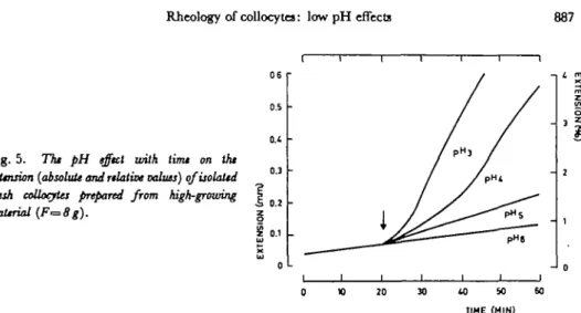
Fig. 5. The pH effect with time on the
exUnsion (absolute and relative values) qfisolaltd fresh collocates prepared from high-growing
material (F=8g).
Our data clearly show that the basal part with an Lr of 0.1 to 0.2 is high-growing material while the basal part with an Lr of 0.8 to 1.0 is low-growing material. Fig. 5 shows that low pH induced extension of isolated fresh collocytes of high-growing material with a lag period of 1-2 min. The response of fresh tissues in-creased with decreasing pH. When pH was changed from an acidic to a neutral value, the extension stopped rapidly; this extension was reversible up to a second extension with an acidic pH (Fig. 6). Other buffers (acetate, glycine) have also been tested and similar results were obtained.
To express the extension rate of collocytes for pHn, the following equation can be proposed:
ERn=dL/dt-l/Lo (expressed in %XlO-4/min) dL/dt: elongation rate for a collocyte bundle.
LQ : initial length of the collocyte bundle.
(II)
Thus, ERj, ER4 and ER3 are the extension rates of collocytes in buffered solu-tions of pH 5, pH 4 and pH 3, respectively. The extension rate was measured 10-20 min after the change of solution or when the extension was at a steady state. The pH effect on the extension rate can be easily expressed:
at pH 5:
at pH 3: JER8=ER3—ERfl
(III) (IV) When calculating the J ER values, the precision limit of the instruments used was 1%X 10~4/min. When a logarithmic scale was used, the zero value of id ER would be transformed in 1% X 10~4/min. As already reported, the pH 3 responses could include a different behavior of extension from that obtained for pH 5 (24).
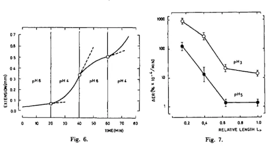
Fig. 7 shows the effects of pH 5 and 3 on Lr. The decrease of the pH responses was correlated with the growth of basal part (see Fig. 3). Note that the same drop was observed for the relative value for both pH 3 and 5 in relation to Lr. However, for Lr= 1.0, the effect of pH 3 was tenfold that of pH 5, which was minimal. Such a difference could be related to the decrease of turgor pressure of the collocytes for treatment at low pH.
OS 01 | 0 3 i a: K 01 ao • pHS PH* /
7
j1^
pHt i pHt too 2 X o 10 cr UJ 1 20 60 70 TIHEtMIN) 0.8 O.e 1.0 RELATIVE LENOIH Lh Fig. 6. Fig. 7.Fig. 6. Effect of pH changes on the extension (absolute values) of isolated fresh colloyctes prepared from
kigh-growing petioles (F=8g).
Fig. 7. 7fa pH effect on the extension rate ( ± standard error) of isolated fresh collocytes in relation to relative
length Lr{F=8g).
The data of Table 1 support this explanation. Frozen-thawed collocytes behaved like fresh collocytes and the extension rate did not significantly differ except for pH 3 for low-growing collocytes. In such case, they exhibited a lower response than fresh collocytes. It has to be noticed that they lost turgor pressure and that biochemical activity, i.e., the ability to synthesize new cell wall material, was reduced (17). However, some enzymes (glucosidases and galactosidases) seem protected against this kind of treatment (23).
Methanol-killed collocytes exhibited a very small response to low pH; this was already observed for epidermic cells (24). But the decrease of their extension
Fig. 8. The pH effect on the extension rat* ( ± standard
error) of frozen-thazotd collocyUs prepared from high-growing (HG) and low-high-growing (LG) material.
10 20 30 FORCE F (9)
Rheology of collocytcs: low pH effects 889
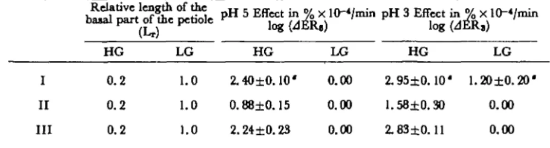
Table 1 pH Effects on fresh (I), methanol-kUUd (II) and frozen-thawed (III) coUenchjma bundles prepared
from Hgh-(HG) and low-growing (LG) petioles at afore* of 10 g
I II III Relative lengt) HG 0.2 0.2 0.2 LG 1.0 1.0 1.0 pHSEffgin^ HG 2.40±0.10' 0.88±0.15 2.24±0.23 FU) LG 0.00 0.00 0.00 nin pH 3 Effect ii log(< HG 2.95±0.10' 1.58±0.30 2.83±0.11 4ER3) LG 1.20±0.20' 0.00 0.00
* Standard deviation given for each value.
ability was (when reported in relative values) identical for both pH 3 and 5. In fact, the methanol-killed samples lost most of their protein content in boiling methanol (24). Thus, the cell wall materials may have been modified by such drastic treatment.
Most tissues (17, 23) show a yield stress for the pH effect on extension when frozen-thawed. Frozen-thawed collocytes of high-growing material displayed similar characteristics (Fig. 8). For low F, only small values were found for A ER$. In contrast, collocytes of low-growing material showed no significant response. This could be explained by the fact that the cell wall is thicker than in high-growing collocytes. The pH effect on collocytes from low- and high-growing materials to the same force or stress is given in Table 2. Stress large enough to induce a pH effect on collocytes of high-growing material had no effect on collocytes of low-growing material. This could be correlated to a change of the cell wall which takes place at the end of the differentiation and which must be in connection with a decrease of permanent strain ep.2 Some observations also indicate that rheological parameters have been related to an increase of cellulose content in the cell wall (7). Other studies on collenchyma cell wall have shown that the increase of differentiation was followed by a large accumulation of cellulose (20). Such data could very well explain, at least partially, the present results.
Table 2 pH Effects on force and stress in frozen-thawed collocytes prepared from high-(HG) and low-growing
(LG) petioles
Force or stress applied F o r c e - 1 0 g Stress = 1 3 . 4 x 1 0 " dynes/cm« HG LG HG LG Total strain (tot 3. 50±0. 30 1.15±0.30 3. 60 ±0.30 2.40±0.45
Standard deviation given for each value.
Strain ratio «ol/«tot 0.45±0.04 0. 72±0.04 0.45±0.04 0. 72±0.04 pH Effect in « log (JER,) 2.42±0.10 0.0 2.42±0.10 0.0 % x 10-</min log (JER,) 2.94±0.11 0.0 2.94±0.11 0.0
The response to low pH decreased when the growth decreased at the end of differ-entiation. It has been previously reported that NAA induces increase in both the length of collocytes and their extensibility (14) and that NAA acts on the cell wall by decreasing the pH as IAA does (10,16). At the end of growth, the wall changes from a low-pH sensitive into a low-pH nonsensitive one (22). In addition, experiments on low-growing material have shown that the isolated collocytes are not sensitive to NAA (unpublished results). However, it has been noticed that pH-sensitive tissues do not necessarily respond to auxin (4). In contrast, fusicoccin induces growth by decreasing the pH of the wall (10) and by acting also on the balance between endogenous IAA and ABA (14, 15). Such pH and growth interactions could probably be better understood by analyzing the comparative effects of NAA and fusicoccin on collocyte extension (work in preparation).
References
( 1) Cleland, R.: Extensibility of isolated cell walls: measurement and change during cell elonga-tion. Planta 74: 197-209 (1967).
( 2) Cleland, R. and D. L. Rayle: Hydrogen ion entry as a controlling factor in the acid-growth response of green pea stem section. Plant Physiol. 55: 547-549 (1975).
( 3) Durand, H. and D. L. Rayle: Physiological evidence for auxin-induced hydrogen ion secretion and the epidermal paradox. Planta 114: 185-193 (1973).
( 4) Evans, M.: Rapid responses to plant hormones. Aim. Rev. Plant Physiology 25:195-223 (1974). ( 5 ) Jaccard, M. and P. E. PUet: Extensibility and rheology of coUenchyma: I. Creep relaxation
and viscoelasticity of young and seneicent cells. Plant & Ctli Physiol. 16: 113-120 (1975).
(6) Jacobs, M. and P. M. Ray: Promotion of xyloglucan metabolism by acid pH. Plant Physiol.
56: 373-376 (1974).
( 7 ) Kawamura, H., S. Kamisaka and Y. Masuda: Regulation of lettuce hypocotyl elongation by gibberellic acid. Correlation between cell elongation, stress relaxation properties of the cell wall and wall polysaccharide content. Plant & Cell Physiology 17: 23-34 (1976).
( 8) Labavitch, J. M. and P. M. Ray: Relationship between promotion of xyloglucan metabolism and induction by indoleacetic acid. Plant Physiol. 54: 499-502 (1974).
( 9 ) Labavitch, J. M. and P. M. Ray: Turnover of cell wall polysaccharides in elongating pea stem segments, ibid. 53: 669-673 (1974).
(10) Marri, E., P. Lado, F. Rasi-Caldognon and R. Colombo: Correlation between proton
ex-trusion and stimulation of cell enlargement Effects of fusicoccin and of cytolcinins on leaf fragment and isolated cotyledons. Plant Sc. Letters 2: 139-150 (1975).
( / / ) Monro, J. A., D. Penny and R. W. Bailey: The organisation and growth of primary cell walls of lupin hypocotyl. Photochemistry 15: 1193-1198 (1976).
(12) Olson, C. A., J. Bonner and D. J. Morre: Force extension analysis of Avena coleoptile cell walls. Planta66: 127-133 (1965).
(13) Pilct, P. E., M. Jaccard and R. Magliocco: Mesure de rextensibilite des cellules de
collen-chyme. C. R. Acad. Sc. 277: 1637-1640 (1973).
(14) Pilet, P. E. and J. Cl. Roland: Growth and extensibility of collenchyme cells. Plant Sri. Letters
2:203-207(1974).
(15) Pilet, P. E.: Action of fusicoccin and abtcuic acid on root growth, ibid. 5: 137-140 (1975). (16) Pilet, P. E.: Fusicoccin and auxin effects on root growth, ibid. 7: 81-84 (1976).
Rheology of collocytes: low pH effects 891
(17) Rayle, D. L. and R. Qeland: The in vitro acid growth response: Relation to in vivo growth
response and auxin action. Planta 104: 282-296 (1972).
(18) Rayle, D. L.: Auxin-induced hydrogen ion secretion in Avena coleoptiles and its implication.
ibid 114:63-73 (1973).
(19) Roelofsen, P. A.: Ultraitructure of the wall in growing cells and its relation to the direction
of growth. Adv. Bot. Rtstarthl: 69-149 (1965).
(20) Roland, J. Cl.: Organisation de la membrane paraplasmique du collenchymc. J. de Micro-scopie 5: 323-348 (1966).
(21) Roland, J. Cl: Recherches en microscopic photonique et en microscopic e'lectronique sur
l'origine de la differentiation des cellules du collenchymc. Ann, dts Sc. Nat., Bot. (Paris) 8: 141-214 (1967).
(22) Roland, J. Cl. and P. E. Pilet: Implications du plasmalemme et de la paroi dans la croissance
dcs cellules vege" tales. Exptricnlia 30: 441 -451 (1974).
(23) Tanimoto, E. and M. Igari: Correlation between ^-galactosidase and auxin-induced
elonga-tion growth in etiolated pea stem. Plant Gt Cell Physiol. 17: 673-682 (1976).
(24) Yamagata, Y., R. Yamamoto and Y. Masuda: Auxin and hydrogen ion action on light-grown
pea epicotyl segment II. Effect of hydrogen ions on extension of isolated epidermis, ibid 15: 873-341 (1974).
(25) Yamamoto, R., K. Maki, Y. Yamagata and Y. Masuda: Auxin and hydrogen ion action on
light-grown pea epicotyl segment. I. Tissue specificity of auxin and hydrogen ion action, ibid. 15: 823-831 (1974).