Annals of Oncology!: 223-225, 1996.
Editorial
Cyclin Dl, another molecule of the year?
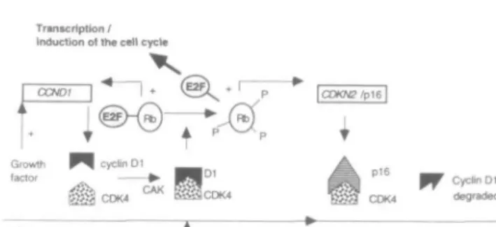
One of the aims of carcinogenesis research is to identify the precise molecular alterations responsible for neo-plastic transformation. Normal cell cycle control is dis-rupted in malignant cells which are unable to regulate their proliferation correctly. Understanding how the cell cycle control operates therefore represents a fun-damental problem in cancer biology. Work in recent years has led to the conclusion that cyclins, and cyctin-dependent kinases (CDKs) together with their inhibi-tors constitute a complicated network of positive and negative regulation circuits. These basic regulatory mechanisms are common to all mammalian cells and act at specific time points during the cell cycle (Figure During the Gl phase, D-type cyclins appear to act as growth factor sensors, driving cells through the first restriction point (START in yeast), beyond which cells are committed to divide. Cyclin Dl together with spe-cific kinases (CDK4, CDK6) and one particular kinase inhibitor (pl6) appears to play a central role in the regulation of the proliferation status. Activated CDK4, through phosphorylation and complex formation with cyclin Dl, inactivates the retinoblastoma protein (pRb). This results in the release from pRb of a transcription factor, E2F, which then activates many genes necessary for cell division. The pl6 protein suppresses this process by competitively binding to the CDK4 mol-ecule (Figure 2). Component genes of this specific con-trol pathway are frequently compromised in malignant cells (for review see [1-5]).
The apparently ever-growing list of cell cycle regula-tors makes it increasingly difficult to keep abreast of this rapidly developing field, let alone for oncologists to think of clinical applications of these basic discoveries. However, some aspects are becoming clearer with respect to cancer, one of the most important proteins is cyclin Dl. It is now well established that cyclin D l has oncogenic functions: constitutive overexpression in rodent cells can shorten the Gl phase [6], and similarly, in breast cancer cells arrested in Gl, cyclin Dl induc-tion is sufficient to complete the cell cycle and to shorten the Gl-S phase [7]. Transfection of the cyclin Dl gene constructs into normal fibroblasts stimulates proliferation by reduction of the Gl-S phase [6], and when transfected into normal fibroblasts with activated
H-ras, it promotes the formation of fibrosarcomas in
nude mice [8]. Moreover, mice carrying an active cyclin D l transgene develop mammary hyperplasia and adenocarcinoma [9]. When overexpression of cyclin Dl is targeted to the lymphoid system, the transgenic mice develop lymphomas in the presence of c-myc
over-cyclin E+CDK2 commitment point (START) cyclin D +CDK4 +CDK6 cyclin A +CDK2 cyclin A and B + CDK1 (cdc2)
Figure I. Combinatorial interactions of cyclins and cyclin
depend-ent kinases (CDK) during the cell cycle.
Transcription / Induction of the cell cycle
Cyclin Dl degraded
G1
T
Commitment point STARTFigure 2. Model for Gl restriction-point control by cyclin Dl:
Growth factors induce the transcription of the cyclin Dl gene
(CCND1) and positively regulate its assembly with CDK4 (and
CDK6, not shown). The cyclin D1-CDK4 complex is activated by cyclin-activating kinase (CAK - cyclin H + CDK7) and the func-tional holoenzyme then phosphorylates the retinoblastoma protein, releasing and thus activating the E2F transcription factor. The
CDKN2 gene (encoding pl6) inactivates CDK4 and induces
accel-erated turnover of unbound cyclin Dl during the S phase.
expression [10, 11]. Very recently, cyclin Dl over-expression has been found at early stages of tumour development, in particular, in carcinoma in situ of the breast [12] and in bronchial epithelia of resection mar-gins of non-small-cell lung cancer that were micro-scopically tumour-free [13].
The region of chromosome I l q l 3 encoding the cyclin Dl gene (CCND1) is frequently altered at the cytogenetic level in malignant disease. Increased gene copy numbers (gene amplification) have been found in breast, ovarian, bladder, esophageal, gallbladder, stom-ach, laryngeal, lung and liver cancers [1]. In esophageal [14], laryngeal [15] and head and neck tumours [16],
CCND1 amplification correlates with poor prognosis, a
high incidence of distant metastasis and frequent recur-rences. In almost all cases, amplification of the I l q l 3 region was associated with overexpression of cyclin Dl. In lymphoproliferative disorders, another mechanism
224
may also lead to cyclin D l overexpression: Transloca-tion between the bcl-1 region, upstream of CCND1, and the immunoglobulin (Ig) heavy chain locus on chromosome 14 [17, 18] results in elevated transcrip-tion of CCND1, with high levels of cyclin Dl [19-22]. The translocation t(ll;14Xql3;q32) occurs in 90% of mantle cell lymphomas and splenic lymphomas with villous lymphocytes as well as in a small percentage of certain aggressive lymphocytic and plasma cell leukae-mias [19-21,23-25].
This issue of Annals of Oncology carries an interest-ing paper expandinterest-ing our knowledge of cyclin Dl in hairy cell leukaemia (HCL). De Boer et al. [26] report on cyclin Dl overexpression in the majority of patients with HCL using both molecular and genetic methods. These findings are in full agreement with the very recently published study by Bosch et al. [27]. Thirty-eight of 40 HCL patients in the two studies showed cyclin D l overexpression. The level of expression is significantly lower than in mantle cell lymphomas; nevertheless, this finding is very interesting and rather unexpected, given the lack of evidence for molecular lesions involving CCND1 in HCL.
Except for some lymphoproliferative disorders with the bcl-1 rearrangement, lymphoid tissues such as ton-sils, spleen and lymph nodes apparently have no or very low levels of cyclin D l expression [19, 28]. De Boer et al. [26] and Bosch et al. [27] were unable to explain the mechanism of cyclin Dl overexpression and CCND1 deregulation in HCL. Neither structural anomalies within I l q l 3 (cytogenetic and molecular techniques) nor gene amplification were detected. Al-though rearrangements cannot be excluded outside the regions examined, translocation appears not to be a main mechanism responsible for cyclin Dl overexpres-sion in HCL. Although the B-cell lineage of HCL is well established [29], there are several lines of evidence indicating some phenotypic and functional similarities between hairy cells and cells of monocyte/macrophage lineage [30], which normally express cyclin Dl [31]. Thus, it remains to be seen whether this cyclin Dl over-expression is at least partially responsible for the malig-nant behaviour of the hairy cells, or whether, as specu-lated by the authors, it might constitute a physiological characteristic shared by hairy cells. Additionally, it may represent an unspecific upregulation occurring as a result of the deregulated cycle network. Further work is therefore needed to establish the role of cyclin Dl in HCL.
For instance, the examination of allele specific
CCND1 expression in HCL would point to a possible
role in malignancy. Were allelic imbalance observed in PCR-amplified cDNA it might be concluded that only one parental allele was responsible for cyclin Dl over-expression. This would lead to the assumption that there had been an alteration, such as a mutation within a CCND1 control region or splice site (RNA proces-sing). This phenomenon was recently demonstrated [32, 33]: Using immunostaining in a series of
non-small-cell lung tumours, cyclin Dl was overexpressed in almost 30% of cases with no apparent structural ab-normalities of CCND1, as assessed by Southern blot-ting and PCR. In these tumours we detected imbal-ances in allele-specific expression by RT-PCR and restriction fragment length polymorphism analysis of a
HaeIR polymorphism. Thus, genetic alteration of CCND1 appears to be a key abnormality in lung
car-cinogenesis. Such a result in HCL would strengthen the evidence of a direct role for cyclin Dl in the disease.
Deregulation or loss of function of genes involved in the control of progression through the Gl restriction point (CCND1, CDK4, CDKN2, RBI) is common in human cancers. Conversely, lesions within the many other genes controlling aspects of the cell cycle (cyclin A, B, E, CDKs or CDK inhibitor genes) is much less frequent. In particular, overexpression of cyclin D l has now been observed in around 50% of the most com-mon and least treatable malignancies, including colo-rectal, head and neck, esophageal, breast, uterus and lung carcinomas, melanomas and sarcomas [14,16, 32, 34-37]. In addition, various studies have demonstrated that blocking the action of cyclin Dl in vitro, with anti-bodies or antisense RNA prevents the growth of many tumour cell lines [6,38]. However, evidence from mice in which both parental cyclin Dl genes have been knocked out suggests that complete loss of cyclin D l function might have only limited effects on overall viability or normal cell proliferation in the adult [39]. These findings raise the exciting possibility that thera-peutic agents directed against elements of this pathway, in particular cyclin Dl, might prove not only successful in the treatment of common malignancies but also have relatively few side effects. Further research is needed to show whether such an approach might be transferred from the laboratory bench to patients. Should this turn out to be successful, then, without doubt, another edi-torial entitled 'Cyclin Dl, the molecule of the century1
will have to be written!
Supported by Krebsforschung, Schweiz, KFS 177-9-1995.
Daniel C. Betticher, M JJ>. Institute of Medical Oncology Inselspital and University of Bern Bern, Switzerland
References
1. Motokura T, Arnold A. Cyclins and oncogenesis. Biochim Bio-phys Acta 1993; 1155:63-78.
2. Hunter T, Pines J. Cyclins and cancer II: Cyclin D and CDK inhibitors come of age. Cell 1994; 79: 573-82.
3. Sherr CJ. D-type cyclins. Trends Biochem Sri 1995; 20:187-90. 4. Sherr CJ, Roberts JM. Inhibitors of mammalian Gl
cyclin-dependent kinases. Genes Dev 1995; 9:1149-63.
5. Hirama T, Koeffler HP. Role of the cyclin-dependent Itinase inhibitors in the development of cancer. Blood 1995; 86: 8 4 1 -54.
225
6. Quelle DE, Ashmun RA, Shurtleff SA et al. Overexpression of mouse D-type cyclins accelerates Gl phase in rodent fibro-blasts. Genes Dev 1993; 7:1559-71.
7. Musgrove EA, Lee CSL, Buckley MF et al. Cyclin Dl induc-tion in breast cancer cells shorten Gl and is sufficient for cells arrested in Gl to complete the cell cycle. Proc Natl Acad Sci USA 1994; 91:8022-6.
8. Lovec H, Sewing A, Lucibello FC et al. Oncogenic activity of cyclin Dl revealed through cooperation with Ha-ras: Link be-tween cell cycle control and malignant transformation. Onco-gene 1994; 9: 323-6.
9. Wang TC, Cardiff RD, Zukerberg L et al. Mammary hyper-plasia and carcinoma in MMTV-cyclin Dl transgenic mice. Nature 1994; 369: 669-71.
10. Bodrug SE, Warner BJ, Bath ML et al. Cyclin Dl transgene impedes lymphocyte maturation and collaborates in lympho-magenesis with the myc gene. EMBO J 1994; 13: 2124-30. 11. Lovec H, Grzeschiczek A, Kowalski MB et al. Cyclin Cl/bcl-1
cooporates with myc genes in the generation of B-cell lym-phoma in transgenic mice. EMBO J 1994; 13: 3487-95. 12. Weinstat-Saslow D, Merino MJ, Manrow RE et al.
Overexpres-sion of cyclin D mRNA distinguishes invasive and in situ breast carcinomas from non-malignant lesions. Nature Med 1995; 1: 1257-60.
13. Betticher DC, Heighway J, Thatcher N et al. Altered CCND1 (cyclin Dl) and Rb (retinoblastoma) genes expression is an early event in non-small cell lung cancer development. Cancer and the Cell Cycle, January 17-20, 1996, Lausanne, Switzer-land, A14.
14. Naitoh H, Shibata J, Kawaguchi A et al. Overexpression and localization of cyclin Dl mRNA and antigen in esophageal cancer. Am JPathol 1995; 146:1161-9.
15. Jares P, Fernandez PL, Campo E et al. PRAD-1/cyclin Dl gene amplification correlates with messenger RNA overexpres-sion and tumor progresoverexpres-sion in human laryngeal carcinomas. Cancer Res 1994; 54:4813-7.
16. Michalides R, Van Veelen N, Hart A et al. Overexpression of cyclin Dl correlates with recurrence in a group of forty-seven operable squamous cell carcinomas of the head and neck. Can-cer Res 1995; 55: 975-8.
17. van den Berghe H, Parloir C, David G et al. A new charac-teristic karyotypic anomaly in lymphoproliferative disorders. Cancer 1979; 44:188-95.
18. de Boer CJ, Loyson S, Kluin PM et al. Multiple breakpoints within the bcl-1 locus in B-cell lymphoma: Rearrangements of the cyclin Dl gene. Cancer Res 1993; 53:4148-52.
19. de Boer CJ, Schuuring E, Dreef E et al. Cyclin Dl protein analysis in the diagnosis of mantle cell lymphoma. Blood 1995; 86:2715-23.
20. Bosch F, Jares P, Campo E et al. PRAD-1/cyclin Dl gene over-expression in chronic lymphoproliferative disorders: A highly specific marker of mantle cell lymphoma. Blood 1994; 84: 2726-32.
21. de Boer CJ, Van Krieken JHJM, Kluin-Neleman HC et al. Cyclin Dl messenger RNA overexpression as a marker for mantle cell lymphoma. Oncogene 1995; 10:1833-40. 22. Williams ME, Nichols GE, Swerdlow SH. In situ hybridization
detection of cyclin Dl mRNA in centrocytic/mantle cell lym-phoma. Ann Oncol 1995; 6: 297-9.
23. Williams ME, Swerdlow SH. Cyclin Dl overexpression in non-Hodgkin's lymphoma with chromosome 11 bcl-1 rearrange-ment Ann Oncol 1994; 5 (Suppl 1): S71-S73.
24. Jadayel D, Matutes E, Dyer MJS et al. Splenic lymphoma with villous lymphocytes: Analysis of BCL-1 rearrangements and expression of the cyclin Dl gene. Blood 1994; 83: 3664-71. 25. Zukerberg LR, Yang WI, Arnold A et al. Cyclin Dl expression
in non-Hodgkin's lymphomas. Am J Clin Pathol 1995; 103: 756-60.
26. de Boer CJ, Kluin-Nelemans JC, Dreef E et al. Involvement of the CCND1 gene in hairy cell leukemia. Ann Oncol 1996;
7: 251-6 (this issue).
27. Bosch F, Campo E, Jares P et al. Increased expression of the PRAD-1/CCND1 gene in hairy cell leukaemia. Br J Haematol 1995; 91:1025-30.
28. Bartkova J, Lukas J, Strauss M et al. Cell cycle-related varia-tion and tissue-restricted expression of human cyclin Dl pro-tein. J Pathol 1994; 172: 237-45.
29. Korsmeyer SJ, Greene WC, Cossman J et al. Rearrangement and expression of immunoglobulin genes and expression of the Tac antigen in hairy cell leukemia. Proc Natl Acad Sci USA 1983; 80:4522-6.
30. Golomb HM, Vardiman JW. Hairy-cell leukemia. In Williams WJ, Beutler E, Erslev AJ, Lichtman MA (eds): Hematology. Fourth edition, New York: McGraw-Hill Publishing Company 1990; 1025-30.
31. Matsushime H, Roussel MF, Ashmun RA et al. Colony-stimu-lating factor 1 regulates novel cyclins during the Gl phase of the cell cycle. Cell 1991; 65: 701-13.
32. Betticher DC, Heighway J, Hasleton PS et al. Prognostic sig-nificance of CCND1 (cyclin Dl) overexpression in primay resected non-small cell lung cancer. Br J Cancer 1996; 73: 294-300.
33. Betticher DC, Thatcher N, Altermatt HJ et al. Alternate splic-ing produces a novel cyclin Dl transcript. Oncogene 1995; 11: 1005-11.
34. Bartkova J, Lukas J, Strauss M et al. Cyclin Dl oncoprotein aberrantly accumulates in malignancies of diverse histogenesis. Oncogene 1995; 10: 775-8.
35. Bartkova J, Lukas J, Strauss M et al. The PRAD-1/Cyclin Dl oncogene product accumulates aberrantly in a subset of colo-rectal carcinomas. Int J Cancer 1994; 58: 568-73.
36. Bartkova J, Lukas J, Miiller H et al. Cyclin Dl protein expres-sion and function in human breast cancer. Int J Cancer 1994; 57:353-61.
37. Gillett C, Fantl V, Smith R et al. Amplification and overexpres-sion of cyclin Dl in breast cancer detected by immunohisto-chemical staining. Cancer Res 1994; 54:1812-7.
38. Zhou P, Jiang W, Zhang YJ et al. Antisense to cyclin Dl inhibits growth and reverses the transformed phenotype of human esophageal cancer cells. Oncogene 1995; 11: 571-80.
39. Sicinski P, Donaher JL, Parker SB et al. Cyclin Dl provides a link between development and oncogenesis in the retina and breast Cell 1995; 82:621-30.