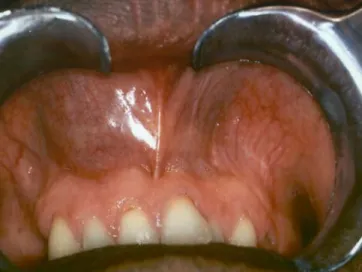
Upper lip swelling caused by a large dentigerous cyst
4
0
0
Texte intégral
Figure
Documents relatifs