Unexpectedly late expression of intracellular CD3ϵ and TCR γδ proteins during adult thymus development
10
0
0
Texte intégral
(2) 1642 Unexpectedly late expression of intracellular CD3ε and TCR γδ sion on immature thymic precursor cells, many groups have utilized molecular methods to follow rearrangement and/or expression of TCR β, γ and δ genes (reviewed in 15,16) as well as CD3 genes (17) in defined subsets. Although these techniques have provided much useful information, they have two major drawbacks in that (i) they are generally not applicable to single cells and (ii) they do not measure the levels of the corresponding protein. In this report we have attempted to overcome these technical problems by undertaking a detailed four-color flow cytofluorometric analysis of the expression of intracellular (ic) CD3ε, TCR β and TCR γδ proteins at defined stages of adult thymus development. Although similar techniques have been utilized previously in the context of fetal thymus development (18–20), the conclusions have been somewhat limited due to the general use of only two- (or occasionally three-) color analysis and to the fact that immature subsets are phenotypically less well defined in the fetal (as opposed to adult) thymus. Our data confirm and extend previous molecular analyses of the timing (and consequences) of icTCR β expression during adult thymus development. Moreover, they provide novel information concerning the timing of both icCD3ε and icTCR γδ protein expression. Methods. Mice and cell suspensions Six-week-old C57BL/6 female mice were obtained from Harlan Olac (Bicester, UK). TCR β–/- and TCR δ–/- mice were obtained from Jackson Laboratories (Bar Harbor, ME) and maintained in our animal facility. DN and DN CD25– thymocytes were prepared as described previously (7,21). Briefly, thymocyte cell suspensions were incubated at 4°C in DMEM/1% FCS containing the appropriate concentrations of IgM isotype antibodies against CD4 (RL172.4), CD8 (31M) and CD25 (7D4) for 20 m. Rabbit complement (Saxon Europe, Suffolk, UK) and DNase I (Boehringer Mannheim, Mannheim, Germany), were added, and the incubation continued at 37°C for 45 min. After washing, viable cells were recovered after density-gradient centrifugation through Lympholyte M (Cedarlane, Hornby, Ontario, Canada). During subsequent FACS analysis and/or sorting, contaminating CD41, CD81, CD251 (where required) and any CD31 (αβ1 or γδ1) cells were positively eliminated where appropriate by staining with a cocktail of FITC-conjugated mAb to these surface markers and ‘live’ gating out all FITC1 cells.. either direct TriColor or CyChrome conjugates, such as TCR γδ–TriColor, CD4–TriColor and CD8α–TriColor (Caltag, Burlingame, CA), CD44–CyChrome (PharMingen, San Diego, CA), or biotinylated-CD25 revealed with TriColor–streptavidin, purchased from Caltag in FL3. For four-color staining, Cy5– CD25, Cy5–CD24 and CD117–allophycocyanin (PharMingen) were used in FL4. All FITC, biotin and Cy5 conjugates were purified and conjugated from hybridomas grown in this laboratory, and have been described elsewhere (7). PE- and/ or CyChrome-conjugates of mAb to TCR β (H57-597), TCR γδ (GL3), CD3ε (17A2 and 145-2C11), and control rat and hamster Ig (PharMingen) were used for intracellular staining. PE conjugates of mAb to TCR Vγ1.1 [clone 11.2 (23)], TCR Vγ2 (UC3-10A6) and TCR Vδ4 (GL2) were prepared in this laboratory using the Phycolink PE-conjugation kit (Prozyme, San Leandro, CA). Surface TCR β, TCR γδ, TCR Vγ1.1, TCR Vγ2, TCR Vδ4 and CD3ε were pre-blocked with unlabeled purified mAb from the same clones as used for intracellular staining. Analysis was performed using Lysys II or CellQuest software.. Cell culture Purified and FACS-sorted CD44loCD25–CD3– DN thymocytes were cultured in DMEM supplemented with HEPES buffer, Glutamine, β2-mercaptoethanol and 5% FCS for 4 h at 37°C. After harvesting and counting the cells were stained and analyzed by four-color FACS either for surface expression of CD4, CD8, TCR β and TCR γδ or for intracellular expression of TCR β and TCR γδ after gating out any mature cells (CD4, CD8, CD3ε, TCR β or TCR γδ) by surface staining with the FITC cocktail described above.. Cell cycle analysis The DNA content of thymus subsets was determined by first sorting the selected populations defined by both surface and intracellular protein expression as described above. Propidium iodide (PI) staining of DNA was performed using standard procedures as described previously (22,24). Briefly, sorted cells were treated with 3N HCl to remove surface markers, washed in PBS and neutralized with 0.1 M Na2B4O7. RNA was removed by incubation in RNase A at 37°C before the addition of PI. Analysis was performed on the FACScan flow cytometer using the doublet discrimination module. Results. Flow cytometry and cell sorting. Intracellular CD3ε protein is first detected in CD251CD44lo CD117lo thymocytes. Thymocyte subsets were analyzed for simultaneous detection of both surface and intracellular proteins as described previously (22), using FACScan (three color) or FACSCalibur (four color) flow cytometers and sorted with a FACStar Plus (Becton Dickinson, San Jose, CA). Briefly, surface staining was performed as usual for one, two or three colours followed by fixation in 2% paraformaldehyde. After permeabilization with 0.5% Saponin, intracellular proteins were detected with mAb directly conjugated to phycoerythrin (PE) and/or CyChrome. Specifically, surface staining was performed using a pool of FITC direct conjugates as described above, together with. In order to establish the timing of CD3ε protein expression during adult thymus development, DN thymocytes were purified by depletion and surface stained in three colors with a cocktail consisting of mAb to CD4, CD8 and CD3 (to remove residual contaminating mature T cells) together with CD25 and either CD44, CD24 or CD117. The fourth color was used for icCD3ε. Surprisingly the CD251 DN population contained a well-defined subset of icCD3ε– cells (Fig. 1A). The proportion of icCD3ε– cells in the CD251 DN subset was highly reproducible (6 6 1 %, N 5 20) and did not depend upon the mAb used to detect CD3ε, since all four mAb used (145-2C11,.
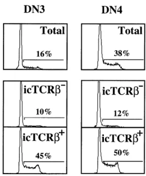
(3) Unexpectedly late expression of intracellular CD3ε and TCR γδ 1643. Fig. 1. Intracellular CD3ε expression marks the DN2 to DN3 transition in adult thymocytes. (A) Expression of icCD3ε and surface CD44 on total CD251 DN thymocytes. (B) Surface expression of CD117, CD44, CD24 and CD25 gated on CD251icCD3ε– (solid line) and CD251icCD3ε1 (fine line) DN thymocytes.. Fig. 2. Intracellular TCR β, TCR γδ and CD3ε expression in immature and mature thymus subsets. Purified DN cells were surface stained with a pool of mAb to remove CD441 early precursors and any CD31, TCR β1 or TCR γδ1 contaminants, together with CD25 to distinguish between DN3 and DN4 subsets. ISP, DP and mature CD41CD8– SP thymocyte subsets were directly gated from total thymus. Intracellular staining for TCR β, CD3ε and TCR γδ is shown on each gated subset. Surface TCR β, TCR γδ or CD3ε were pre-blocked with an excess of the appropriate unlabeled, purified mAb. Overlays are the same cell populations stained with control Ig. Numbers represent the percentage positive cells (6 SD where shown).. Intracellular TCR β protein is first detected in the DN3 subset. 17A2, 13D5 and 500-A2) yielded similar results (data not shown). By four-color analysis the icCD3ε– subset of CD251 DN cells homogeneously expressed much higher levels of CD44, CD25 and CD117 (c-kit R) as compared to the icCD3ε– CD251 population (Fig. 1B). However, expression levels of CD24 (HSA) were similar in both subsets. These data indicate that CD3ε protein is expressed later in adult thymus development than was originally believed based on mRNA expression studies (17). Indeed the homogeneous phenotype of icCD3ε– CD251 DN cells (CD441CD1171) corresponds to the accepted definition of the DN2 subset (6), thus indicating that expression of CD3ε marks the DN2 to DN3 transition. Analysis of icCD3ε expression during subsequent stages of adult thymus development indicated that essentially 100% of cells were icCD3ε1 as expected (Fig. 2), with the exception of a small subset of DN4 cells that probably do not belong to the T cell lineage (see below).. Semi-quantitative PCR and Southern blotting techniques have shown that TCR β rearrangement is initiated during the transition from the DN2 to DN3 stages of intrathymic development (6,10). However, the point at which productive TCR β protein can be first detected has been difficult to determine by conventional FACS analysis due to the extremely low amount expressed at the surface of immature thymocytes. In order to define the earliest DN subset in which icTCR β protein is expressed we have used four-color FACS analysis as described above. The results shown in Fig. 3 demonstrate clearly that icTCR β protein is first detected in a subset of DN3 thymocytes and not at all in DN2 thymocytes. These results confirm and extend previous studies (6,8,10,17), which show that full-length VDJCβ transcripts can be first detected by Northern blot analysis in the DN3 subset. As development proceeds, the percentage of icTCR β1 cells increases rapidly from ~25% in DN3 to 80% at the DN4 stage, after which essentially 100% of the cells show high levels of icTCR β (Fig. 2)..
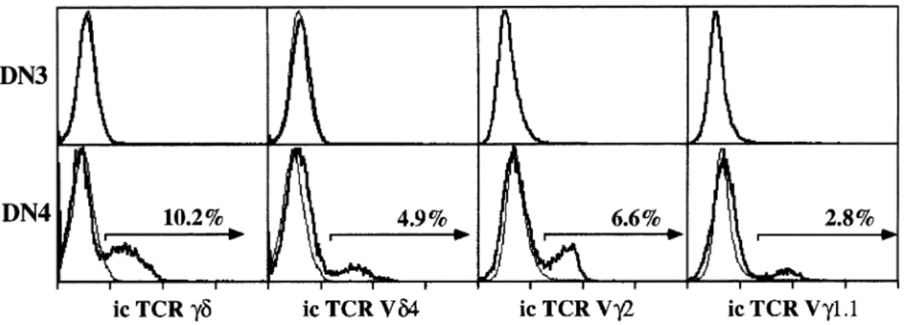
(4) 1644 Unexpectedly late expression of intracellular CD3ε and TCR γδ. Fig. 4. Correlation between icTCR β expression and proliferation in immature thymus subsets. Purified DN thymocytes were stained as for Figure 2. DN3 and DN4 cells were sorted into icTCR β1 or icTCR β– subsets and the percentage of cells in S 1 G2/M phases of the cell cycle was determined by PI staining. Control populations are total sorted DN3 and DN4 cells.. Fig. 3. Intracellular TCR β is first expressed in DN3 adult thymocytes. Expression of icTCR β or icTCR γδ and surface CD44 gated on total CD251 DN thymocytes.. Immature DN thymocytes expressing intracellular TCR β are proliferating. It is generally accepted that immature DN3 thymocytes which have undergone successful TCR β rearrangement express the pre-TCR (TCR β, pTα and the CD3 complex) and that subsequent signaling through this complex results in ‘βselection’ (reviewed in 2–4,14,25). A major consequence of β-selection is proliferation. In the absence of any one of the components of the pre-TCR, this proliferative effect is absent or severely reduced (reviewed in 14–16). To determine directly whether expression of icTCR β protein correlates with proliferation, icTCR β1 and icTCR β– DN3 and DN4 thymocytes were purified by cell sorting and analyzed for DNA content by PI staining. As shown in Fig. 4 almost half (45%), of the icTCR β1 cells in DN3 have a DNA content that corresponds to the S 1 G2/M phases of the cell cycle compared with only 10% in the icTCR β– DN3 population and 16% in the unfractionated DN3 population. Similar results were obtained for the DN4 subset where 50% of the icTCR β1 cells are in cycle compared to only 12% in the icTCR β– subset and 38% in the unfractionated population (Fig. 4). Thus there is a strict correlation between icTCR β expression and proliferation in both the DN3 and DN4 subsets of adult thymocytes. A similar correlation was observed previously for thymocytes developing in fetal thymic organ culture (FTOC) (12,26).. Intracellular TCR γδ protein is only detected in the DN4 subset Until now, γδ lineage precursor T cells have not been able to be distinguished from their αβ counterparts by surface phenotype due to the lack of a unique surface marker. To search for possible γδ cell precursors we analyzed DN thymus subsets for icTCR γδ expression in the same manner as for icTCR β. As shown in Figs 2 and 3, in contrast to icTCR β expression, icTCR γδ protein is detected in only a small percentage (~7–10%) of the DN4 subset and is undetectable in any subset either earlier or later in thymus development. Since the GL3 mAb used to detect icTCR γδ protein in this study apparently only binds the TCR δ chain when associated with a TCR γ chain (27), we also stained DN3 and DN4 cells intracellularly with the available mAb against TCR Vγ and TCR Vδ chains (TCR Vγ1.1, Vγ2 and Vδ4). As shown in Fig. 5, a significant percentage of the icTCR γδ1 cells in the DN4 subset can be stained with each of the anti-V region mAb tested. However, none of the mAb showed detectable staining of cells in the DN3 subset.. TCR γδ1 thymocytes can be produced from DN4 icTCR γδ1 thymocytes in vitro In order to determine whether icTCR γδ1 thymocytes are indeed precursors of TCR γδ1 cells, purified and FACS-sorted DN4 thymocytes were cultured at 37°C for 4 h. As it is not possible to culture cells after intracellular staining due to the fixation and permeabilization protocol, DN4 icTCR γδ1 cells could not be specifically isolated to do a direct precursor to product study. Therefore the cultured DN4 cells were monitored in parallel for the appearance of surface TCR γδ1 cells and the disappearance of icTCR γδ1 cells. Cell loss after 4 h.
(5) Unexpectedly late expression of intracellular CD3ε and TCR γδ 1645. Fig. 5. Intracellular TCR Vγ and Vδ expression is first detected in DN4 thymocytes. Purified DN thymocytes were stained as for Fig. 2. Intracellular staining for TCR γδ, TCR Vδ4, TCR Vγ2 and TCR Vγ1.1 is shown on gated DN3 and DN4 thymocytes. Overlays (thin lines) are isotype control stainings of the same gated subsets.. Fig. 6. Production of surface TCR γδ1 cells from DN4 thymocytes in vitro. (A) Appearance of surface TCR γδ1 cells from sorted DN4 thymocytes after 4 h in culture. Gated DN cells were stained for surface TCR β versus TCR γδ (or isotype controls). (B) Disappearance of icTCR γδ1 cells from sorted DN4 thymocytes after 4 h in culture. Gated DN4 cells were stained for icTCR β versus icTCR γδ (or isotype controls). Untreated controls in both cases were FACS sorted DN4 cells incubated at 4°C.. in culture was minimal (~15%). As shown in Fig. 6, a significant proportion of surface TCR γδ1 cells appeared after 4 h at 37°C and icTCR γδ1 cells virtually disappeared. Importantly the absolute number of surface TCR γδ1 cells produced in this time period corresponded to the number of icTCR γδ1 cells that were present prior to culture and which had disappeared after 4 h at 37°C (Table 1). These data provide compelling evidence that icTCR γδ1 DN4 cells are immediate precursors of thymic γδ T cells.. Most icTCR γδ1 DN4 thymocytes are not ‘β-selected’ To determine if icTCR γδ1 cells within the DN4 subset also expressed icTCR β and could therefore be ‘β-selected’, we performed two-color intracellular staining for these two TCR proteins after gating out all non-DN4 and mature T cells with the third color. The contour plot shown in Fig. 7 demonstrates. Table 1. Quantitative conversion of icTCR γδ1 DN4 thymocytes to surface TCR γδ1 cells in vitro Experiment. 1 2. icTCR γδ1 cells. Surface TCR γδ1 cells. Pre. Post. Pre. Post. 120,000 151,000. ,10,000 15,000. – –. 126,000 152,000. DN4 thymocytes were purified by cell sorting and cultured for 4 h at 37°C in the absence of cytokines. The absolute number of ic TCR γδ1 and surface TCR γδ1 cells was calculated pre- and post-culture according to independent staining reactions (see Fig. 6). Data represent two independent experiments..
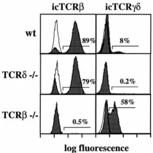
(6) 1646 Unexpectedly late expression of intracellular CD3ε and TCR γδ. Fig. 7. Correlation of icTCR β and icTCR γδ staining of DN4 thymocytes with proliferation status. (A) Purified DN CD25– thymocytes were surface stained with a pool of mAb to CD44, CD3ε, TCR β and TCR γδ to remove contaminating early precursors and mature T cells. The DN4 subset so defined was further stained with icTCR γδ together with icTCR β. Numbers are the percentage cells in each quadrant. (B) Percentage of cells in S 1 G2/M phases of the cell cycle in the four subsets defined in Fig. 5(A).. that the majority of icTCR γδ1 DN4 cells are icTCR β–, although a small but reproducible fraction (~15%) are icTCR β1. The four DN4 subsets defined by icTCR β and icTCR γδ staining were then sorted and analyzed for their cell cycle status by PI staining (Fig. 7). Interestingly, only 10% of icTCR γδ1β– cells were found to be in the S 1 G2/M phases of the cell cycle, whereas 23% of icTCR γδ1β1 cells were in cycle. As expected from analyses of the total DN4 subset (Fig. 4), 46% of icTCR γδ–β1 cells were in cycle as compared to 3% of icTCR γδ–β– cells. Taken together these data indicate that icTCR γδ1 DN4 cells are in general icTCR β– and not cycling, so therefore not ‘β-selected’.. Increased frequency of icTCR γδ1 DN4 thymocytes in TCR β-deficient mice Mice deficient for either the TCR β chain or pTα have essentially normal development of γδ cells despite a dramatic reduction (50- to 100-fold) in αβ T cell development (28,29). It was therefore of interest to investigate whether the percentage and absolute number of icTCR γδ1 DN4 cells present in TCR β–/– mutant mice differs from wild-type animals. As shown in Fig. 8, the proportion of icTCR γδ1 cells in the DN4 subset of TCR β-deficient mice is greatly increased (~58%). However, the number of DN4 thymocytes is correspondingly decreased in the absence of TCR β, such that the absolute number of icTCR γδ1 DN4 cells is similar to that found in wild-type mice (data not shown). Discussion. Timing of CD3ε expression during normal adult T cell development The data presented here demonstrate clearly that icCD3ε protein is not expressed at detectable levels until both CD44 and CD117 are down-regulated in CD251 DN adult thymocytes. Thus icCD3ε expression marks the transition between the DN2 and DN3 thymocyte subsets. Previous studies of. Fig. 8. Expression of icTCR β or icTCR γδ in DN4 thymocytes from normal or mutant mice. Purified DN CD25– thymocytes from C57BL/ 6 (wt), TCR β–/- or TCR δ–/- mice were surface stained as in Fig. 7, followed by staining for icTCR β or icTCR γδ (filled histograms). Negative controls (overlays) are the equivalent cells purified from TCR β–/- mice (icTCR β) or TCR δ–/- mice (icTCR γδ) and stained in the same manner.. icCD3ε expression in the fetal thymus have been controversial. Whereas essentially all CD251 fetal thymocytes were found to be icCD3ε1 in one report (18), ~40% of CD251CD1171 fetal thymocytes were shown to be icCD3ε– in another study (20). The apparent discrepancy between these data and our own may be related to differences in the timing of CD3ε expression in the fetal and adult thymus. Alternatively, since CD25 expression was not directly correlated with both CD44 and CD117 in the earlier studies, it is possible that a discrete.
(7) Unexpectedly late expression of intracellular CD3ε and TCR γδ 1647 (but homogeneous) subset of icCD3ε–CD251CD441 CD1171 fetal thymocytes was overlooked. The relatively late expression of icCD3ε protein observed in this study is surprising in view of our previous analysis of CD3ε mRNA expression in adult thymus subsets (17). Whereas CD3ε mRNA could be detected in a large fraction of DN1 and DN2 thymocytes by single-cell in situ hybridization, no detectable icCD3ε protein can be observed prior to the DN3 stage. This discrepancy could reflect an intrinsic instability of the CD3ε protein in more immature thymocytes. Alternatively it is formally possible that icCD3ε protein can only be detected by the mAb used here (17A2 and 1452C11) when complexed to other proteins first expressed at the DN3 stage such as TCR β and/or pTα. However, the latter explanation seems unlikely in view of the fact that icCD3ε can be readily detected in immature thymocytes of TCR β–/- and pTα–/- mice (data not shown). Although icCD3ε protein expression strictly correlates with the DN2 to DN3 transition in the adult thymus it does not represent a critical check-point for further T cell development. Indeed in CD3ε-deficient mice development does not arrest until the DN3 stage, and TCR β, γ and δ genes undergo apparently normal rearrangement (30). Nevertheless it is possible that signaling via CD3ε may influence the level of expression of rearranged TCR β genes, as suggested by a recent report (31).. Timing of icTCR β expression during normal adult T cell development Although it has been known for some time that icTCR β protein is expressed early during fetal thymus development (18,19,26), the data presented here provide the first quantitative analysis of icTCR β protein expression at defined stages of normal adult thymus development. Previous mRNA studies at the population level have shown that 1.3 kb TCR β transcripts (corresponding to VDJβ rearrangements) are first detected at the DN3 stage (5,6,8,17), although shorter (DJβ) and longer (presumably germline) transcripts can already be observed in DN2 cells (6,17). Intracellular staining directly confirms and extends these earlier studies to the single-cell level by demonstrating that TCR β protein is undetectable in DN2 cells but present in a fraction (~25%) of DN3 cells. Moreover, icTCR β1 cells increase in frequency during subsequent thymus development, reaching levels of ~80% in DN4 cells, and 100% in the subsequent ISP and DP stages. The heterogeneous expression of icTCR β protein observed in the DN4 subset reflects the presence of several distinct cell populations. In particular, the 20% icTCR β– DN4 cells are composed of ~10% icCD3ε– cells (presumably non-T cell contaminants resulting from the negative gating criteria used to define this subset) and ~10% icTCR γδ1 cells, presumably γδ lineage precursors (see below). Thus it appears that essentially all αβ lineage-committed cells in the DN4 subset express icTCR β. In contrast to DN4, the DN3 subset contains 75% icTCR β– cells that are homogeneously icCD3ε1 and icTCR γδ–. Many of these icTCR β– DN3 cells most likely represent αβ lineage-committed thymocytes that have either not yet attempted VDJβ rearrangement or undergone non-productive rearrangement on one or both alleles. Since under normal. circumstances 44% (four of nine) of precursor cells that attempt VDJβ rearrangement on both alleles are unsuccessful, the simplest interpretation of our data is that ~45% of DN3 cells have already undergone VDJβ rearrangement (25% productively and 20% non-productively) while the remaining 55% have not. Obviously this estimate does not take into account the dynamics of cell death and proliferation within the DN3 subset (9,32–34). In this context DN3 cells with non-productive VDJβ rearrangements might be preferentially eliminated via programmed cell death, whereas selective proliferation of productively rearranged DN3 cells (see below) might lead to an overestimate of their frequency. Irrespective of these caveats it appears clear that VDJβ rearrangement in immature thymocytes does not initiate synchronously during the DN2 to DN3 transition, but rather occurs progressively throughout the DN3 stage. Progression in VDJβ rearrangement during thymus development has also been shown by quantitative Southern blot analysis using a Vβ probe (10); however, the estimated frequency of VDJβ rearrangements in DN3 cells in that study was too low to account for our observed frequency of icTCR β1 cells.. Intracellular TCR β expression and ‘β-selection’ The concept that productive TCR β rearrangement and the ensuing expression of a pre-TCR (composed of TCR β, pTα and CD3) allows progression of DN3 thymocytes to the DN4 stage as well as their subsequent expansion is based on two independent lines of evidence. First analysis of mutant mice lacking the ability to form a pre-TCR (TCR β–/-, RAG–/-, pTα–/and CD3ε–/-) has revealed that development is largely (or completely) blocked at the DN3 stage (28–30,35,36). Moreover re-introduction of a TCR β transgene in mice deficient for TCR β (TCR β–/- or RAG–/-) releases this block and allows development to proceed (reviewed in 3,15). In a second approach PCR-restriction length fragment polymorphism analysis of the frequency of productive (in-frame) VDJβ rearrangements in DN3 and DN4 cells has revealed a clear progression from essentially random to preferentially productive rearrangements (9,10,37). Moreover separation of DN3 thymocytes on the basis of forward scatter has revealed that large (cycling) DN3 cells exhibit preferentially in-frame VDJβ rearrangements, whereas small (non-cycling) DN3 cells exhibit random VDJβ joints (38). Taken together these results have led to the conclusion that DN3 thymocytes expressing productive TCR β rearrangements (and hence a pre-TCR) are selected to proliferate and proceed to the DN4 stage, a process which has been termed ‘β-selection’ (2). The data presented here on the relationship between proliferation and icTCR β protein expression in adult thymocyte subsets together with related studies carried out in FTOC (12,19,26) provide unequivocal evidence that ‘β-selection’ plays a critical role in thymus development. In particular icTCR β1 cells in both DN3 and DN4 subsets were enriched by 4- to 5-fold in cycling cells as compared to their icTCR β– counterparts. Interestingly, the icTCR β1 DN3 subset had a similar proportion of cells in cycle as the icTCR β1 DN4 subset despite the fact that only a minority of DN3 cells express icTCR β. This result demonstrates that the consequences of ‘β-selection’ (at least insofar as cell cycle progression is concerned) are manifested rapidly as soon as.
(8) 1648 Unexpectedly late expression of intracellular CD3ε and TCR γδ icTCR β is expressed. This in turn implies that icTCR β expression in DN3 cells is most likely the limiting component in the assembly of a pre-TCR complex and consequently that pTα must already be present. In this regard we have previously observed that pTα mRNA (assessed by Northern blot) is first detectable at the DN3 stage (17). In contrast to αβ T cell development the potential significance of β-selection in γδ lineage cells remains controversial (reviewed in 4,39). In this context it is interesting to note that a small but distinct subset of icTCR γδ1 DN4 thymocytes expresses TCR β. Moreover, the frequency of cycling cells in this icTCR β1γδ1 subset was 2-fold higher than in the corresponding icTCR β–γδ1 DN4 population. Assuming that icTCR γδ1 DN4 thymocytes belong to the γδ lineage (see below), these data imply that expression of icTCR β protein confers a proliferative advantage on immature γδ cells. Interestingly a similar correlation between icTCR β expression and cell cycle has been observed for mature thymic γδ cells (22,40). Whether the increased cycling of icTCR β1 γδ lineage cells reflects pre-TCR signaling or some other unknown function of the TCR β chain remains to be established. Clearly analysis of pTα expression in these cells will be required to resolve this issue.. αβ/γδ lineage divergence An important new finding in this study is the identification of a distinct subset of thymocytes that express γδ protein intracellularly but not on the cell surface. Interestingly icTCR γδ1 thymocytes are found exclusively in the DN4 subset. Although it is formally possible that icTCR γδ1 DN4 thymocytes represent mature γδ T cells which have internalized the γδ TCR or ‘δ-selected’ αβ lineage-committed cells (41,42), that do not express a pre-TCR but have been rescued by expression of in-frame TCR δ and TCR γ rearrangements, we consider it much more likely that they are the immediate precursors of mature thymic γδ T cells for the following reasons. (i) Mature thymic γδ cells and icTCR γδ1 DN4 cells are indistinguishable in surface phenotype (except for γδ TCR expression) (43). (ii) Although present in the DN4 subset, icTCR γδ1 cells are completely absent in the subsequent ISP and DP subsets that are universally accepted to be αβ lineage committed. (iii) In contrast to what would be expected for ‘δ-selected’ αβ cells, icTCR γδ1 DN4 cells contain a significant fraction of cells that actually co-express icTCR β. Importantly a similar proportion of icTCR β1 cells (~10–15%) is found among mature γδ cells in both mice (22,40) and rats (44). (iv) Short-term culture of purified DN4 thymocytes results in the appearance of surface TCR γδ1 cells and a concomitant reduction in the number of icTCR γδ1 cells. The latter argument in particular provides compelling support for the hypothesis that a precursor–product relationship exists between icTCR γδ1 DN4 cells and mature thymic γδ cells. If icTCR γδ1 thymocytes are indeed the immediate precursors of γδ cells, it is of considerable interest that they are apparently restricted to the DN4 subset. In this regard, although the precise timing of TCR β, γ and δ rearrangements during adult thymus development remains controversial (reviewed in 1,4,10, 15,37,45), it is generally accepted that all three TCR loci are extensively rearranged in DN3 cells. Nevertheless no icTCR γδ protein was observed in the DN3 subset.. At least three hypotheses could explain our failure to detect icTCR γδ1 DN3 cells. First, it is possible that the vast majority of DN3 cells represent αβ lineage-committed precursors that do not express TCR γ protein due to the activation of a lineage-specific transcriptional silencer (46–50). The few remaining bona fide γδ lineage-committed DN3 cells that productively rearrange and transcribe both TCR γ and TCR δ genes may be too infrequent to be detected by icTCR γδ staining. Alternatively the formation of a γδ TCR in γδ lineagecommitted DN3 cells may signal a rapid down-regulation of CD25, thus resulting in the observed DN4 phenotype of icTCR γδ1 thymocytes. This latter scenario would be consistent with the reported under-representation of productive TCR δ rearrangements in DN3 cells (51). A third formal possibility would be that γδ cells may not follow the same DN1 to DN4 developmental sequence as αβ cells. For example, immature γδ precursor cells could branch off from this pathway at the DN1 and/or DN2 stages. Following productive TCR γδ rearrangement and down-regulation of CD44 and/or CD25 these precursor cells would subsequently give rise to an icTCR γδ1 DN4 developmental intermediate and ultimately to mature γδ cells. Finally it should be noted that the unexpectedly late expression of icCD3ε protein during adult thymus development places certain constraints upon models of αβ/γδ lineage divergence that depend upon TCR signaling events. In this regard recent data indicate that TCR γ and TCR δ rearrangements initiate at the DN2 stage of adult thymus development (10,52,53). However, the absence of icCD3ε in the DN2 subset implies that such TCR rearrangements presumably cannot impinge upon the lineage commitment process until a later developmental stage.. Acknowledgements The authors wish to thank Pablo Pereira for providing the anti-Vγ1.1 mAb, Isabel Ferrero for helpful discussions, Pierre Zaech for FACS sorting and Ce´line Mare´chal for excellent technical assistance. This work was in part supported by a HFSP grant (A. W.).. Abbreviations ic DN DP FTOC ISP PE PI SP. intracellular double negative double positive fetal thymic organ culture immature single positive phycoerythrin propidium iodide single positive. References 1 Shortman, K. and Wu, L. 1996. Early T lymphocyte progenitors. Annu. Rev. Immunol. 14:29. 2 Godfrey, D. I. and Zlotnik, A. 1993. Control points in early T cell development. Immunol. Today 14:547. 3 Fehling, H. J. and von Boehmer, H. 1997. Early αβ T cell development in the thymus of normal and genetically altered mice. Curr. Biol. 9:263. 4 Kang, J. and Raulet, D. H. 1997. Events that regulate the differentiation of αβ TCR1 and γδ TCR1 T cells from a common precursor. Semin. Immunol. 9:171..
(9) Unexpectedly late expression of intracellular CD3ε and TCR γδ 1649 5 Pearse, M., Wu, L., Egerton, M., Wilson, A., Shortman, K. and Scollay, R. 1988. A murine early thymocyte developmental sequence is marked by transient expression of the interleukin 2 receptor. Proc. Natl Acad. Sci. USA 1989:1614. 6 Godfrey, D. I., Kennedy, J., Mombaerts, P., Tonegawa, S. and Zlotnik, A. 1994. Onset of TCR-β rearrangement and selection during thymocyte development in adult mice. J. Immunol. 152:4783. 7 Wilson, A., Held, W. and MacDonald, H. R. 1994. Two waves of recombinase gene expression in developing thymocytes. J. Exp. Med. 179:1355. 8 Wilson, A., de Villartay, J.-P. and MacDonald, H. R. 1996. T cell receptor δ gene rearrangement and T Early α (TEA) expression in immature αβ lineage thymocytes: implications for αβ/γδ lineage commitment. Immunity 4:37. 9 Petrie, H. T., Livak, F., Burtrum, D. and Mazel, S. 1995. T cell receptor gene recombination patterns and mechanisms: cell death, rescue, and T cell production. J. Exp. Med. 182:121. 10 Tourigny, M., Mazel, S., Burtrum, D. B. and Petrie, H. T. 1997. T cell receptor (TCR)-β gene recombination: dissociation from cell cycle regulation and developmental progression during T cell ontogeny. J. Exp. Med. 185:1549. 11 Petrie, H. T., Scollay, R. and Shortman, K. 1992. Commitment to the T cell receptor-αβ or -γδ lineages can occur just prior to the onset of CD4 and CD8 expression among immature thymocytes. Eur. J. Immunol. 22:2185. 12 Falk, I., Levelt, C. N. and Eichmann, K. 1993. Lineage relationships of the fetal thymocyte subset that expresses the β chain of the interleukin-2 receptor. Eur. J. Immunol. 23:3373. 13 Godfrey, D. I., Kennedy, J., Suda, T. and Zlotnik, A. 1993. A developmental pathway involving four phenotypically and functionally distinct subsets of CD3–CD4–CD8– triple-negative adult mouse thymocytes defined by CD44 and CD25 expression. J. Immunol. 150:4244. 14 von Boehmer, H. and Fehling, H. J. 1997. Structure and function of the pre-T cell receptor. Annu. Rev. Immunol. 15:433. 15 Fehling, H. J., Gilfillan, S. and Ceredig, R. 1999. αβ/γδ lineage commitment in the thymus of normal and genetically manipulated mice. Adv. Immunol. 71:1. 16 Rodewald, H.-R. and Fehling, H. J. 1998. Molecular and cellular events in early thymocyte development. Adv. Immunol. 69:1. 17 Wilson, A. and MacDonald, H. R. 1995. Expression of genes encoding the pre-TCR and CD3 complex during thymus development. Int. Immunol. 7:1659. 18 Levelt, C. N., Carsetti, R. and Eichmann, K. 1993. Regulation of thymocyte development through CD3. II. Expression of T cell receptor β CD3ε and maturation to the CD4181 stage are highly correlated in individual thymocytes. J. Exp. Med. 178:1867. 19 Falk, I., Biro, J. Kohler, H. and Eichmann, K. 1996. Proliferation kinetics associated with T cell receptor-β chain selection of fetal murine thymocytes. J. Exp. Med. 184:2327. 20 Hozumi, K., Kobori, A., Sato, T., Nishimura, T. and Habu, S. 1996 Transcription and demethylation of TCR β gene initiate prior to the gene rearrangement in c-kit1 thymocytes with CD3 expression: evidence of T cell commitment in the thymus. Int. Immunol. 8:1473. 21 Wilson, A., Petrie, H. T., Scollay, R. and Shortman, K. 1989. The acquisition of CD4 and CD8 during the differentiation of early thymocytes in short-term culture. Dev. Immunol. 2:85. 22 Wilson, A. and MacDonald, H. R. 1998. A limited role for ‘β-selection’ during γδ T cell development. J. Immunol. 161:5851. 23 Pereira, P., Gerber, D., Huang, S. Y. and Tonegawa, S. 1995. Ontogenic development and tissue distribution of Vγ1-expressing γδ T lymphocytes in normal mice. J. Exp. Med. 182:1921. 24 Renno, T., Hahne, M. and MacDonald, H. R. 1995. Proliferation is a pre-requisite for bacterial superantigen-induced T cell apoptosis in vivo. J. Exp. Med. 181:2283. 25 Malissen, B. and Malissen, M. 1996. Functions of TCR and preTCR subunits: lessons from gene ablation. Curr. Biol. 8:383. 26 Parkin, I. G., Owen, J. J. T. and Jenkinson, E. J. 1988. Proliferation of thymocytes in relation to T-cell receptor β chain expression. Immunology. 64:97.. 27 Goodman, T. and LeFrancois, L. 1989. Intraepithelial lymphocytes: anatomical site, not T cell receptor form, dictates phenotype and function. J. Exp. Med. 170:1569. 28 Mombaerts, P., Clarke, A. R., Rudnicki, M. A., Iacomini, J., Itohara, S., Lafaille, J. J., Wang, L., Ichikawa, Y., Jaenisch, R., Hooper, M. L. and Tonegawa, S. 1992. Mutations in T-cell antigen receptor genes α and β block thymocyte development at different stages. Nature 360:225. 29 Fehling, H. J., Krotova, A., Saint-Ruf, C. and von Boehmer, H. 1995. Crucial role of the pre-T cell receptor α gene in the development of αβ but not γδ T cells. Nature 375:795. 30 Malissen, M., Gillet, M., Ardouin, L., Bouvier, G., Trucy, J., Ferrier, P., Vivier, E. and Malissen, B. 1995. Altered T cell development in mice with a targeted mutation of the CD3-ε gene. EMBO J. 14:4641. 31 Wurch, A., Biro, J., Potocnik, A. J., Falk, I., Mossmann, H. and Eichmann, K. 1998. Requirement of CD3 complex-associated signaling functions for expression of rearranged T cell receptor β VDJ genes in early thymic development. J. Exp. Med. 188:1669. 32 Egerton, M., Shortman, K. and Scollay, R. 1990. The kinetics of immature thymocyte development in vivo. Int. Immunol. 2:501. 33 Shortman, K., Egerton, M. and Scollay, R. 1990. The generation and fate of thymocytes. Semin. Immunol. 2:3. 34 Penit, C., Lucas, B. and Vasseur, F. 1995. Cell expansion and growth arrest phases during the transition from precursor (CD4– 8–) to immature (CD4181) thymocytes in normal and genetically modified mice. J. Immunol. 154:5103. 35 Mombaerts, P., Iacomini, J., Johnson, R. S., Herrup, K., Tonegawa, S. and Papaioannou, V. E. 1992. Rag-1 deficient mice have no mature B and T lymphocytes. Cell 68:869. 36 Shinkai, Y., Rathburn, G., Lam, K.-P., Stewart, V., Mendelsohn, M., Charron, J., Datta, M., Young, F., Stall, A. M. and Alt, F. W. 1992. Rag-2 deficient mice lack mature lymphocytes owing to their inability to initiate V(D)J rearrangemant. Cell 68:855. 37 Dudley, E. C., Petrie, H. T., Shah, L. M., Owen, M. J. and Hayday, A. C. 1994. T cell receptor β chain gene rearrangement and selection during thymocyte development in adult mice. Immunity 1:83. 38 Hoffman, E. S., Passoni, L., Crompton, T., Leu, T. M. J., Schatz, D. G., Koff, A., Owen, M. J. and Hayday, A. C. 1996. Productive T-cell receptor β-chain gene rearrangement: coincident regulation of cell cycle and clonality during development in vivo. Genes Dev. 10:948. 39 MacDonald, H. R. and Wilson, A. 1998. The role of the Tcell receptor (TCR) in αβ/γδ lineage commitment: clues from intracellular staining. Immunol. Rev. 165:87. 40 Aifantis, I., Azogui, O., Feinberg, J., Saint-Ruf, C., Buer, J. and von Boehmer, H. 1998. On the role of the pre-T cell receptor in αβ versus γδ T lineage commitment. Immunity 9:649. 41 Livak, F., Wilson, A., MacDonald, H. R. and Schatz, D. G. 1997. αβ lineage-committed thymocytes can be rescued by the γδ T cell receptor (TCR) in the absence of TCR β chain. Eur. J. Immunol. 27:2948. 42 Passoni, L., Hoffman, E. S., Kim, S., Crompton, T., Pao, W., Dong, M.-Q., Owen, M. J. and Hayday, A. C. 1997. Intrathymic δ selection events in γδ cell development. Immunity 7:83. 43 Zorbas, M. I. and Scollay, R. 1995. The γδ T lymphocytes of the perinatal murine thymus. Dev. Immunol. 4:93. 44 Bischof, A., Park, J.-H. and Hu¨nig, T. 1999. Expression of T-cell receptor β chain mRNA and protein in γ/δ T cells from euthymic and athymic rats: implications for T cell lineage divergence. Dev. Immunol., in press. 45 Zuniga-Pflu¨cker, J. C. and Lenardo, M. J. 1996. Regulation of thymocyte development from immature progenitors. Curr. Opin. Immunol. 8:215. 46 Bonneville, M., Ishida, I., Mombaerts, P., Katsuki, M., Verbeek, S., Berns, A. and Tonegawa, S. 1989. Blockage of αβ T-cell development by TCR γδ transgenes. Nature 342:931. 47 Ishida, I., Verbeek, S., Bonneville, M., Itaharo, S., Berns, A. and Tonegawa, S. 1990. T cell receptor γδ and γ transgenic mice suggest a role of a γ gene silencer in the generation of αβ T cells and programmed rearrangements of γδ TCR genes. Cell 72:337..
(10) 1650 Unexpectedly late expression of intracellular CD3ε and TCR γδ 48 Haas, W. and Tonegawa, S. 1992. Development and selection of γδ T cells. Curr. Opin. Immunol. 4:147. 49 Sim, G.-K., Olsson, C. and Augustin, A. 1995. Commitment and maintenance of the αβ and γδ T cell lineages. J. Immunol. 154:5821. 50 Kang, J., Fehling, H. J., Laplace, C., Malissen, M., Cado, D. and Raulet, D. H. 1998. T cell receptor γ gene regulatory sequences prevent the function of a novel TCRγ/pTα pre-T cell receptor. Immunity 8:713. 51 Dudley, E. C., Girardi, M., Owen, M. J. and Hayday, A. C. 1995.. αβ and γδ T cells can share a late common precursor. Curr. Biol. 5:659. 52 Capone, M., Hockett, R. D., Jr and Zlotnik, A. 1998. Kinetics of T cell receptor β, γ and δ rearrangements during adult thymic development: T cell receptor rearrangements are present in CD441 CD251 pro thymocytes. Proc. Natl Acad. Sci. USA 95:12522. 53 Livak, F., Tourigny, M., Schatz, D. G. and Petrie, H. T. 1999. Characterization of TCR gene rearrangements during adult murine T cell development. J. Immunol. 162:2575..
(11)
Figure

Documents relatifs