HAL Id: hal-03154898
https://hal-univ-tlse3.archives-ouvertes.fr/hal-03154898
Submitted on 1 Mar 2021
HAL is a multi-disciplinary open access
archive for the deposit and dissemination of
sci-entific research documents, whether they are
pub-lished or not. The documents may come from
teaching and research institutions in France or
abroad, or from public or private research centers.
L’archive ouverte pluridisciplinaire HAL, est
destinée au dépôt et à la diffusion de documents
scientifiques de niveau recherche, publiés ou non,
émanant des établissements d’enseignement et de
recherche français ou étrangers, des laboratoires
publics ou privés.
Degeneration and Therapeutic Implications
Roland Liblau, Lidia Yshii, Chloé Bost
To cite this version:
Roland Liblau, Lidia Yshii, Chloé Bost. Immunological Bases of Paraneoplastic Cerebellar
Degenera-tion and Therapeutic ImplicaDegenera-tions. Frontiers in Immunology, Frontiers, 2020, Frontiers in
Immunol-ogy, 11 (article 991), pp.1-14. �10.3389/fimmu.2020.00991�. �hal-03154898�
doi: 10.3389/fimmu.2020.00991
Edited by: Bruno Stankoff, Sorbonne Universités, France Reviewed by: Noel G. Carlson, University of Utah School of Medicine, United States Bonaventura Casanova, University and Polytechnic Hospital of La Fe, Spain *Correspondence: Roland Liblau roland.liblau@inserm.fr
†These authors have contributed
equally to this work
Specialty section: This article was submitted to Multiple Sclerosis and Neuroimmunology, a section of the journal Frontiers in Immunology Received: 29 February 2020 Accepted: 27 April 2020 Published: 02 June 2020 Citation: Yshii L, Bost C and Liblau R (2020) Immunological Bases of Paraneoplastic Cerebellar Degeneration and Therapeutic Implications. Front. Immunol. 11:991. doi: 10.3389/fimmu.2020.00991
Immunological Bases of
Paraneoplastic Cerebellar
Degeneration and Therapeutic
Implications
Lidia Yshii
1†, Chloé Bost
1,2†and Roland Liblau
1,2*
1INSERM U1043, CNRS UMR 5282, Université Toulouse III, Center for Pathophysiology Toulouse Purpan, Toulouse, France, 2Department of Immunology, Purpan University Hospital Toulouse, Toulouse, France
Paraneoplastic cerebellar degeneration (PCD) is a rare immune-mediated disease that
develops mostly in the setting of neoplasia and offers a unique prospect to explore the
interplay between tumor immunity and autoimmunity. In PCD, the deleterious adaptive
immune response targets self-antigens aberrantly expressed by tumor cells, mostly
gynecological cancers, and physiologically expressed by the Purkinje neurons of the
cerebellum. Highly specific anti-neuronal antibodies in the serum and cerebrospinal
fluid represent key diagnostic biomarkers of PCD. Some anti-neuronal antibodies such
as anti-Yo autoantibodies (recognizing the CDR2/CDR2L proteins) are only associated
with PCD. Other anti-neuronal antibodies, such as anti-Hu, anti-Ri, and anti-Ma2,
are detected in patients with PCD or other types of paraneoplastic neurological
manifestations. Importantly, these autoantibodies cannot transfer disease and evidence
for a pathogenic role of autoreactive T cells is accumulating. However, the precise
mechanisms responsible for disruption of self-tolerance to neuronal self-antigens in the
cancer setting and the pathways involved in pathogenesis within the cerebellum remain
to be fully deciphered. Although the occurrence of PCD is rare, the risk for such severe
complication may increase with wider use of cancer immunotherapy, notably immune
checkpoint blockade. Here, we review recent literature pertaining to the pathophysiology
of PCD and propose an immune scheme underlying this disabling disease. Additionally,
based on observations from patients’ samples and on the pre-clinical model we
recently developed, we discuss potential therapeutic strategies that could blunt this
cerebellum-specific autoimmune disease.
Keywords: paraneoplastic cerebellar degeneration, anti-neuronal antibodies, T cell, autoimmunity, immunotherapy, animal model
INTRODUCTION
The central nervous system (CNS) can be the target of deleterious cellular and humoral immune
responses in context of infectious, degenerative, or autoimmune diseases (
1
–
4
). Among these
immune-mediated CNS disorders, autoimmune diseases are wide and heterogeneous, occurring
both in paraneoplastic and non-paraneoplastic context (
1
). Paraneoplastic neurological disorders
are characterized by acute or subacute neurological manifestations associated with autoantibodies
against antigens expressed physiologically by neural cells as
well as by tumor cells, so-called “onconeuronal antigens”
(
5
,
6
). Although the autoantibodies are considered faithful
diagnostic biomarkers of paraneoplastic neurological disorders,
their pathogenic contribution, when the target antigens are
intracellular, is uncertain (
7
,
8
). In these cases,
antigen-specific cytotoxic CD8 T cells that recognize epitopes derived
from intracellular proteins in the context of MHC class I
presentation are considered the main players causing the
neuronal damage (
9
,
10
).
Paraneoplastic cerebellar degeneration (PCD), one of the most
common paraneoplastic neurological syndromes (
11
), represents
a heterogeneous group that differs in clinical features, prognosis,
associated tumor and associated antibody (
7
) (Table 1).
Cerebellar degeneration is the dominant presentation but
neocortex, limbic system, basal ganglia, spinal cord and
the peripheral nervous system can be involved (
88
). The
clinical presentation is partially correlated to the pattern of
expression of the target autoantigen in the CNS. Antibodies
are mainly directed against intracellular neuronal antigens
and, contrary to autoimmune encephalitis associated with
antibodies targeting cell surface proteins (such as the NMDA
receptor), the pathogenic immune response involves cellular
immune mechanisms and irreversible neuronal death. This
neuronal death leads to severe and irreversible neurological
impairment. Highly specific anti-neuronal autoantibodies in the
sera and/or cerebrospinal fluid (CSF) are the key diagnostic
biomarkers of PCD. About 50% of PCD cases are related with
anti-Yo antibodies, also known as CDR2/CDR2L (cerebellar
degeneration-related antigen), making anti-Yo antibodies the
predominant autoantibody associated with PCD among the 37
other neural antibodies described (Table 1), such as
anti-Hu, anti-Tr, anti-Ri, anti-Ma2, anti-P/Q-type calcium channel, or
anti-CV2/CRMP5 (
89
–
91
).
The anti-CDR2/CDR2L antibody-associated PCD will be the
main focus of the current article. This type of PCD develops
mostly in female patients with gynecologic (ovarian and breast)
carcinomas that express the Purkinje neuron-specific CDR2
protein (
9
) and its paralog CDR2L (
14
), but also in patients
with other types of cancers including endometrial, digestive and
lung (
37
,
92
–
95
).
EXPRESSION AND ROLE OF CDR2/CDR2L
PROTEINS IN PHYSIOLOGICAL CONTEXT
CDR2 and CDR2L are members of the cerebellar degeneration
related (CDR) protein family. The Cdr2 gene is widely
transcribed and encodes a cytoplasmic leucine zipper protein.
The RNA is expressed in almost all tissues but the protein has
only been found to be expressed in cerebellar Purkinje neurons,
some brainstem neurons, ovarian and mammary tissue, prostate,
testis and spermatogonia (
96
–
99
). These results suggest that
the tissue-specific expression of CDR2 is regulated at a
post-transcriptional level.
The biological function of CDR2 remains ill defined, and
that of CDR2L even less known. It has been shown that
CDR2 inhibits the functions of the oncogene c-Myc, a master
regulator of cellular growth and cellular metabolism, through
its sequestration in the cytoplasm (
100
). CDR2 interacts
with other proteins involved in signal transduction and gene
transcription such as cell cycle-related proteins, and
acid-activated serine/threonine protein kinase (
100
–
104
). A similar
interaction was found with NF-kB, a transcription factor involved
in neuronal development and synaptic plasticity (
105
); and with
MRGXm, a transcriptional regulator involved in cell growth and
apoptosis (
103
). These data indicate that CDR2 may be involved
in the regulation of cell cycle, at least in part through interactions
with c-Myc. Since cells display an impaired proliferation capacity
upon CDR2 knockdown, it can be reasoned that CDR2 is
required for appropriate mitotic function (
101
).
Anti-Yo antibodies react against CDR2 (62 kDa protein
consisting of 454 amino acids) and CDR2L (a CDR2 paralog
consisting of 465 amino acids). CDR2L has a 44.7% sequence
identity with CDR2 and contains three potential coiled-coil
regions (
14
). In the cerebellum, both CDR2 and CDR2L
are present in the cytoplasm and proximal dendrites of
Purkinje cells (
96
,
100
). Recent data, combining co-staining on
human cerebellar sections and cultured cancer cells, protein
immunoprecipitation and pre-absorption experiments, strongly
suggest that under native conditions CDR2L, rather than or in
addition to CDR2, is the major target of serum and CSF Yo
antibodies (
14
,
106
). However, there is currently no evidence
indicating that immunity toward CDR2L actually causes Purkinje
cell death.
DISRUPTION OF IMMUNE TOLERANCE
AND INITIATION OF THE AUTOIMMUNE
PROCESS IN PCD
Immune Tolerance and PCD
The expression of CDR2 appears tightly restricted to “immune
privileged” sites whereas CDRL2 is transcribed also in the
digestive tract. CDR2 has been found to be poorly immunogenic
even when expressed in tumors (
107
,
108
). During lymphocytes
development in the thymus, tolerance mechanisms delete
most autoreactive T cells with high affinity or redirect them
to a regulatory phenotype (
109
,
110
). Through “ectopic”
expression of a number of tissue-associated self-antigens, the
autoimmune regulator (AIRE) acts as a master regulator
of central T-cell tolerance by preventing the development
of pathogenic autoreactive T cells (
111
,
112
). Interestingly,
transcription of both Cdr2 and Cdr2l has been reported
in thymic medullary epithelial cells (
113
). Whether this
expression is dependent on AIRE and whether it results
in central T-cell tolerance remains unknown at this time.
In this context, the observed up-regulation of AIRE mRNA
expression in ovarian tumors associated with anti-Yo PCD
as compared to other ovarian tumors is puzzling (
114
).
Moreover, genes transcriptionally regulated by AIRE are enriched
among those differentially expressed between anti-Yo-associated
vs. control ovarian tumors, although the immunological
consequences of this differential expression is unknown (
114
).
TABLE 1 | Main autoantibodies reported in paraneoplastic cerebellar degeneration.
Autoantibodies Target, role, and localization Main associated tumor References PCD with associated autoantibodies
Anti-Purkinje cell cytoplasmic antibody-type 1 (PCA1) (anti-Yo)
CDR2 and its paralogue CDR2L, putative neuronal signal transduction proteins, in the cytoplasm of Purkinje cells
Ovarian tumor, breast cancer (12–14) ANNA-1 (anti-Hu/HuD) RNA-binding protein, cytoplasmic, neuronal nuclei SCLC, other neuroendocrine tumors (15–17) Anti-GAD65 Enzyme expressed intracellularly, allowing the conversion
of glutamate to GABA in CNS neurons and pancreatic islet cells
Rarely paraneoplastic if not associated with other neuronal autoantibodies: SCLC, neuroendocrine, thymoma, breast cancer, non-Hodgkin lymphoma
(18–22)
Anti-Ca/RhoGTPase-activating protein 26 (anti-ARHGAP26)
Also referred to as oligophrenin-like protein, GTPase-activating protein involved in numerous pathways (in particular in endocytic pathway). In cytosol and at the membrane of Purkinje cells, stellar cells, basket cells, Golgi cells and the granular cells in cerebellum as well as in a subset of neurons in the hippocampus
One case with ovarian carcinoma, one with history of breast cancer and malignant melanoma, one with a B-cell lymphoma, one with prostate cancer, one with gastric
adenocarcinoma
(23–27)
Anti-glial fibrillary acidic protein (anti-GFAP)
Major intermediate filament protein of mature astrocytes, localized in the cytoplasm.
Ovarian teratoma, adrenal carcinoma, and others
(28) Anti-CV2/CRMP5 Cytosolic protein involved in brain ontogenesis by
relaying semaphorin 3A signaling. Located predominantly in dendrites of cortical pyramidal neurons, hippocampal CA1 pyramidal cells, Purkinje cells and oligodendrocytes
SCLC, thymoma, gynecological cancer
(29–32)
Anti-metabotropic glutamate receptor 1 (anti-mGluR1)
Main glutamate metabotropic receptor at the cell surface of Purkinje cells; but also widely expressed in the CNS
Hodgkin’s lymphoma, prostate adenocarcinoma
(33–36) Anti-voltage-gated calcium channel
(anti-P/Q-type VGCC +/- anti-N-type VGCC)
Membrane high-voltage threshold-activated cation channel, mediating P- and Q-type Ca2+ currents. Important role in glutamatergic neurotransmission. Expressed on Purkinje cells somata and dendrites and abundantly throughout the CNS
SCLC (60%) (37–39)
Anti-amphiphysin Thought to regulate exocytosis in synapses and to control the properties of the membrane associated cytoskeleton. It is a cytoplasmic synaptic vesicle-associated protein
When not associated with other neuronal autoantibodies: breast cancer and lung carcinoma
(40,41)
Anti-dipeptidyl peptidase-like protein 6 (anti-DPPX)
Extracellular regulatory subunit of the Kv4.2 potassium channels at the cell surface of neurons
B cell neoplasm in some patients (42–45) Anti-contactin-associated protein 2
(anti-caspr2)
Transmembrane protein. Essential for the clustering of the VGKC subunits Kv1.1 and Kv1.2 at juxtaparanodal regions of myelinated axons and at the axon hillock. Highly expressed in the axons of the granule neurons of the cerebellum
Rarely, thymoma (46–48)
Anti-γ-aminobutyric acid B receptor antibodies (anti-GABAbR)
Receptor of the main inhibitory neurotransmitter, localized on the neuronal membrane
Around 50% of cases: SCLC, neuroendocrine tumor
(49–51) AGNA/Anti-SOX1 Developmental transcription factor; preferentially
expressed in Bergman glial cell nuclei
Lung cancer (52,53)
Anti-Ma2/Ta (PNMA2) Ma2 could play a role in mRNA biogenesis, localized to structures that resemble nuclear bodies.
Testicular germ-cell tumors. (54–56) Anti-Ma1 (PNMA1) Ma1 could play a role in mRNA biogenesis, localized to
structures that resemble nuclear bodies.
Variable: SCLC and non-small cell lung cancer, colon cancer, non-Hodgkin lymphoma, breast cancer
(54,57)
ANNA-2 (Anti-Ri) Neuron-specific RNA-binding proteins, widely express in the CNS
Breast and gynecological cancer, SCLC
(58–60) Anti-Purkinje cell antibody 2
(anti-PCA-2)
Target antigen not known SCLC (10 cases) (61)
ANNA-3 Target antigen not known; localized in the neuronal nuclei SCLC (62) Anti-Zic4 C2H2-type zinc finger proteins acting as a transcriptional
activator during neurogenesis; in neuronal nuclei.
Hodgkin’s lymphoma, SCLC (63,64)
TABLE 1 | Continued
Autoantibodies Target, role, and localization Main associated tumor References Anti-Zic2 C2H2-type zinc finger proteins acting as a transcriptional
activator during neurogenesis; in neuronal nuclei.
Mostly associated with anti-Zic4 (65) Anti-Zic1 C2H2-type zinc finger proteins acting as a transcriptional
activator during neurogenesis; in neuronal nuclei.
Always associated with anti-ZIc4 (65) Anti-Homer protein homolog 3
(anti-Homer-3)
Linking mGluR1 and Homer-3; mainly in the cytosol of Purkinje cells.
One lung cancer but only four cases described
(66,67) Anti-Sj/inositol 1,4,5-trisphosphate
receptor (anti-Sj/ITPR1)
Mediates intracellular Ca2+ release from the ER calcium storage after activation by mGluR1; mainly located in the membrane of the ER of Purkinje cells.
Five patients described: one breast cancer and no data available for the other four patients
(68,69)
Anti-carbonic anhydrase-related protein VIII (anti-CARP VIII)
Limit Ca2+ efflux from the ER by reducing the affinity of ITPR1 for inositol 1,4,5-trisphosphate, in Purkinje cells (intracellular).
One melanoma and one ovarian cancer
(70,71)
Anti-Tr/delta notch-like epidermal growth factor-related receptor (anti-Tr/DNER)
Transmembrane protein involved in the Notch pathway. Highly expressed in Purkinje cell body and dendrites but also in the hippocampus and cortex.
Hodgkin’s lymphoma (72–76)
Anti-tripartite motif-containing protein 46 (anti-TRIM46)
Protein involved in axon specification and outgrowth during early brain development and in the maintenance of microtubules. Specifically localized to the proximal axon.
2 SCLC and one without tumor (77)
Anti-tripartite motif-containing protein 9 (anti-TRIM9)
Protein expressed widely in the CNS and localizes to cytoplasmic bodies that may be involved in axon guidance.
Lung adenocarcinoma (78)
Anti-tripartite motif-containing protein 67 (anti-TRIM67)
Expressed in the cytoplasm, exclusively in the cerebellum and retina, involved in neuritogenesis.
Lung adenocarcinoma and melanoma (78,79) Anti- glucose-regulated protein 78
(anti-GRP78)
Plays a role in proliferation, apoptosis and inflammation; expressed on the endothelial cell surface.
SCLC (80)
Anti-Plasticity-Related Gene 5 Transmembrane protein involved in neurite outgrowth and dendritic spines formation; enriched in hippocampus and cerebellum
Squamous cell lung carcinoma (81)
Anti-neurochondrin Leucine-rich neuronal cytoplasmic protein probably involved in signal transduction, in the nervous system
one uterine cancer (82,83) Anti-septin-5 Guanosine triphosphate (GTP)-binding neural protein
involved in neurotransmitter exocytosis
non paraneoplastic but only four cases described
(84) Example of studies with PCD and without identified autoantibodies
Lung, breast, lymphoma, gastronintestinal, ovary cancer (85)
9 patients with PCD and SCLC (65)
one thymic carcinoma (86)
39 cases with lymphoma, non-SCLC and genitourinary cancers (87)
Collectively, these findings suggest that defects in immune
tolerance induction could be implicated in the pathogenesis
of PCD.
Central tolerance is only partial since lymphocytes
capable of recognizing autoantigens are prevalent, even in
healthy individuals (
115
). Therefore, in order to prevent
deleterious
autoimmune
reactions,
peripheral
tolerance
mechanisms are necessary, among which regulatory FOXP3+
CD4 T cells play a major role. Break of immune tolerance
resulting in autoimmunity usually requires a failure of
one or several tolerance checkpoints. The autoimmune
response against CDR2/CDR2L antigens in the context of
PCD is likely multifactorial, involving high CDR2/CDR2L
expression in the tumor, a genetic predisposition, and a
productive, chronic immune response toward tumor cells, as
detailed below.
CDR2/CDR2L Protein Expression in Tumor
and T Cells
The tumor inflammatory microenvironment has been suggested
to facilitate the release of intracellular antigens resulting in
abnormal exposure of self-antigens to the immune system;
this provides an explanation for the numerous autoantibodies
produced against intracellular antigens in cancer patients (
116
).
However, PCD is rare even though all ovarian cancer subtypes,
regardless of their association with anti-Yo antibodies and
PCD, express CDR2/CDR2L (
98
,
99
,
117
). Therefore, the mere
expression of CDR2/CDR2L by tumor cells is insufficient to
trigger autoimmunity against Purkinje cells. The low incidence
of PCD may also be linked to the lymphocyte expression of
CDR2 (
118
).
Recently, it has been shown that ovarian tumors associated
with PCD and anti-Yo antibodies differ from other ovarian
tumors. Indeed, Small and colleagues showed that tumor cells
from all 25 PCD patients with anti-Yo antibodies exhibited
(likely somatic) mutations and/or gains in CDR2 and/or
CDR2L genes, leading to higher protein expression and/or
expression of proteins with missense mutations. This high rate
of genetic alterations is characteristic of tumors from patients
with PCD and anti-Yo antibodies, as they have not been
reported in 841 other ovarian carcinomas (
119
). Moreover, this
study demonstrates massive infiltration of PCD tumors with
anti-Yo antibodies by activated immune effector cells. This
suggests that genetic alterations in tumor cells trigger immune
tolerance breakdown and initiation of the autoimmune disease.
Comforting this hypothesis, ovarian tumors associated with PCD
and anti-Yo antibodies are characterized by a higher and more
frequent immune cell infiltration, including CD8 T cells, B cells,
plasma cells and mature Lamp+ dendritic cells (DC), known to
be associated with more efficient T cell antitumor response (
120
).
The characterization of such DC and whether they contribute to
onconeuronal antigen presentation remain to be determined.
Genetic Basis of PCD
The infrequency of anti-Yo antibody-associated PCD among
patients with gynecological cancers could also reflect
predisposing factors such as a genetic susceptibility. Hillary
and colleagues conducted high resolution HLA class I and
class II genotyping in 40 patients with PCD vs. ethnically
matched controls (
11
). They provided evidence for association
of the DRB1
∗13:01∼DQA1
∗01:03∼DQB1
∗06:03 haplotype with
ovarian cancer-associated, but not breast cancer-associated,
PCD (present in 9 of 29 cases). As HLA class II molecules
present antigenic peptides to CD4 T cells, the data suggest that
this T cell subset could be a major player in the onset of PCD.
Significant findings were also observed with several HLA class
I alleles, especially within the HLA-C locus (C
∗03:04, C
∗04:01,
and C
∗07:01). These data indirectly suggest the involvement of
CD8 T cells or NK cells in PCD pathogenesis. Other genes, in
particular immune-related genes, may be at play. A genome-wide
association study would be greatly beneficial in identifying these
genes which may play varied roles in PCD pathogenesis; albeit,
challenging, given the low prevalence of PCD.
BLOOD-BRAIN BARRIER
TRANSMIGRATION INTO THE
CEREBELLUM
Circulating immune cells have to cross the blood-brain barrier
(BBB) to get into the CNS, involving distinct trafficking
molecules at the surface of the BBB endothelial cells and
on immune cells for the sequential transmigration steps:
tethering, rolling, capture, adhesion and diapedesis (
121
). Several
surface molecules expressed by T cells, such as P-selectin
glycoprotein ligand-1 (PSGL1), activated leucocyte cell adhesion
molecule (CD6) and integrins, contribute to these steps. PSGL1
bind to P/E-selectin on endothelial cells and mediates the
initial rolling and tethering of CD4 and CD8 T cells (
122
).
Furthermore, the α4β1 integrin interacts with vascular cell
adhesion protein 1 (VCAM 1) to form strong adhesion between
T cells and the endothelium (
123
,
124
). Under inflammatory
conditions, the BBB-endothelial cells up-regulate the expression
of adhesion molecules (selectins and cell adhesion molecules
of the immunoglobulin superfamily) (
121
). In experimental
autoimmune encephalomyelitis (EAE), a classical animal model
of multiple sclerosis, BBB-endothelial cells express CCL2, CCL19,
and CCL21, which mediate firm arrest of CCR2+ monocytes and
DC as well as CCR7+ CD4 T cells (
125
). Stimulating chemokine
receptors also results in a conformational change of the cell
surface integrin molecules providing increased affinity for their
ligands (
126
).
Although cumulative evidence highlights the key role of
CD8 T cells in several inflammatory CNS disorders such
as PCD, the molecular cues responsible for trafficking of
CD8 T cells into the CNS are less known. Interaction between
PSGL1 and P-selectin contributes to the recruitment of CD8 T
cells from multiple sclerosis patients to brain vessels (
127
).
However, CD8 T-cell transmigration is not affected by blocking
interactions between αLβ2/ICAM-1, PECAM-1/PECAM-1, or
CCL2/CCR2 (
128
). Using a murine model of CNS autoimmune
neuroinflammation, we showed that the migration of cytotoxic
CD8 T cells to the CNS relies on the α4β1-integrin and that
VCAM-1 and JAM-B expressed by BBB endothelial cells are likely
implicated in this process (
129
). Targeting this pathway of
T-cell trafficking to the CNS may hold promise in neurological
diseases other than multiple sclerosis. Indeed, Natalizumab, a
humanized mAb against α4 integrin, was tested in a patient with
immune checkpoint inhibitor-induced encephalitis resulting in
neurological improvement after 2 months of treatment (
130
)
and in a few patients with Susac syndrome (
131
). It is yet to be
determined if the pathogenic immune cells require α4 integrin
expression to penetrate into the cerebellum during PCD.
Regional peculiarities may exist for T and B cell migration
within the CNS. For example, in 2-day-old piglets the cerebellum
was more permeable than the cortical regions to bilirubin
(
132
). Moreover, it has been suggested that the expression
of P-glycoprotein, a transporter essential in preventing the
BBB penetration of substrates (
133
), is lower in the BBB of
the cerebellum than in the cortex (
134
). It could therefore
be proposed that structural or molecular peculiarities of the
cerebellum may contribute to local transmigration of T cells
during PCD.
IMMUNE MECHANISMS OF PCD
PCD is characterized by the selective and extensive loss of
cerebellar Purkinje neurons associated with local inflammatory
infiltrates reported in several studies (
7
,
135
,
136
). Depopulation
of Purkinje cell axons and secondary demyelination is also
prevalent. In some patients, immune infiltrates and microglial
activation extend beyond the cerebellum. In the cerebellum
of PCD patients, the inflammatory infiltrates are composed
of CD8 T cells, macrophages, and activated microglia that
can form nodules (
137
–
139
). CD4 T cells and B cells are
either absent or found in small numbers around blood vessels
(
137
). No IgG deposition or complement activation is found
in relation to the Purkinje cells (
138
,
139
). In PCD CD8 T
cells exhibited an activated phenotype with granzyme B- and
perforin-containing cytolytic granules, which are sometimes
polarized toward the targeted neurons (
140
,
141
). The fact that
Purkinje cells can up-regulate the expression of MHC class I
molecules during an inflammatory process may provide the
opportunity for CD8 T cells to recognize antigens presented by
these neurons (
140
,
142
,
143
). We hypothesized that
interferon-γ
(IFNγ) is likely a part of the local inflammatory milieu
as suggested by local up-regulation and nuclear translocation
of phosphorylated STAT1 (
143
). Pre-clinical data have yet to
show Purkinje cell destruction by T cells specific for
PCD-associated autoantigens. Indeed, Ma1-specific Th1 CD4 T cells
can induce encephalomyelitis but failed to induce neuronal
degeneration (
144
). However, in mice, CD8 T cells specific for a
model onco-neuronal antigen can kill neurons upon help from
antigen-specific CD4 cells (
145
), with accumulative evidence
indicating that cytotoxic T cells are likely final mediators of
neuronal injury (
146
). However, in some instances, complete
elimination of Purkinje cells is found in the absence of immune
cell infiltration, which is reminiscent of burned out lesions (
12
,
139
). Despite these data strongly arguing that CD8 T cells are
the final effector cells involved in Purkinje cell demise, definitive
proof is lacking.
A possible direct role of autoantibodies directed against
intracellular target has been evoked in early studies (
5
,
147
).
However, passive transfer of anti-Yo or anti-Hu antibodies,
including through the intracerebro-ventricular route, failed
to transfer disease in animal models (
5
,
148
–
151
). More
recently, a direct neurotoxic role of autoantibodies was
documented on cerebellar organotypic slice cultures upon
incubation with anti-CDR2/anti-CDR2L or anti-Hu antibodies
(
13
,
100
,
152
–
154
). Collectively, the studies indicate that,
under these experimental conditions, uptake of anti-neuronal
antibodies by neurons is possible and may result in neuronal
death. From a mechanistic standpoint, both human and
rabbit anti-CDR2/CDR2L antibodies applied on cerebellar
organotypic slice culture were rapidly internalized by Purkinje
cells and led to increased expression levels of
voltage-gated calcium channel Cav2.1, protein kinase C gamma
and calcium-dependent protease, calpain-2; this resulted in
the decrease of arborizations of Purkinje cells (
13
). It was
therefore suggested that this autoantibody internalization causes
deregulation of cell calcium homeostasis. This, in turn, leads
to neuronal dysfunction, ultimately resulting in destruction
of diseased Purkinje neurons (
13
). Another team highlighted
also on rat cerebellar slice cultures that application of
anti-Yo positive IgG resulted in marked Purkinje cell death
(
155
). This effect was reversed after adsorption of the
anti-Yo antibodies with their 62kDa target antigen (
153
). As
neuronal death preceded mononuclear cell infiltration, the
autoantibodies appeared to have a direct pathogenic role.
These data raise the question of whether there is anti-Yo
antibody penetration across the blood-brain-barrier, or from
CSF to tissue, and then inside Purkinje cells in patients
with PCD.
PCD AS A SIDE EFFECT OF CANCER
IMMUNOTHERAPY
As already underlined the occurrence of PCD is infrequent
(
85
), with about 10 cases/year in France (
119
). The risk for
paraneoplastic disease appears to increase with application of
immunotherapies for cancer, most notably with use of immune
checkpoint blockers (
156
,
157
). Increasing numbers of cases
of autoimmune encephalomyelitis developing within days after
treatment with anti-PD1 mAb (either as a monotherapy or in
combination with anti-CTLA-4) have been recently reported in
patients harboring melanoma or other types of cancers (
130
,
158
,
159
), possibly identifying paraneoplastic neurological disorders
as a side effect of immune checkpoint inhibitors. Moreover,
we recently evaluated, experimentally, the possibility to induce
PCD after CTLA-4 blockade in a mouse model in which a
neo-self-antigen was expressed in both Purkinje cells and implanted
breast tumor cells (
146
). In this context, an enhanced tumor
control was obtained at the expense of autoimmune PCD. We
showed that the immune checkpoint therapy in this mouse
model of PCD elicits T cell migration into the cerebellum
and subsequent killing of Purkinje cells (
146
). Therefore,
by blocking an essential inhibitory immunological signal in
the mouse model, it is possible to elicit PCD. Interestingly,
our recent results indicate that while 84% of
anti-CTLA-4-treated mice develop PCD, a much lower proportion of mice
developed PCD upon anti-PD1 mAb therapy (unpublished).
Recently, it was demonstrated that both PD1 and
anti-CTLA-4 antibodies target a subset of tumor-infiltrating T
cell populations, resulting in the expansion of exhausted-like
CD8 T cells (
160
). Remarkably, anti-CTLA-4 mAb, but not
anti-PD1 mAb, modulated the CD4 effector compartment,
specifically inducing the expansion of Th1-like CD4 effector
cells (
160
). These CD4 effector T cells elicited by anti-CTLA-4
mAb improved anti-tumor responses by enhancing CD8 T cell
infiltration, and cytolytic CD8 activity, demonstrating that PD-1
and CTLA-4 attenuate T cell activation though distinct molecular
and cellular mechanisms.
Building on these observations, it is tempting to hypothesize
that patients developing PCD carry polymorphisms, or that
tumors harbor alterations, in genes related to the immune
regulation pathway. A detailed next-generation sequencing
analysis of ovarian or breast tumors associated with PCD-related
to anti-Yo antibodies and in circulating T cells could explore
whether there are alterations in immune regulation, either locally
or systemically, in patients with PCD. In that respect, the
transcriptomic profile of 12 ovarian cancers from anti-Yo PCD
was compared with public data of 733 control ovarian tumor
transcriptomes from The Cancer Genome Atlas database. A total
of 5,634 genes were differentially expressed between anti-Yo
PCD ovarian tumors and control ovarian tumors; among these
genes, two members of the CD28 family—CTLA4 and ICOS—
were significantly down-regulated, implying that suppressive
functions of T cells could be altered within the anti-Yo PCD
ovarian tumor microenvironment (
114
).
OVERALL IMMUNOLOGICAL SCENARIO
FOR THE INITIATION AND DEVELOPMENT
OF PCD (FIGURE 1)
Key questions regarding the site of priming and the antigenic
specificity of the pathogenic cerebellum-infiltrating CD8 T
cells remain to be answered. Regarding the site of priming,
pathological studies indicate the presence of moderate to
profound immune infiltrates in the primary tumor or metastases
of PCD-associated tumors (
12
,
119
,
135
,
140
). Importantly,
these infiltrates were composed of CD8 T cells with cytotoxic
potential, B cells, plasmabasts, and DC-LAMP+ dendritic cells
(
119
). Intriguingly, those tumor infiltrates could sometimes form
tertiary lymphoid structures, a feature recently associated with
better prognosis and reponse to immune checkpoint blockade
(
161
,
162
). The identification of a high expression of CDR2
and CDR2L within anti-Yo PCD-associated tumors as well as a
very high rate of CDR2 and CDR2L mutations within the tumor
strengthen the hypothesis of local adaptive immune activation
against tumor antigens, mutated or not. A similar scenario has
been described for patients with paraneoplasic scleroderma, in
whom genetic alterations of the POLR3A locus and resulting
T and B cell responses against the POLR3A gene product were
detected in 75% of tumors but were absent from control tumors
(
163
). In that regard, the identification of CDR2 antigen-specific
CD8 T cells in the blood and CSF of PCD patients favors
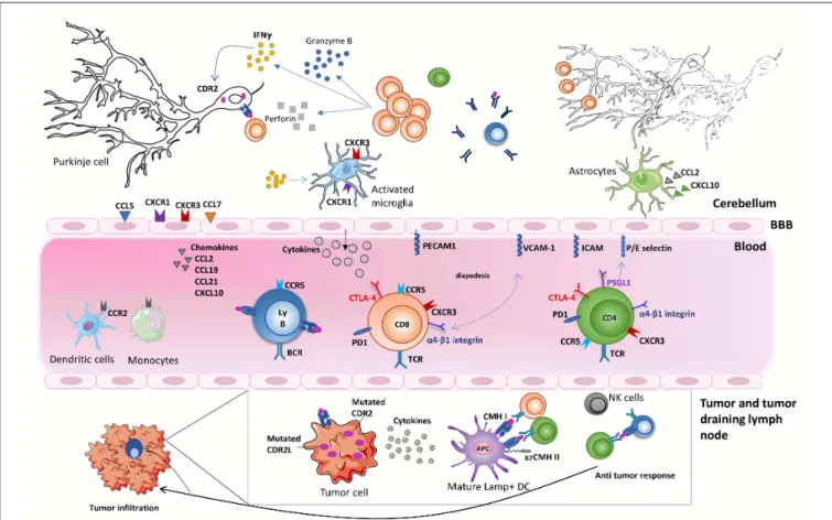
FIGURE 1 | Onconeural antigens such as CDR2 and CDR2L, mutated or not, can be the target of an anti-tumor immune response. Moderate to profound immune infiltrates are found in the primary tumor or metastases of anti-Yo PCD-associated tumors. These infiltrates sometimes form tertiary lymphoid structures. Priming of onconeural antigen-specific CD4 T cells may occur in the tumor-draining lymph nodes or the tumor itself following presentation by local DCs. Differentiation and recruitment of tumor antigen-specific cytotoxic CD8 T-cell likely contribute to the partial tumor control described in patients with PCD. B cells and plasma cells are also found in the tumor. The onconeural antigen-specific T cells reach the CNS after crossing the blood-brain barrier, through a multistep process involving selectins, integrins, and chemokine receptors. The preferential infiltration of the cerebellum in PCD patients could relate to presentation of the onconeural antigens by local APCs and/or to regional peculiarities. In the cerebellum of PCD patients, the inflammatory infiltrates are mostly composed of CD8 T cells, macrophages, and activated microglia. Purkinje neurons are naturally expressing onconeural antigen, such as CDR2 and CDR2L, and up-regulate their expression of MHC class I molecules during an inflammatory process providing the opportunity for CD8 T cells to recognize antigens presented by these neurons. Local CD8 T cells exhibit an activated phenotype with granzyme B- and perforin-containing cytolytic granules. They also secrete locally IFNγ, which has a number of disease-enhancing properties and, thus, is likely a key part of PCD pathogenesis. In particular, IFNγ increases the expression of MHC class I molecules on Purkinje neurons enhances VCAM1 expression on brain endothelial cells and elicits secretion of CXCL10, a ligand of CXCR3 expressed on Th1 cells and activated CD8 T cells. Although antibodies against intracellular onconeural antigens do not appear pathogenic in vivo, they could promote antigen presentation to pathogenic T cells.
the hypothesis that these T cells arise as a consequence of
anti-tumor immunity (
9
,
164
). However, the TCR repertoire
and antigenic specificity of cerebellum-infiltrating CD8 T cells
in PCD are still elusive. Leveraging the latest molecular tools
to address these questions appears to be the next logical
step (
4
,
165
).
Several studies in PCD have focused on CD8 T cells, with
much less emphasis given to CD4 T cells. In other context CD4 T
cells orchestrate functional immune responses by coordinating
immune activity (
166
). CD4 T cells optimize during the priming
the cytotoxic response both in quality and scale, by increasing the
cytotoxic CD8 T cell motility and migratory capacities (
166
). The
expansion of Th1-like CD4 T cells following blockage of
CTLA-4 improves anti-tumor responses by enhancing CD8 infiltration,
cytotoxic CD8 T cells activity, and T cell memory formations
(
160
). It is tempting to speculate that in PCD antigen-specific
Th1-type CD4 T cells are responsible for initiating the disease,
as documented in a rat model (
144
), and as a consequence, these
CD4 T cells are able to recruit and enhance the cytotoxic CD8
T-cell responses.
We have shown in animal models as well as in cerebellar
samples from PCD patients that IFNγ signaling occurs locally
in Purkinje neurons and surrounding cell types (
143
). Moreover,
CXCL10, an IFNγ-induced chemokine, is present at high level in
the CSF of patients with PCD (
167
). We, therefore, hypothesize
that IFNγ stimulates a disease-promoting cascade of events in
the cerebellum and could represent a promising therapeutic
target in PCD. Indeed, autoantigen-reactive CD8 T cells (and
maybe other immune cell types) produce IFNγ locally, which
has a number of disease-enhancing properties. IFNγ increases
the expression of MHC class I molecules on neurons, including
Purkinje cells, which can therefore present self-peptides to the
cytotoxic CD8 T cells, promoting their own destruction (
143
,
168
,
169
). In addition, IFNγ promotes local immune cell recruitment
by enhancing VCAM1 expression on brain endothelial cells
and by eliciting secretion of the chemokine CXCL10, a ligand
of CXCR3 expressed on Th1 cells and activated CD8 T cells.
Importantly, the levels of CXCL10, but not those of CCL2, are
elevated in the CSF of PCD patients indirectly suggesting that the
CXCL10/CXCR3 axis may contribute to the trafficking of T cells
into the cerebellum (
167
).
The role played by activated microglia and microglia
nodules in the disease process is currently unknown. In
Rasmussen encephalitis, microglial nodules have been associated
with neuronal phagocytosis, following CD8 T cell-mediated
brain neuron destruction (
170
). In human PCD, similar to
Rasmussen encephalitis, IFNγ-mediated signaling (STAT1
phosphorylation) occurs in microglial cells, which can further
amplify cytokine/chemokine release (
143
,
170
). Further
investigation on the role of microglia as an amplifier of
inflammation and an executor of neuronal death is needed
in PCD.
As
already
discussed,
antibodies
against
neuronal
autoantigens such as anti-Yo antibodies are extremely useful
diagnostic biomarkers. Their direct pathogenic potential
documented in in vitro models is still unproven in vivo
(
5
,
13
,
149
,
153
,
171
). However, autoreactive B cells could
participate in the development of the disease. They can
uptake (in an antigen-specific manner due to their surface
immunoglobulin), process and present autoantigens to
pathogenic T cells. Their scarcity in the cerebellum would,
however, rather favor an alternative scenario. For instance,
autoantibodies entering the CNS can be up-taken together
with their target antigen by resident antigen-presenting
phagocytes, a phenomenon that enhances the activation
of the incoming effector T cells (
172
). Therefore, the
autoantibodies could cooperate with the T cells and support local
autoimmune neuroinflammation.
POTENTIAL THERAPEUTIC IMPLICATIONS
To date, treatment of PCD is empirical and usually relies
on 2 pillars: treatment of the underlying cancer and general
immunosuppressive drugs. In the absence of large clinical trials,
most of the therapeutics conclusions come from observational
clinical studies and case reports. Concerning PCD associated
with anti-Yo antibodies, corticoids seem ineffective, whereas
plasma exchange and rituximab may have provided some benefit
(
173
–
176
). The efficacy of intravenous immunoglobulins is
controversial: suggested in a small proportion of patients for
some studies (
177
,
178
) but not confirmed in a larger study
(
89
). Since there is currently no evidence arguing for a direct
role of anti-Yo antibodies in Purkinje cell death, we do not
address here potential therapeutic strategies aiming at reducing
antibodies levels.
Due to the intracellular localization of the CDR2/CDR2L and
Hu antigens, as well as the identification of CD8 T cells in close
proximity to neuronal cells, cytotoxic T cells are considered
to be the final effectors responsible for neuronal loss in PCD
(
9
,
140
,
146
,
164
,
179
). These T cells likely contribute to the
tumor control outside of the CNS. Therefore, the conundrum
is how to selectively target the CNS-targeting immune cells
while preserving (as much as possible) the tumor-controlling
counterparts. Uncoupling these two simultaneous immune
responses is, theoretically, possible provided that the molecular
bases of immune cell migration or effector mechanisms at the two
sites differ. It is paramount that the treatment of PCD should be
started as early as possible since Purkinje cells are post-mitotic
cells that do not renew.
The α4β1 integrin is important for both CD4 and CD8 T
cell migration to the CNS and a monoclonal antibody targeting
the α4 subunit provides important benefits for the treatment
of persons with multiple sclerosis. Therefore, one approach
could be to initiate anti-α4 integrin therapy early on in the
disease process in order to preserve as many Purkinje neurons
as possible. One conceptual obstacle is that blood-borne T cells
may not be needed to fuel ongoing tissue destruction by
tissue-resident T cells. This may explain why, in our mouse model of
PCD, blocking α4 integrin early in the disease process did not
yield significant benefit (
143
). Along the same lines, blocking
CXCR3 with either monoclonal antibodies or pharmacological
compounds is tempting, given the evidence of a role for the
CXCL10/CXCR3 axis in PCD. However, to our knowledge, this
approach has not yet reached the clinical setting.
We argued earlier that IFNγ secretion by CD8 T cells could
sustain a feed forward loop by rendering Purkinje cells vulnerable
to direct killing by autoantigen-specific CD8 T cells. If IFNγ plays
a non-redundant role in the progression of PCD, this molecule
could be targeted therapeutically. Our recent pre-clinical data
showing that blocking IFNγ strongly reduces PCD development
without eliciting tumor growth rebound are encouraging (
143
).
Moreover, administration of anti-IFNγ antibodies has been
tested recently in clinical trials. For instance, Emapalumab, a
fully human monoclonal neutralizing anti-IFNγ antibody has
been approved for the treatment of primary hemophagocytic
lymphohistiocytosis, a syndrome of excessive immune activation
and progressive immune-mediated organ damage due to genetic
defects in cell-mediated cytotoxicity (
180
). Taken together, the
accumulating evidence from human samples and the mouse
model as well as the previous development of approved
anti-IFNγ antibody in humans should facilitate the testing of this
strategy in patients with PCD.
CONCLUSION
The precise mechanisms and pathways involved in the
pathogenesis of PCD need to be explored in greater depth.
This can be done with the use of bodily fluids (blood and
CSF) and tissue from PCD patients and further validated in
reductionist mouse models. Key questions remain regarding
the antigenic specificity, phenotype, migration of T and B cells
infiltrating the tumor and the cerebellar tissue. The molecular
dissection of the steps involved in pathogenesis is a
pre-requisite for rational development of new therapeutic strategies.
In depth investigation of the immune changes in patients
suffering from paraneoplastic neurological disorders in the
frame of immune checkpoint blockade should provide clues in
that respect.
AUTHOR CONTRIBUTIONS
LY and CB drafted the work. RL designed the concept and
revised the manuscript critically. All authors made substantial
contributions to the conception or design of the work and
contributed to manuscript revision, read, and approved the
submitted version.
FUNDING
This work of the authors was supported by the RHU BETPSY,
the Foundation pour la Recherche Médicale (FRM), the French
Research Agency (ANR T-cell Mig), ERA-NET NEURON
(Meltra-BBB), and Bristol Myers-Squibb Foundation.
ACKNOWLEDGMENTS
We would like to thank Leila Khajavi and Drs. Abdelhadi Saoudi
for their valuable input on the manuscript.
REFERENCES
1. Waisman A, Liblau RS, Becher B. Innate and adaptive
immune responses in the CNS. Lancet Neurol. (2015) 14:945– 55. doi: 10.1016/S1474-4422(15)00141-6
2. Sulzer D, Alcalay RN, Garretti F, Cote L, Kanter E, Agin-Liebes J, et al. T cells of Parkinson’s disease patients recognize α-synuclein peptides. Nature. (2017) 546:656–61. doi: 10.1038/nature22815
3. Latorre D, Kallweit U, Armentani E, Foglierini M, Mele F, Cassotta A, et al. T cells in patients with narcolepsy target self-antigens of hypocretin neurons. Nature. (2018) 562:63–8. doi: 10.1038/s41586-018-0540-1
4. Gate D, Saligrama N, Leventhal O, Yang AC, Unger MS, Middeldorp J, et al. Clonally expanded CD8 T cells patrol the cerebrospinal fluid in Alzheimer’s disease. Nature. (2020) 577:399–404. doi: 10.1038/s41586-019-1895-7 5. Graus F, Illa I, Agusti M, Ribalta T, Cruz-Sanchez F, Juarez
C. Effect of intraventricular injection of an anti-Purkinje cell antibody (anti-Yo) in a guinea pig model. J Neurol Sci. (1991) 106:82–7. doi: 10.1016/0022-510X(91)90198-G
6. Furneaux HM, Rosenblum MK, Dalmau J, Wong E, Woodruff P, Graus F, et al. Selective expression of Purkinje cell antigens in tumor tissue from patients with paraneoplastic cerebellar degeneration. N Engl J Med. (1990) 322:1844–51. doi: 10.1056/NEJM199006283222604
7. Dalmau J, Rosenfeld MR. Paraneoplastic syndromes of the CNS. Lancet Neurol. (2008) 7:327–40. doi: 10.1016/S1474-4422(08)70060-7
8. Zou W, Wolchok JD, Chen L. PD-L1 (B7-H1) and PD-1
pathway blockade for cancer therapy. Sci Transl Med. (2016) 8:328rv4. doi: 10.1126/scitranslmed.aad7118
9. Albert ML, Darnell JC, Bender A, Francisco LM, Bhardwaj N, Darnell RB. Tumor-specific killer cells in paraneoplastic cerebellar degeneration. Nat Med. (1998) 4:1321–4. doi: 10.1038/3315
10. Roberts WK, Deluca IJ, Thomas A, Fak J, Williams T, Buckley N, et al. Patients with lung cancer and paraneoplastic Hu syndrome harbor
HuD-specific type 2 CD8+ T cells. J Clin Invest. (2009) 119:2042– 51. doi: 10.1172/JCI36131
11. Hillary RP, Ollila HM, Lin L, Desestret V, Rogemond V,
Picard G, et al. Complex HLA association in paraneoplastic
cerebellar ataxia with anti-Yo antibodies. J Neuroimmunol. (2018) 315:28–32. doi: 10.1016/j.jneuroim.2017.12.012
12. Peterson K, Rosenblum MK, Kotanides H, Posner JB. Paraneoplastic cerebellar degeneration : I. A clinical analysis of 55 anti-Yo antibody-positive patients. Neurology. (1992) 42:1931–7. doi: 10.1212/WNL.42.10.1931 13. Schubert M, Panja D, Haugen M, Bramham CR, Vedeler CA. Paraneoplastic
CDR2 and CDR2L antibodies affect Purkinje cell calcium homeostasis. Acta Neuropathol. (2014) 128:835–52. doi: 10.1007/s00401-014-1351-6 14. Eichler TW, Totland C, Haugen M, Qvale TH, Mazengia K, Storstein A, et al.
CDR2L antibodies : a new player in paraneoplastic cerebellar degeneration. PLoS ONE. (2013) 8:e66002. doi: 10.1371/journal.pone.0066002
15. Lucchinetti CF, Kimmel DW, Lennon VA. Paraneoplastic and oncologic profiles of patients seropositive for type 1 antineuronal nuclear autoantibodies. Neurology. (1998) 50:652–7. doi: 10.1212/WNL.50.3.652 16. Graus F, Keime-Guibert F, Rene R, Benyahia B, Ribalta T, Ascaso C,
et al. Anti-Hu-associated paraneoplastic encephalomyelitis : analysis of 200 patients. Brain. (2001) 124:1138–48. doi: 10.1093/brain/124.6.1138
17. Dalmau J, Graus F, Rosenblum M, Posner JB.
Anti-Hu-associated paraneoplastic encephalomyelitis/sensory neuronopathy
: a clinical study of 71 patients. Medicine (Baltimore). (1992) 71:59–72. doi: 10.1097/00005792-199203000-00001
18. Honnorat J, Saiz A, Giometto B, Vincent A, Brieva L, de Andres C, et al. Cerebellar ataxia with anti-glutamic acid decarboxylase antibodies: study of 14 patients. Arch Neurol. (2001) 58:225–30. doi: 10.1001/archneur.58.2.225 19. Jarius S, Stich O, Speck J, Rasiah C, Wildemann B, Meinck HM, et al.
Qualitative and quantitative evidence of anti-glutamic acid decarboxylase-specific intrathecal antibody synthesis in patients with stiff person syndrome. J Neuroimmunol. (2010) 229:219–24. doi: 10.1016/j.jneuroim.2010.07.019
20. Kono S, Miyajima H, Sugimoto M, Suzuki Y, Takahashi Y, Hishida A. Stiff-person syndrome associated with cerebellar ataxia and high glutamic acid decarboxylase antibody titer. Intern Med. (2001) 40:968– 71. doi: 10.2169/internalmedicine.40.968
21. Piccolo G, Tavazzi E, Cavallaro T, Romani A, Scelsi R, Martino
G. Clinico-pathological findings in a patient with progressive
cerebellar ataxia, autoimmune polyendocrine syndrome, hepatocellular
carcinoma and anti-GAD autoantibodies. J Neurol Sci. (2010)
290:148–9. doi: 10.1016/j.jns.2009.12.006
22. Saiz A, Blanco Y, Sabater L, González F, Bataller L, Casamitjana R, et al. Spectrum of neurological syndromes associated with glutamic acid decarboxylase antibodies: diagnostic clues for this association. Brain. (2008) 131:2553–63. doi: 10.1093/brain/awn183
23. Jarius S, Wandinger KP, Horn S, Heuer H, Wildemann B. A new Purkinje cell antibody (anti-Ca) associated with subacute cerebellar ataxia: immunological characterization. J Neuroinflammation. (2010) 7:21. doi: 10.1186/1742-2094-7-21
24. Jarius S, Martínez-García P, Hernandez AL, Brase JC, Borowski K, Regula JU, et al. Two new cases of anti-Ca (anti-ARHGAP26/GRAF) autoantibody-associated cerebellar ataxia. J Neuroinflammation. (2013) 10:7. doi: 10.1186/1742-2094-10-7
25. Doss S, Nümann A, Ziegler A, Siebert E, Borowski K, Stöcker
W, et al. Anti-Ca/anti-ARHGAP26 antibodies associated with
cerebellar atrophy and cognitive decline. J Neuroimmunol. (2014) 267:102–4. doi: 10.1016/j.jneuroim.2013.10.010
26. Bartels F, Prüss H, Finke C. Anti-ARHGAP26 autoantibodies are associated with isolated cognitive impairment. Front Neurol. (2018) 9:656. doi: 10.3389/fneur.2018.00656
27. Wallwitz U, Brock S, Schunck A, Wildemann B, Jarius S, Hoffmann F. From dizziness to severe ataxia and dysarthria: new cases of anti-Ca/ARHGAP26 autoantibody-associated cerebellar ataxia suggest a broad clinical spectrum. J Neuroimmunol. (2017) 309:77–81. doi: 10.1016/j.jneuroim.2017.05.011 28. Shan F, Long Y, Qiu W. Autoimmune glial fibrillary acidic protein
astrocytopathy: a review of the literature. Front Immunol. (2018) 9:2802. doi: 10.3389/fimmu.2018.02802
29. Yu Z, Kryzer TJ, Griesmann GE, Kim KK, Benarroch EE,
Lennon VA. CRMP-5 neuronal autoantibody: marker of lung
cancer and thymoma-related autoimmunity. Ann Neurol. (2001) 49:146–54. doi: 10.1002/1531-8249(20010201)49:2<146::aid-ana34>3.0.co 30. Antoine JC, Honnorat J, Vocanson C, Koenig F, Aguera M, Belin MF,
et al. Posterior uveitis, paraneoplastic encephalomyelitis and auto-antibodies reacting with developmental protein of brain and retina. J Neurol Sci. (1993) 117:215–23. doi: 10.1016/0022-510X(93)90176-Y
31. Honnorat J, Cartalat-Carel S, Ricard D, Camdessanche JP, Carpentier AF, Rogemond V, et al. Onco-neural antibodies and tumour type determine survival and neurological symptoms in paraneoplastic neurological syndromes with Hu or CV2/CRMP5 antibodies. J Neurol Neurosurg Psychiatry. (2009) 80:412–6. doi: 10.1136/jnnp.2007.138016
32. Honnorat J, Antoine JC, Derrington E, Aguera M, Belin MF. Antibodies to a subpopulation of glial cells and a 66 kDa developmental protein in patients with paraneoplastic neurological syndromes. J Neurol Neurosurg Psychiatry. (1996) 61:270–8. doi: 10.1136/jnnp.61.3.270
33. Lopez-Chiriboga AS, Komorowski L, Kümpfel T, Probst C, Hinson SR, Pittock SJ, et al. Metabotropic glutamate receptor type 1 autoimmunity : clinical features and treatment outcomes. Neurology. (2016) 86:1009– 13. doi: 10.1212/WNL.0000000000002476
34. Marignier R, Chenevier F, Rogemond V, Sillevis Smitt P, Renoux C, Cavillon G, et al. Metabotropic glutamate receptor type 1 autoantibody–associated cerebellitis. Arch Neurol. (2010) 67:627–30. doi: 10.1001/archneurol.2010.51 35. Sillevis-Smitt P, Kinoshita M, De Leeuw B, Moll W, Coesmans M, Jaarsma D, et al. Paraneoplastic cerebellar ataxia due to autoantibodies against glutamate receptor. N Engl J Med. (2000) 342:21–7. doi: 10.1056/NEJM200001063420104
36. Iorio R, Damato V, Mirabella M, Vita MG, Hulsenboom E, Plantone D, et al. Cerebellar degeneration associated with mGluR1 autoantibodies as a paraneoplastic manifestation of prostate adenocarcinoma. J Neuroimmunol. (2013) 263:155–8. doi: 10.1016/j.jneuroim.2013.07.015
37. Mason WP, Graus F, Lang B, Honnorat J, Delattre JY, Valldeoriola F, et al. Small-cell lung cancer, paraneoplastic cerebellar degeneration and the Lambert-Eaton myasthenic syndrome. Brain. (1997) 120:1279– 300. doi: 10.1093/brain/120.8.1279
38. Graus F, Lang B, Pozo-Rosich P, Saiz A, Casamitjana R, Vincent A. P / Q type calcium- channel antibodies in paraneoplastic cerebellar degeneration with lung cancer. Neurology. (2002) 59:764–6. doi: 10.1212/WNL.59.5.764 39. Bürk K, Wick M, Roth G, Decker P, Voltz R. Antineuronal
antibodies in sporadic late-onset cerebellar ataxia. J Neurol. (2010) 257:59–62. doi: 10.1007/s00415-009-5262-8
40. Pittock SJ, Lucchinetti CF, Parisi JE, Benarroch EE, Mokri B, Stephan CL, et al. Amphiphysin autoimmunity: paraneoplastic accompaniments. Ann Neurol. (2005) 58:96–107. doi: 10.1002/ana.20529
41. Moon J, Lee S-T, Shin J-W, Byun J-I, Lim J-A, Shin Y-W, et al.
Non-stiff anti-amphiphysin syndrome: clinical manifestations
and outcome after immunotherapy. J Neuroimmunol. (2014)
274:209–14. doi: 10.1016/j.jneuroim.2014.07.011
42. Balint B, Jarius S, Nagel S, Haberkorn U, Probst C, Blöcker
IM, et al. Progressive encephalomyelitis with rigidity and
myoclonus: a new variant with DPPX antibodies. Neurology. (2014) 82:1521–8. doi: 10.1212/WNL.0000000000000372
43. Boronat A, Gelfand JM, Gresa-Arribas N, Jeong HY, Walsh M, Roberts K, et al. Encephalitis and antibodies to dipeptidyl-peptidase-like protein-6, a subunit of Kv4.2 potassium channels. Ann Neurol. (2013) 73:120– 8. doi: 10.1002/ana.23756
44. Tobin WO, Lennon VA, Komorowski L, Probst C, Clardy SL, Aksamit AJ, et al. DPPX potassium channel antibody: Frequency, clinical accompaniments, and outcomes in 20 patients. Neurology. (2014) 83:1797–803. doi: 10.1212/WNL.0000000000000991
45. Stoeck K, Carstens PO, Jarius S, Raddatz D, Stöcker W, Wildemann B, et al. Prednisolone and azathioprine are effective in DPPX antibody-positive autoimmune encephalitis. Neurol Neuroimmunol NeuroInflamm. (2015) 2:e86. doi: 10.1212/NXI.0000000000000086
46. Becker EBE, Zuliani L, Pettingill R, Lang B, Waters P, Dulneva
A, et al. Contactin-associated protein-2 antibodies in
non-paraneoplastic cerebellar ataxia. J Neurol Neurosurg Psychiatry. (2012) 83:437–40. doi: 10.1136/jnnp-2011-301506
47. Balint B, Regula JU, Jarius S, Wildemann B. Caspr2 antibodies in limbic encephalitis with cerebellar ataxia, dyskinesias and myoclonus. J Neurol Sci. (2013) 327:73–4. doi: 10.1016/j.jns.2013.01.040
48. Kannoth S, Nambiar V, Gopinath S, Anandakuttan A, Mathai A, Rajan PK. Expanding spectrum of contactin-associated protein 2 (CASPR2) autoimmunity—syndrome of parkinsonism and ataxia. Neurol Sci. (2018) 39:455–60. doi: 10.1007/s10072-017-3222-0
49. Boronat A, Sabater L, Saiz A, Dalmau J, Graus F. GABA(B)
receptor antibodies in limbic encephalitis and
anti-GAD-associated neurologic disorders. Neurology. (2011) 76:795–
800. doi: 10.1212/WNL.0b013e31820e7b8d
50. Lancaster E, Lai M, Peng X, Hughes E, Constantinescu R, Raizer J, et al. Antibodies to the GABA(B) receptor in limbic encephalitis with seizures: case series and characterisation of the antigen. Lancet Neurol. (2010) 9:67– 76. doi: 10.1016/S1474-4422(09)70324-2
51. Jarius S, Steinmeyer F, Knobel A, Streitberger K, Hotter B, Horn S, et al. GABA B receptor antibodies in paraneoplastic cerebellar ataxia. J Neuroimmunol. (2013) 256:94–6. doi: 10.1016/j.jneuroim.2012.12.006 52. Graus F, Vincent A, Pozo-Rosich P, Sabater L, Saiz A, Lang B,
et al. Anti-glial nuclear antibody: marker of lung cancer-related
paraneoplastic neurological syndromes. J Neuroimmunol. (2005)
165:166–71. doi: 10.1016/j.jneuroim.2005.03.020
53. Titulaer MJ, Klooster R, Potman M, Sabater L, Graus F, Hegeman IM, et al. SOX antibodies in small-cell lung cancer and Lambert-Eaton myasthenic syndrome: frequency and relation with survival. J Clin Oncol. (2009) 27:4260–7. doi: 10.1200/JCO.2008.20.6169
54. Hoffmann LA, Jarius S, Pellkofer HL, Schueller M, Krumbholz M, Koenig F, et al. Anti-Ma and anti-Ta associated paraneoplastic neurological syndromes: 22 newly diagnosed patients and review of previous cases. J Neurol Neurosurg Psychiatry. (2008) 79:767–73. doi: 10.1136/jnnp.2007.118588
55. Dalmau J, Graus F, Villarejo A, Posner JB, Blumenthal D, Thiessen B, et al. Clinical analysis of anti-Ma2-associated encephalitis. Brain. (2004) 127:1831–44. doi: 10.1093/brain/awh203
56. Voltz R, Gultekin SH, Rosenfeld MR, Gerstner E, Eichen J, Posner JB, et al. A serologic marker of paraneoplastic limbic and brain-stem encephalitis in patients with testicular cancer. N Engl J Med. (1999) 340:1788– 95. doi: 10.1056/NEJM199906103402303
57. Dalmau J, Gultekin SH, Voltz R, Hoard R, DesChamps T, Balmaceda C, et al. Ma1, a novel neuron- and testis-specific protein, is recognized by the serum of patients with paraneoplastic neurological disorders. Brain. (1999) 122:27–39. doi: 10.1093/brain/122.1.27
58. Pittock SJ, Lucchinetti CF, Lennon VA. Anti-neuronal nuclear autoantibody type 2: paraneoplastic accompaniments. Ann Neurol. (2003) 53:580– 7. doi: 10.1002/ana.10518
59. Budde-Steffen C, Anderson NE, Rosenblum MK, Graus F, Ford D, Synek BJL, et al. An antineuronal autoantibody in paraneoplastic opsoclonus. Ann Neurol. (1988) 23:528–31. doi: 10.1002/ana.410230518
60. O’Leary CG, Battley JE, O’Reilly S. Anti-Ri antibody associated
small cell lung carcinoma. Ir J Med Sci. (2016) 186:265–
7. doi: 10.1007/s11845-016-1402-1
61. Vernino S, Lennon VA. New Purkinje cell antibody (PCA-2): marker of lung cancer-related neurological autoimmunity. Ann Neurol. (2000) 47:297–305. doi: 10.1002/1531-8249(200003)47:3<297::AID-ANA4>3.0.CO;2-4 62. Chan KH, Vernino S, Lennon VA. ANNA-3 anti-neuronal nuclear antibody:
marker of lung cancer-related autoimmunity. Ann Neurol. (2001) 50:301– 11. doi: 10.1002/ana.1127
63. Bataller L, Wade DF, Fuller GN, Rosenfeld MR, Dalmau J. Cerebellar degeneration and autoimmunity to zinc-finger proteins of the cerebellum. Neurology. (2002) 59:1985–7. doi: 10.1212/01.WNL.0000038352.01415.CE
64. Bataller L, Wade DF, Graus F, Stacey HD, Rosenfeld MR,
Dalmau J. Antibodies to Zic4 in paraneoplastic neurologic
disorders and small-cell lung cancer. Neurology. (2004) 62:778– 82. doi: 10.1212/01.WNL.0000113749.77217.01
65. Sabater L, Bataller L, Suarez-Calvet M, Saiz A, Dalmau J, Graus F. ZIC antibodies in paraneoplastic cerebellar degeneration and
small cell lung cancer. J Neuroimmunol. (2008) 201–2:163–
5. doi: 10.1016/j.jneuroim.2008.01.018
66. Zuliani L, Sabater L, Saiz A, Baiges J, Giometto B, Graus F. Homer 3 autoimmunity in subacute idiopathic cerebellar ataxia. Neurology. (2007) 68:239–40. doi: 10.1212/01.wnl.0000251308.79366.f9
67. Höftberger R, Sabater L, Ortega A, Dalmau J, Graus F. Patient with homer-3 antibodies and cerebellitis. JAMA Neurol. (2013) 70:506– 9. doi: 10.1001/jamaneurol.2013.1955
68. Jarius S, Scharf M, Begemann N, Stöcker W, Probst C, Serysheva II, et al. Antibodies to the inositol 1,4,5-trisphosphate receptor type 1 (ITPR1) in cerebellar ataxia. J Neuroinflammation. (2014) 11:206. doi: 10.1186/s12974-014-0206-3
69. Berzero G, Hacohen Y, Komorowski L, Scharf M, Dehais C, Leclercq D, et al. Paraneoplastic cerebellar degeneration associated with anti-ITPR1 antibodies. Neurol Neuroimmunol NeuroInflammation. (2017) 4:e326. doi: 10.1212/NXI.0000000000000326
70. Bataller L, Sabater L, Saiz A, Serra C, Claramonte B, Graus F. Carbonic anhydrase-related protein VlII: autoantigen in paraneoplastic cerebellar degeneration. Ann Neurol. (2004) 56:575–9. doi: 10.1002/ana.20238 71. Höftberger R, Sabater L, Velasco F, Ciordia R, Dalmau J, Graus F.
Carbonic anhydrase-related protein VIII antibodies and paraneoplastic cerebellar degeneration. Neuropathol Appl Neurobiol. (2014) 40:650– 3. doi: 10.1111/nan.12118
72. de Graaff E, Maat P, Hulsenboom E, van Den Berg R, van Den Bent M, Demmers J, et al. Identification of delta/notch-like epidermal growth factor-related receptor as the Tr antigen in paraneoplastic cerebellar degeneration. Ann Neurol. (2012) 71:815–24. doi: 10.1002/ana.23550
73. Probst C, Komorowski L, de Graaff E, van Coevorden-Hameete M, Rogemond V, Honnorat J, et al. Standardized test for anti-Tr/DNER in patients with paraneoplastic cerebellar degeneration. Neurol Neuroimmunol Neuroinflamm. (2015) 2:e68. doi: 10.1212/NXI.0000000000000068 74. Graus F, Dalmau J, Valldeoriola F, Ferrer I, Reñé R, Marin C, et al.
Immunological characterization of a neuronal antibody (anti-Tr) associated with paraneoplastic cerebellar degeneration and Hodgkin’s disease. J
75. Trotter JL, Hendin BA, Osterland K. Cerebellar degeneration
with Hodgkin’s disease. Arch Neurol. (1976) 33:660–
1. doi: 10.1001/archneur.1976.00500090066014
76. Bernal F, Shams’ili S, Rojas I, Sanchez-Valle R, Saiz A, Dalmau
J, et al. Anti-Tr antibodies as markers of paraneoplastic
cerebellar degeneration and Hodgkin’s disease. Neurology. (2003) 60:230–4. doi: 10.1212/01.WNL.0000041495.87539.98
77. van Coevorden-Hameete MH, van Beuningen SFB, Perrenoud M, Will LM, Hulsenboom E, Demonet JF, et al. Antibodies to TRIM46 are associated with paraneoplastic neurological syndromes. Ann Clin Transl Neurol. (2017) 4:680–6. doi: 10.1002/acn3.396
78. Do LD, Gupton SL, Tanji K, Bastien J, Brugière S, Couté Y, et al. TRIM9 and TRIM67 are new targets in paraneoplastic cerebellar degeneration. Cerebellum. (2019) 18:245–54. doi: 10.1007/s12311-018-0987-5
79. Larman HB, Zhao Z, Laserson U, Li MZ, Ciccia A, Gakidis MAM, et al. Autoantigen discovery with a synthetic human peptidome. Nat Biotechnol. (2011) 29:535–41. doi: 10.1038/nbt.1856
80. Shimizu F, Takeshita Y, Sano Y, Hamamoto Y, Shiraishi H, Sato T, et al. GRP78 antibodies damage the blood-brain barrier and relate to cerebellar degeneration in Lambert-Eaton myasthenic syndrome. Brain. (2019) 142:2253–64. doi: 10.1093/brain/awz168
81. van Coevorden-Hameete MH, de Graaff E, Titulaer MJ, Hulsenboom
E, Sabater L, Hoogenraad CC, et al. Plasticity-related gene
5: a novel surface autoantigen in paraneoplastic cerebellar
degeneration. Neurol Neuroimmunol NeuroInflamm. (2015)
2:e156. doi: 10.1212/NXI.0000000000000156
82. Shelly S, Kryzer TJ, Komorowski L, Miske R, Anderson MD, Flanagan EP, et al. Neurochondrin neurological autoimmunity. Neurol Neuroimmunol
Neuroinflamm. (2019) 6:e612. doi: 10.1212/NXI.00000000000
00612
83. Miske R, Gross CC, Scharf M, Golombeck KS, Hartwig M, Bhatia U, et al. Neurochondrin is a neuronal target antigen in autoimmune cerebellar degeneration. Neurol Neuroimmunol NeuroInflamm. (2017) 4:e307. doi: 10.1212/NXI.0000000000000307
84. Honorat JA, Sebastian Lopez-Chiriboga A, Kryzer TJ,
Fryer JP, Devine M, Flores A, et al. Autoimmune septin-5
cerebellar ataxia. Neurol Neuroimmunol NeuroInflamm. (2018)
5:e474. doi: 10.1212/NXI.0000000000000474
85. Vogrig A, Gigli GL, Segatti S, Corazza E, Marini A, Bernardini A, et al. Epidemiology of paraneoplastic neurological syndromes: a population-based study. J Neurol. (2020) 267:26–35. doi: 10.1007/s00415-019-09544-1 86. Garcia Cuartero I, Rodriguez Galdeano M, Perez Pinar M, Solis Garcia
del Pozo J. Thymic carcinoma associated with cerebellar degeneration. Eur J Case Rep Intern Med. (2018) 5:000780. doi: 10.12890/2018_ 000780
87. Ducray F, Demarquay G, Graus F, Decullier E, Antoine JC, Giometto B, et al. Seronegative paraneoplastic cerebellar degeneration: the PNS Euronetwork experience. Eur J Neurol. (2014) 21:731–5. doi: 10.1111/ene.12368 88. Joubert B, Rostásy K, Honnorat J. Immune-mediated ataxias. Handb
Clin Neurol. (2018) 155:313–32. doi: 10.1016/B978-0-444-64189-2.
00021-4
89. Shams’ili S, Grefkens J, de Leeuw B, van den Bent M, Hooijkaas H, van der Holt B, et al. Paraneoplastic cerebellar degeneration associated with antineuronal antibodies: analysis of 50 patients. Brain. (2003) 126:1409– 18. doi: 10.1093/brain/awg133
90. Jarius S, Wildemann B. “Medusa-head ataxia”: the expanding spectrum of Purkinje cell antibodies in autoimmune cerebellar ataxia. Part 1: Anti-mGluR1, anti-Homer-3, anti-Sj/ITPR1 anti-CARP VIII. J Neuroinflammation. (2015) 12:166. doi: 10.1186/s12974-015-0356-y 91. Venkatraman A, Opal P. Paraneoplastic cerebellar degeneration with
anti-Yo antibodies – a review. Ann Clin Transl Neurol. (2016) 3:655– 63. doi: 10.1002/acn3.328
92. Drlicek M, Bianchi G, Bogliun G, Casati B, Grisold W, Kolig C, et al. Antibodies of the anti-Yo and anti-Ri type in the absence of paraneoplastic neurological syndromes: a long-term survey of ovarian cancer patients. J Neurol. (1997) 244:85–9. doi: 10.1007/s004150050054
93. Rana AQ, Rana AN, Adlul A. Acute ataxia due to anti-Yo antibody paraneoplastic cerebellar degeneration 4 months prior