HAL Id: hal-03095909
https://hal.archives-ouvertes.fr/hal-03095909
Submitted on 2 Feb 2021
HAL is a multi-disciplinary open access
archive for the deposit and dissemination of
sci-entific research documents, whether they are
pub-lished or not. The documents may come from
teaching and research institutions in France or
abroad, or from public or private research centers.
L’archive ouverte pluridisciplinaire HAL, est
destinée au dépôt et à la diffusion de documents
scientifiques de niveau recherche, publiés ou non,
émanant des établissements d’enseignement et de
recherche français ou étrangers, des laboratoires
publics ou privés.
Pertussis Toxin Reduces the Number of Splenic Foxp3 +
Regulatory T Cells
Cécile Cassan, Eliane Piaggio, Jacques Zappulla, Lennart Mars, Nicolas
Couturier, Florence Bucciarelli, Sabine Desbois, Jan Bauer, Daniel
Gonzalez-Dunia, Roland Liblau
To cite this version:
Cécile Cassan, Eliane Piaggio, Jacques Zappulla, Lennart Mars, Nicolas Couturier, et al.. Pertussis
Toxin Reduces the Number of Splenic Foxp3 + Regulatory T Cells. Journal of Immunology, Publisher :
Baltimore : Williams & Wilkins, c1950-. Latest Publisher : Bethesda, MD : American Association of
Immunologists, 2006, 177 (3), pp.1552-1560. �10.4049/jimmunol.177.3.1552�. �hal-03095909�
of September 17, 2018.
This information is current as
Regulatory T Cells
+
Splenic Foxp3
Pertussis Toxin Reduces the Number of
Liblau
Desbois, Jan Bauer, Daniel Gonzalez-Dunia and Roland S.
T. Mars, Nicolas Couturier, Florence Bucciarelli, Sabine
Cécile Cassan, Eliane Piaggio, Jacques P. Zappulla, Lennart
http://www.jimmunol.org/content/177/3/1552
doi: 10.4049/jimmunol.177.3.1552
2006; 177:1552-1560; ;
J Immunol
References
http://www.jimmunol.org/content/177/3/1552.full#ref-list-1
, 44 of which you can access for free at:
cites 73 articles
This article
average *
4 weeks from acceptance to publication
Fast Publication!
•
Every submission reviewed by practicing scientists
No Triage!
•
from submission to initial decision
Rapid Reviews! 30 days*
•
Submit online.
?
The JI
Why
Subscription
http://jimmunol.org/subscription
is online at:
The Journal of Immunology
Information about subscribing to
Permissions
http://www.aai.org/About/Publications/JI/copyright.html
Submit copyright permission requests at:
Email Alerts
http://jimmunol.org/alerts
Receive free email-alerts when new articles cite this article. Sign up at:
Print ISSN: 0022-1767 Online ISSN: 1550-6606.
Immunologists All rights reserved.
Copyright © 2006 by The American Association of
1451 Rockville Pike, Suite 650, Rockville, MD 20852
The American Association of Immunologists, Inc.,
is published twice each month by
The Journal of Immunology
by guest on September 17, 2018 http://www.jimmunol.org/ Downloaded from by guest on September 17, 2018 http://www.jimmunol.org/ Downloaded from
Pertussis Toxin Reduces the Number of Splenic Foxp3
ⴙ
Regulatory T Cells
1
Ce´cile Cassan,* Eliane Piaggio,* Jacques P. Zappulla,* Lennart T. Mars,* Nicolas Couturier,*
Florence Bucciarelli,* Sabine Desbois,* Jan Bauer,
†Daniel Gonzalez-Dunia,* and
Roland S. Liblau*
Pertussis toxin (PTx) is a bacterial toxin used to enhance the severity of experimental autoimmune diseases such as experimental autoimmune encephalomyelitis. It is known to promote permeabilization of the blood-brain barrier, maturation of APC, activation of autoreactive lymphocytes and alteration of lymphocyte migration. In this study, we show that i.v. injection of PTx in mice induces a decrease in the number of splenic CD4ⴙCD25ⴙregulatory T cells (Treg cells). Furthermore, PTx not only induces a depletion of the dominant CD4ⴙCD25ⴙFoxp3ⴙsubpopulation of splenic Treg cells, but also reduces to a similar extent the CD4ⴙCD25ⴚFoxp3ⴙsubpopulation. On a per cell basis, the suppressive properties of the remaining Treg cells are not modified by PTx treatment. The reduction in splenic Treg cells is associated with preferential migration of these cells to the liver. Addi-tionally, Treg cells exhibit a high sensitivity to PTx-mediated apoptosis in vitro. Finally, in vivo depletion of Treg cells by injection of an anti-CD25 Ab, and PTx treatment, present synergistic experimental autoimmune encephalomyelitis exacerbating effects. Therefore, we identify a new effect of PTx and provide an additional illustration of the influence of microbial components on the immune system affecting the balance between tolerance, inflammation and autoimmunity. The Journal of Immunology, 2006, 177: 1552–1560.
P
ertussis toxin (PTx)3has been used for over three decadesas an adjuvant allowing to enhance experimental autoim-mune encephalomyelitis (EAE) severity in permissive strains and even to render resistant strains susceptible (1, 2). Its disease-enhancing properties are not restricted to CNS autoimmu-nity, but also apply to other animal models of autoimmunity such as orchitis (3), uveitis (4, 5), or inflammatory myopathy (6). This 105-kDa protein complex is produced by Bordetella pertussis, the bacterium responsible for whooping cough in humans. PTx is immu-nogenic and involved in the pathogenesis of B. pertussis infection (7, 8) and, therefore, has been included in some acellular pertussis vac-cines in a chemically or genetically detoxified form (9).
The five proteins that compose PTx (S1, S2, S3, S4, and S5) are encoded by genes organized in a single operon (10). PTx belongs to the A-B class of exotoxins. The B subunit, which is a pentamer (S2, S3, S4, and S5 with a 1:1:2:1 stoichiometry) presents se-quence homologies with selectins. It binds glycoproteins (N-linked oligosaccharides and sialylated glycoconjugates) and glycolipids (glucosylceramide, lactosylceramide) expressed on the surface of many eukaryotic cell types (11–13). The A subunit, a single
pro-tein (S1), is then released into the cytoplasm and mediates ADP-ribosylation of the␣-subunit of Gi proteins. Thereby, it interferes with the inhibitory activity of Gi proteins on adenylate cyclase, inducing an increase of intracellular cAMP level, and uncouples G-coupled receptors from their signaling pathway (14, 15).
Numerous immunological effects have been attributed to PTx. On the innate immune system, PTx decreases the production of IL-6 and IL-10 by mast cells (16), promotes maturation of APC leading to the up-regulation of MHC class II or costimulatory mol-ecules and production of IL-12 (4, 9, 17, 18). On the adaptive immune system, PTx increases both Th1 and Th2 responses (19 – 21), and inhibits chemokine-induced lymphocyte migration (22, 23). Classically, the exacerbating effect of PTx on EAE was at-tributed to increased sensitization to histamine (24, 25), and per-meabilization of the blood-brain barrier (24, 26). More recently, PTx was shown to enhance rolling and adhesion of activated T cells on pial vessels. This effect was related to cerebrovascular induction of P-selectin expression, and depended on TLR4 signal-ing (27, 28). Thus, followsignal-ing administration of PTx, several mech-anisms concur to exacerbate autoimmune diseases through en-hanced activation of autoaggressive T cells (29) and increased infiltration of the target tissue.
Given the pleiotropic effects of PTx on the immune system, we hypothesized that it might affect CD4⫹CD25⫹regulatory T (Treg) cells and thereby contribute to its EAE-enhancing properties. In-deed, Treg cells represent a natural population of regulatory cells that plays a major role in the maintenance of peripheral tolerance (30). They develop in the thymus and the transcription factor Foxp3 is essential for Treg lineage specification and function (30, 31). They have been shown to play an important role in the pro-tection against EAE (32–35). Moreover, altered functions of Treg cells from blood of multiple sclerosis patients have recently been identified, inferring that they could also be crucial in regulating immune responses in this disease (36, 37). We thus investigated whether PTx could quantitatively or qualitatively affect Treg cells *Institut National de la Sante´ et de la Recherche Me´dicale (INSERM) Unite´ 563,
Centre de Physiopathologie de Toulouse Purpan (CPTP), Toulouse, France; and
†
Brain Research Institute, University of Vienna, Vienna, Austria
Received for publication February 3, 2006. Accepted for publication May 17, 2006. The costs of publication of this article were defrayed in part by the payment of page charges. This article must therefore be hereby marked advertisement in accordance with 18 U.S.C. Section 1734 solely to indicate this fact.
1
This work was supported by the Association pour la Recherche sur la Scle´rose en Plaques, INSERM, the European Union (Neuropromise; LSHM-CT-2005-018637), and the Region Midi-Pyre´ne´es.
2
Address correspondence and reprint requests to Dr. Roland S. Liblau, INSERM U563, CPTP, Baˆt. B, CHU Purpan, BP3028, 31024 Toulouse cedex 3, France. E-mail: rolandliblau@hotmail.com
3
Abbreviations used in this paper: PTx, pertussis toxin; EAE, experimental autoim-mune encephalomyelitis; Treg, regulatory T; PI, propidium iodide; MFI, mean fluo-rescence intensity.
The Journal of Immunology
Copyright © 2006 by The American Association of Immunologists, Inc. 0022-1767/06/$02.00
by guest on September 17, 2018
http://www.jimmunol.org/
and whether this effect could contribute to its adjuvant activity in EAE.
Materials and Methods
MiceFemale C57BL/6 mice of 6 –12 wk of age were purchased from Charles River Laboratories and were bred at the IFR30 animal facility under spe-cific pathogen-free conditions. Congenic CD45.1 mice were either pur-chased from CDTA (Centre National de la Recherche Scientifique) or from our animal house facility (IFR30, Toulouse). All experimental protocols were approved by the local ethic committee on animal experimentation and are in compliance with European Union guidelines.
Abs and PTx
PTx (⬃99% pure) was from List Biological Laboratories. Anti-CD4-FITC (RM4-5), CD8-FITC (53-6.7), CD45.2-FITC (104), and anti-B220-PE (RA3-6B2) mAbs were obtained from BD Pharmingen. Anti-CD25-PE (PC61), anti-CD4-PE (CT-CD4) mAbs, and Streptavidin Tri-Color conjugate were purchased from Caltag Laboratories. Anti-CD4-allophycocyanin (RM4-5) and anti-Foxp3-PE (FJK-16s) mAbs were ob-tained from eBioscience. Abs used for cell purification were produced in the laboratory (anti-CD8, anti-B220, anti-MHC class II, and anti-Mac-1).
Cell purification
For the purification of CD4⫹CD25⫹or CD4⫹CD25⫺T cells, spleen and peripheral lymph nodes were recovered. Negative selection of CD4⫹cells was performed by incubating the single cell suspension with anti-CD8 (53-6.7), B220 (RA3-6B2), MHC class II (M5–114), and anti-Mac-1 (M1/70) mAbs, followed by depletion with magnetic beads coated with anti-rat IgG (Dynal Biotech). Cells were then incubated with a bio-tinylated anti-CD25 mAb (7D4), and positively selected using streptavidin-conjugated microbeads from Miltenyi Biotec. Cell separation was per-formed on MS columns from Miltenyi Biotec. Purity was⬃80% for the CD4⫹CD25⫺population or the CD4⫹CD25⫹population.
For the in vitro assessment of Treg cell function, CD4⫹CD25brightcells
were purified from PBS- or PTx-treated mice. To this end, negatively se-lected CD4⫹cells were incubated with PE-conjugated anti-CD25 (PC61; BD Pharmingen), washed, and then incubated with anti-PE-conjugated mi-crobeads (Miltenyi Biotec). After enrichment on MS columns, CD4⫹CD25brightisolation was achieved based on CD25-PE and CD4-FITC
labeling on a High Speed Cell Sorter Epics ALTRA (Beckman Coulter). Purity was above 98% for CD4⫹CD25⫹T cells isolated from either PBS-or PTx-treated mice.
Flow cytometry analyses
Splenocytes or lymph node cells (3⫻ 106cells/50l) were incubated in
FACS buffer (PBS containing 10 g/L BSA and 0.2 g/L NaN3) with
fluo-rochrome-conjugated Abs for 30 min on ice and then washed. For blood cell staining, 50l of peripheral blood was incubated for 30 min on ice with a mix of Abs; RBC were lysed with FACS Lysing solution (BD Biosciences), and samples were washed three times in FACS buffer. For intranuclear staining using anti-Foxp3-PE mAb, we followed the manu-facturer’s instructions.
For annexin V and propidium iodide (PI) staining, fractionated CD4⫹T cells were recovered after 4 h of incubation at 37°C in the presence or absence of PTx, washed in FACS buffer, and stained with PE-conjugated anti-CD4 Ab. T cells were then washed and incubated with 50l of An-nexin VFITC(Annexin V-FLUOS Staining kit; Roche) for 15 min at room
temperature. Cells were washed, resuspended in FACS buffer, and ana-lyzed on the cytometer within 2 h. PI was added at 170 ng/ml just before sample collection.
Data were collected on an Epics XL4C (Beckman Coulter) or a FACSCalibur (BD Biosciences) instrument, and analysis was performed using the CELLQuest software (BD Biosciences).
In vitro T cell proliferation and suppression assays
25,000 CD4⫹CD25⫺T cells purified from naive mice were stimulated with 0.5g/ml anti-CD3 mAb (145 2c11) and 200,000 irradiated (2,500 rad) spleen cells (APC) in the presence of increasing numbers (from 0 to 25,000) of highly purified CD4⫹CD25brightT cells isolated from
PBS-treated or PTx-PBS-treated mice. Cultures were performed in round-bottom 96-well plates in complete medium: RPMI 1640 (Invitrogen Life Technolo-gies) supplemented with 10% heat-inactivated FBS (Invitrogen Life Technologies), 2 mM L-glutamine (Invitrogen Life Technologies), 100 U/ml penicillin-streptomycin (Eurobio), 10 mM HEPES (Invitrogen Life
Technologies), 1 mM sodium pyruvate (Invitrogen Life Technologies), and 50M 2-ME (Sigma-Aldrich Chimie). After 72 h of culture, 1 Ci of [3H]thymidine (Amersham Biosciences) was added to the wells for the last
12 h of culture. Cultures were harvested onto glass fiber filters (Packard), and [3H]thymidine uptake was measured with a MATRIX 9600 Direct
Beta Counter (Packard).
Cytokine detection by quantitative RT-PCR
The expression of several cytokines was determined by quantitative RT-PCR on cDNA prepared from highly purified CD4⫹CD25brightT cells.
RNA was extracted with the RNeasy Mini kit (Qiagen) and genomic DNA was digested with the RNase-free DNA Set kit (Qiagen). First-strand cDNA was synthesized with SuperScript III (Invitrogen Life Technolo-gies). PCR was performed with the ABI Prism 7000 Sequence Detection System (Applied Biosystems) using the manufacturer’s protocol, and SYBR Green Reagent (Eurogentec). Primer sequences were the following: for IFN-␥, 5⬘-TCAAGTGGCATAGATGTGGAAGAA-3⬘ and 5⬘-TGGC TCTGCAGGATTTTCATG-3⬘; for TGF-1: 5⬘-TGACGTCACTGGAG TTGTACGG-3⬘ and 5⬘-GGTTCATGTCATGGATGGTGC-3⬘; for IL-10, 5⬘-GGTTGCCAAGCCTTATCGGA-3⬘ and 5⬘-ACCTGCTCCACTGCCT TGCT-3⬘; for IL-2, CCTGAGCAGGATGGAGAATTACA-3⬘ and 5⬘-TCCAGAACATGCCGCAGAG-3⬘;for-actin,5⬘-CAATAGTGATGACC TGGCCGT-3⬘ and 5⬘-CACTGCCGCATCCTCTTCCTCCC-3⬘ (Proligo). Conditions for PCR were 2 min at 50°C, 10 min at 95°C, followed by 40 cycles consisting of 15 s at 95°C, 1 min at 60°C.
The⌬⌬Ct was calculated as following: ⌬⌬Ct ⫽ (Ct target gene ⫺ Ct -actin)x⫺ (Ct target gene ⫺ Ct-actin)ywhere x⫽ sample to analyze
and y⫽ sample arbitrarily chosen to normalize. Results were expressed as N-fold changes in target gene copies, according to the following equation: amount of target⫽ 2⫺⌬⌬Ct. Two independent experiments were conducted for each gene and sample, and in each experiment, each sample was run in duplicate.
Analysis of tissue lymphocytes
Mice were perfused intracardially with 20 ml of PBS 3 days after the first injection of either PBS or PTx. Bone marrow was recovered by flushing medium through one femur. Brains were successively passed through a 150- and a 60-m cell strainer. Cells were resuspended in 20 ml of RPMI 1640 containing 30% Percoll (Amersham Biosciences) and deposited on 25 ml of RPMI 1640 containing 70% Percoll. After centrifugation for 20 min at 3000 rpm, mononuclear cells were recovered at the interface. Livers were mechanically dissociated, passed through a 70-m cell strainer, and lymphocytes were isolated after Percoll separation. Lungs were cut and in-cubated in RPMI 1640, 0.5 mg/ml Liberase TI (Roche Diagnostic), and 0.5 mg/ml DNase I (Sigma-Aldrich Chimie) at 37°C for 1 h. The suspension was passed through a 70-m cell strainer and washed in complete medium. Colons were washed in HBSS, cut, and incubated at room temperature for 15 min in HBSS containing 1 mM dithiotreitol. The tissue was then incu-bated in HBSS and 0.75 mM EDTA for 90 min at 37°C; the suspension was passed through a 70-m cell strainer and intraepithelial lymphocytes were recovered in the filtrate. The remaining tissue was washed in HBSS, and incubated for 6 h at 37°C in complete medium and 0.05 mg/ml collagenase A (Sigma-Aldrich Chimie). The suspension was then filtered to recover lamina propria lymphocytes. Results are presented as pooled analyses of intraepithelial and lamina propria lymphocytes.
In vivo depletion of CD25⫹cells
C57BL/6 mice were injected i.p. with 0.5 or 1 mg of anti-CD25 mAb (PC61), or control rat IgG (Sigma-Aldrich) diluted in PBS. Where indi-cated, i.p. injection of PC61 or control rat IgG was performed 7 days before EAE induction.
Induction and scoring of EAE
Female C57BL/6 mice were injected s.c. at the base of the tail with 200l of an emulsion containing 100g of MOG35–55peptide in CFA (Difco
Laboratories) supplemented with 500g of Mycobacterium tuberculosis H37RA (Difco Laboratories). The MOG35–55peptide was synthesized by
Mimotopes (Clayton Victoria) with a purity of 93%. Mice received an i.v. injection of 400 ng of PTx in PBS on the day of immunization and another injection of 200 ng on day 2. Clinical signs of EAE were monitored daily using a score graded as follows: 0, no clinical disease; 0.5, loss of tail tonicity; 1, tail paralysis; 2, hind limbs weakness; 3, hind limbs paralysis; 4, quadriplegia; and 5, moribund or dead animals. Moribund mice were sacrificed.
1553 The Journal of Immunology
by guest on September 17, 2018
http://www.jimmunol.org/
Histology
Immunized mice were sacrificed by injection of a lethal dose of sedatives, and perfused intracardially with 20 ml of 4% paraformaldehyde in PBS. Brain and spinal cord were embedded in paraffin, and 13-m spinal cord sections were stained with H&E. The inflammation index represents the average number of inflammatory infiltrates per spinal cord section, with 10 –15 sections studied per mouse.
Statistical analysis
Data are presented as means⫾ SEM. Statistical analysis was performed using Student’s t test. When values displayed a non-Gaussian distribution, the Mann-Whitney U test was used. For EAE experiments, the log-rank test was used to assess differences in kinetics and prevalence of disease. A p value of⬍0.05 was considered significant.
Results
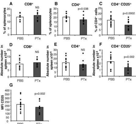
PTx treatment selectively reduces the size of the splenic CD4⫹CD25⫹T cell population
PTx has been shown to induce pleiotropic effects on the immune system. To assess whether PTx could have an influence on Treg cells, we injected C57BL/6 mice i.v. with 400 ng PTx at day 0, and 200 ng at day 2, a regimen used for EAE induction. Control mice received PBS with the same schedule. Mice were sacrificed on day 3 and the cellular composition of lymphoid organs was analyzed by FACS. Total numbers of splenocytes did not display any sig-nificant variation upon PTx treatment (133⫾18 ⫻ 106 in
PTx-treated mice, n⫽ 11, vs 110 ⫾ 11 ⫻ 106in PBS-treated mice, n⫽
11; p⫽ 0.2). Similarly, no differences were found between groups in the percentage or absolute numbers of CD8⫹splenocytes (Fig. 1, A and D), whereas there was a modest but significant decrease in CD4⫹T cell frequency ( p⫽ 0.038) but not absolute numbers ( p⫽ 0.35) (Fig. 1, B and E). However, we found a 33% decrease in the percentage of CD4⫹splenocytes expressing CD25 in PTx-treated mice ( p ⫽ 0.0002) and a 41% decrease in the absolute numbers of CD4⫹CD25⫹splenocytes ( p⫽ 0.002) (Fig. 1, C and F). This decrease was associated with a reduced expression level of CD25 on the remaining CD4⫹CD25⫹T cells (mean fluores-cence intensity (MFI) 177⫾ 18 vs 214 ⫾ 23, p ⫽ 0.002) (Fig.
1G). In contrast, the absolute numbers of CD4⫹CD25⫺ spleno-cytes was unaffected by PTx treatment (13.4⫾ 2.8 ⫻ 106in
PTx-treated vs 14.0⫾ 1.7 ⫻ 106cells in PBS-treated mice, p⫽ 0.79).
Similarly, the percentage of CD4⫹CD25⫺ cells among spleno-cytes was not significantly altered by PTx treatment (10.5⫾ 0.8 vs 12.6⫾ 0.8%, p ⫽ 0.10), suggesting that the decrease in frequency of CD4⫹splenocytes can at least partially be explained by the deletion in CD4⫹CD25⫹cells.
We also showed that the bioactive PTx holotoxin, but not the heat-inactivated PTx nor the purified B subunit, could induce a reduction of splenic Treg cell numbers (data not shown). These results strongly argue that bioactive PTx is responsible for the described effect, and suggest that this effect depends on the ADP-ribosyltransferase activity of PTx.
In agreement with published data (22), lymph nodes from PTx-treated mice showed a great decrease in total cell numbers (3.2⫾ 0.7⫻ 106, n⫽ 7, vs 14.6 ⫾ 2.2 ⫻ 106, n⫽ 7; p ⫽ 0.002) as well
as in the number of CD4⫹CD25⫹T cells, although a modest in-crease in the percentage of CD4⫹CD25⫹T cells (13.2 ⫾ 1.1%, n⫽ 9, vs 11.3 ⫾ 0.7%, n ⫽ 9; p ⫽ 0.036) was found. No signif-icant differences were seen in frequency of CD4⫹CD25⫹T cells among CD4⫹ blood cells nor CD4⫹CD8⫺thymocytes of PTx-treated and PBS-PTx-treated mice (data not shown).
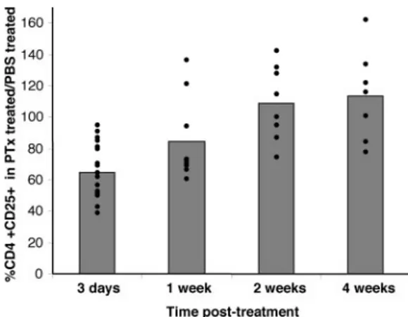
We then investigated the duration of the depletion of CD4⫹CD25⫹ T cells in spleen following PTx treatment. Mice were treated as above and the size of the CD4⫹CD25⫹splenic compartment was analyzed at day 3, and 1, 2, or 4 wk after the first PTx injection. After a significant decrease at day 3, the proportion of splenic CD4⫹CD25⫹T cells increased from day 7. At 4 wk, there was a full recovery and even a modest increase in CD4⫹CD25⫹frequency (113⫾ 11%) in PTx-treated relative to control mice (Fig. 2).
PTx treatment depletes regulatory CD4⫹Foxp3⫹T cell populations
The expression of the transcription factor Foxp3 in T cells is tightly associated with regulatory functions. Indeed, whereas
FIGURE 1. Reduction in the relative and abso-lute numbers of CD4⫹CD25⫹T cells in the spleen of PTx-treated mice. Mice injected i.v. with 400 ng of PTx or PBS at day 0 and 200 ng of PTx or PBS at day 2 were sacrificed on day 3. Spleen cells were stained with anti-CD4 and anti-CD25, or anti-CD8 mAbs. Percentages (A) and absolute numbers (D) of CD8⫹cells. Percentages (B) and absolute numbers (E) of CD4⫹cells. C, Percentages of CD25⫹among CD4⫹cells. F, Absolute numbers of CD4⫹CD25⫹ cells. G, MFI of CD25 on splenic CD4⫹CD25⫹ cells from PTx- or PBS-treated mice. Results are from 9 to 13 mice/group.
1554 EFFECTS OF PTx ON SPLENIC Treg CELLS
by guest on September 17, 2018
http://www.jimmunol.org/
CD4⫹CD25⫹Foxp3⫺ T cells are activated effector cells, both CD4⫹CD25⫹Foxp3⫹ and CD4⫹CD25⫺Foxp3⫹ T cell subsets display regulatory properties (31). To ensure that PTx was indeed depleting Treg cells, we performed anti-Foxp3 intranuclear stain-ing on splenocytes from PTx- or PBS-treated mice. Intereststain-ingly, a clear decrease (⬎50%) in the percentage of both CD4⫹CD25⫹Foxp3⫹ and CD4⫹CD25⫺Foxp3⫹ T cell subsets was observed at day 3 post PTx-treatment (Fig. 3). We observed a 26% decrease in the MFI of Foxp3 staining in Treg cells from PTx-treated mice. A profound decrease was also found in the ab-solute numbers of both CD4⫹CD25⫹Foxp3⫹(0.48⫾ 0.10 ⫻ 106
in PTx-treated vs 1.27 ⫾ 0.17 ⫻ 106in PBS-treated mice, p⫽
0.0025) and CD4⫹CD25⫺Foxp3⫹(0.26 ⫾ 0.03 ⫻ 106 in
PTx-treated vs 0.62⫾ 0.06 ⫻ 106in PBS-treated mice, p⫽ 0.0002)
Treg populations. Contrasting with these results, the number of CD4⫹CD25⫹Foxp3⫺ cells in PTx-treated mice were not de-creased, and even slightly inde-creased, as compared with PBS-treated mice, consistent with the previously described effects of PTx on T cells (19, 38).
Collectively, our data show that PTx treatment induces a reduc-tion in both the percentage and the absolute numbers of splenic Treg cells, which applies to CD4⫹Foxp3⫹Treg cells regardless of their CD25 expression.
PTx does not affect the in vitro functional properties of CD4⫹CD25⫹Treg cells
We next examined the influence of in vivo PTx treatment on the functional properties of highly purified CD4⫹CD25brightT cells.
CD4⫹CD25brightT cells purified by FACS sorting from PBS- or
PTx-treated mice were stimulated with anti-CD3 and APC alone (Fig. 4A) or in coculture with CD4⫹CD25⫺responder T cells (Fig. 4B). CD4⫹CD25bright T cells from both PBS- and PTx-treated
mice were hyporesponsive in vitro to TCR triggering. Moreover, the addition of CD4⫹CD25brightT cells from either group of mice
strongly inhibited CD4⫹CD25⫺T cell proliferation with similar dose-response suppressive activities. These results indicate that the function of the remaining CD4⫹CD25bright Treg cells is not
af-fected by the PTx treatment. Consistent with this statement, their cytokine mRNA content, as determined by real-time quantitative RT-PCR, did not reveal important differences between CD4⫹CD25⫹T cells from PBS vs PTx-treated mice (Fig. 4C). Investigation of the mechanisms of PTx-induced reduction of splenic Treg cells
The apparent PTx-induced depletion of splenic Treg cells could be attributed to several mechanisms: 1) loss of expression of their characteristic markers; 2) direct or indirect toxicity of PTx on Treg cells; and/or 3) modification of their migratory properties.
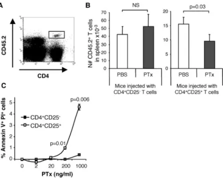
To investigate whether the observed reduction of splenic Treg cells is due to decreased CD25 expression or to a physical loss of Treg cells, we transferred either CD4⫹CD25⫹or CD4⫹CD25⫺T cells from CD45.2 mice into CD45.1 congenic mice before PTx or PBS treatment. Recipient mice were sacrificed on day 3 and the transferred cells were identified based on CD45.2 expression (Fig. 5A). Of note, CD45.2 expression levels on transferred cells (CD4⫹CD25⫹and CD4⫹CD25⫺T cells) was unaffected by PTx treatment (data not shown). The absolute numbers of donor CD45.2⫹CD4⫹CD25⫹T cells was reduced by 38⫾ 11% in the spleen of PTx-treated mice compared with PBS-treated mice, whereas there was no difference between groups regarding the number of CD4⫹CD25⫺ T cells of donor origin (Fig. 5B). As expected, there was a reduction of endogenous CD45.2⫺CD4⫹CD25⫹T cells in both groups of PTx-treated re-cipient mice (data not shown). These data, using a congenic sur-face marker unaffected by PTx treatment, confirm the reduction of Treg cells in the spleen of PTx-treated mice and show that this reduction results from a loss of cells and is not merely attributed to a down-regulation of the CD25 molecule.
We next investigated whether this physical loss of Treg cells could be due to apoptosis induction by PTx. CD4⫹CD25⫹ or CD4⫹CD25⫺T cells purified from untreated mice were incubated in vitro with increasing concentrations of PTx and stained for an-nexin V and PI. A short incubation of 4 h was selected since, upon longer incubations, Treg cells exhibited poor survival capacities in vitro (39). Treg cells were significantly more sensitive than
FIGURE 2. PTx-induced CD4⫹CD25⫹T cell depletion in the spleen is a transient phenomenon. Mice were treated with PTx or PBS as described in Fig. 1 and were sacrificed 3 days (n⫽ 19/group), 1 wk (n ⫽ 9), 2 wk (n⫽ 8), or 4 wk (n ⫽ 7) later. Spleen cells were stained with anti-CD4 and anti-CD25 mAbs. Data are shown as the ratio of percentages of CD4⫹T cells expressing CD25 in PTx-treated mice and PBS-treated mice.
FIGURE 3. Decrease in Foxp3⫹CD4⫹T cells following PTx treatment. Mice were treated with PTx (■) or PBS (䡺) as described in Fig. 1, and the percentage of CD4⫹CD25⫺Foxp3⫹ (A), CD4⫹CD25⫹Foxp3⫹ (B), and CD4⫹CD25⫹Foxp3⫺(D) among splenic CD4 T cells was determined, 3 and 7 days after treatment, by a combination of surface and intranuclear FACS staining. C, A representative CD25 and Foxp3 staining gated on CD4⫹splenocytes illustrating the cell populations analyzed in A, B, and D. Six PTx-treated and 5 PBS-treated mice were analyzed at day 3, and 11 PTx-treated and 11 PBS-treated mice were analyzed at day 7.
1555 The Journal of Immunology
by guest on September 17, 2018
http://www.jimmunol.org/
CD4⫹CD25⫺T cells to PTx-induced apoptosis in vitro (Fig. 5C). Therefore, PTx can directly promote apoptosis of Treg cells in vitro.
To evaluate whether differential tissular migration of the Treg cells could explain their reduction from the spleen after PTx treat-ment, infiltrating lymphocytes were isolated 3 days after treatment from a large number of tissues, and stained for CD4, CD25 and Foxp3. These experiments revealed a significant decrease in CD4⫹CD25⫹Foxp3⫹ cells in both spleen and bone marrow of PTx- vs PBS-treated mice (Table I). In addition, an increase in the absolute numbers of CD4⫹CD25⫹Foxp3⫹cells was found in the liver ( p⬍ 0.005), and in the lungs and brain (not reaching statis-tical significance, possibly as a result of error) of PTx-treated animals (Table I).
Synergistic effect of PTx and PC61 treatment on EAE
Previous studies have underlined the important role played by Treg cells in the control of EAE (32–35). Because our results indicated that PTx induces a partial depletion in Treg cells, we investigated the contribution of this PTx-induced reduction of Treg cells on EAE induction, relative to the other disease-potentiating effects of PTx.
We first evaluated the effect of a profound in vivo depletion of Treg cells on the course of EAE. A single i.p. injection of 500g of the anti-CD25 mAb PC61 in C57BL/6 mice induced a durable depletion of CD4⫹CD25⫹T cells (by 83%) in blood of treated mice detectable as early as day 2 after injection and lasting for ⬎17 days (Fig. 6A). Then, C57BL/6 mice were treated with 500 g of PC61 or control rat IgG or PBS 7 days before a full protocol of EAE induction. PC61-treated mice displayed a marked increase in the incidence and severity of the disease but not an earlier onset (Fig. 6B) in agreement with previous work (40, 41). Moreover, upon analysis at day 32, the CNS of PC61 ascitis-treated animals showed much higher infiltrates than untreated controls (inflamma-tion index: 3.6⫾ 0.3, n ⫽ 2 treated mice, vs 0.1 ⫾ 0.0, n ⫽ 3 untreated mice, p⫽ 0.0006). Taken together, these results indicate a major role for Treg cells in controlling inflammation and tissue damage during CNS autoimmunity.
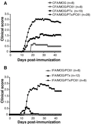
We next evaluated whether the EAE-enhancing effect of PTx was dependent on its action on Treg cells. To this end, mice pre-injected with PC61 or control IgG were immunized with MOG35–55in CFA and were treated or not with PTx. In the ab-sence of PTx, PC61 treatment induced a moderate disease (Fig. 7A), despite profound and durable Treg cell depletion (Fig. 6A). PTx treatment alone, although it resulted in a less pronounced Treg cell reduction, promoted a more severe disease. Importantly, PC61 administration and PTx treatment acted in a synergistic manner on EAE severity (Fig. 7A) indicating that they are acting on nonre-dundant pathways lowering the threshold for clinical autoimmu-nity. This synergy was even more striking when we used IFA instead of CFA for MOG35–55immunization (Fig. 7B). Indeed, in this context, PC61 administration alone failed to induce clinical disease. Similarly, PTx only induced a minor and transient disease in 25% of the mice. However, the combined treatment with PTx and PC61 induced a severe disease in 100% of the mice (Fig. 7B). These results indicate that enhancement of EAE by PTx treat-ment does not solely rely on its effect on CD4⫹CD25⫹Treg cells. In addition, they reveal a potent synergistic effect of PTx treatment and in vivo depletion of Treg cells using the PC61 Ab.
Discussion
In this report, we show that administration of PTx to C57BL/6 mice, following a regimen used to induce EAE, caused a specific depletion of both CD4⫹CD25⫹Foxp3⫹and CD4⫹CD25⫺Foxp3⫹ Treg subpopulations from the spleen. As a consequence, both the percentage and absolute numbers of splenic Treg cells were de-creased following PTx injection. Similar results were obtained in BALB/c mice (data not shown). This decrease in Treg cells did not affect evenly all tissues, involving mostly the spleen and to a lesser extent the bone marrow, whereas the liver showed higher Treg cell numbers after PTx treatment. This phenomenon was associated with a preferential sensitivity of Treg cells to the toxicity of PTx in vitro. Experiments in which induction of EAE was performed with or without PTx and/or PC61 treatment indicated that deple-tion of CD4⫹CD25⫹T cells is not the major mechanism by which PTx participates to EAE development. However, our results show a clear synergy between PC61, Mycobacterium and PTx for the
FIGURE 4. PTx treatment does not affect the functional properties of CD4⫹CD25⫹T cells. A, CD4⫹CD25brightor CD4⫹CD25⫺were purified
from PTx- or PBS-treated mice 3 days after treatment, and the proliferative capacity of each cell population (25,000 cells/well) was assessed following stimulation with 0.5g/ml anti-CD3 and 200,000 APC. B, A total of 25,000 responder CD4⫹CD25⫺T cells from untreated mice was stimulated with 0.5g/ml anti-CD3 and 200,000 APC in the presence of increasing numbers of highly purified CD4⫹CD25brightT cells from PTx-treated (gray
symbols) or PBS-treated (white symbols) mice. Unstimulated proliferation was minimal, ranging from 13 to 35 cpm. These experiments are repre-sentative of four independently performed experiments. C, Expression of IFN-␥, TGF-1, IL-10, or IL-2 mRNA in CD4⫹CD25⫹or CD4⫹CD25⫺ freshly purified from PBS (䡺)- or PTx (■)-treated mice. Results are pre-sented as fold increase relative to CD4⫹CD25⫹ cells from PTx-treated mice.ⴱ, p ⬍ 0.05; ⴱⴱ, p ⬍ 0.005; ⴱⴱⴱ, p ⬍ 0.0005.
1556 EFFECTS OF PTx ON SPLENIC Treg CELLS
by guest on September 17, 2018
http://www.jimmunol.org/
induction and severity of EAE, illustrating how multiple adjuvants can maximize activation of the immune system.
PTx led to a decreased expression of the CD25 molecule on splenic CD4⫹T cells. This decrease in CD25 MFI (Fig. 1G) may be explained either by a preferential elimination of CD4⫹CD25bright T cells, or by a down-regulation of CD25
ex-pression on Treg cells following PTx treatment. The adoptive transfer experiments indicate physical loss of Treg cells following PTx administration on the basis of a congenic marker whose ex-pression is not modified upon PTx treatment. Second, down-mod-ulation of the CD25 molecule would result in an increase, rather than the observed decrease, of the CD4⫹CD25⫺Foxp3⫹ subpopu-lation. Moreover, our in vitro studies suggested that the PTx-in-duced reduction of Treg cells might in part be related to the pref-erential sensitivity of these cells to direct PTx toxicity. However, the relative contribution of this PTx-induced apoptosis to the Treg cell depletion observed in vivo is at present unclear. PTx-induced apoptosis of Treg cells in vivo might be difficult to assess because of rapid elimination of dying cells.
Our results suggest that depletion of Treg cells is not the major mechanism by which PTx participates in EAE induction or
aggra-vation (Fig. 7). However, a contribution of this effect in EAE en-hancement cannot be excluded. Indeed, whereas PC61 only de-pletes the dominant CD25⫹Treg population, PTx reduces to the same extent both the CD25⫹and the CD25⫺Foxp3⫹regulatory populations (Fig. 3, A and B).
By interfering with Gi protein activity, PTx can affect both sur-vival and migration of Treg cells. The susceptibility of Treg cells to apoptosis is controversial. Human Treg cells were shown to be apoptosis-prone because of lower expression of the anti-apoptotic molecule Bcl-2 than conventional lymphocytes (39). On the other hand, murine Treg cells are resistant to apoptosis induced by Fas (42) as well as by dexamethasone (43). Why should Treg cells be more sensitive to PTx toxicity than conventional T cells? An in-teresting hypothesis could be that the Treg cell survival specifi-cally depends on a PTx-sensitive receptor. Two major molecules are considered essential to Treg cell survival: TGF- and IL-2. TGF- plays an important role in induction, maintenance (44, 45) and activity of Treg cells (46, 47), and a recent study reveals a lack of response to TGF- in G␣i2-deficient mice (48). Thus, a possible explanation for PTx-induced apoptosis of Treg cells could be an inhibition of survival signals provided by TGF-. Disruption of
Table I. Quantitative analysis of Treg cells in several tissues following PTx treatmenta
Spleen Thymusb Bone marrowc Blood % CD25⫹Foxp3⫹ PBS 10.22⫾ 0.25 2.30⫾ 0.14 32.75⫾ 0.95 2.82⫾ 0.38 PTx 5.76⫾ 0.60*** 2.54⫾ 0.15 22.70⫾ 0.60*** 2.96⫾ 0.30 Absolute numbers of PBS 1.22⫾ 0.12 ⫻ 106 1.68⫾ 0.24 ⫻ 105 4.55⫾ 0.54 ⫻ 104 CD25⫹Foxp3⫹CD4⫹cells PTx 0.66⫾ 0.10 ⫻ 106** 1.04⫾ 0.11 ⫻ 105* 3.03⫾ 0.67 ⫻ 104 Peritoneum Braind Livere Lungs Colon % CD25⫹Foxp3⫹ PBS 9.85⫾ 2.81 7.01⫾ 0.41 3.49⫾ 0.17 5.44⫾ 0.33 8.80⫾ 1.43 PTx 8.98⫾ 0.73 8.30⫾ 1.06 4.41⫾ 0.37 4.69⫾ 0.69 5.88⫾ 1.04 Absolute numbers of PBS 6.09⫾ 1.65 ⫻ 102 3.32⫾ 1.04 ⫻ 104 8.56⫾ 2.01 ⫻ 103 1.08⫾ 0.68 ⫻ 102 CD25⫹Foxp3⫹CD4⫹cells PTx 19.82⫾ 5.74 ⫻ 102 6.92⫾ 1.40 ⫻ 104** 17.09⫾ 5.07 ⫻ 103 0.13⫾ 0.04 ⫻ 102 a
Numbers of mice analyzed per group: spleen, n⫽ 16; thymus, n ⫽ 8; bone marrow, n ⫽ 8; blood, n ⫽ 8; peritoneum, n ⫽ 3; brain, three experiments with two to three pooled mice per group; liver, n⫽ 7; lungs, n ⫽ 7; colon, n ⫽ 7. ⴱ, p ⬍ 0.05; ⴱⴱ, p ⬍ 0.005; ⴱⴱⴱ, p ⬍ 0.0005.
b
Gated on CD4⫹CD8⫺single-positive thymocytes.
c
Gated on CD4⫹TCR⫹cells; absolute numbers given for one femur.
d
Absolute numbers per mouse.
e
Gated on CD4highTCRhigh
cells.
FIGURE 5. PTx preferentially depletes CD4⫹CD25⫹ T cells. CD45.1 congenic C57BL/6 mice received 2– 4⫻ 106CD4⫹CD25⫹or CD4⫹CD25⫺T cells from CD45.2
mice and were treated with PTx or PBS 3 days later. Recipient mice were sacrificed on day 3 after treatment and spleen cells were stained with CD45.2, anti-CD4, and anti-CD25 mAbs. A, Identification of trans-ferred cells based on CD45.2 staining. B, Numbers of transferred cells present in spleen per million of injected cells. Results are pooled from three independent exper-iments and groups were compared using one-tailed Stu-dent’s t test. C, CD4⫹CD25⫹T cells are more sensitive than CD4⫹CD25⫺ T cells to PTx-induced cell death. 200,000 CD4⫹CD25⫹(white symbols) or CD4⫹CD25⫺ (black symbols) cells were incubated in vitro with dif-ferent concentrations of PTx for 4 h. Cells were then recovered and stained with annexin V and PI. The per-centages of Annexin V⫹PI⫹ cells are presented after subtraction of the background apoptosis present in me-dium only cultures. This experiment is representative of three independent experiments.
1557 The Journal of Immunology
by guest on September 17, 2018
http://www.jimmunol.org/
IL-2R signaling in Treg cells could represent a second mechanism explaining this effect. Indeed, IL-2 has proved to be crucial in the survival of Treg cells in the periphery (49, 50). Although the IL-2R is not directly coupled to a Gi protein, a PTx-sensitive GTP-bind-ing protein has been shown to be activated followGTP-bind-ing the ligation of IL-2 on lymphocytes (51). This signaling pathway has to our knowledge never been explored further, but its inhibition by PTx may have consequences on T cell biology.
Our quantification of Treg cells in an variety of tissues indicates that there is no generalized reduction of Treg cells following PTx injection, but rather that this decrease is selective, affecting mostly the spleen, and is accompanied by migration and/or retention of Treg cells in the liver. Whether the Treg cells accumulating in the liver following PTx injection are destined to die (52) or are only transiently trapped in this tissue is currently uncertain.
Interestingly, several PTx-sensitive chemokine receptors, such as CCR2 (53), CCR4 (54), CCR5 (55), CCR6 (56), and CCR7 (57), seem important for Treg cell function and recruitment in tissues. However, the consequences of PTx-induced impairment of chemokines-induced signaling remain ambivalent. Indeed, the in-hibition of chemokine receptors by PTx can lead to a reduced migration of pathogenic T cells to their target tissue and thus to an inhibition of inflammatory diseases (23, 58). Nevertheless, we pro-pose that as PTx inhibits the entry of all lymphocytes in lymph nodes, and induces a decreased frequency of Treg cells in spleen, the probability of encounter between Treg cells and the population of conventional lymphocytes, that contains autoaggressive cells, is limited, so the balance is biased toward activation of effector cells.
Interestingly, this effect of PTx may be linked to PTx-induced cell death, because chemokines receptors can be implicated in both apoptotic signaling pathway and migratory patterns (59, 60).
An alternative hypothesis that could explain the effects of PTx on Treg cells involves the inhibition of the PTx-sensitive sphin-gosine 1-phosphate receptors S1P1and S1P4signaling. Indeed, the
triggering of these receptors on T cells by their ligand leads to increased survival, and increased or decreased chemotaxis depend-ing on the ligand concentration (61). Strikdepend-ingly, treatment of mice with a pharmacological agonist of this receptor, FTY720, doubles the number of Treg cells in the spleen, and thus appears to differ-entially affect the homing properties of Treg cells (62).
A synergistic effect on EAE severity was exerted by PTx and PC61 treatment (Fig. 7), a phenomenon that has also recently been described in a passive model of EAE (63). We also noted a synergy between CFA and PTx on EAE triggering. Thus, development of EAE results from the integration of an autoantigen-specific acti-vation signal acting on T cells, and of several non-Ag-specific signals. Among the non-Ag-specific signals, PTx has been shown to promote clonal expansion and differentiation of autoreactive T cells, possibly by providing maturation signals to APC (19 –21). Permeabilization of the blood-brain barrier by PTx would then facilitate infiltration of pathogenic cells into the CNS, where they are reactivated, promote recruitment of other types of immune cells and start tissue injury. It is interesting to note that both ad-juvants (Mycobacterium tuberculosis and PTx) used to potentiate EAE are bacterial compounds. Other microbial molecules have been shown to influence EAE susceptibility or severity (64 – 67). The mechanisms of these EAE-aggravating effects of infectious
FIGURE 6. Depletion of CD4⫹CD25⫹ T cells exacerbates EAE. A, Mice were injected i.p. with PBS (〫), 500g of control rat IgG (䡺), or 500g (F) or 1 mg (■) of PC61 mAb at day 1. Blood was collected at different time points, and FACS staining was performed using anti-CD4 and anti-CD25 (7D4) mAbs. The percentage of CD25⫹among CD4⫹cells from serially sampled mice was plotted. B, Mice were treated with PBS (〫), 500g of rat IgG (䡺), or PC61 (F) and were immunized 7 days later to induce EAE. Pooled results from six experiments are shown. The log-rank test analysis showed that the PBS-treated and the IgG-treated mice display similar EAE curves, whereas the PC61-treated mice have exacer-bated disease compared with either PBS or IgG-treated mice (p⬍ 0.001).
FIGURE 7. Synergy between Ab-mediated depletion of CD4⫹CD25⫹ T cells and PTx administration for EAE induction. A, Mice that have been pretreated or not with PC61 7 days earlier were immunized subcutaneously with 100g of MOG35–55in CFA. These mice further received either PTx
or PBS treatment. B, A similar protocol was performed except that we substituted IFA for CFA. In this series of experiments, the IFA/MOG35–55
group (without PTx nor PC61 injection) was omitted because it consis-tently fails to induce EAE (21, 65, 67).
1558 EFFECTS OF PTx ON SPLENIC Treg CELLS
by guest on September 17, 2018
http://www.jimmunol.org/
agents are thought to implicate TLR triggering. Indeed, direct ac-tivation of TLR9 by CpG oligonucleotides, or TLR4 by LPS, in-creases the incidence of EAE in a proteolipid protein-TCR trans-genic mouse model, however to a lower degree than PTx (66). Besides their known consequences on dendritic cells, the trigger-ing of TLRs could also affect the biology of Treg cells. Actually, Treg cells express TLR4, TLR5, TLR7, and TLR8 (68); ligands of TLR8 have been shown to reverse Treg cell suppressive activity (69), while CpG, ssRNA, or LPS have been shown to modify Treg cell activity following TLR ligation, although the effect varies greatly depending on the experimental models (68 –71). Interest-ingly, a recent report demonstrates that PTx can signal through TLR4, leading to increased permeabilization of the pial vessels to leukocytes (27, 28). In addition, it has been shown that the trig-gering of TLR4 induces a refractoriness of conventional lympho-cytes to the suppressive activity of Treg cells, by a mechanism dependent on IL-6 secretion by APC (70). Thus, although it has been suspected for a long time that signals provided by microbial agents can favor self tolerance disruption leading to autoimmune diseases (72), accumulating evidence indicate that they may target Treg cells directly, particularly through TLR, in animal models and possibly in humans.
To conclude, we have revealed an unknown selective effect of PTx on Treg cells, which could participate in the pathogenesis of autoimmune diseases in animal models. PTx-induced selective elimination of Treg cells, reduces the relative and absolute num-bers of Treg cells in the spleen of PTx-treated mice. The discovery of this new effect of PTx, among its multiple ones on the organism, highlights the numerous and complex consequences that environ-mental agents can display on the control of immunity and the de-velopment of autoimmune diseases.
Note added in proof. While this manuscript was undergoing the
reviewing process, an independent study similarly reported a de-crease in splenic Treg cells following PTx treatment (73).
Acknowledgments
We thank Dr. Abdelhadi Saoudi for critical reading of the manuscript and helpful discussions, and Dr. Hans Lassmann for analysis of histological tissue sections. We acknowledge Maryline Calise for excellent manage-ment of the animal house and Fatima L’Faqihi Olive for cell separation on the Beckman Coulter sorter.
Disclosures
The authors have no financial conflict of interest.
References
1. Munoz, J. J., C. C. Bernard, and I. R. Mackay. 1984. Elicitation of experimental allergic encephalomyelitis (EAE) in mice with the aid of pertussigen. Cell.
Im-munol. 83: 92–100.
2. Linthicum, D. S. 1982. Development of acute autoimmune encephalomyelitis in mice: factors regulating the effector phase of the disease. Immunobiology 162: 211–220.
3. Kohno, S., J. A. Munoz, T. M. Williams, C. Teuscher, C. C. Bernard, and K. S. Tung. 1983. Immunopathology of murine experimental allergic orchitis.
J. Immunol. 130: 2675–2682.
4. Hou, W., Y. Wu, S. Sun, M. Shi, Y. Sun, C. Yang, G. Pei, Y. Gu, C. Zhong, and B. Sun. 2003. Pertussis toxin enhances Th1 responses by stimulation of dendritic cells. J. Immunol. 170: 1728 –1736.
5. Agarwal, R. K., S. H. Sun, S. B. Su, C. C. Chan, and R. R. Caspi. 2002. Pertussis toxin alters the innate and the adaptive immune responses in a pertussis-depen-dent model of autoimmunity. J. Neuroimmunol. 129: 133–140.
6. Hart, M. N., D. S. Linthicum, M. M. Waldschmidt, S. K. Tassell, R. L. Schelper, and R. A. Robinson. 1987. Experimental autoimmune inflammatory myopathy.
J. Neuropathol. Exp. Neurol. 46: 511–521.
7. Nencioni, L., M. G. Pizza, G. Volpini, M. T. De Magistris, F. Giovannoni, and R. Rappuoli. 1991. Properties of the B oligomer of pertussis toxin. Infect. Immun. 59: 4732– 4734.
8. Mascart, F., V. Verscheure, A. Malfroot, M. Hainaut, D. Pierard, S. Temerman, A. Peltier, A. S. Debrie, J. Levy, G. Del Giudice, and C. Locht. 2003. Bordetella pertussis infection in 2-month-old infants promotes type 1 T cell responses. J.
Im-munol. 170: 1504 –1509.
9. Tonon, S., S. Goriely, E. Aksoy, O. Pradier, G. Del Giudice, E. Trannoy, F. Willems, M. Goldman, and D. De Wit. 2002. Bordetella pertussis toxin in-duces the release of inflammatory cytokines and dendritic cell activation in whole blood: impaired responses in human newborns. Eur. J. Immunol. 32: 3118 –3125. 10. Nicosia, A., M. Perugini, C. Franzini, M. C. Casagli, M. G. Borri, G. Antoni, M. Almoni, P. Neri, G. Ratti, and R. Rappuoli. 1986. Cloning and sequencing of the pertussis toxin genes: operon structure and gene duplication. Proc. Natl.
Acad. Sci. USA 83: 4631– 4635.
11. Brennan, M. J., J. L. David, J. G. Kenimer, and C. R. Manclark. 1988. Lectin-like binding of pertussis toxin to a 165-kilodalton Chinese hamster ovary cell glyco-protein. J. Biol. Chem. 263: 4895– 4899.
12. Tuomanen, E., H. Towbin, G. Rosenfelder, D. Braun, G. Larson, G. C. Hansson, and R. Hill. 1988. Receptor analogs and monoclonal antibodies that inhibit ad-herence of Bordetella pertussis to human ciliated respiratory epithelial cells.
J. Exp. Med. 168: 267–277.
13. Witvliet, M. H., D. L. Burns, M. J. Brennan, J. T. Poolman, and C. R. Manclark. 1989. Binding of pertussis toxin to eucaryotic cells and glycoproteins. Infect.
Immun. 57: 3324 –3330.
14. Katada, T., T. Amano, and M. Ui. 1982. Modulation by islet-activating protein of adenylate cyclase activity in C6 glioma cells. J. Biol. Chem. 257: 3739 –3746. 15. Katada, T., and M. Ui. 1982. Direct modification of the membrane adenylate
cyclase system by islet-activating protein due to ADP-ribosylation of a membrane protein. Proc. Natl. Acad. Sci. USA 79: 3129 –3133.
16. Mielcarek, N., E. H. Hornquist, B. R. Johansson, C. Locht, S. N. Abraham, and J. Holmgren. 2001. Interaction of Bordetella pertussis with mast cells, modulation of cytokine secretion by pertussis toxin. Cell Microbiol. 3: 181–188. 17. Darabi, K., A. Y. Karulin, B. O. Boehm, H. H. Hofstetter, Z. Fabry,
J. C. LaManna, J. C. Chavez, M. Tary-Lehmann, and P. V. Lehmann. 2004. The third signal in T cell-mediated autoimmune disease? J. Immunol. 173: 92–99. 18. He, J., S. Gurunathan, A. Iwasaki, B. Ash-Shaheed, and B. L. Kelsall. 2000.
Primary role for Gi protein signaling in the regulation of interleukin 12 produc-tion and the inducproduc-tion of T helper cell type 1 responses. J. Exp. Med. 191: 1605–1610.
19. Ryan, M., L. McCarthy, R. Rappuoli, B. P. Mahon, and K. H. Mills. 1998. Pertussis toxin potentiates Th1 and Th2 responses to co-injected antigen: adju-vant action is associated with enhanced regulatory cytokine production and ex-pression of the co-stimulatory molecules B7-1, B7-2 and CD28. Int. Immunol. 10: 651– 662.
20. Shive, C. L., H. Hofstetter, L. Arredondo, C. Shaw, and T. G. Forsthuber. 2000. The enhanced antigen-specific production of cytokines induced by pertussis toxin is due to clonal expansion of T cells and not to altered effector functions of long-term memory cells. Eur. J. Immunol. 30: 2422–2431.
21. Hofstetter, H. H., C. L. Shive, and T. G. Forsthuber. 2002. Pertussis toxin mod-ulates the immune response to neuroantigens injected in incomplete Freund’s adjuvant: induction of Th1 cells and experimental autoimmune encephalomyelitis in the presence of high frequencies of Th2 cells. J. Immunol. 169: 117–125. 22. Cyster, J. G., and C. C. Goodnow. 1995. Pertussis toxin inhibits migration of B
and T lymphocytes into splenic white pulp cords. J. Exp. Med. 182: 581–586. 23. Alt, C., M. Laschinger, and B. Engelhardt. 2002. Functional expression of the
lymphoid chemokines CCL19 (ELC) and CCL 21 (SLC) at the blood-brain bar-rier suggests their involvement in G-protein-dependent lymphocyte recruitment into the central nervous system during experimental autoimmune encephalomy-elitis. Eur. J. Immunol. 32: 2133–2144.
24. Linthicum, D. S., J. J. Munoz, and A. Blaskett. 1982. Acute experimental auto-immune encephalomyelitis in mice. I. Adjuvant action of Bordetella pertussis is due to vasoactive amine sensitization and increased vascular permeability of the central nervous system. Cell. Immunol. 73: 299 –310.
25. Linthicum, D. S., and J. A. Frelinger. 1982. Acute autoimmune encephalomyelitis in mice. II. Susceptibility is controlled by the combination of H-2 and histamine sensitization genes. J. Exp. Med. 156: 31– 40.
26. Bruckener, K. E., A. el Baya, H. J. Galla, and M. A. Schmidt. 2003. Permeabi-lization in a cerebral endothelial barrier model by pertussis toxin involves the PKC effector pathway and is abolished by elevated levels of cAMP. J. Cell Sci. 116: 1837–1846.
27. Kerfoot, S. M., E. M. Long, M. J. Hickey, G. Andonegui, B. M. Lapointe, R. C. Zanardo, C. Bonder, W. G. James, S. M. Robbins, and P. Kubes. 2004. TLR4 contributes to disease-inducing mechanisms resulting in central nervous system autoimmune disease. J. Immunol. 173: 7070 –7077.
28. Racke, M. K., W. Hu, and A. E. Lovett-Racke. 2005. PTX cruiser: driving au-toimmunity via TLR4. Trends Immunol. 26: 289 –291.
29. Kamradt, T., P. D. Soloway, D. L. Perkins, and M. L. Gefter. 1991. Pertussis toxin prevents the induction of peripheral T cell anergy and enhances the T cell response to an encephalitogenic peptide of myelin basic protein. J. Immunol. 147: 3296 –3302.
30. Sakaguchi, S. 2004. Naturally arising CD4⫹regulatory T cells for immunologic self-tolerance and negative control of immune responses. Annu. Rev. Immunol. 22: 531–562.
31. Fontenot, J. D., J. P. Rasmussen, L. M. Williams, J. L. Dooley, A. G. Farr, and A. Y. Rudensky. 2005. Regulatory T cell lineage specification by the forkhead transcription factor foxp3. Immunity. 22: 329 –341.
32. Olivares-Villagomez, D., A. K. Wensky, Y. Wang, and J. J. Lafaille. 2000. Rep-ertoire requirements of CD4⫹T cells that prevent spontaneous autoimmune en-cephalomyelitis. J. Immunol. 164: 5499 –5507.
33. Kohm, A. P., P. A. Carpentier, H. A. Anger, and S. D. Miller. 2002. Cutting edge: CD4⫹CD25⫹regulatory T cells suppress antigen-specific autoreactive immune responses and central nervous system inflammation during active experimental autoimmune encephalomyelitis. J. Immunol. 169: 4712– 4716.
1559 The Journal of Immunology
by guest on September 17, 2018
http://www.jimmunol.org/
34. Hori, S., M. Haury, A. Coutinho, and J. Demengeot. 2002. Specificity require-ments for selection and effector functions of CD25⫹4⫹regulatory T cells in anti-myelin basic protein T cell receptor transgenic mice. Proc. Natl. Acad. Sci.
USA 99: 8213– 8218.
35. Reddy, J., Z. Illes, X. Zhang, J. Encinas, J. Pyrdol, L. Nicholson, R. A. Sobel, K. W. Wucherpfennig, and V. K. Kuchroo. 2004. Myelin proteolipid protein-specific CD4⫹CD25⫹regulatory cells mediate genetic resistance to experimental autoimmune encephalomyelitis. Proc. Natl. Acad. Sci. USA 101: 15434 –15439. 36. Viglietta, V., C. Baecher-Allan, H. L. Weiner, and D. A. Hafler. 2004. Loss of functional suppression by CD4⫹CD25⫹regulatory T cells in patients with mul-tiple sclerosis. J. Exp. Med. 199: 971–979.
37. Haas, J., A. Hug, A. Viehover, B. Fritzsching, C. S. Falk, A. Filser, T. Vetter, L. Milkova, M. Korporal, B. Fritz, et al. 2005. Reduced suppressive effect of CD4⫹CD25high
regulatory T cells on the T cell immune response against myelin oligodendrocyte glycoprotein in patients with multiple sclerosis. Eur. J. Immunol. 35: 3343–3352.
38. Wakatsuki, A., P. Borrow, K. Rigley, and P. C. Beverley. 2003. Cell-surface bound pertussis toxin induces polyclonal T cell responses with high levels of interferon-␥ in the absence of interleukin-12. Eur. J. Immunol. 33: 1859–1868. 39. Taams, L. S., J. Smith, M. H. Rustin, M. Salmon, L. W. Poulter, and A. N. Akbar. 2001. Human anergic/suppressive CD4⫹CD25⫹T cells: a highly differentiated and apoptosis-prone population. Eur. J. Immunol. 31: 1122–1131.
40. Montero, E., G. Nussbaum, J. F. Kaye, R. Perez, A. Lage, A. Ben-Nun, and I. R. Cohen. 2004. Regulation of experimental autoimmune encephalomyelitis by CD4⫹, CD25⫹ and CD8⫹ T cells: analysis using depleting antibodies.
J. Autoimmun. 23: 1–7.
41. Stephens, L. A., D. Gray, and S. M. Anderton. 2005. CD4⫹CD25⫹regulatory T cells limit the risk of autoimmune disease arising from T cell receptor crossre-activity. Proc. Natl. Acad. Sci. USA 102: 17418 –17423.
42. Banz, A., C. Pontoux, and M. Papiernik. 2002. Modulation of Fas-dependent apoptosis: a dynamic process controlling both the persistence and death of CD4 regulatory T cells and effector T cells. J. Immunol. 169: 750 –757.
43. Chen, X., T. Murakami, J. J. Oppenheim, and O. M. Howard. 2004. Differential response of murine CD4⫹CD25⫹and CD4⫹CD25⫺T cells to dexamethasone-induced cell death. Eur. J. Immunol. 34: 859 – 869.
44. Chen, W., W. Jin, N. Hardegen, K. J. Lei, L. Li, N. Marinos, G. McGrady, and S. M. Wahl. 2003. Conversion of peripheral CD4⫹CD25⫺naive T cells to CD4⫹CD25⫹ regulatory T cells by TGF- induction of transcription factor Foxp3. J. Exp. Med. 198: 1875–1886.
45. Schramm, C., S. Huber, M. Protschka, P. Czochra, J. Burg, E. Schmitt, A. W. Lohse, P. R. Galle, and M. Blessing. 2004. TGF regulates the CD4⫹CD25⫹T-cell pool and the expression of Foxp3 in vivo. Int. Immunol. 16: 1241–1249.
46. Huber, S., C. Schramm, H. A. Lehr, A. Mann, S. Schmitt, C. Becker, M. Protschka, P. R. Galle, M. F. Neurath, and M. Blessing. 2004. Cutting edge: TGF- signaling is required for the in vivo expansion and immunosuppressive capacity of regulatory CD4⫹CD25⫹T cells. J. Immunol. 173: 6526 – 6531. 47. Marie, J. C., J. J. Letterio, M. Gavin, and A. Y. Rudensky. 2005. TGF-1
main-tains suppressor function and Foxp3 expression in CD4⫹CD25⫹regulatory T cells. J. Exp. Med. 201: 1061–1067.
48. Wu, J. Y., Y. Jin, R. A. Edwards, Y. Zhang, M. J. Finegold, and M. X. Wu. 2005. Impaired TGF- responses in peripheral T cells of G␣i2⫺/⫺mice. J. Immunol.
174: 6122– 6128.
49. D’Cruz, L. M., and L. Klein. 2005. Development and function of agonist-induced CD25⫹Foxp3⫹regulatory T cells in the absence of interleukin 2 signaling. Nat.
Immunol. 6: 1152–1159.
50. Fontenot, J. D., J. P. Rasmussen, M. A. Gavin, and A. Y. Rudensky. 2005. A function for interleukin 2 in Foxp3-expressing regulatory T cells. Nat. Immunol. 6: 1142–1151.
51. Evans, S. W., S. K. Beckner, and W. L. Farrar. 1987. Stimulation of specific GTP binding and hydrolysis activities in lymphocyte membrane by interleukin-2.
Na-ture 325: 166 –168.
52. Crispe, I. N. 2003. Hepatic T cells and liver tolerance. Nat. Rev. Immunol. 3: 51– 62.
53. Bruhl, H., J. Cihak, M. A. Schneider, J. Plachy, T. Rupp, I. Wenzel, M. Shakarami, S. Milz, J. W. Ellwart, M. Stangassinger, et al. 2004. Dual role of CCR2 during initiation and progression of collagen-induced arthritis: evidence for regulatory activity of CCR2⫹T cells. J. Immunol. 172: 890 – 898.
54. Lee, I., L. Wang, A. D. Wells, M. E. Dorf, E. Ozkaynak, and W. W. Hancock. 2005. Recruitment of Foxp3⫹T regulatory cells mediating allograft tolerance depends on the CCR4 chemokine receptor. J. Exp. Med. 201: 1037–1044. 55. Wysocki, C. A., Q. Jiang, A. Panoskaltsis-Mortari, P. A. Taylor, K. P. McKinnon,
L. Su, B. R. Blazar, and J. S. Serody. 2005. Critical role for CCR5 in the function of donor CD4⫹CD25⫹regulatory T cells during acute graft-versus-host disease.
Blood 106: 3300 –3307.
56. Kleinewietfeld, M., F. Puentes, G. Borsellino, L. Battistini, O. Rotzschke, and K. Falk. 2005. CCR6 expression defines regulatory effector/memory-like cells within the CD25⫹CD4⫹T-cell subset. Blood 105: 2877–2886.
57. Szanya, V., J. Ermann, C. Taylor, C. Holness, and C. G. Fathman. 2002. The subpopulation of CD4⫹CD25⫹splenocytes that delays adoptive transfer of dia-betes expresses L-selectin and high levels of CCR7. J. Immunol. 169: 2461–2465. 58. Su, S. B., P. B. Silver, M. Zhang, C. C. Chan, and R. R. Caspi. 2001. Pertussis toxin inhibits induction of tissue-specific autoimmune disease by disrupting G protein-coupled signals. J. Immunol. 167: 250 –256.
59. Vlahakis, S. R., A. Villasis-Keever, T. Gomez, M. Vanegas, N. Vlahakis, and C. V. Paya. 2002. G protein-coupled chemokine receptors induce both survival and apoptotic signaling pathways. J. Immunol. 169: 5546 –5554.
60. Rot, A., and U. H. von Andrian. 2004. Chemokines in innate and adaptive host defense: basic chemokinese grammar for immune cells. Annu. Rev. Immunol. 22: 891–928.
61. Rosen, H., and E. J. Goetzl. 2005. Sphingosine 1-phosphate and its receptors: an autocrine and paracrine network. Nat. Rev. Immunol. 5: 560 –570.
62. Sawicka, E., G. Dubois, G. Jarai, M. Edwards, M. Thomas, A. Nicholls, R. Albert, C. Newson, V. Brinkmann, and C. Walker. 2005. The sphingosine 1-phosphate receptor agonist FTY720 differentially affects the sequestration of CD4⫹/CD25⫹T regulatory cells and enhances their functional activity. J.
Im-munol. 175: 7973–7980.
63. Zelenay, S., T. Lopes-Carvalho, I. Caramalho, M. F. Moraes-Fontes, M. Rebelo, and J. Demengeot. 2005. Foxp3⫹CD25⫺CD4 T cells constitute a reservoir of committed regulatory cells that regain CD25 expression upon homeostatic ex-pansion. Proc. Natl. Acad. Sci. USA 102: 4091– 4096.
64. Brabb, T., A. W. Goldrath, P. von Dassow, A. Paez, H. D. Liggitt, and J. Goverman. 1997. Triggers of autoimmune disease in a murine TCR-transgenic model for multiple sclerosis. J. Immunol. 159: 497–507.
65. Billiau, A., and P. Matthys. 2001. Modes of action of Freund’s adjuvants in experimental models of autoimmune diseases. J. Leukocyte Biol. 70: 849 – 860. 66. Waldner, H., M. Collins, and V. K. Kuchroo. 2004. Activation of antigen-pre-senting cells by microbial products breaks self tolerance and induces autoimmune disease. J. Clin. Invest. 113: 990 –997.
67. Visser, L., H. Jan de Heer, L. A. Boven, D. van Riel, M. van Meurs, M. J. Melief, U. Zahringer, J. van Strijp, B. N. Lambrecht, E. E. Nieuwenhuis, and J. D. Laman. 2005. Proinflammatory bacterial peptidoglycan as a cofactor for the development of central nervous system autoimmune disease. J. Immunol. 174: 808 – 816.
68. Caramalho, I., T. Lopes-Carvalho, D. Ostler, S. Zelenay, M. Haury, and J. Demengeot. 2003. Regulatory T cells selectively express Toll-like receptors and are activated by lipopolysaccharide. J. Exp. Med. 197: 403– 411. 69. Peng, G., Z. Guo, Y. Kiniwa, K. S. Voo, W. Peng, T. Fu, D. Y. Wang, Y. Li,
H. Y. Wang, and R. F. Wang. 2005. Toll-like receptor 8-mediated reversal of CD4⫹regulatory T cell function. Science 309: 1380 –1384.
70. Pasare, C., and R. Medzhitov. 2003. Toll pathway-dependent blockade of CD4⫹CD25⫹ T cell-mediated suppression by dendritic cells. Science 299: 1033–1036.
71. Kubo, T., R. D. Hatton, J. Oliver, X. Liu, C. O. Elson, and C. T. Weaver. 2004. Regulatory T cell suppression and anergy are differentially regulated by proin-flammatory cytokines produced by TLR-activated dendritic cells. J. Immunol. 173: 7249 –7258.
72. Buljevac, D., H. Z. Flach, W. C. Hop, D. Hijdra, J. D. Laman, H. F. Savelkoul, F. G. van Der Meche, P. A. van Doorn, and R. Q. Hintzen. 2002. Prospective study on the relationship between infections and multiple sclerosis exacerbations.
Brain 125: 952–960.
73. Chen, X., R. T. Winkler-Pickett, N. H. Carbonetti, J. R. Ortaldor, J. J. Oppen-heim, and O. M. Howard. 2006. Pertussis toxin as an adjuvant suppresses the number and function of CD4⫹CD25⫹T regulatory cells. Eur. J. Immunol. 36: 671– 680.
1560 EFFECTS OF PTx ON SPLENIC Treg CELLS
by guest on September 17, 2018
http://www.jimmunol.org/
![[PDF] Cours sur la programmation impérative avec Python](data:image/gif;base64,R0lGODlhAQABAIAAAP///wAAACH5BAEAAAAALAAAAAABAAEAAAICRAEAOw==)