HAL Id: hal-02263895
https://hal.umontpellier.fr/hal-02263895
Submitted on 1 Nov 2020HAL is a multi-disciplinary open access archive for the deposit and dissemination of sci-entific research documents, whether they are pub-lished or not. The documents may come from teaching and research institutions in France or abroad, or from public or private research centers.
L’archive ouverte pluridisciplinaire HAL, est destinée au dépôt et à la diffusion de documents scientifiques de niveau recherche, publiés ou non, émanant des établissements d’enseignement et de recherche français ou étrangers, des laboratoires publics ou privés.
Miocene mesotheriid (Mammalia, Notoungulata) from
the Altiplano of Bolivia: Palaeoecological inferences
Marcos Fernandez-Monescillo, Pierre-Olivier Antoine, Bernardino Quispe,
Philippe Munch, Rubén Flores, Laurent Marivaux, Francois Pujos
To cite this version:
Marcos Fernandez-Monescillo, Pierre-Olivier Antoine, Bernardino Quispe, Philippe Munch, Rubén Flores, et al.. Multiple skeletal and dental pathologies in a late Miocene mesotheriid (Mammalia, No-toungulata) from the Altiplano of Bolivia: Palaeoecological inferences. Palaeogeography, Palaeoclima-tology, Palaeoecology, Elsevier, 2019, 534, pp.109297. �10.1016/j.palaeo.2019.109297�. �hal-02263895�
Multiple skeletal and dental palaeopathologies in a mesotheriid individual
1
(Mammalia, Notoungulata) and palaeoecological inferences in the upper Miocene
2
of the Bolivian Altiplano.
3
4
Marcos Fernández-Monescillo a,*,Pierre-Olivier Antoine b,Bernardino Mamani Quispe c,
5
Philippe Münch d,Rubén Andrade Flores c,Laurent Marivaux b, François Pujos a 6
7
a Instituto Argentino de Nivología, Glaciología y Ciencias Ambientales (IANIGLA),
8
CCT–CONICET–Mendoza, Avda. Ruíz Leal s/n, Parque Gral. San Martín, 5500
9
Mendoza, Argentina
10
b Institut des Sciences de l’Evolution, cc64, Université de Montpellier, CNRS, IRD,
11
EPHE, F-34095 Montpellier, France
12
c Departamento de Paleontología, Museo Nacional de Historia Natural, Calle 26 s/n, Cota
13
Cota, La Paz, Estado Plurinacional de Bolivia
14
d Géosciences Montpellier, Université de Montpellier, CNRS, F-34095 Montpellier,
15
France
16
17
* Corresponding author e-mail address : mfernandezmonescillo@gmail.com (M.
18 Fernández-Monescillo) 19 20 Abstract 21
We report here the first case of bilateral mandibular hypodontia for a notoungulate, 22
further associated with exostosis on various limb bones. We describe a partial skeleton 23
of a palaeopathological individual of the notoungulate mesotheriid Plesiotypotherium 24
achirense, from the Upper Miocene site of Achiri, Bolivian Altiplano. The main
pathology is hypodontia of two first molars on both sides of the jaw. Other 26
craniomandibular affliction, likely related to the latter pathology, are a striking 27
overgrowth of two first upper molars on both sides and the anomalous development of 28
masticatory muscle insertions (m. masseter, pterygoideus medialis, temporalis, and 29
temporalis pars profundis). The pathological sequence of the masticatory apparatus was 30
reconstructed in this individual according to distinctive wear of ever-growing crowns of 31
two first upper molars, and the unequal lower alveoli reabsorption. Additional 32
pathologies are exostoses observed on several articular surfaces of limb bones, from the 33
scapula to distal phalanges. They likely decreased the range of movements during 34
locomotion of this individual. Given its multiple pathologies, we might have expected 35
this abnormal individual to be a potential easy prey. However, the long-time survival of 36
this animal suggests a low predatory pressure in this area at that time, a hypothesis 37
which is consistent with the virtual absence of flesh-eating vertebrates in the Achiri 38
fossil record. 39
Key words: South America, late Neogene, Typotheria, Plesiotypotherium,
40
palaeopathology, hypodontia, exostosis. 41
1. Introduction
42
Notoungulata are among the most successful and diversified South American 43
mammalian clades in the Cenozoic, with a late Palaeocene–Holocene range (e.g., 44
Simpson, 1948; McKenna and Bell 1997; Croft, 1999). Within species-rich rodent-like 45
notoungulates (Typotheria; Reguero and Prevosti, 2010), Mesotheriidae include 46
Trachytheriinae (Oligocene; Billet et al., 2008) and Mesotheriinae (Miocene– 47
Pleistocene; Paz et al., 2011). In the Bolivian Altiplano, the Upper Miocene locality of 48
Achiri (Pacajes Province, La Paz Department) was first reported by Hoffstetter et al. 49
(1972). The most common taxon in Achiri is the mesotheriine Plesiotypotherium 50
achirense, with ca. 64% of the mammalian fossil specimens recovered (45 out of 70)
51
(Fernández García, 2018). The excavations carried out in the Achiri area during the last 52
decade have allowed for recovering cranial, mandibular, and postcranial remains of P. 53
achirense, including a partial skeleton of a striking individual displaying multiple dental
54
and skeletal pathologies (MNHN-Bol-V 12617). These palaeopathologies are mainly 55
observed in the masticatory apparatus and in the postcranial skeleton. Pathologies were 56
so far virtually undocumented among Notoungulata, with the noticeable exceptions of a 57
few dental defects (enamel hypoplasia; Toxodon sp.; Braun et al., 2014), and postcranial 58
exostoses (Toxodon platensis; Guérin and Faure, 2013) in Pleistocene toxodontids. To 59
our knowledge, the individual MNHN-Bol-V 12617 is the first case of multiple osteo-60
dental pathologies formally described among Mesotheriidae. Noticeably, an individual 61
of the late Oligocene mesotheriid Trachytherus alloxus with multiple broken limb bones 62
subsequently healed has been recently reported (Croft, 2016), but it has not been 63
described yet. In this paper we (1) describe the pathologies of this individual of P. 64
achirense with respect to numerous asymptomatic specimens from the same taxon and
65
locality, and (2) provide hypothetical palaeobiological inferences based on the long-66
lasting survival of this abnormal mesotheriine individual, and the scarce flesh-eating 67
taxa evidence at Achiri at this time. 68
69
2. Material and methods
70
2.2. Material
71
2.2.1. Institutional abbreviations
72
MNHN-Bol, Museo Nacional de Historia Natural, La Paz, Bolivia; MNHN, 73
Muséum national d’Histoire naturelle, Paris, France. 74
2.2.2. Plesiotypotherium achirense specimens
The pathological specimen MNHN-Bol-V 12617 of Plesiotypotherium achirense 76
was unearthed at the top of the Cerro Pisakeri (Fig. 1A–B). All asymptomatic specimens 77
used for comparison come from the same geological unit (Mauri Formation [Fm.], Unit 78
IV), also in the Achiri area (Fig. 1B). 79
2.2.3. Systematic palaeontology
80
Order Notoungulata Roth 1903 81
Suborden Typotheria Zittel 1893 82
Family Mesotheriidae Alston 1876 83
Subfamily Mesotheriinae Alston 1876 84
Genus Plesiotypotherium Villarroel 1974 85
Plesiotypotherium achirense Villarroel 1974
86
(Figs. 2–5) 87
2.2.4. Measurements
88
We have used a digital calliper of 150 mm (precision ±0.1 mm). Measurement 89
abbreviations (craniomandibular, dental, and postcranial) and the summary of the 90
measurements of the study specimen and other P. achirense specimens appears in 91
Supplementary Material 1. 92
2.2.5. X-ray tomography and 3D surface rendering
93
The skull and mandible of the pathological individual of P. achirense (MNHN-94
Bol-V 12617) and other asymptomatic cranial specimen (MNHN-Bol-V 8507) and 95
mandible (MNHN-Bol-V 12669) were scanned in the MEDICENTRO clinic of La Paz, 96
Bolivia, using a Phillips MX 8000 clinical CT Scanner (140Kv and 300 mA; 0.26 mm 97
pixel size and 0.75 mm interslice). The digital surfaces (skull and mandible) were 98
extracted using AVIZO 10.0 (FEI Visualization Sciences Group). The skull and 99
mandible were generated by thresholding tool, while the dental reconstruction was made 100
manually slice by slice. Finally, the 3D surfaces were generated using unconstrained 101
smoothing option with a value of ca. 3.5–4. 102
2.2.6. Anatomical description
103
The osteological terms follow the terminology of the Nomina Anatomica 104
Veterinaria (Wible et al., 2005). As for orientation, we used the terms: anterior, posterior 105
(occipital), dorsal, medial, and lateral (skull and mandible); mesial, distal, lingual, and 106
labial (in teeth); medial (sagittal plane), lateral, cranial (dorsal in the manus and pes), 107
caudal (palmar in the manus and plantar in the pes), proximal, and distal (appendicular 108
skeleton). The pelvis and Mt I had not been previously reported for this taxon. 109
2.2.7. Dating
110
The fossil-yielding sedimentary series at Cerro Pisakeri includes five volcanic 111
tuffs. We dated two tuffs immediately bracketing the pathological specimen: sample 112
ACH-TUF3 and sample ACH-TUF4, which are located five metres beneath and ten 113
metres above the MNHN-Bol-V 12617 specimen, respectively. We performed step-114
heating 40Ar/39Ar experiments on feldspar micro-populations (detailed methodology in 115
online Supplementary Material 2). For the sample ACH-TUF3, we obtained a plateau 116
age of 10.35 ± 0.07 Ma, corresponding to 57.4% of 39Ar released (five steps; 117
Supplementary Material 3). We also calculated for all steps an inverse isochron age of 118
10.42 ± 0.09 Ma with a MSWD = 4.55 and an initial 40Ar/36Ar ratio of 299.9 ± 2.3 119
(Supplementary Material 4), indicating that the trapped 40Ar/36Ar is indistinguishable 120
from the atmospheric 40Ar/36Ar. We retained the inverse isochron age at 10.42 ± 0.09 121
Ma for the tuff below the MNHN-Bol-V 12617 specimen. For the sample ACH-TUF4, 122
we obtained a plateau age of 9.42 ± 0.1 Ma corresponding to 99.39% of 39Ar released 123
(Supplementary Material 5). These 40Ar/39Ar datings confirm that the fossil assemblage 124
from the Cerro Pisakeri, including the pathological specimen, is Upper Miocene in age 125
(late Mayoan–early Chasicoan South American Land Mammal ages [SALMA], based 126
on Gradstein et al., 2012). 127
128
2.3. Description of the pathologies
129
2.3.1. Pathological descriptions
130
We use the term hypodontia considered as the absence of teeth (less than six 131
teeth expect the third molars, primary or definitive dentition; Pemberton et al. 2005), 132
and related absent of teeth development (Al-Ani et al. 2017). The taxon P. achirense, as 133
member of the Mesotheriinae subfamily is characterized by hypselodontia (ever-134
growing teeth; Gomes Rodrigues et al. 2017), therefore the teeth development is 135
increased during the life of the animal. 136
The term exostosis is used to refer to any outgrowth of a bone (bony spur) from 137
the cortical surface and is a more general term than the entity of osteochondroma, or 138
osseocartilaginous exostosis (Khurana 2008). 139
2.3.2. Skull and upper teeth
140
The skull of the pathological individual (MNHN-Bol-V 12617) does not show 141
any particular osteological deformation in its dorsal, anterior and occipital aspects (Fig. 142
1 Supplementary Material 1 A–D, Fig. 2A–D). In lateral views (Fig.1 Supplementary 143
Material 1 C–D), overgrown M1s and M2s stand out on both sides. The M1-M2s are not 144
pathological teeth per se (no unusual outline or cusp Bauplan), but unworn hypselodont 145
teeth instead, characterised by a striking overgrowth due to the absence of occlusion 146
with their missing lower counterparts (hypodonty of both m1 and m2; Fig.1 147
Supplementary Material 1 C–D, Fig. 2A–D). The CT-Scan M1-M2 reconstruction 148
discard any post-dead teeth ejection from the alveolus (Fig. 2 A-D). The M1s further 149
present an anomalous wear, with oblique mesial wear at the mesial border or protoloph 150
(not at the same occlusal plane than the crista 2-crochet or metaloph; Fig. 2A–D). The 151
M2s show also an oblique wear at the distal border, affecting the metaloph and crista 2-152
crochet (not as the same plane than the protoloph; Fig. 2A–D). The M3s have developed 153
a generic wear (Fig. 2A–D) with respect to specimens from other individuals (i.e. 154
MNHN-Bol-V 8507; Fig. 2E–H). 155
2.3.3. Mandible and lower teeth
156
The mandible is the main pathological element of MNHN-Bol-V 12617 (Fig. 2 157
Supplementary Material 1 A–E) with respect to normal individuals (i.e. MNHN-Bol-V 158
12669, Fig. 2 Supplementary Material 1 F–G). The identified pathologies are: (1) a 159
bilateral hypodontia of m1-m2 (Fig. 3A–D); (2) anomalous bone structures in the 160
ventrolateral and ventromedial border of the right mandibular ramus (Fig. 2 161
Supplementary Material 1 A, C); (3) a rostrocaudal elongation of the left coronoid 162
process (Fig. 2 Supplementary Material 1 B); and (4) an anomalous and rounded 163
overgrowth of the enamel of the right i1 (Fig. 2 Supplementary Material 1 D). The 164
alveoli of left and right m1s are closed, whereas those of m2s are still open but shows 165
initial alveolar bone resorption (Fig. 3 C-D). Compared with other referred specimens 166
(Fig. 2 Supplementary Material 1 4F–G), we further noticed a dorsal alveolar bone 167
resorption, which affects the ventrodorsal height of the right mandibular ramus (Fig. 2 168
A-D). The p4 shows an oblique (distoventral orientation) wear, finishing in a 169
remarkable mesial tip (Fig. 3A–D), absent in other assigned specimens (Fig. 3E–H). The 170
m3 shows a normal occlusal wear, except for a slight rostroventral orientation (Fig. 3A–
171
B), lacking in other individuals (Fig. 3E–F). 172
2.3.4. Postcranial skeleton (Axial and appendicular)
173
MNHN-Bol-V 12617 is well represented by postcranial remains: axis, C3 174
vertebra, both scapulae, both radii, left ulna, left Mc IV, right Mc V, left scaphoid (Fig. 3 175
Supplementary Material 1), fused sacrum and pelvis, left Mt I-III, V, left navicular, left 176
cuboids and left ectocuneiform (Fig.4 Supplementary Material 1). On the right scapula, 177
the suprahamatus process (metacromion; see Fernández-Monescillo et al. 2018) shows 178
an ossified callus, which could correspond to a fracture subsequently healed (Fig. 4A). 179
Most postcranial remains available show slight exostosis in their articular surfaces: (1) 180
the coronoid process and glenoid cavity of the scapula; (2) the lateral and medial 181
borders of the caput radii (radius head) (Fig. 4B–C); (3) both lateral and medial sides of 182
the proximal articular surface of Mc IV, Mc V, Mt II, Mt III and Mt V (Fig. 4E); (4) the 183
coronoid process and anconeus process edges on the fragmentary left ulna (Fig. 4D); (5) 184
the articular borders of carpals (scaphoid) and tarsals (navicular, ectocuneiform, and 185
cuboids); and (6) also the surrounded external articular borders in the proximal 186
epiphysis of the first phalanges (not second or third phalanges) of manus and pes (Fig. 187 4F). 188 189 3. Discussion 190
3.1. Masticatory apparatus pathology
191
In humans, hypodontia is congenital and it seems to have a genetic component. 192
Although the genetic origin of this anomaly remains unknown, polymorphism in 5’ 193
flanking region of the PAX9 gene (Peres et al., 2005) and AXIN2 (Mostowska et al., 194
2006) have been associated with non-syndromic hypodontia in humans (Pemberton et 195
al., 2005, Al Ani et al. 2017). Hypodontia is considered as the most common dental 196
anomaly in any human populations (e.g., Pemberton et al., 2005; Altug-Atac and 197
Erdem, 2007; Al-Abdallah, 2015). Hypodontia affects different teeth or dental regions, 198
and this affliction differs according to the ethnic groups of humans: second mandibular 199
premolars in North American children (Clayton, 1956), lateral maxillary incisors in 200
Saudi Arabian children (Al-Emran, 1990), second maxillary premolar in European 201
children (Grahnén, 1956), or lateral maxillary incisors followed by premolars in Turkish 202
population (Altug-Atac and Erdem, 2007). Lavelle and Moore (1973) indicated 203
primarily molar region affliction for humans. In human populations, the hypodontia 204
occurs more often bilaterally than unilaterally (Silverman and Ackerman, 1979; Polder 205
et al., 2004) or with almost similar percentage affliction (Al-Abdallah, 2015), and 206
furthermore it is more common in the mandible (Wisth et al., 1974). Contrastingly, 207
hypodontia affects mostly premolars and molars in other mammals (e.g., Cuesta Ruíz-208
Colmenares et al., 2004; Dacre, 2006). This dental pathology has been documented in 209
domestic mammals like cats (Mestrinho et al., 2018), dogs (Pavlica et al., 2001) and 210
equids (Dixon et al., 1999; Ramzan, 2001; Dacre, 2006; Easley, 2006). 211
In wild animals, hypodontia has been deeply documented in: (1) artiodactyls 212
such as bighorn sheep (Lyman, 2010), Spanish wild goats (Vigal and Machordom, 1985; 213
Gómez-Olivencia et al., 2011), mountain goat (Cowan and McCrory, 1970), wild 214
Japanese serow (Natsume et al., 2005); and (2) primates, notably in Cercopithecoidea 215
(Lavelle and Moore, 1973) and especially in colobines (Jablonski, 1992). By contrast, 216
hypodontia has so far remained poorly documented in extinct mammals, with the 217
exception of hypodontia reported on P4 of a lophiodontid perissodactyl from the Eocene 218
of Spain (Cuesta Ruíz-Colmenares et al., 2004). 219
Hypodontia or dental affliction negatively impacts the masticatory function and 220
global masticatory apparatus, as it can disrupt dental occlusion and constrain chewing 221
movements as a result (Dixon and Dacre, 2005; Brown et al., 2008; Ardila and 222
Montoya, 2009; Ali et al. 2014). It is documented in MNHN-Bol-V 12617 through the 223
osteological anomaly noticed on the mandibular ramus, affecting the concerned 224
muscular insertion. Indeed, the anomalous bone structure located on the ventrolateral 225
and ventromedial borders of the mandible likely impacted the insertion of the m. 226
masseter and pterygoideus medialis, respectively (Fernández García, 2018). The 227
rostrocaudal elongation of the coronoid process would also have affected the insertion 228
of the m. temporalis laterally, and of the m. temporalis pars profundis medially 229
(Fernández García, 2018). Accordingly, an anomalous chewing cycle can be inferred for 230
this mesotheriid specimen. Given these observations, it may be expected that 231
hypodontia pathology would have affected more severely the masticatory apparatus of 232
taxa with hypselodont (i.e., mesotheriines) than hypsodont or brachydont dentition, due 233
to extreme overgrowth of non-opposed counterparts. 234
The dental formula of adult mesotheriines is 1023/2013 (Francis, 1965; Thenius, 235
1989). The M3s erupt faster than permanent upper premolars (Gomes Rodrigues et al., 236
2017). The presence of DP2, DP3 and DP4 is confirmed by several authors (e.g., 237
Kraglievich, 1934; Francis, 1965; Gomes Rodrigues et al., 2017). In mesotheriids, 238
distinct patterns of dental development are documented in hypsodont trachytheriines 239
and hypselodont mesotheriines (Gomes Rodrigues et al., 2017). Several studies have 240
been focussed to identified the specific genes involved into the tooth development (Al-241
Ani et al. 2017). Over 300 genes are expressed and involved in tooth morphogenesis 242
(Kapadia et al. 2007; Küchler et al. 2013; Alves-Ferreira et al. 2014). In the case of 243
hypselodont (ever-growing) teeth observed in mousses the continuous tooth renewal 244
capacity relies on epithelial and mesenchymal stem cells (Renvois and Michon 2014). 245
The term “crown-to-root transition” designated for hypselodont teeth, is fundamental to 246
keep the balance between the suppling cells of the crown and that of the root to holding 247
the tooth in place (Renvois and Michon 2014). In murine molars (hypselodont) is 248
evidence loss of epithelial Notch and mesenchymal FGF (fibroblast growth factor) 249
signal, helping in the continuous growth of the molar crown (Harada et al. 2002, 250
Yokohama-Tamaki et al. 2008). 251
MNHN-Bol-V 12617 exhibits a complete adult upper and lower dental formula 252
(without deciduous teeth), except for the absence of m1-m2. This dental anomaly is 253
identified as a bilateral hypodontia that occurred with already definitive molars erupted. 254
The absence of osseous displacement or bridging callous presence of the mandibular 255
ramus would discard an osseous fracture event. Also, agenesis (no teeth formation) of 256
m1-m2 is discarded by effectively wear trilobed pattern of upper molars counterparts. In 257
addition, the alveolus of m2 still open, the smooth aspects of the surface of the 258
surrounded areas of m1 and m2 alveoli, and the absence of any osseous tissue swelling, 259
and its bilateral and symmetrical aspects most likely discard infection as a potential 260
cause for this pathology. Thus, would tentatively relate it with genetical factors provably 261
affecting the hypselodont epithelial stem cells or the molecular regulation. 262
According to the distinctive wear pattern of the crowns of hypselodont M1-M2, 263
his distinct occlusal level reached, and the unequal alveolar reabsorption of m1-m2, the 264
pathology of the masticatory apparatus can be sequentially reconstructed as follows: 265
(1) Initially, the upper and lower teeth occlusion would be non-pathological, 266
this is inferred according to the trilobed occlusal pattern of the upper molars (M1-M3), 267
with no closed crown (Fig. 5A); 268
(2) Posteriorly, mandibular hypodontia likely appeared first for m1. This is 269
suggested taking into account the major M1 overgrowth (crista 2-crochet and metaloph) 270
in comparison to M2, and a closed alveolus of the m1 (right and left; Fig. 5B); 271
(3) Finally, hypodontia would have occurred for m2, a last step inferred from 272
the major crown wear of the protoloph and medial crista 2-crochet on M2 (compared to 273
M1), and the maintain of an open alveolus of the m2 (in early stages of alveolar 274
reabsorption) (Fig. 5C). 275
According to the pathological stages of the occlusal dental surfaces, it can be 276
inferred that P3, P4, and the protoloph of M1 were partially worn by p4, and that the 277
crista 2-crochet and entoloph of M2-M3 were eroded by m3. This growing sequence is 278
likely to offer key information for morphofunctional studies of the masticatory 279
apparatus in mesotheriids. On MNHN-Bol-V 12617, there is no evidence of enamel 280
hypoplasia that is usually recognised as a sign of starvation or feeding stresses in 281
mammals (Mead, 1999). Therefore, a long-time starvation for this individual is not 282
suggested, although the craniomandibular pathologies may have limited the food intake. 283
Indeed, m1-m2 hypodontia (during life time) associated with the extreme M1-M2 284
overgrowth have involved constrained/abnormal chewing movements, as highlighted by 285
the anomalous muscular insertions of some maxillomandibular muscles, which have 286
probably affected a normal feeding supply. 287
3.2. Postcranial pathologies
288
In mammals, postcranial pathologies identified as exostoses are identified in: 289
(1) modern wild animals such as bovids (e.g., Tragelaphus sp., Addax 290
nasomaculatus, Bison bison, Oryx gazella; Greer et al., 1977), cervids (e.g., Odocoileus
291
virginianus, Cervus sp.; Greer et al., 1977) felids (e.g., Panthera leo; Greer et al., 1977),
292
canids (Canis lupus; Greer et al., 1977), mustelids (e.g., Gulo gulo; Greer et al., 1977, 293
or Lutra lutra; Prummel, 1987), ailurids (Ailurus fulgens; Lynch et al., 2002), and also 294
ursids (Ursus arctos; Bartosiewicz, 2002); 295
(2) modern domesticated animals such as cats (Pool and Carrig, 1972), dogs 296
(Dingwall et al., 1970; Silver et al., 2001), and horses (Bertoni et al., 2012); 297
(3) and extinct animals such as cave bears (Torres et al., 2005), machairodontine 298
felid carnivorans (Salesa et al., 2014), and South-American endemic mammals like 299
ground sloths (McDonald, 1989; Pujos et al., 2016), glyptodonts (Glyptodon sp.; 300
Gillette and Ray, 1981), and notoungulates (Toxodon platensis; Guérin and Faure, 301
2013). 302
When severe, an exostosis on a bone articulation decreases the range of 303
movements and causes pain (Bertoni et al., 2012; Gavanier and Blum, 2017). The 304
pathological condition of exostosis observed on MNHN-Bol-V 12617 is categorised as 305
level 2 (i.e., “showing a minor irregular bulging of bone”) according to the classification 306
of Stilton et al. (2016). Although slight, the numerous exostoses observed on the 307
articular surfaces of the available postcranial elements of MNHN-Bol-V 12617 indicate 308
that this pathological individual of Plesiotypotherium achirense may have presented 309
such symptoms. 310
3.3. Palaeoecological inferences
311
The vertebrate fauna from the Mauri 6 Fm. is dominated by Plesiotypotherium, 312
considered as a “guide fossil” for upper Miocene deposits of the Bolivian Altiplano 313
(Marshall et al., 1983). In Achiri, P. achirense is the dominant species with a prevalence 314
of ca. 64%, among other notoungulates (Toxodontidae and Hegetotheriidae), 315
xenarthrans (sloths and cingulates), and rodents. 316
Palaeo-floras from Cerro Jakko Kota are closely located (ca. 18 km) and almost 317
coeval to the Cerro Pisakeri assemblage (ca. 10.66 ± 0.06 Ma; Gregory-Wodzicki, 318
2002). They indicate mean annual temperatures (MAT) of 21.5ºC, mean annual 319
precipitations of 550 ± 180 mm, and mean growing season precipitations of 580 ± 160 320
mm (Gregory-Wodzicki, 2002). Moreover, these palaeo-floras allow estimating a 321
palaeo-elevation of 1,160 ± 600 m, which is almost 2,800 m lower than the current 322
elevation (Gregory-Wodzicki, 2002), and consistent with estimates of 1,200 ± 1,000 m 323
(Garzione et al., 2008) and 1,400 ± 400 m (Lamb, 2016) determined from isotopic 324
palaeo-elevation proxies. The upper Miocene palaeo-floras of the northern Altiplano 325
and from other locations in the Central Andes suggest that a large portion of this region 326
was covered by subtropical-dry forests under a subtropical-dry climate at moderate 327
elevations (Gregory-Wodzicki, 2002 and references therein). Such palaeo-ecological 328
conditions may have favoured the large development of mesotheriine notoungulates in 329
the northern Altiplano during Upper Miocene times, more specifically at ca. 10 Ma. 330
These conditions drastically changed during the Pliocene, with a noticeable cooling 331
(MAT 8–9ºC; Gregory-Wodzicki et al., 1998) associated with a substantial rise of that 332
region, up to modern height at ca. 4,000 m above sea level (Garzione et al., 2008; Lamb, 333
2016). These drastic abiotic changes affected mammalian communities of the northern 334
Altiplano as demonstrated by the major faunal turnover recorded around the Miocene– 335
Pliocene transition: mesotheriines (primarily Plesiotypotherium) were dominant during 336
the upper Miocene whereas ground sloths became dominant during the Pliocene 337
(Marshall et al., 1983; Pujos et al., 2016). 338
The only flesh-eating taxon attested in the Achiri assemblage is the 339
sparassodontan metatherian Borhyaenidium altiplanicus, documented by a single 340
specimen (MNHN-Bol-V 011889=ACH-0243, holotype; Villarroel and Marshall, 1983). 341
In addition, there is no record either for prey birds, snakes, or crocodylians at Achiri 342
(Villarroel and Marshall, 1983). Moreover, no predation mark has been identified on the 343
available bone sample (old and new collects). Predators are known to mainly hunt 344
substandard individuals (Temple, 1987; Genovart et al., 2010), as would be the case for 345
the Plesiotypotherium achirense individual (MNHN-Bol-V 12617). According to that, 346
due to its numerous pathologies of the chewing-cycle abnormalities, mostly evidenced 347
in mandibular muscles insertion and extreme M1-M2 overgrowth, the normal feeding 348
intake would be affected. Also, the postcranial pathologies (exostoses) would suggest a 349
restricted appendicular movement, that would identify as preferential prey for predators. 350
The inferred long-termed multiple pathologies would have increased the probability for 351
this mesotheriine of being hunted. Strikingly, no predation evidence is visible in this 352
individual, and no other precise cause can be evoked for its death. This, together with 353
the almost inexistent presence of flesh-eating taxa at Achiri at this time would suggest a 354
very low predation pressure, thus confirming other observations in pre-GABI Cenozoic 355
stages of South America with respect to other continental regions (e.g., Holarctic or 356
Australian regions; Croft, 2006; Croft et al., 2018; Prevosti and Forasiepi, 2018). 357
358
4. Conclusions
359
The palaeo-ecological conditions inferred at the late Mayoan-early Chasicoan 360
SALMAs (10 Ma) at the Bolivian Altiplane clearly favoured the dominant faunas 361
(Plesiotypotherium, Mesotheriinae), while dramatical climatic change at the Mio-362
Pliocene transition identified the major faunal turnover with the major prevalence of 363
ground sloths during the Pliocene. 364
The surprising encounter of the study sample MNHN-Bol-V 12617 of P. 365
achirense, characterized by numerous and long-termed multiple paleopathologies
366
(craniomandibular, dental, and postcranial) would identify as potential prey with 367
increased chances to be hunted. This, together with the almost inexistent presence of 368
flesh-eating taxa at Achiri at this time would suggest and confirming other observations 369
in other Cenozoic stages of South America compared with other Australian or Holarctic 370
regions. 371
372
Acknowledgments: We thank all the team members (M. A. Abello, S. Adnet, G. Billet,
373
and M. B. Prámparo) who participated to the 2010-2015 field campaigns in Achiri. We 374
thank the staff of the MEDICENTRO clinic in La Paz (Bolivia) for providing access to 375
CT Scan facilities. We warmly thank to Juan Esteban Rodriguez 376
(http://juanestebanartwork.com/) because his exceptional reconstruction of 377
Plesiotypotherium achirense. We are indebted to the people and local authorities from
378
Achiri for facilitating our fieldwork during the 2010–2018 campaigns. We thank A. 379
Iemmolo for his technical support during 40Ar/39Ar analyses. This project was made 380
possible according to the cooperation agreement (Nº 864/2014) between the MNHN-381
Bol (Bolivia), the ISE-M (France), and the CONICET (Argentina). This work was 382
financially supported by the ECOS-FonCyT program (A14U01), the National 383
Geographic Society (NGS 9971–16), and by the “Investissements d’Avenir” grant 384
managed by the “Agence Nationale de la Recherche” (CEBA, ANR–10–LABX–0025–
385 01). 386 387 References 388
Al-Abdallah, M., 2015. Prevalence and Gender Distribution of Permanent Tooth 389
Agenesis among Jordanian Dental Patients: A Cross-Sectional Survey. Jordan 390
Medical Journal 49:241–251. 391
Al-Ani, A.H., Antoun, J.S., Thomson, W,T., Merriman, T,R; Farella, M., 2017. 392
Hypodontia: An Update on Its Etiology, Classification, and Clinical 393
Management. BioMed Research International. 9378325, 1-9. 394
Al-Emran, S., 1990. Prevalence of hypodontia and developmental malformation of 395
permanent teeth in Saudi Arabian schoolchildren. British Journal of 396
Orthodontics 17:115–118. 397
Ali, S.A., Hussain M., Naqvi, K., Khan, M.M., 2014. Myofacial Pain Dysfunction 398
Syndrome (MPDS). Journal of the Pakistan Dental Association 23: 15-18. 399
Alston, E.R., 1876. On the classification of the Order Glires. Proceedings of The 400
Zoological Society 44:61–98. 401
Altug-Atac, A.T., D. Erdem., 2007. Prevalence and distribution of dental anomalies in 402
orthodontic patients. American Journal of Orthodontics and Dentofacial 403
Orthopedics 131:510–514. 404
Alves-Ferreira, M; Pinho, T., Sousa, A; Sequeiros, J; Lemos, C., Alonso, I., 2014. 405
Indentification of genetic risk factors formaxillary lateral incisor agenesis. 406
Journal of Dental Research 93: 452-458. 407
Ardila, C.M., L. Montoya., 2009. Desórdenes bucales equinos. Revista de Salud Animal 408
31:143–151. 409
Bartosiewicz, L. 2002., Pathological Lesions on Prehistoric Animal Remains from 410
Soutwest Asia; pp. 320–360 in H. Buitenhuis, M. Mashkour, A. M. Choyke, and 411
A H. Al-shyyab (eds.), Archaeozoology of the Near East V. ARC Publicatioes, 412
Groningen. 413
Bertoni, L., Forresu, D., Coudry, V., F. Audigie, F., Denoix, J-M., 2012. Exostoses on 414
the palmar or plantar aspects of the diaphysis of the third metacarpal or 415
metatarsal bone in horses: 16 cases (2001-2010). Journal of the American 416
Veterinary Medical Association 240:740–747. 417
Billet, G., C. De Muizon., Mamani Quispe, B., 2008. Late Oligocene mesotheriids 418
(Mammalia, Notoungulata) from Salla and Lacayani (Bolivia): implications for 419
basal mesotheriid phylogeny and distribution. Zoological Journal of the Linnean 420
Society 152:153-200. 421
Braun, P.R., Ribeiro, A.M., Ferigolo, J., 2014. Microstructural defects and enamel 422
hypoplasia in teeth of Toxodon Owen 1837 from the Pleistocene of Southern 423
Brazil, Lethaia 47:418-431. 424
Brown, S.L., Arkins, S., D. J. Shaw., Dixon, P.M., 2008. Occlusal angles of cheek teeth 425
in normal horses with dental disease. The Veterinary Record 162:807–808. 426
Clayton, J.M., 1956. Congenital dental anomalies occurring in 3557 children”, ASDC 427
Journal of Dentristy for Children 23:206–208. 428
Cowan, I.M., McCrory, W., 1970. Variation in the Mountain Goat, Oreamnos 429
americanus (Blainville). Journal of Mammalogy 51:60–73.
430
pp. 431
Croft, D. A., 1999. Placentals: endemic American ungulates; pp. 890–906 in R. Singer 432
(ed), Encyclopedia of Paleontology. Fitzroy Dearborn Publishers. Chicago. 433
Croft, D. A., 2006. Do marsupials make good predators? Insight from predato-prey 434
diversity ratios. Evolutionary Research 8: 1193–1214. 435
Croft, D. A., 2016. Horned Armadillos and Rafting Monkeys. The Fascinating Fossil 436
Mammals of South America (Life of The Past). Indiana University Press. 320. 437
Croft, D. A., Engelman, R.K., Dolgushina, T., Wesney, G., 2018. Diversity and disparity 438
of sparassodonts (Metatheria) reveal non-analogue nature of ancient South 439
American mammalian carnivore guilds. Proceedins B of the Linnean Society. 440
285:20172012. 441
Cuesta Ruíz-Colmenares, M.A., Jiménez Fuentes, E., Pérez Pérez, P. J., 2004. Un caso 442
de Hipodoncia en un lofiodóntido (Perissodactyla, Mammalia) del Eoceno 443
Medio de la Cuenca del Duero (Castilla y León, España). Interpretación a luz de 444
la agenesia dentaria humana. Revista Española de Paleontología 19:145–150. 445
Dacre, K., 2006. Applied Equine Dental Development. American Association of Equine 446
Practitioners, Indianapolis. 447
Dingwall, J.S., Pass, D.A., Pennock, P.W., Cawley, A.J., 1970. Case report. Multiple 448
cartilaginous exostoses in a dog. The Canadian Veterinary Journal 11:114–119. 449
Dixon, P.M., Dacre, I., 2005. A review of equine dental disorders. Veterinary Journal 450
169:165–187. 451
Dixon, P.M., Tremaine, W.H., Pickles, K., Kuhns, L., Hawe, C., Maccan, J., McGorum, 452
B.C., Railton, D.R., Brammer, S., 1999. Equine dental disease part 2: a long-453
term study of 400 cases: disorders of development and eruption and variations in 454
position of the cheek teeth. Equine Veterinary Journal 31:519–528. 455
Easley, J. 2006. Equine Dental Developmental Abnormalities. American Association of 456
Equine Practitioners. Indianapolis. 457
Fernández García, M. 2018. Descripción dentaria y osteológica, anatomía funcional, 458
paleoneurología, sistemática y filogenia de la familia Mesotheriidae (Mammalia, 459
Notoungulata). PhD dissertation, Universidad Nacional de Cuyo, Mendoza, 460
Argentina, 385 pp. 461
Fernández-Monescillo, M., Mamani Quispe, B., François, P., Antoine, P-O., 2018. 462
Functional Anatomy of the Forelimb of Plesiotypotherium achirense 463
(Mammalia, Notoungulata, Mesotheriidae) and Evolutionary Insights at the 464
Family level. Journal of Mammalian Evolution. 25: 197-211. 465
Francis, J. C. 1965. Los géneros de la subfamilia mesotheriinae (Typotheria, 466
Notoungulata) de la República Argentina. Boletín del Laboratorio de 467
Paleontología de Vertebrados 1:7–31. 468
Garzione, C.N., Hoke, G.D., Libarkin, J.C., Withers, S., Macfadden, B., Eiler, J., Ghosh, 469
J.P, Mulch, A., 2008. Rise of the Andes. Science 320:1304–1307. 470
Gavanier, M., A. Blum, A., 2017. Imaging of benign complications of exostoses of the 471
shoulder, pelvic girdles and appendicular skeleton. Diagnostic and Interventional 472
Imaging 98:21–28. 473
Genovart, M., Negre, N., Tavecchia, G., Bistuer, A., Parpal, L., Oro, D. 2010., The 474
Young, the Weak and the Sick: Evidence of Natural Selection by Predation. 475
PLoS One 5:e9774. 476
Gillette, D.D., Ray, C.E., 1981. Glyptodonts of North America. Smithsonian 477
Contitributions Paleobiology 255. 478
Gomes Rodrigues, H., Herrel, A., G. Billet, G., 2017. Ontogenetic and life history trait 479
changes associated with convergent ecological specializations in extinct 480
ungulate mammals. Proceedings of the National Academy of Sciences of the 481
United States of America 114:1069–1074. 482
Gómez-Olivencia, A., Arcederedillo, D., Rios-Garaizar, J., Garate, D., Iriarte, E., 483
Ziortza San Pedro., 2011. Dental Anomalies in the Mandible of Capra 484
pirenaica: Presence of Two Permanent fourth Premolars in a Pleistocene Wild
485
Goat from Arlanpe Cave (Bizkaia, Northern Spain). International Journal of 486
Osteoarchaeology 23:737–745. 487
Gradstein, F.M., Ogg, J.G., Schmitz, M.D., Ogg, G.M., 2012. The Geological Time 488
Scale 2012. Volume 2. Amsterdam, Elsevier. 489
Grahnén, H., 1956. Hypodontia in the permanent dentition. A clinical and genetical 490
investigation. Odontologisk Revy 7:5–100. 491
Greer, M., Greer,J.K., Gilligham, J., 1977. Osteoarthritis in Selected Wild Mammals. 492
Proceedings of the Oklahoma Academy of Science 57:39–43. 493
Gregory-Wodzicki, K.M., Mcintosh, W.C., Velasquez, K., 1998. Climatic and tectonic 494
implications of the late Miocene Jakokkota flora, Bolivian Altiplano. Journal of 495
South American Earth Sciences 11:533–560. 496
Gregory-Wodzicki, K. M., 2002. A late Miocene subtropical-dry flora from the northern 497
Altiplano, Bolivia. Palaeogeography, Palaeoclimatology, Palaeoecology 498
180:331–348. 499
Guérin, C., Faure, M., 2013. Un Nouveau Toxodontidae (Mammalia, Notoungulata) du 500
Pléistocène supérieur du Nordeste du Brésil. Geodiversitas 36:155–205. 501
Hoffstetter, R; C. Martinez; M., and P. Tomasi. 1972. Nouveaux gisement de 502
Mammifères néogènes dans les couches rouges de l´Altiplano bolivien. Comptes 503
Rendus de l’Académie des Sciences Paris 275:739-742. 504
Jablonski, N.G., 1992. Dental agenesis as evidence of possible genetic isolation on the 505
colobine monkey, Rhinopithecus roxellana. Primates 33:371–376. 506
Kapadia, H., Mues, G., D’Souza, R., 2007. Genes affection tooth morphogenesis. 507
Orthodontics and Craniofacial Research 93: 237-244. 508
Khurana, J.S. Bone Pathology. Humana Press. 416 pp. 509
Kraglievich, L., 1934. La antigüedad pliocena de las faunas de Monte Hermoso y 510
Chapadmalalal deducidas de su comparación con las que le precedieron y 511
sucedieron. Imprenta El Siglo ilustrado. Montevideo 939:1- 136. 512
Küchler, E.C., A. Lips, A., Tannure, P.N., Ho, B.; Costa, M.C., Granjeiro, J.M., Vieira, 513
A.R. Tooth agenesis association with self-reported family history of cancer. 514
Journal of Dental Research 92: 149-155. 515
Lamb, S. 2016., Cenozoic uplift of the Central Andes in northern Chile and Bolivia– 516
reconciling paleoaltimetry with the geological evolution. Canadian Journal of 517
Earth Sciences 53:1227–1245. 518
Lavelle, C.L., Moore, W.J., 1973. The Incidence of Agenesis and Polygenesis in the 519
Primate Dentition. American Journal of Physical Anthropology 38:671-679. 520
Lyman, R.L., 2010. Mandibular Hypodontia and Osteoarthritis in Prehistoric Bighorn 521
Sheep (Ovis canadensis) in Eastern Washington State. USA. International 522
Journal of Osteoarchaeology 20:396–404. 523
Lynch M., McCraken, H., Scolocombe, R., 2002. Hyperostotic bone disease in red 524
pandas (Ailurus fulgens). Journal of Zoo and Wildlife Medicine 33:263–271. 525
Marshall, L. G., Hoffstetter, R., Pascual, R., 1983. Mammals and Stratigraphy: 526
Geochronology of the Continental Mammal-Bearing Tertiary of South America. 527
Palaeovertebrata 1-93. 528
McDonald, H. G. 1989., Not all ground sloth bones are pathological: but some are. 529
Journal of Vertebrate Paleontology 9:32A. 530
McKenna, M., Bell, S.K., 1997. Classification of Mammals: Above the Species Level. 531
Columbia University Press, New York, 640 pp. 532
Mead, A.J., 1999. Enamel hypoplasia in Miocene rhinoceroses (Teleoceras) from 533
Nebraska: evidence of severe physiological stress. Journal of Vertebrate 534
Paleontology 19:391–397. 535
Mestrinho, L.A., Louro, J.M., Gordo, I.S., Niza, M.M.R.E., Requicha, J. F., Force, J.G., 536
Gawor, P., 2018. Oral and dental anomalies in purebred, brachycephalic Persian 537
and Exotic cats. Journal of the American Veterinary Medical Association 538
253:66–72. 539
Mostowska, A., Briedzia, B., Jagodziniski, P.P., 2006. Axis inhibition protein (AXIN2) 540
polymorphisms may be a risk factor for selective tooth agenesis. Journal of 541
Human Genetic 51:262-266. 542
Natsume, A., Koyasu, K., Hanamura, H., Nakagaski, H., Oda, S., 2005. Variation in the 543
number of teeth in wild Japanese serow (Naemorhedus crispus). Archives of 544
Oral Biology 50:849–860. 545
Pavlica, Z., Erjavec, V., Petelin, M., 2001. Teeth abnormalities in the dog. Acta 546
Veterinaria Brno 70:65–72. 547
Paz, R., Kramarz, A., Bond, M., 2011. Mesotheriid (Mammalia, Notoungulata) remains 548
from the Colhuehuapian Beds (Early Miocene) of Chichinales Formation, Río 549
Negro Province, Argentina. Ameghiniana 48:264–269. 550
Pemberton, T.J., Das, P., Patel, P.I., 2005. Hypodontia: genetics and future perspectives. 551
Brazilian Journal of Oral Sciences 4:695–706. 552
Peres, R.C.R., Scarel-Caminaga, R.M., Santo, A.R.R., Line S.R.P., 2005. Association 553
between PAX-9 promoter polymorphisms and hypodontia in humans. Archives 554
of Oral Biology 50:861–871. 555
Polder M.A., Van’t Hof, M.A., Van Der Linden, F.P., Kuijpers-Jagtman, A.M., 2004. A 556
meta-analysis of the prevalence of dental agenesis of permanent teeth. 557
Community Dentistry and Oral Epidemiology 32:217–226. 558
Pool, R.R., Carrig, C.B., 1972. Multiple Cartilaginous Exostoses in a Cat. Vertebrate 559
Pathology 9:350–359. 560
Prevosti, F.J., Forasiepi, A.M., 2018. Evolution of South American Mammalian 561
Predators During the Cenozoic: Paleobiogeographic and Paleoenvironmental 562
Contingencies. Springer Geology. 196 pp. 563
Prummel, W., 1987. The faunal remains from the Neolithic site of Hekelingen III. 564
Helinium 27:190–258. 565
Pujos, F., De Iuliis, G., Mamani Quispe, B., Adnet, S., Andrade Flores, R., Billet, G., 566
Fernández-Monescillo, M., Marivaux, L., Münch, P., Prámparo, M.B., and 567
Antoine, P-O., 2016. A new nothrotheriid xenarthran from the early Pliocene of 568
Pomata-Ayte (Bolivia): new insights into the caniniform-molariform transition in 569
sloths. Zoologial Journal of the Linnean Society 178:679-712. 570
Ramzan, P.H., Dixont, P. M., Kempson, S.A., Rossdale, P.D., 2001. Dental dysplasia 571
and oligodontia in a throughbred colt. Equine Veterinary Journal 33:99–104. 572
Reguero. M.A., Prevosti, F.J., 2010. Rodent-like notoungulates (Typotheria) from Gran 573
Barranca, Chubut Province, Argentina: phylogeny and systematics; pp. 148–165 574
in Madden, R. H. Carlini, H. H., Vucetich, M. G, and Kay, R. F (eds). The 575
Paleontology of Gran Barranca: Evolution and Environmental Change through 576
the Middle Cenozoic of Patagonia. Cambridge University Press, Cambridge. 577
Roth, S., 1903. Los ungulados sudamericanos. Anales del Museo de La Plata. Sección 578
Paleontologia, 5:1–36. 579
Salesa M.J., Anton, M., Siliceo, G., Pesquero, M.D., Alcala, L., 2014. First evidence of 580
pathology in the forelimb of the late Miocene saber-toothed felid 581
Promegantereon ogygia (Machairodontinae, Smilodontini). Anatomical Record
582
297:1090–1095. 583
Silver, G.M., Bargley, R.S., Gavin, P.R., Kippenes, H., 2001. Radiographic diagnosis: 584
cartilaginous exostoses in a dog. Veterinary Radiology and Ultrasound 42:231– 585
234. 586
Silverman N.E., Ackerman, J.L., 1979. Oligodontia: a study of its prevalence and 587
variation in 4032 children. Journal of Dentistry for Children 46:470–477. 588
Simpson G.G., 1948. The beginning of the age of mammals in South America Part 1. 589
Systematics: Marsupialia, Edentata, Condylarthra, Litopterna and 590
Notioprogonia, Bulletin American Museum of Natural History 91:1–232. 591
Stilton K.T., Hopkins, S.S.B., Davis, E.B., 2016. Osteopathology in Rhinocerotidae 592
from 50 Million Years to the Present. PLoS One 11:e0146221. 593
Temple S.A., 1987. Do Predators Always Capture Substandard Individuals 594
Disproportionately From Prey Populations?. Ecology 68:669–674. 595
Thenius, E., 1989. Mammalia: Zähne ung Gebiss der Säugetiere in J. Niethammer, H. 596
Schliemann, and D. Starck (eds.) Handbuch der Zooologie, Volume 8. Verlag 597
Walter de Gruyter and Co. Berlin. 513 pp. [German 1–164]. 598
Torres, T de., Ortiz, J.E., Cobo, R, Julià, R., Camacho, A., Puch, C., Llamas, J.F., 2005. 599
Presence of two cave bear species in La Lucia cave (Lamasón, Cantabria, N 600
Spain): Ursus deningeri von Reichenau and Ursus spelaeus Rosenmüller-601
Heinroth. MUNIBE Antropologia-Arkeologia 57;103–122. 602
Vigal, C. R., Machordom, A., 1985. Tooth Eruption and Replacement in the Spanish 603
Wild Goat. Acta Theriologica 30:305–320. 604
Villarroel, C., 1974. Les mésothérinés (Notoungulata, Mammalia) du Pliocène de 605
Bolivie et leurs rapport avec ceux d’Argentine. Annales de Paléontologie 606
60:245–281. [French 245–281] 607
Villarroel, C., Marshall, L. G., 1983. Two New Late tertiary Marsupials 608
(Hathlyacyninae and Sparassocyninae) from the Bolivian Atiplano. Journal of 609
Paleontology 57:1061–1066. 610
Waibl, H., Gasse,H., Hashimoto, Y., 2005. Nomina Anatomica Veterinaria. Hannover: 611
International Committee on Veterinary Gross Anatomical Nomenclature, World 612
Association of Veterinary Anatomis, Hannover. 613
Wisth, P.J., Thunold, K., Böe, O.E., 1974. Frequency of hypodontia in relation to tooth 614
size and dental arch width. Acta Odontologica Scandinavica 32: 201-206. 615
Zittel, K.A., 1893. Handbuch der Palaeontologie, Volume IV. Vertebrata (Mammalia). 616
Druck und Verlag von R. Oldenbourg, Munchen und Leipzig, 590 pp. 617
618
Figure Captions
620
Figure 1. Location map of Achiri, Pacajes Province, La Paz Department, Bolivia. 621
General map of Bolivia (a), detailed map showing the location of the Mauri and Ulloma 622
formations, and hills (‘cerros’) surrounding the Achiri Village (b). [prepared for page 623
width] 624
625
Figure 2. Skulls of Plesiotypotherium achirense with digital reconstruction of the teeth. 626
Pathological skull (MNHN-Bol-V12617) (a-d), non-pathological skull (MNHN-Bol-V 627
8507) in mirror view (e-h). Lateral right views (a-b, e-f); ventral views (c-d, g-h). In 628
pink color and “º” the upper premolars (P3-P4), in red the upper molars (M1-M2), and 629
“*” appear the deformed overgrown M1-M2. In grey appears the usual aspect of upper 630
teeth of Plesiotypotherium achirense. Scale bar represents 5 cm. [prepared for page 631
width] 632
633
Figure 3. Mandible of Plesiotypotherium achirense with digital reconstruction of the 634
teeth. Pathological mandible (MNHN -Bol-V 12617) (a-d); mandible of 635
Plesiotypotherium achirense (MNHN-Bol-V 12669) (e-f). Right lateral views (a-b, e-f);
636
occlusal views (c-d, g-h). In red color and “*” are figured the pathological p4. In grey 637
appears the usual aspects of lower teeth. Scale bar represents 5 cm. [prepared for full 638
page width] 639
640
Figure 4. Skeletal reconstruction of the pathological individual of Plesiotypotherium 641
achirense from Achiri, with distinct osteological pathologies of postcranial elements
642
(MNHN-Bol-V 12617); blue denotes osteological remains that were unearthed. Lateral 643
view of the right scapula showing the osteological callus in the hamatus process 644
(acromion) (a); Head of the radius in cranial and proximal views respectively (b-c); 645
Proximal epiphysis of the ulna in medial view (d); Mt III in proximal view (e); Second 646
phalanx of the pes in dorsal view (f). Exostosis as visible in the articular edges (b-d); in 647
addition, show a weak cortical bone structure in their broken areas (c-d). Reconstruction 648
of Plesiotypotherium achirense made by Juan Esteban Rodriguez 649
(http://juanestebanartwork.com/). [prepared for page width] 650
651
Figure 5. Pathological occlusal sequence reconstruction (upper left molar premolar 652
series and lower left molar-premolar series) during the life time of Plesiotypotherium 653
achirense (MNHN-Bol-V 12617). Early stages of occlusion (typical stage) (a);
654
hypodontia of m1 and overgrowth of the crista 2-crochet and metaloph of the M1 (b); 655
hypodontia of m2 and overgrowth of the mesial part (protoloph and parastyle) area of 656
the M2 (c). Black dotted lines and “*” show the M1-M2 overgrown. [prepared for 657 column width] 658 659 660 661
of caudomedial border of the mandibular ramus (c); ventrorostral border (d); dorsal view (e). Non-pathological mandible (MNHN-Bol-V 12669) (f-g) in lateral right (f), medial views (g). Scale bar represents 5 cm (C and D are not to scale).
12617). Left scapula in lateral and cranial views respectively (a,b); right radius in cranial view (c); left radius in cranial view (d); proximal epiphysis of left ulna (e); left Mc IV in medial and dorsal views respectively (f-g); right Mc V in dorsal and medial views respectively (h-i); left scaphoid in dorsal and distal views respectively (j-k); axis and third cervical vertebra (C3) in dorsal view (l). Scale bar represents 5 cm.
cranial and right lateral views respectively (a-b); left Mt I in medial and plantar views respectively (c-d); right Mt II in dorsal and medial views respectively (e-f); left Mt III in dorsal and medial views respectively(g-h); left navicular in dorsal and proximal views respectively (i-j); left
ectocuneiform in dorsal and distal views respectively (k-l); left cuboid in dorsal and proximal views respectively (m-n); right Mt V in dorsal and lateral views respectively (o-p); second right phalanx of the digit V in lateral and dorsal views respectively (q-r); third right phalanx of the digit V in lateral and dorsal views respectively (s-t). Scale bar represents 5 cm. [prepared for page width]
dorsal, ventral and lateral views, respectively (c-d); Mandible in lateral view (d); upper teeth in occlusal view (e); lower teeth in occlusal
N W Nasal width
F W Maximal frontal width C W Maximal skull caudal width BL Basal skull length
Mo L Upper molar series length
SC Length of the posterior portion of the skull, measured from the occipital condyle to the distal m3 border SB Occipital height, measured from the base of the foramen magnum to the top of the occipital region SD Depth of the face under the orbit measured from the limit between molar and premolar tooth sequence to the nearest point of the orbit. Z L Maximal length of the zygomatic arch
L I1-I1 I1-I1 incisor length, measured from the rostral contact of both I1 to the caudal border between both I1 W I1-I1 I1-I1 incisor width
D L Upper diastema length
W M3-M3 Width between the both M3 parastyles AMAZ Maximal width of the zygomatic arch OC W Occipital condyle width
Pal-I1 L Maximal length of the palate to the labial border of the I1. d L Lower diastema length
mo L Lower molar sequence length den L Lower dental length
m2 H Mandibular body height at m2 level
Co H Height of the mandibular condyle to the base of the mandibular ramus
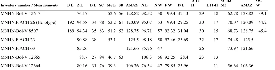
Inventory number / Measurements B L Z L D L SC Mo L SB AMAZ N L N W F W D L W I1-I1 L I1-I1 W M3-M3 AMAZ OC W MNHN-Bol-V 12617 76.17 52.6 56 128.82 98.52 50 99.4 32.13 29 18 62.78 128.82 39.1 MNHN.F.ACH 26 (Holotype) 192 94.58 34 88 53.2 61 120.09 95.07 53 99.4 29.25 30 17 70.07 120.09 44.2 MNHN-Bol-V 8507 189 94.34 35 83 51.2 52 128.75 96.71 57 92.32 31.04 30 15 68.73 128.75 45.4 MNHN.F.ACH 23 90.88 38 53.1 125.5 98.18 50 92.46 25.69 32 17 74.48 125.5 MNHN.F.ACH 63 85.26 121.66 85.76 47 26 73.97 121.66 MNHN-Bol-V 12665 88.7 27 94 46.7 63 106.3 56 92.25 28.4 23 13 MNHN-Bol-V 12664 80.16 31 76 39.3 106.36 76.54 47 79.85 25.96 11 56.64 106.36 Table 2 Supplementary material 1. Skull measurements (in mm) of Plesiotypotheirum achirense. For abbreviations see Figure 5 Supplementary material 1.
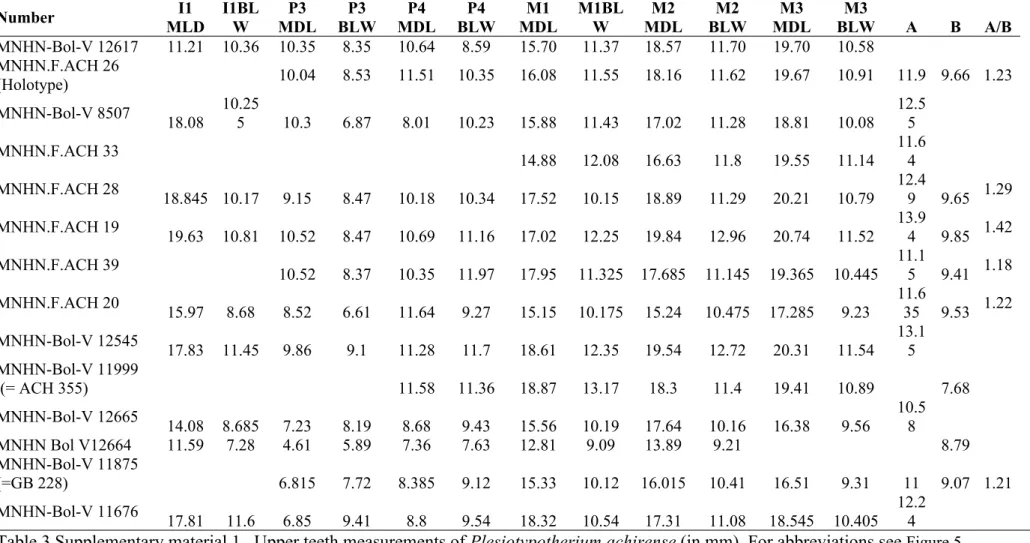
MNHN-Bol-V 12617 11.21 10.36 10.35 8.35 10.64 8.59 15.70 11.37 18.57 11.70 19.70 10.58 MNHN.F.ACH 26 (Holotype) 10.04 8.53 11.51 10.35 16.08 11.55 18.16 11.62 19.67 10.91 11.9 9.66 1.23 MNHN-Bol-V 8507 18.08 10.25 5 10.3 6.87 8.01 10.23 15.88 11.43 17.02 11.28 18.81 10.08 12.5 5 MNHN.F.ACH 33 14.88 12.08 16.63 11.8 19.55 11.14 11.6 4 MNHN.F.ACH 28 18.845 10.17 9.15 8.47 10.18 10.34 17.52 10.15 18.89 11.29 20.21 10.79 12.4 9 9.65 1.29 MNHN.F.ACH 19 19.63 10.81 10.52 8.47 10.69 11.16 17.02 12.25 19.84 12.96 20.74 11.52 13.9 4 9.85 1.42 MNHN.F.ACH 39 10.52 8.37 10.35 11.97 17.95 11.325 17.685 11.145 19.365 10.445 11.1 5 9.41 1.18 MNHN.F.ACH 20 15.97 8.68 8.52 6.61 11.64 9.27 15.15 10.175 15.24 10.475 17.285 9.23 11.6 35 9.53 1.22 MNHN-Bol-V 12545 17.83 11.45 9.86 9.1 11.28 11.7 18.61 12.35 19.54 12.72 20.31 11.54 13.1 5 MNHN-Bol-V 11999 (= ACH 355) 11.58 11.36 18.87 13.17 18.3 11.4 19.41 10.89 7.68 MNHN-Bol-V 12665 14.08 8.685 7.23 8.19 8.68 9.43 15.56 10.19 17.64 10.16 16.38 9.56 10.58 MNHN Bol V12664 11.59 7.28 4.61 5.89 7.36 7.63 12.81 9.09 13.89 9.21 8.79 MNHN-Bol-V 11875 (=GB 228) 6.815 7.72 8.385 9.12 15.33 10.12 16.015 10.41 16.51 9.31 11 9.07 1.21 MNHN-Bol-V 11676 17.81 11.6 6.85 9.41 8.8 9.54 18.32 10.54 17.31 11.08 18.545 10.405 12.24
Table 3 Supplementary material 1. Upper teeth measurements of Plesiotypotherium achirense (in mm). For abbreviations see Figure 5 Supplementary material 1.
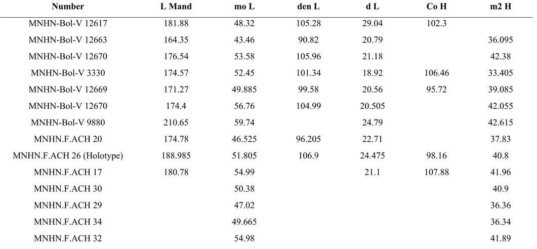
MNHN-Bol-V 12663 164.35 43.46 90.82 20.79 36.095 MNHN-Bol-V 12670 176.54 53.58 105.96 21.18 42.38 MNHN-Bol-V 3330 174.57 52.45 101.34 18.92 106.46 33.405 MNHN-Bol-V 12669 171.27 49.885 99.58 20.56 95.72 39.085 MNHN-Bol-V 12670 174.4 56.76 104.99 20.505 42.055 MNHN-Bol-V 9880 210.65 59.74 24.79 42.615 MNHN.F.ACH 20 174.78 46.525 96.205 22.71 37.83 MNHN.F.ACH 26 (Holotype) 188.985 51.805 106.9 24.475 98.16 40.8 MNHN.F.ACH 17 180.78 54.99 21.1 107.88 41.96 MNHN.F.ACH 30 50.38 40.9 MNHN.F.ACH 29 47.02 36.36 MNHN.F.ACH 34 49.665 36.34 MNHN.F.ACH 32 54.98 41.89
Table 4 Supplementary material 1. Mandible measurements (in mm) of Plesiotypotherium achirense. For abbreviations see Figure 5 Supplementary material 1.
MNHN-Bol-V 12662 10.35 6.27 10.63 7.42 13.355 8.595 15.705 7.94 21.135 7.025 MNHN-Bol-V 12664 6.5 4.78 10.89 7.5 18.49 7.245 MNHN-Bol-V 12663 8.855 5.315 5.765 4.3 8.74 6.055 11.965 7.715 13.275 7.375 18.775 6.58 MNHN-Bol-V 12641 7.64 5.71 5.35 MNHN-Bol-V 12670 10.76 6.61 7.52 4.7 11.9 7.77 13.97 9.57 15.69 9.67 MNHN-Bol-V 3330 10.99 6.655 5.15 4 10.38 6.75 13.955 8.33 16.045 7.22 19.28 7.1 MNHN-Bol-V 3334 11.66 6.92 13.55 8.45 14.81 7.63 MNHN-Bol-V 3755 9.79 7.12 11.7 8.23 MNHN-Bol-V 11677 13.24 8.04 MNHN-Bol-V 12669 10.03 6.555 7.085 5 9.925 7.975 13.685 9.785 15.52 9.63 22.11 8.16 MNHN-Bol-V 12670 10.76 6.61 7.52 4.7 11.9 7.77 13.97 9.57 15.69 9.67 MNHN-Bol-V 9880 12.11 7.47 8.44 4.9 11.935 8.385 16.23 10.09 17.23 9.53 23.64 9.37 MNHN-Bol-V 12642 13.08 7.85 12.22 7.24 MNHN.F.ACH 20 9.1 5.775 6.41 4.3 9.93 6.815 12.76 8.445 15 8.155 19.76 7.02 MNHN.F.ACH 26 10.655 6.45 7.215 5.1 11.315 7.89 13.78 9.55 15.485 9.03 22.265 8.19 MNHN.F.ACH 17 11.215 7.665 14.73 9.43 15.94 8.98 23.165 7.49 MNHN.F.ACH 30 MNHN.F.ACH 29 9.4 5.84 6.61 4.5 9.94 6.65 13.365 8.46 13.99 7.685 18.455 6.9 MNHN.F.ACH 36 12.26 8.69 15.59 9.97
Table 5 Supplementary material 1. Lower teeth measurements (in mm) of Plesiotypotherium achirense. For abbreviations see Figure 5 Supplementary material 1.
metatarsal measurements (d-e); Right scaphoid measurements in dorsal (f), and distal view (g); Left cuboid measurements in dorsal (H) and in distal view (i); Left navicular measurements in dorsal (h), and in proximal view (i); Left ectocuneiform measurements in dorsal (l), and in distal view (m).
CC-Diast R Craniocaudal width of radius diaphysis
LM-Diast R Lateromedial width of the radius diaphysis
CC-Prox R Craniocaudal width of the radius head
LM-Prox R Lateromedial width of the radius head
CC-Fac Dist R Craniocaudal width of the fossa carpi radialis of radius
LM-Fac Dist R Lateromedial width of the fossa carpi radialis of radius
LMc/Mt Length of the Mc or Mt
LM-Prox W Lateromedial width of the proximal epiphysis of the Mc or Mt
PD-Prox H Plantodorsal width of the proximal epiphysis of the Mc or Mt
LM Diast Mc/Mt Lateromedial width of the diastema of the Mc or Mt
PD-Diast Mc/Mt Plantodorsal width of the diastema of the Mc or Mt
PrDist L Proximodistal lenght (carpal or tarsal)
Med Lat W Mediolateral width (carpal or tarsal)
P Dor H Palmodorsal or plantodorsal height (carpal or tarsal)
Table 7 Supplementary material 1. Abbreviation, description and summary of postcranial measurements (in mm) of Plesiotypotherium
achirense. Number Element LR CC-Diast R LM-Diast R CC-Prox R LM-Prox R CC-Fac Dist R LM-Fac Dist R MNHN-Bol-V 12617 Radius (R) 141.38 10.4 27.20 15.59 27.24 15.85 22.38 MNHN-Bol-V 12617 Radius (L) 142.08 10.6 27.34 15.11 27.34 14.5 21.08 MNHN-Bol-V 12760 Radius (R) 148.5 10.98 15.72 15.6 21.35 26.02 MNHN.F.ACH 18 (Paratype) Radius 170.64 13.4 21.76 28.08 15.3 16.72 Table 8 Supplementary material 1. Radius measurements (in mm). For abbreviations see Table 7 Supplementary material 1.
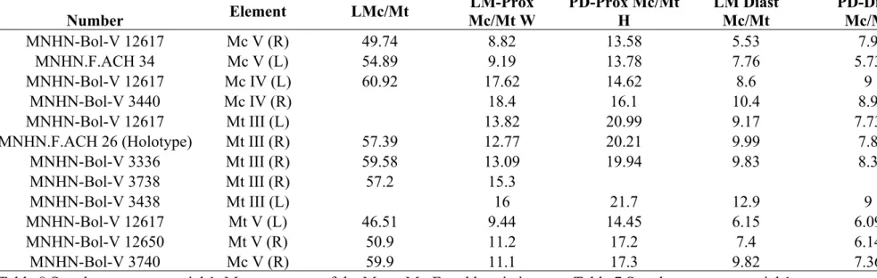
MNHN-Bol-V 12617 Mc V (R) 49.74 8.82 13.58 5.53 7.9 MNHN.F.ACH 34 Mc V (L) 54.89 9.19 13.78 7.76 5.73 MNHN-Bol-V 12617 Mc IV (L) 60.92 17.62 14.62 8.6 9
MNHN-Bol-V 3440 Mc IV (R) 18.4 16.1 10.4 8.9
MNHN-Bol-V 12617 Mt III (L) 13.82 20.99 9.17 7.73 MNHN.F.ACH 26 (Holotype) Mt III (R) 57.39 12.77 20.21 9.99 7.8
MNHN-Bol-V 3336 Mt III (R) 59.58 13.09 19.94 9.83 8.3 MNHN-Bol-V 3738 Mt III (R) 57.2 15.3 MNHN-Bol-V 3438 Mt III (L) 16 21.7 12.9 9 MNHN-Bol-V 12617 Mt V (L) 46.51 9.44 14.45 6.15 6.09 MNHN-Bol-V 12650 Mt V (R) 50.9 11.2 17.2 7.4 6.14 MNHN-Bol-V 3740 Mc V (R) 59.9 11.1 17.3 9.82 7.36 Table 8 Supplementary material 1. Measurements of the Mc or Mt. For abbreviations see Table 7 Supplementary material 1.
Samples were crushed and sieved and a 100–200 μm grain size was retained for feldspar
separation. After magnetic separation, feldspar grains were selected under a
stereomicroscope. The grains were leached with HNO3 (1N) for a few minutes and then
repeatedly cleaned ultrasonically in distilled water and alcohol. Samples were packed in
aluminium foil for irradiation in the core of the Triga Mark II nuclear reactor of Pavia (Italy)
with several aliquots of the Taylor Creek sanidine standard (28.619 ± 0.034 Ma in Renne et
al., 2011) as flux monitor. Argon isotopic interferences on K and Ca were determined by
irradiation of KF and CaF2 pure salts from which the following correction factors were
obtained: (40Ar/39Ar) K = 0.00969 ± 0.00038, (38Ar/39Ar) K = 0.01297 ± 0.00045,
(39Ar/37Ar)Ca = 0.0007474 ± 0.000021 and (36Ar/37Ar)Ca = 0.000288 ± 0.000016. 40Ar/39Ar
step-heating analyses were performed at Géosciences Montpellier (France). The gas
extraction and purification line consist of (a) an IR-CO2 laser of 100 kHz used at 3-20%
power to heat samples during 60 seconds, (b) a lenses system for beam focusing, (c) a steel
chamber maintained at 10-8 - 10-9 bar, with a copper holder in which 2 mm-diameter blind
holes were milled, and (d) two Zr-Al getters for purification of gases. Argon isotopes are
analysed with an ArgusVI multi-collection mass spectrometre (with 4 faradays for masses
40Ar-37Ar and ion counting on 36Ar). One minute was allowed for equilibration before
analysis. Mass discrimination of machines is followed daily. Mass discrimination was
monitored daily using an automated air pipette and provided a mean value of 0.99985 ±
0.00274 per dalton. Micropopulations of feldspar crystals were distributed in holes of the
copper holder and were step heated. Blank analyses were performed every three sample
analyses. Raw data of each step and blank were processed and ages were calculated using the
ArArCALC-software (Koppers, 2002). Isotopic ratios were corrected for irradiation
interferences and air contamination using a mean air value (40Ar/36Ar)
released spectra and inverse isochrones. Plateau ages are calculated from at least three consecutive 39Ar release steps comprising up to 50% of total 39Ar
K released and overlapping
at the 2σ confidence level (Fleck et al., 1977). Isochrone ages are accepted when mean square
weighted deviation (MSWD) is close to 1 and the 40Ar/36Ar intercept within 2 from the
(40Ar/36Ar)
atmvalue. The analytical data are reported in the tables ACH TUF 4A
A9.detailed.xls, ACH TUF 3A A10 plateau.detailed.xls and ACH TUF 3A A10
isochrone.detailed.xls. All errors are quoted at the 2σ level uncertainty including the error on
the irradiation factor J.
Fleck, R.J., J. F. Sutter, and D. H. Elliot. 1977. Interpretation of discordant 40Ar/39Ar age- spectra of mesozoic tholeiites from Antarctica: Geochimica et Cosmochimica Acta 41: 15–32.
Lee J-Y, K. Marti, J. P. Severinghaus, K. Kawamura, H. S. Yoo, J. B. Lee, and J. S. Kim. 2006. A redetermination of the isotopic abundances of atmospheric Ar: Geochimica et Cosmochimica Acta 70:4507–4512.
Renne, P.R., G. Balco, K. R. Ludwig, R. Mundil, and K. Min. 2011. Response to the comment by W.H. Schwarz et al. on “Joint determination of 40K decay constants and 40Ar*/40K for the Fish Canyon sanidine standard, and improved accuracy for 40Ar/39Ar geochronology” by P.R. Renne et al. (2010): Geochimica et Cosmochimica Acta 75:5097–5100.
Renne, P.R., W. S. Cassata, and L. E. Morgan. 2009. The isotopic composition of atmospheric argon and 40Ar/39Ar geochronology: time for a change: Quaternary Geochronology 4:288-298.