HAL Id: hal-03036079
https://hal.archives-ouvertes.fr/hal-03036079
Submitted on 21 Dec 2020
HAL is a multi-disciplinary open access
archive for the deposit and dissemination of
sci-entific research documents, whether they are
pub-lished or not. The documents may come from
teaching and research institutions in France or
abroad, or from public or private research centers.
L’archive ouverte pluridisciplinaire HAL, est
destinée au dépôt et à la diffusion de documents
scientifiques de niveau recherche, publiés ou non,
émanant des établissements d’enseignement et de
recherche français ou étrangers, des laboratoires
publics ou privés.
infected erythrocyte permeability
Guillaume Bouyer, Daniela Barbieri, Florian Dupuy, Anthony Marteau,
Abdoulaye Sissoko, Marie-Esther N’dri, Gaelle Neveu, Laurianne Bedault,
Nabiha Khodabux, Diana Roman, et al.
To cite this version:
Guillaume Bouyer, Daniela Barbieri, Florian Dupuy, Anthony Marteau, Abdoulaye Sissoko, et al..
Plasmodium falciparum sexual parasites regulate infected erythrocyte permeability. Communications
Biology, Nature Publishing Group, 2020, 3 (1), �10.1038/s42003-020-01454-7�. �hal-03036079�
ARTICLE
Plasmodium falciparum sexual parasites regulate
infected erythrocyte permeability
Guillaume Bouyer
1,2,7
, Daniela Barbieri
2,3,7
, Florian Dupuy
2,3
, Anthony Marteau
2,3
, Abdoulaye Sissoko
2,4
,
Marie-Esther N
’Dri
2,3
, Gaelle Neveu
2,3
, Laurianne Bedault
2,3
, Nabiha Khodabux
2,3
, Diana Roman
2,4
,
Sandrine Houzé
2,4
, Giulia Siciliano
5
, Pietro Alano
5
, Rafael M. Martins
6
, Jose-Juan Lopez-Rubio
6
,
Jérome Clain
2,4
, Romain Duval
2,4
, Stéphane Egée
1,2,8
& Catherine Lavazec
2,3,8
✉
To ensure the transport of nutrients necessary for their survival, Plasmodium falciparum
parasites increase erythrocyte permeability to diverse solutes. These new permeation
pathways (NPPs) have been extensively characterized in the pathogenic asexual parasite
stages, however the existence of NPPs has never been investigated in gametocytes, the
sexual stages responsible for transmission to mosquitoes. Here, we show that NPPs are still
active in erythrocytes infected with immature gametocytes and that this activity declines
along gametocyte maturation. Our results indicate that NPPs are regulated by cyclic AMP
(cAMP) signaling cascade, and that the decrease in cAMP levels in mature stages results in a
slowdown of NPP activity. We also show that NPPs facilitate the uptake of artemisinin
derivatives and that phosphodiesterase (PDE) inhibitors can reactivate NPPs and increase
drug uptake in mature gametocytes. These processes are predicted to play a key role in P.
falciparum gametocyte biology and susceptibility to antimalarials.
https://doi.org/10.1038/s42003-020-01454-7
OPEN
1Sorbonne Université, CNRS UMR 8227, Station Biologique de Roscoff, Roscoff, France.2Laboratoire d’excellence GR-Ex, Paris, France.3Université de
Paris, Inserm U1016, CNRS UMR 8104, Institut Cochin, Paris, France.4Université de Paris, IRD 261, MERIT, Paris, France.5Istituto Superiore di Sanita,
Roma, Italy.6Université de Montpellier 1 & 2, CNRS 5290, IRD 224, MIVEGEC, Montpellier, France.7These authors contributed equally: Guillaume Bouyer,
Daniela Barbieri.8These authors jointly supervised this work: Stéphane Egée, Catherine Lavazec. ✉email:catherine.lavazec@inserm.fr
123456789
M
alaria remains a major public health problem with more
than 200 million cases and almost half a million deaths
annually. The clinical symptoms of malaria are
attrib-uted to Plasmodium asexual stages, whereas parasite transmission
from humans to mosquitoes relies on the gametocytes, the
spe-cialized sexual cells formed by a fraction of parasites which cease
asexual
propagation.
Plasmodium
falciparum
gametocyte
maturation requires about ten days, and is classically divided into
five developmental stages based upon morphological features
1. If
eradication of malaria obviously requires compounds targeting
asexual stages, transmission remains the Achilles heel of the
strategies implemented today
2. However, most antimalarial drugs
target the asexual stages, but they are less effective against
gametocytes
3,4. As a consequence, infected individuals remain a
source of transmission even after they are cured. It remains
unclear why gametocytes become less sensitive to artemisinin as
their maturation progresses from stage I to stage V. Thus,
understanding the biology of gametocyte development within
erythrocytes is crucial for successful malaria elimination.
Following P. falciparum invasion, the infected erythrocyte
displays important alterations of its membrane properties. For
instance, asexual parasites increase erythrocyte permeability to
diverse solutes to ensure the transport of nutrients and waste
products necessary for their replication and survival. Mature
asexual parasites activate weakly selective anion channels in the
erythrocyte membrane to generate new permeability pathways
(NPPs) that render infected erythrocytes more permeable to a
range of nutrients
5,6and to several antimalarials
7–10. Key proteins
involved in NPPs have been characterized, like parasitic proteins
CLAG3/RhopH1, RhopH2 and RhopH3 or endogenous
con-stituent of the peripheral-type benzodiazepine receptor
11–14.
However, the full identity of the NPPs has never been
con-clusively established
15. Although their regulatory mechanisms
also remain unclear, cyclic AMP (cAMP)/Protein Kinase A
(PKA) pathway seems to play a key part in regulating ion
channels in the membrane of erythrocytes infected with asexual
stages
16. Despite the vital role of NPPs in asexual stages, the level
of NPP activity in gametocyte-infected erythrocytes (GIE) is
unknown, nor is the role of NPPs in antimalarials uptake by GIE.
Refractoriness of GIE to lysis upon short exposure to isosmotic
sorbitol solution has led to the dogma that NPPs are totally
absent in gametocyte stages
17. However, this assumption is not
supported by the fact that during their 10-day maturation,
gametocytes also need to absorb nutrients from the plasma and
get rid of toxic waste products they generate upon hemoglobin
digestion, two major roles of NPPs at asexual stages. In addition,
the absence of NPPs in GIE is not consistent with the expression
profile of members of the RhopH complex that are synthesized in
mature intracellular parasites and then secreted upon egress onto
the erythrocyte targeted for invasion
18,19. Therefore, all known
NPP components should be present at the surface of newly
invaded GIE.
In this study, we performed isosmotic lysis, electrophysiology,
fluorescence tracer uptake and viability experiments to evaluate
NPP activity during P. falciparum gametocytogenesis. We found
that NPP activity is regulated by the cycling AMP signaling
cascade, and interfering with this pathway can reactivate
ery-throcyte permeability and facilitate uptake of artemisinin
deri-vatives by mature gametocytes.
Results
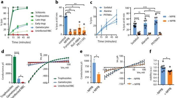
NPPs are still active in immature gametocytes. To evaluate the
NPP activity in GIE, we
first performed measurements of
iso-smotic lysis of immature GIE in sorbitol, a sugar alcohol
per-meant through NPPs
6. Sorbitol uptake was drastically reduced in
stage II GIE compared to trophozoites and schizonts; however,
about 36% GIE were lysed after 60 min in sorbitol, suggesting that
erythrocyte permeability is modified by immature gametocytes
(Fig.
1
a). Lysis kinetics in stage II GIE were similar to that of late
rings 16 h-post-invasion, when NPPs start to be expressed during
the asexual cycle
20. GIE lysis was significantly inhibited by the
general anion channel inhibitors
5-nitro-2-(3-phenylpropyla-mino)benzoic acid (NPPB) and furosemide, by the
benzodiaze-pine Ro5-4864 and the isoquinoline PK11195, which have all
been shown to inhibit NPPs (Fig.
1
b)
11,21. Equivalent lysis
kinetics observed in alanine or phenyltrimethylammonium
(PhTMA
+) isosmotic solutions, with a slightly greater lysis in
PhTMA
+than in sorbitol, confirmed that uptake occurred
through NPPs (Fig.
1
c). Measurement of membrane currents
using whole-cell patch-clamp strengthened observations obtained
with isosmotic lysis. Quantification of total ion fluxes across the
host membrane of individual infected erythrocytes indicated that
membrane conductance at
−100 mV was sixfold lower in stage II
GIE than in trophozoites (Fig.
1
d and Supplementary Fig. S1).
However, conductance in early GIE showed anion selectivity,
inward rectification and NPPB sensitivity (Fig.
1
e and
Supple-mentary Fig. S1), which are three fundamental characteristics of
the correlate membrane currents of NPP activity
22. These
observations are consistent with detection of the NPP component
RhopH2
12in immature GIE by immunostaining (Supplementary
Fig. S1). Pharmacological inhibition of NPPs is expected to lead
to nutrient starvation and accumulation of toxic metabolic wastes
eventually leading to parasite death. Thus, we measured viability
of early gametocytes (stages I−II) upon NPPs inhibition by using
a luciferase-based assay with a transgenic parasite expressing
luciferase under the control of the gametocyte-specific promoter
Pfs16
23. We observed a slight but significant decrease in early
gametocytes viability 48 h after a 3-h exposure to 100 µM NPPB,
consistent with a vital role of NPP activity (Fig.
1
f). Fewer
tran-sition to later stages gametocytes after exposure to NPPB was also
quantified by Giemsa staining (Supplementary Fig. S1).
There-fore, organic solute uptake, patch-clamp and viability studies
indicate that NPPs are weaker than in trophozoites but are still
active and necessary in early GIE.
NPPs decline along gametocytogenesis. To further analyze the
evolution of this permeability along gametocytogenesis, we
per-formed isosmotic lysis experiments in sorbitol, alanine and
PhTMA
+for synchronous cultures of stage I, II, III, IV, and V
gametocytes (Fig.
2
a–c). A transgenic parasite expressing GFP
under the control of the Pfs16 promoter was used to discriminate
stage I gametocytes from asexual stages
24. From stage I to stage V,
we observed a progressive decrease in permeability for the three
different solutes, leading to undetectable NPP activity in mature
GIE. Electrophysiological experiments using whole-cell
config-uration performed on GIE from all stages confirmed that
mem-brane conductance drops along gametocyte maturation, leading
to currents in mature GIE corresponding to levels usually
recorded in uninfected erythrocytes (Fig.
2
d and Supplementary
Fig. S2). Altogether, these data indicate that NPP activity declines
along gametocytogenesis.
NPP activity is regulated by cAMP-signaling. We then aimed to
determine the mechanisms underlying the regulation of NPP
activity in GIE. We have previously observed that cAMP/PKA
pathway activates ion channels in the membrane of erythrocytes
infected with asexual stages
16, suggesting that cAMP-signaling
may also modulate NPPs during gametocytogenesis. To address
this hypothesis, we performed sorbitol lysis experiments on early
GIE preincubated with KT5720 and H89, two independent and
widely used PKA inhibitors that have already been shown to
inhibit PKA activity in P. falciparum
25,26. We found that both
compounds ablated GIE permeability (Fig.
3
a), whereas the
cAMP analog 8-Bromide-cyclic adenosine-monophosphate
(8Br-cAMP) significantly increased the sorbitol-induced lysis (Fig.
3
b).
As an alternative way to investigate the effects of inhibiting PKA
activity, we used a transgenic parasite that overexpresses the
regulatory subunit of PKA using an episome selected by the
antimalarial drug pyrimethamine (pHLpfpkar)
16,26. The
down-regulation of PKA activity in this parasite line also resulted in a
significant decrease in permeability of early GIE (Fig.
3
c). This
phenotype was reverted to the levels of wild-type parasites upon
incubation with 8Br-cAMP, or in a revertant parasite line that has
shed the overexpressing episome
26(Fig.
3
c). We observed a
similar inhibition of alanine- or PhTMA
+-induced isosmotic lysis
of stage II GIE for the pHLpfpkar line (Supplementary Fig. S3).
These results indicate that PKA activity contributes to NPPs in
immature GIE, as described for asexual stages
16.
These observations suggest that the decline in NPP activity
during gametocytogenesis likely results from the previously
described drop in cAMP concentration in GIE due to the rising
expression of PfPDEδ in mature gametocytes
26. Thus, we
hypothesized that interfering with cAMP pathway with molecules
that raise cAMP levels may turn on NPP activity in stage V GIE.
As expected, raising cAMP levels in mature GIE upon incubation
with 8Br-cAMP or with the PDE inhibitors sildenafil and tadalafil
restored the sorbitol-induced lysis to the levels observed in early
GIE, whereas tadalafil did not trigger any significant effect in early
GIE (Fig.
3
d, e and Supplementary Fig. S4). Consistently, the loss
of PDE activity in a transgenic parasite in which the Pfpdeδ gene
had been deleted
27also resulted in a drastic increase in
permeability of mature GIE (Fig.
3
f and Supplementary Fig. S3).
These
findings show that activating the cAMP/PKA pathway can
reactivate NPPs in mature stages.
NPPs contribute to the uptake of artemisinin derivatives by
immature GIE. P. falciparum mature gametocytes are known to
become less sensitive to several antimalarials, including
artemi-sinin derivatives, as their maturation progresses from stage I to
stage V
28,29. Although lower metabolic activity and complete
hemoglobin digestion in mature stages have been suggested to
contribute to this decrease in chemosensitivity
4,30, we
hypothe-sized that this decrease may also be linked to the decline of host
membrane permeability during gametocyte maturation. To
address this hypothesis, we
first evaluated whether NPP activity is
required for uptake of artemisinin derivatives by immature GIE.
For this purpose, we used Fluo-DHA, a
fluorescent probe
mimicking the clinical antimalarials dihydroartemisinin (DHA)
and artemether
31. This probe was synthesized from DHA and a
4-nitrobenzoxadiazole
(NBD)
fluorophore (Supplementary
Fig. 1 NPPs are still active in immature gametocytes. a Kinetics of sorbitol-induced isosmotic lysis of erythrocytes infected with early rings (4 h-post-invasion (hpi)), late rings (16 hpi), trophozoites (28 hpi), schizonts (40 hpi), stage II gametocytes and uninfected erythrocytes during 60 min. n = 3 independent experiments.b % lysis of stage II GIE in sorbitol at 60 min with or without 100µM NPPB, Furosemide, Ro5-4864, or PK11195. c Lysis kinetics (left) and % lysis at 60 min (right) of stage II GIE in sorbitol, alanine or PhTMA+.d Patch-clamp experiments on erythrocytes infected with trophozoites, (green bar, number of cells= 14), stage II gametocytes (blue bar, number of cells = 14) or uninfected erythrocytes (red bar, number of cells = 11). Left: whole-cell conductance calculated at−100 mV. Right: I−V plot from patch-clamp experiments. e Patch-clamp experiments on stage II GIE in presence or absence of NPPB (number of cells= 14 and 5, respectively). Left: whole-cell conductance calculated at −100 mV. Right: I−V plot from patch-clamp experiments.f Viability (luciferase activity) of early gametocytes of the NF54-cg6-pfs16-CBG99 line 48 h after a 3-h incubation with or without 100µM NPPB. The graph shows relative viability normalized by the average luciferase activity of control (without NPPB). a.u. arbitrary units. In (b, c, f), circles indicate the number of independent experiments. In (d, e), the data distribution is shown in Supplementary Fig. S1. Error bars show the standard error of the mean (SEM). Statistical significance is determined by a Mann−Whitney test (d−f) or by one-way ANOVA with Dunnet correction (b) or Sidak correction (c) for multiple comparisons. ****p < 0.0001, ***p < 0.001, **p < 0.01, *p < 0.05, ns: non-significant difference.
Fig. S5). First, we validated that Fluo-DHA was detectable in the
gametocyte cytoplasm after 2 h incubation and exhibited
anti-plasmodial activity against early gametocytes as assessed by a
luciferase-based viability assay (Supplementary Fig. S5). Flow
cytometry quantification showed that Fluo-DHA was taken up by
stage II GIE, whereas uninfected erythrocytes were all negative
(Fig.
4
a, b and Supplementary Fig. S6). The pharmacological
specificity of this probe was validated by a competition assay with
an excess of DHA (Fig.
4
c). Importantly, preincubation with the
NPP inhibitors NPPB and furosemide induced a significant
decrease in the mean
fluorescence intensity, indicating that
Fluo-DHA uptake is partly mediated by NPPs in early GIE (Fig.
4
d).
About 50% of
fluorescence remained upon NPPB treatment,
suggesting that Fluo-DHA uptake may also partly occur by
Fig. 2 NPPs decline along gametocytogenesis. a–c Sorbitol-induced (a), alanine-induced (b) and PhTMA+-induced (c) isosmotic lysis of GIE from stage I to stage V. Left: Kinetics of isosmotic lysis during 60 min. Right: % lysis at 60 min with (orange) or without (blue) 100µM NPPB. Circles indicate the number of independent experiments and error bars show the SEM. Statistical significance is determined by one-way ANOVA with Sidak correction for multiple comparisons.d Left: I−V plot from patch experiments on GIE from stage I to stage V. Right: Whole-cell conductance calculated at −100 mV on GIE from stage I to stage V with (orange) or without (blue) 100µM NPPB. Circles indicate the number of independent experiments and error bars show the SEM. Statistical significance is determined by a Mann−Whitney test at each gametocyte stage and by ANOVA test for trend between stage 1 and stage 5, p = 0.0013, slope −134.7 ± 40.03 pS/stage transition. ****p < 0.0001, ***p < 0.001, **p < 0.01, *p < 0.05, ns: non-significant difference.
diffusion through the erythrocyte membrane or by another route.
Moreover, the viability at 48 h of early GIE following a 3-h pulse
exposure to 150 nM Fluo-DHA or 700 nM artemisinin was
slightly but significantly increased upon preincubation with
NPPB, suggesting that NPP inhibition impacts the transport of
these antimalarials into the infected erythrocytes (Fig.
4
e). These
results indicate that NPPs facilitate the uptake of artemisinin
derivatives, and substantiate the idea that the slowdown of NPP
activity in mature stages may account for their refractoriness to
these drugs. To address this hypothesis, we evaluated Fluo-DHA
uptake in synchronous cultures of stage II, III, IV, and V
game-tocytes. As observed for the decline in NPP activity, Fluo-DHA
uptake also decreased during gametocyte maturation (Fig.
4
f and
Supplementary Fig. S6). The mean
fluorescence intensity in GIE
slowed down in stage III and reached the same level as that of
NPPB-treated cells in stage V GIE. This profile is consistent with
the increased IC
50of Fluo-DHA in mature gametocytes
(Sup-plementary Fig. S5) and correlates with the previously described
chemosensitivity shift occurring at the transition from stage III to
stage IV
28,32. Therefore, the decline in NPP activity and in drug
uptake along gametocytogenesis parallels the decrease in
game-tocytes sensitivity to several antimalarials
4,28. Although we cannot
rule out that the decrease in Fluo-DHA activity in mature stages
reflects the slowdown of hemoglobin breakdown required for the
activation of artemisinin
33, our results provide evidence that
NPPs significantly contribute to the uptake of artemisinin
deri-vatives in GIE.
cAMP-mediated reactivation of NPPs increases uptake of
artemisinin. Next, we analyzed whether the uptake of artemisinin
derivatives is also regulated by cAMP-signaling. In early GIE, we
observed that both pharmacological and genetic inhibition of
PKA activity decreased the mean
fluorescence intensity of
Fluo-DHA in treated cells (Fig.
4
g, h), suggesting that PKA activity
facilitates Fluo-DHA uptake in immature GIE. Thus, we
hypo-thesized that a reactivation of NPP activity in mature GIE may
increase their susceptibility to these drugs. First, we addressed
whether activating the cAMP pathway may enhance drug uptake
by stage V GIE. Preincubation of mature GIE with the PDE
inhibitors sildenafil and tadalafil strongly increased the mean
fluorescence intensity of Fluo-DHA in treated cells, reaching the
level observed in early GIE (Fig.
4
i and Supplementary Fig. S7).
The uptake of Fluo-DHA by mature GIE was significantly
increased with 10 µM tadalafil, which approximately corresponds
to tenfold the reported peak serum concentration reached
in humans after 60 min following 20 mg oral dose (C
max378 ng/
ml)
34. As expected, preincubation of stage II GIE with tadalafil
did not trigger any significant effect (Supplementary Fig. S7).
To investigate the gametocytocidal effect of combining PDE
inhibitors and artemisinin, we performed a gamete egress assay
based on the ability of functionally viable mature gametocytes to
undergo a temperature-dependent release of gametes from
their host erythrocytes
35. We observed that the incubation of
mature GIE with a combination of 5 µM artemisinin and 30 µM
tadalafil drastically reduced the proportion of egressed gametes
compared to tadalafil or artemisinin alone, indicating that
phosphodiesterase inhibitors potentiate the effect of artemisinin
in mature gametocytes (Fig.
4
j). Therefore, these results show
that cAMP-mediated reactivation of NPPs in mature stages
enhances the uptake of artemisinin, and importantly this
mechanism can increase mature gametocyte sensitivity to this
antimalarial.
Fig. 3 NPP activity is regulated by cAMP-signaling. a, b Sorbitol-induced isosmotic lysis of stage II GIE with 100µM H89 or 10 µM KT5720 (a), or with 100µM 8Br-cAMP (b). c Sorbitol-induced isosmotic lysis of stage II GIE from the NF54 isolate (Control) and the transgenic pHLpfpkar line, cultivated with or without pyrimethamine (Pyri), or preincubated with 100µM 8Br-cAMP. d, e Sorbitol-induced isosmotic lysis of stage V GIE with 100 µM 8Br-cAMP (d), sildenafil (e) or tadalafil (e). f Sorbitol-induced isosmotic lysis of stage V gametocytes from the NF54 isolate and the transgenic line PDEδ−. All experiments were performed in the presence (orange) or absence (blue) of 100µM NPPB. Circles indicate the number of independent experiments and error bars show the SEM. Statistical significance is determined by one-way ANOVA with Sidak correction for multiple comparisons. ****p < 0.0001, ***p < 0.001, **p < 0.01, *p < 0.05, ns: non-significant difference.
Discussion
It has been known for long that P. falciparum asexual stages
modify their host erythrocyte to make them more permeable to
supplementary nutrient uptake from the plasma and for removal
of toxic waste by activating NPPs in the erythrocyte membrane.
In this study we unravel how these NPPs are regulated during
sexual parasite development and we propose that activating these
pathways may facilitate artemisinin uptake by mature
gameto-cytes. Our results raise a new model (Fig.
5
) where NPP activity in
immature GIE depends on the PKA-mediated phosphorylation of
one or several proteins directly or indirectly involved in channel
activity. According to this model, NPPs contribute to the uptake
Fig. 4 NPPs contribute to the uptake of artemisinin derivatives. a Diagram illustrating the uptake assay. b Left: Quantification of Fluo-DHA uptake in uninfected erythrocytes and in early GIE byflow cytometry. Right: scatter plots showing the gating strategy for Fluo-DHA uptake. c Competition assay between Fluo-DHA and DHA, and control of HPA-NBD and DHA-inducedfluorescence levels. d Inhibition of Fluo-DHA uptake in early GIE upon 100 µM NPPB or Furosemide incubation.e Viability (luciferase activity) of early gametocytes of the NF54-cg6-pfs16-CBG99 line 48 h after a 3-h incubation with 150 nM Fluo-DHA or 5µM artemisinin, with or without 100 µM NPPB. The graph shows the ratio of luciferase activity (drug-treated/control) and is normalized to the condition without NPPB. a.u. arbitrary units.f Quantification of Fluo-DHA uptake during gametocytogenesis with (orange) or without (blue) 100µM NPPB. g Fluo-DHA uptake in early GIE upon 100 µM H89 or 10 µM KT5720 incubation. h Fluo-DHA uptake in early GIE from NF54 (Control) and the transgenic pHLpfpkar line, cultivated with or without pyrimethamine (Pyri), or preincubated with 100 µM 8Br-cAMP. i Fluo-DHA uptake in stage V GIE upon 0, 10, 30 and 100µM tadalafil incubation. j Left: % inhibition of gamete egress after a 24-h incubation with 5 µM artemisinin, with or without 30 µM tadalafil. Right: gamete egress observed by IFAs. Samples were co-stained with mouse anti-glycophorin A (GPA, red) and rabbit anti-Pfg27 (green) IgG. DIC differential interference contrast. Arrow: mature GIE with an intact erythrocyte membrane, arrowhead: egressed gamete. Scale bars: 5μm. Circles indicate the number of independent experiments and error bars show the SEM. Statistical significance is determined by one-way ANOVA with Dunnet correction (b−d, g, i) or with Sidak correction (f, h, j) for multiple comparisons or by a Mann−Whitney test (e). ****p < 0.0001, ***p < 0.001, **p < 0.01, *p < 0.05, ns: non-significant difference.
of artemisinin and derivatives by immature GIE. Then in mature
GIE, PfPDEδ, whose catalytic domain is predicted to be exposed
outwards of the parasite
36, is highly expressed and degrades
cAMP
26. The resulting drop of cAMP level in the
para-sitophorous vacuole and in the host cell may reduce the
phos-phorylation of these proteins and consequently decrease NPP
activity and drug uptake. Importantly, pharmacological inhibition
of PfPDEδ can reactivate NPP activity and restore uptake of
artemisinin derivatives by mature gametocytes, leading to an
increased susceptibility to these drugs. Altogether these results
show that P. falciparum gametocytes regulate infected erythrocyte
permeability and that this permeability may facilitate uptake of
some antimalarials.
Our results showing that NPPs are still active in immature GIE
contradict the paradigm that NPP activity is abolished in sexual
stages. This dogma was based on a single study showing
refrac-toriness of GIE to lysis upon short exposition to isosmotic
sor-bitol solution
17. In contrast, our data report that GIE were lysed
after longer exposition to sorbitol, indicating the presence,
although slower than in trophozoites, of NPP activity in
imma-ture GIE. In support of the existence of NPPs in sexual stages,
recent RNA sequencing analyzes highlighted that sexually
com-mitted parasites show higher transcription of RhopH and clag
genes
37, whose products are involved in NPP activity
12–14. These
observations suggest that members of the RhopH complex
syn-thesized in sexually committed parasites may be discharged upon
egress onto the membrane of the erythrocyte targeted for
inva-sion, within which a gametocyte will develop. Accordingly, our
data show the presence of at least one member of the RhopH
complex in GIE. This hypothesis would be consistent with the
fact that gametocytes should absorb nutrients from the
extra-cellular medium, such as panthotenate or isoleucine, for which
NPPs is the major route
21. In addition, NPPs have been proposed
to play a role in exporting amino acids liberated by the digestion
of hemoglobin from the infected erythrocyte, thereby protecting
the cell against the osmotic challenge posed by elevated
intra-cellular amino acid levels
20. Thus, the slowdown of NPP activity
in mature GIE may result from the completion of hemoglobin
digestion in these stages
38. Absence of channel activity in mature
stages may allow the infected erythrocyte to decrease its osmotic
fragility and thus to persist longer in the blood circulation. This
process appears to be tightly regulated by cAMP-signaling
pathway, as previously observed for asexual stages
16. These
results are in accordance with the detection of CLAG3/RhopH1,
one of the major component of NPPs, in a global
phospho-proteomic analysis of P. falciparum parasites
39. As part of the
cAMP-signaling cascade, the PfPDEδ enzyme, whose expression
increases in mature gametocytes
26, plays an instrumental role
in the NPP decline. Interestingly, this enzyme has also been
reported to govern the switch in GIE deformability that
enables mature gametocytes to circulate several days in the
bloodstream and avoid clearance by the spleen
26,40,41. Therefore,
by decreasing GIE osmotic fragility and increasing GIE
deform-ability, PfPDEδ appears to be the key regulator of mature
gametocytes persistence in the blood circulation. Importantly, our
present work suggests that targeting this enzyme with the
FDA-approved drug tadalafil may enhance artemisinin uptake by
mature gametocytes, thereby decreasing their refractoriness to
this antimalarial.
As PDE inhibitors also render mature GIE rigid and hence may
promote their clearance by the spleen
26, they represent novel
drug leads potentially capable of blocking malaria transmission by
impacting on both gametocytes circulation and susceptibility to
artemisinin derivatives.
Methods
Parasite culturing and gametocyte production. The P. falciparum NF54 strain, the B10 clone and the transgenic lines pHLpfpkar, PfPDEδ−,
NF54-cg6-pfs16-CBG99 and NF54-pfs47-pfs16-GFP have been described elsewhere16,23,27,42.
Parasites were cultivated in vitro under standard conditions using RPMI 1640 medium supplemented with 10% heat-inactivated human serum and human ery-throcytes at a 5% hematocrit. To obtain synchronous asexual stages, parasites were synchronized by the isolation of schizonts by magnetic isolation using a MACS depletion column (Miltenyi Biotec) in conjunction with a magnetic separator, and placed back into culture. After invasion of merozoites, a second magnetic isolation was used for the selection of ring-stage parasites to obtain a tighter window of synchronization. Synchronous production of specific gametocytes stages was achieved by treating synchronized cultures at the ring stage (10–15% parasitemia, day 0) with 50 mM N-acetylglucosamine (NAG) for 5 days to eliminate asexual parasites. Gametocyte preparations were enriched in different experiments by magnetic isolation. Stage I GIE were collected at day 1 after initiating NAG treatment, stage II GIE were collected at days 2 and 3, stage III GIE were collected Fig. 5 Model for cAMP-mediated regulation of NPP activity. Left: In early GIE, PfPDEδ expression is low, resulting in high cAMP levels in the parasitophorous vacuole and in the host cell, thereby activating the human PKA. PKA phosphorylates one or several proteins directly or indirectly involved in NPP activity that contributes to the uptake of drugs. Middle: In mature GIE, PfPDEδ is highly expressed and degrades cAMP, leading to a decrease in PKA phosphorylation and NPP activity. Right: In mature GIE, inhibition of PfPDEδ by tadalafil results in increased levels of cAMP that reactivate PKA and NPPs, thereby restoring uptake of drugs.
at days 4 and 5, stage IV GIE were collected at days 6 and 7, and stage V GIE were collected at day 8 onwards.
Isosmotic lysis. 500 µl of P. falciparum cultures (containing gametocytes or asexual stages) with a parasitemia >0.5% were washed once with RPMI and incubated in 1.5 ml tubes for 60 min at 37 °C in 500 µl isosmotic solution con-taining either 300 mM sorbitol, 300 mM Alanine or 150 mM PhTMA+ supple-mented with 10 mM Hepes, 5 mM glucose and with a pH adjusted to 7.4. For each experiment,five tubes were prepared including one tube containing 100 µM NPPB, Furosemide, Ro5-4864, or PK11195 (all purchased from Sigma-Aldrich). At each sampling time, one tube was centrifuged and smears were prepared and stained with Giemsa R solution (RAL diagnostics). Parasitemia was estimated for each point by counting infected cells out of at least 4000 erythrocytes, and lysis per-centage was calculated using the formula: % lysis (t)= [1 − (parasitemia (t)/ parasitemia (t0))] × 100.
Microscopy examination of Giemsa-stained smears allowed to morphologically confirm that only gametocytes, but not contaminating asexual parasites, were counted. The percentage of lysis of stage I GIE was determined using the NF54-pfs47-pfs16-GFP line24byfluorescence microscopy at ×40 magnification on a Leica
DMi8 microscope. Addressing the role of cAMP-signaling in isosmotic lysis, stage II GIE (days 2 and 3 post NAG treatment) were preincubated 30 min with 100 µM H89, 10 µM KT5720 or 100 µM 8Br-cAMP, and mature GIE (days 8–11 post NAG treatment) were preincubated 30 min with tadalafil or sildenafil at different concentrations (from 10 to 100 µM). All inhibitors were purchased from Sigma-Aldrich or Euromedex.
For standard semi-quantitative isosmotic lysis assays on uninfected erythrocytes, hemoglobin release was used to estimate lysis. Erythrocytes were washed three times in culture medium without serum and resuspended at 50% hematocrit. Time courses started with the addition of a 10 µl packed cells suspension to 1 ml of the sorbitol isosmotic solution. Experiments were performed in triplicate. At predetermined intervals (0, 15, 30, 60 min), microcentrifuge tubes were centrifuged for 30 s and 200 µl of the supernatant solution was transferred into 96-well plates for spectrophotometric estimation of hemoglobin
concentration by absorption at a wavelength of 540 nm (A540). In all experiments, the A540 value corresponding to full hemolysis of erythrocytes was estimated from thefinal A540 value achieved in the supernatant solution from a suspension where 5 µl of Triton X-100 is added. Data analyses were carried out as previously described.
Patch-clamp. Patch-clamp experiments were performed at room temperature using the whole-cell configuration. Pipettes were pulled using a DMZ Universal Puller (Zeitz Instruments, Germany) from borosilicate glass capillaries (GC150F-10, Harvard apparatus, UK) to obtain a tip resistance of 10–15 MΩ. Pipette solution was: 145 mM KCl, 1.85 mM CaCl2, 1.2 mM MgCl2, 5 mM ethylene
glycol-bis(β-aminoethyl ether)-N,N,N′,N′-tetraacetic acid (EGTA), 10 mM Hepes, 10 mM, pH 7.2. Bath solution was: 145 mM NaCl, 5 mM KCl, 1 mM CaCl2, 1 mM
MgCl2, 10 mM Hepes, 10 mM glucose, pH 7.4. Seal resistance were 4–20 GΩ, and
whole-cell configuration was obtained by a brief electrical pulse (zap) and was assessed by the development of small capacitance transient currents and reduction of access resistance. Whole-cell current were recorded using either an Axopatch 200B (Molecular Devices, USA) or a RK400 (Biologic, France) amplifier, with voltage command protocols and current analysis done with pclamp10 suite soft-ware (Molecular Devices, USA) or WinWCP4.7 softsoft-ware (J. Dempster, Strathclyde University, UK), respectively. Currents were elicited by generating a series of membrane potentials from+100 to −100 mV in −10-mV steps for 500 ms, from a holding potential of 0 mV. For stage I GIE, the gametocytefluorescent NF54-pfs47-pfs16-GFP line24was used to easily distinguish by microscopy between asexual and
early sexual stages.
Fluorescence microscopy. To visualize the uptake of Fluo-DHA, GIE were incubated 2 h with Fluo-DHA or dimethyl sulfoxide (DMSO) 0.1% at 37 °C. Cells were stained with Hoechst 33342 (1/20,000, Thermo Fisher Scientific) for 10 min at 37 °C. After one wash with phosphate-buffered-saline (PBS) 1×, cells werefixed for 10 min at room temperature with 1% paraformaldehyde (PFA) (Electron Micro-scopy Science) and 0.25% glutaraldehyde (Sigma-Aldrich) in PBS 1×. After three washes with PBS 1×, infected cells were observed on a glass slide at ×100 mag-nification using a Leica DMi8 microscope.
For immunofluorescence analysis, smears were prepared with NF54 culture containing immature GIE and asexual stages. Slides were air-dried,fixed for 5 min in 75% acetone/25%methanol at−20 °C, washed three times in PBS, preincubated for 2 h in 1× PBS/2% bovine serum albumin, incubated overnight with a mouse antibody directed against RhopH2 (1/50)43and then 1 h with a rabbit antibody
raised again the Pf11-1 protein (1/5,000)44. Then, slides were incubated with Alexa
Fluor 488-conjugated goat anti-rabbit (1/2,000), Alexa Fluor 594-conjugated goat anti-mouse antibody (1/2,000) and Hoechst 33342 (1/20,000) (Thermo Fisher) for 1 h at room temperature. All samples were observed at ×100 magnification using a Leica DMi8 microscope. The RhopH2-specific mouse serum was obtained from C. Braun-Breton and the Pf11-1-specific rabbit serum was obtained from O. Mercereau-Puijalon.
Quantification of Fluo-DHA uptake assay. The synthesis of the Fluo-DHA probe was described in Sissoko et al.31. Quantification of Fluo-DHA uptake was
per-formed usingflow cytometry. GIE were preincubated or not with 100 µM NPPB or with 100 µM Furosemide for 30 min and then incubated with 1 µM of Fluo-DHA for 2 h at 37 °C, 5% CO2and 5% O2. To address the role of cAMP-signaling in
Fluo-DHA uptake, early GIE (day 2 post NAG treatment) were preincubated 30 min with 100 µM H89, 10 µM KT5720 or 100 µM 8Br-cAMP, and mature GIE (days 8–11 post NAG treatment) were preincubated 30 min with tadalafil or sil-denafil at different concentrations (from 10 to 100 µM). To assess the specificity of Fluo-DHA uptake, early GIE were preincubated or not with 20 µM DHA for 2 h and then incubated with Fluo-DHA (1 µM) for 2 h. DHA alone (20 µM) and HPA-NBD (1 µM) were used as control. Twenty minutes before the end of incubation, GIE were stained with Hoechst 33342 (1/10,000). Cells were then washed with PBS 1× andfixed for 10 min at room temperature with PBS, 1% PFA and 0.25% glutaraldehyde. The percentage of Fluo-DHA-positive cells was quantified using Fortessa (BD Biosciences) cytometer.
Gametocyte survival assay. To calculate the IC50for Fluo-DHA on early or
mature gametocytes, 2 × 10⁵MACS-purified early GIE (day 2 post NAG
treat-ment) or mature GIE (days 8–11 post NAG treattreat-ment) from the NF54-cg6-Pfs16-CBG99 line were incubated with serial dilutions of Fluo-DHA for 3 h, then GIE were washed and incubated with complete medium without drugs for 72 h. To perform viability assays with drugs and inhibitors, early GIE were preincubated at 37 °C for 30 min with 100 µM NPPB or 0.1% DMSO in complete medium and then incubated with the same inhibitors supplemented with 700 nM or 5 µM artemisinin (Sigma-Aldrich), 150 nM Fluo-DHA or 0.1% DMSO for 3 h. GIE were then washed and incubated with complete medium without inhibitors and drugs for 48 h. Cell viability was evaluated by adding a non-lysing formulation of 0.5 mMD-Luciferin substrate23(Sigma-Aldrich) and measuring luciferase activity for
1 s on a plate Reader Infinite 200 PRO (Tecan®). All experiments were performed in triplicate on 96-well plates.
Gamete egress assay. NF54 cultures containing 2−5% stage V gametocytes were incubated for 24 h at 37 °C with 5 µM artemisinin supplemented or not with 30 µM tadalafil. Controls comprising gametocytes exposed to the same final concentration of DMSO (0.1%) were processed in parallel. After the incubation, 100 µl samples from each condition were pelleted at 1000 × g for 1 min and rapidly resuspended in 50 µl human serum at room temperature for 30 min during which gametogenesis took place. Samples were then pelleted at 1000 × g for 1 min, smeared on glass slides, methanol-fixed and co-stained with mouse anti-glycophorin A (GPA, 1/ 1000, Santa Cruz Biotechnology) and rabbit anti-Pfg2745(1/2000) followed by
anti-rabbit Alexa 488- and anti-mouse Alexa 594-conjugated IgG (1/2000, Life Tech-nologies). Samples were observed at ×100 magnification using a Leica DMi8. At least 100 GIE were analyzed for each sample. Percent gamete egress was deter-mined by calculating the % of GPA-negative round gametes in the total GIE population detected by Pfg27 staining.
Statistics and reproducibility. Group data are presented as mean ± s.e.m. Sta-tistics for all datasets were performed using GraphPad Prism. Statistical sig-nificance is determined by one-way ANOVA with Dunnet correction or with Sidak correction for multiple comparisons or by a Mann−Whitney test or paired t test. Sample sizes and replicate details are described in the relevantfigure descriptions.
Data availability
All experimental data generated or analyzed during this study are included in this published article (and its Supplementary Informationfiles: Supplementary Information and Source Data).
Received: 7 February 2020; Accepted: 30 October 2020;
References
1. Hawking, F., Wilson, M. E. & Gammage, K. Evidence for cyclic development and short-lived maturity in the gametocytes of Plasmodium falciparum. Trans. R. Soc. Trop. Med. Hyg. 65, 549–559 (1971).
2. mal, E. R. A. R. C. Po. TfM. E. malERA: an updated research agenda for diagnostics, drugs, vaccines, and vector control in malaria elimination and eradication. PLoS Med. 14, e1002455 (2017).
3. Birkholtz, L. M., Coetzer, T. L., Mancama, D., Leroy, D. & Alano, P. Discovering new transmission-blocking antimalarial compounds: challenges and opportunities. Trends Parasitol. 32, 669–681 (2016).
4. Plouffe, D. M. et al. High-throughput assay and discovery of small molecules that interrupt malaria transmission. Cell Host Microbe 19, 114–126 (2016).
5. Desai, S. A., Bezrukov, S. M. & Zimmerberg, J. A voltage-dependent channel involved in nutrient uptake by red blood cells infected with the malaria parasite. Nature 406, 1001–1005 (2000).
6. Ginsburg, H., Krugliak, M., Eidelman, O. & Cabantchik, Z. I. New permeability pathways induced in membranes of Plasmodium falciparum infected erythrocytes. Mol. Biochem. Parasitol. 8, 177–190 (1983). 7. Baumeister, S. et al. Fosmidomycin uptake into Plasmodium and
Babesia-infected erythrocytes is facilitated by parasite-induced new permeability pathways. PLoS ONE 6, e19334 (2011).
8. Biagini, G. A. et al. Heme binding contributes to antimalarial activity of bis-quaternary ammoniums. Antimicrob. Agents Chemother. 47, 2584–2589 (2003).
9. Dana, S. et al. Potent antimalarial activity of acriflavine in vitro and in vivo. ACS Chem. Biol. 9, 2366–2373 (2014).
10. Stead, A. M. et al. Diamidine compounds: selective uptake and targeting in Plasmodium falciparum. Mol. Pharm. 59, 1298–1306 (2001).
11. Bouyer, G. et al. Erythrocyte peripheral type benzodiazepine receptor/voltage-dependent anion channels are upregulated by Plasmodium falciparum. Blood 118, 2305–2312 (2011).
12. Counihan, N. A. et al. Plasmodium falciparum parasites deploy RhopH2 into the host erythrocyte to obtain nutrients, grow and replicate. Elife 6, e23217 (2017).
13. Nguitragool, W. et al. Malaria parasite clag3 genes determine channel-mediated nutrient uptake by infected red blood cells. Cell 145, 665–677 (2011).
14. Sherling, E. S. et al. The Plasmodium falciparum rhoptry protein RhopH3 plays essential roles in host cell invasion and nutrient uptake. Elife 6, e23239 (2017).
15. Gilson, P. R., Chisholm, S. A., Crabb, B. S. & de Koning-Ward, T. F. Host cell remodelling in malaria parasites: a new pool of potential drug targets. Int. J. Parasitol. 47, 119–127 (2017).
16. Merckx, A. et al. Plasmodium falciparum regulatory subunit of cAMP-dependent PKA and anion channel conductance. PLoS Pathog. 4, e19 (2008). 17. Saul, A., Graves, P. & Edser, L. Refractoriness of erythrocytes infected with
Plasmodium falciparum gametocytes to lysis by sorbitol. Int. J. Parasitol. 20, 1095–1097 (1990).
18. Kaneko, O. et al. The high molecular mass rhoptry protein, RhopH1, is encoded by members of the clag multigene family in Plasmodium falciparum and Plasmodium yoelii. Mol. Biochem. Parasitol. 118, 223–231 (2001). 19. Kaneko, O. et al. Apical expression of three RhopH1/Clag proteins as
components of the Plasmodium falciparum RhopH complex. Mol. Biochem. Parasitol. 143, 20–28 (2005).
20. Krugliak, M., Zhang, J. & Ginsburg, H. Intraerythrocytic Plasmodium falciparum utilizes only a fraction of the amino acids derived from the digestion of host cell cytosol for the biosynthesis of its proteins. Mol. Biochem. Parasitol. 119, 249–256 (2002).
21. Kirk, K., Horner, H. A., Elford, B. C., Ellory, J. C. & Newbold, C. I. Transport of diverse substrates into malaria-infected erythrocytes via a pathway showing functional characteristics of a chloride channel. J. Biol. Chem. 269, 3339–3347 (1994).
22. Staines, H. M. et al. Electrophysiological studies of malaria parasite-infected erythrocytes: current status. Int. J. Parasitol. 37, 475–482 (2007).
23. Cevenini, L. et al. Multicolor bioluminescence boosts malaria research: quantitative dual-color assay and single-cell imaging in Plasmodium falciparum parasites. Anal. Chem. 86, 8814–8821 (2014).
24. Neveu, G. et al. Plasmodium falciparum sexual parasites develop in human erythroblasts and affect erythropoiesis. Blood 136, 1381–1393 (2020). 25. Leykauf, K. et al. Protein kinase a dependent phosphorylation of apical
membrane antigen 1 plays an important role in erythrocyte invasion by the malaria parasite. PLoS Pathog. 6, e1000941 (2010).
26. Ramdani, G. et al. cAMP-signalling regulates gametocyte-infected erythrocyte deformability required for malaria parasite transmission. PLoS Pathog. 11, e1004815 (2015).
27. Taylor, C. J., McRobert, L. & Baker, D. A. Disruption of a Plasmodium falciparum cyclic nucleotide phosphodiesterase gene causes aberrant gametogenesis. Mol. Microbiol. 69, 110–118 (2008).
28. Lucantoni, L., Fidock, D. A. & Avery, V. M. Luciferase-based, high-throughput assay for screening and profiling transmission-blocking compounds against Plasmodium falciparum gametocytes. Antimicrob. Agents Chemother. 60, 2097–2107 (2016).
29. Delves, M. J., Angrisano, F. & Blagborough, A. M. Antimalarial transmission-blocking interventions: past, present, and future. Trends Parasitol. 34, 735–746 (2018).
30. Adjalley, S. H. et al. Quantitative assessment of Plasmodium falciparum sexual development reveals potent transmission-blocking activity by methylene blue. Proc. Natl Acad. Sci. USA 108, E1214–1223 (2011).
31. Sissoko, A. et al. A chemically stablefluorescent mimic of dihydroartemisinin, artemether, and arteether with conserved bioactivity and specificity shows
high pharmacological relevance to the antimalarial drugs. ACS. Infect. Dis. 6, 1532–1547 (2020).
32. Wang, Z. et al. Aflow cytometry-based quantitative drug sensitivity assay for all Plasmodium falciparum gametocyte stages. PLoS ONE 9, e93825 (2014). 33. Klonis, N. et al. Artemisinin activity against Plasmodium falciparum requires
hemoglobin uptake and digestion. Proc. Natl Acad. Sci. USA 108, 11405–11410 (2011).
34. Smith, W. B. II et al. PDE5 inhibitors: considerations for preference and long-term adherence. Int. J. Clin. Pr. 67, 768–780 (2013).
35. Suarez-Cortes, P., Silvestrini, F. & Alano, P. A fast, non-invasive, quantitative staining protocol provides insights in Plasmodium falciparum gamete egress and in the role of osmiophilic bodies. Malar. J. 13, 389 (2014).
36. Wentzinger, L. et al. Cyclic nucleotide-specific phosphodiesterases of Plasmodium falciparum: PfPDEalpha, a non-essential cGMP-specific PDE that is an integral membrane protein. Int. J. Parasitol. 38, 1625–1637 (2008). 37. Brancucci, N. M. B. et al. Probing Plasmodium falciparum sexual commitment
at the single-cell level. Wellcome Open Res. 3, 70 (2018).
38. Hanssen, E. et al. Soft X-ray microscopy analysis of cell volume and hemoglobin content in erythrocytes infected with asexual and sexual stages of Plasmodium falciparum. J. Struct. Biol. 177, 224–232 (2012).
39. Solyakov, L. et al. Global kinomic and phospho-proteomic analyses of the human malaria parasite Plasmodium falciparum. Nat. Commun. 2, 565 (2011).
40. Tiburcio, M. et al. A switch in infected erythrocyte deformability at the maturation and blood circulation of Plasmodium falciparum transmission stages. Blood 119, e172–e180 (2012).
41. Naissant, B. et al. Plasmodium falciparum STEVOR phosphorylation regulates host erythrocyte deformability enabling malaria parasite transmission. Blood 127, e42–e53 (2016).
42. Lavazec, C., Sanyal, S. & Templeton, T. J. Expression switching in the stevor and Pfmc-2TM superfamilies in Plasmodium falciparum. Mol. Microbiol. 64, 1621–1634 (2007).
43. Vincensini, L., Fall, G., Berry, L., Blisnick, T. & Braun Breton, C. The RhopH complex is transferred to the host cell cytoplasm following red blood cell invasion by Plasmodium falciparum. Mol. Biochem. Parasitol. 160, 81–89 (2008).
44. Scherf, A. et al. Gene inactivation of Pf11-1 of Plasmodium falciparum by chromosome breakage and healing: identification of a gametocyte-specific protein with a potential role in gametogenesis. EMBO J. 11, 2293–2301 (1992). 45. Olivieri, A. et al. The Plasmodium falciparum protein Pfg27 is dispensable for gametocyte and gamete production, but contributes to cell integrity during gametocytogenesis. Mol. Microbiol. 73, 180–193 (2009).
Acknowledgements
The authors thank D. Baker (LSHTM) for providing the PfPDEδ−line, C. Braun-Breton
for providing the anti-RhopH2 antibodies and O. Mercereau-Puijalon for providing the anti-Pf11-1 antibodies. The authors acknowledge T. Guilbert at the imaging core facility Imag’IC and the Flow Cytometry core facility CYBIO of the Institut Cochin for technical help. This study was supported by grants from Laboratory of Excellence GR-Ex, reference ANR-11-LABX-0051. The labex GR-Ex is funded by the IdEx program“Investissements d’avenir” of the French National Research Agency, reference ANR-18-IDEX-0001. C.L., D.B., F.D., A.M., M.-E.N., G.N., and L.B. acknowledge thefinancial support from the Cnrs, Inserm and the Fondation pour la Recherche Médicale (“Equipe FRM” grant EQ20170336722).
Author contributions
G.B., S.E. and C.L. conceived the project. G.B., D.B., J.C., R.D., S.E. and C.L. designed and interpreted the experiments. G.B., D.B., F.D., A.M., A.S., M.-E.N., G.N., L.B., N.K., D.R. and C.L. performed the experiments. S.H., G.S., P.A., R.M.M., J.-J.L.-R. and R.D. con-tributed resources or data. C.L. wrote the article, with major input from G.B., J.C., R.D. and S.E.
Competing interests
The authors declare no competing interests.
Additional information
Supplementary informationis available for this paper at https://doi.org/10.1038/s42003-020-01454-7.
Correspondenceand requests for materials should be addressed to C.L.
Reprints and permission informationis available athttp://www.nature.com/reprints
Publisher’s note Springer Nature remains neutral with regard to jurisdictional claims in published maps and institutional affiliations.
Open Access This article is licensed under a Creative Commons Attribution 4.0 International License, which permits use, sharing, adaptation, distribution and reproduction in any medium or format, as long as you give appropriate credit to the original author(s) and the source, provide a link to the Creative Commons license, and indicate if changes were made. The images or other third party material in this article are included in the article’s Creative Commons license, unless indicated otherwise in a credit line to the material. If material is not included in the article’s Creative Commons license and your intended use is not permitted by statutory regulation or exceeds the permitted use, you will need to obtain permission directly from the copyright holder. To view a copy of this license, visithttp://creativecommons.org/ licenses/by/4.0/.