Pulmonary blastoma: report of five cases and identification of clinical
features suggestive of the disease
John Robert
a,*, Jean-Claude Pache
b, Yodit Seium
c, Marc de Perrot
a, Anastase Spiliopoulos
a aClinic of Thoracic Surgery, University Hospital, Geneva, Switzerland
b
Division of Clinical Pathology, University Hospital, Geneva, Switzerland
c
Oncosurgery, University Hospital, Geneva, Switzerland
Received 22 May 2002; received in revised form 9 August 2002; accepted 21 August 2002
Abstract
Objective: Identification of clinical features suggestive of pulmonary blastoma (PB) through a retrospective comparison with cases of non-small cell lung cancer (NSCLC) operated during the same period. Methods: Between 1977 and 1999, five patients were operated for PB at Geneva University Hospital (four women and one man, aged 32–46 years – mean 36.8) versus 1913 consecutive patients (1558 men and 355 women, mean age 61.2) for primary NSCLC. In the PB subgroup (0.3%), the pulmonary tumor was single, located in an upper lobe in all but one instance, and measured between 5 and 13 cm (mean 9.6), whereas in the total NSCLC group, 27% of patients had tumors ,3 cm (T1), evenly distributed in both lungs. All but one PB patients were symptomatic, compared to 45% in the NSCLC group. Results: The five patients with PB underwent curative pulmonary excisions (lobectomy in three and pneumonectomy in two) with mediastinal lymph node sampling. Pathological examination revealed extensive tumor necrosis in four, and N2 lymph node metastases in four (in the total NSCLC group, N2 disease was diagnosed in 21%). Postoperatively, three PB patients received radio- and/or chemotherapy. Four patients died between six and 30 months after the operation (mean 15), whereas 5-year survival in the NSCLC group was 32%, with a median survival of 3.7 years; the fifth patient is alive 28 months later, without any sign of recurrence. Conclusions: Compared to operated NSCLC, PB are rare, large, and symptomatic tumors; they affect younger patients and carry a worse prognosis. q 2002 Elsevier Science B.V. All rights reserved.
Keywords: Pulmonary blastoma; Non-small cell lung cancer
1. Introduction
Dismal outlooks in five young cases of pulmonary blas-toma (PB) operated in Geneva have prompted us to see whether such a perspective was regularly as grim in other reported cases of PB, and in what respects PB differed from the bulk of non-small cell lung cancers (NSCLC). In order to hasten diagnosis in PB (and hopefully treatment as well), attention was focused on readily available date, essentially clinical and radiological. The following features were compared in both groups of tumors: age, gender, presence of symptoms, tumor size, mediastinal lymph node involve-ment, treatinvolve-ment, and survival. Should some of these features be relied upon to hasten surgery in a rare condition, but treacherous enough to occasionally mimic other benign conditions?
2. Methods
Between January 1977 and December 1999, 1913 conse-cutive patients (1558 men and 355 women, aged between 27 and 83 – mean 61.2) were operated on at the Unit of Thor-acic Surgery of Geneva University Hospital for primary NSCLC (Table 1). Five other patients suffered from PB; there were four women and one man, aged between 32 and 46 years (mean 37). In the latter subgroup, the pulmon-ary tumor was single, located in an upper lobe in all but one instance, and measured between 5 and 13 cm (mean 9.6) (Fig. 1), whereas in the whole group, 27% of patients had tumors ,3 cm (T1), evenly distributed between upper and lower lobes.
In the blastoma group, four were heavy smokers (mean 15 pack-years); their personal histories were non-contributive, except for a 34-year old man who suffered from von Reck-linghausen’s disease, and a 46-year old woman who was operated on 2 years earlier for an anal squamous carcinoma (without signs of recurrence at the time of PB). The latter was the only asymptomatic patient: the four others
European Journal of Cardio-thoracic Surgery 22 (2002) 708–711
1010-7940/02/$ - see front matter q 2002 Elsevier Science B.V. All rights reserved. PII: S 1 0 1 0 - 7 9 4 0 ( 0 2 ) 0 0 5 2 9 - 8
www.elsevier.com/locate/ejcts
* Corresponding author. Tel.: 141-22-372-7881; fax: 141-22-372-7880. E-mail address: john.h.robert@hcuge.ch (J. Robert).
complained of chest pain, cough, fatigue, and/or weight loss.
3. Results
The five patients with PB all underwent pulmonary exci-sions which were considered macroscopically curative at the time of surgery (a lobectomy in three and a pneumo-nectomy in two, one with a partial diaphragm resection). Mediastinal lymph nodes were sampled in all cases and removed when enlarged.
Pathological examination revealed extensive tumor necrosis (due to their rapid growth) in four patients, and N2 lymph node metastases in four (in the whole NSCLC group, N2 disease was diagnosed in 21% – Table 2). Post-operatively, three patients received radio- and/or chemotherapy.
Four patients died between 6 and 30 months after the operation (mean 15), whereas 5-year survival in the NSCLC group was 32%, with a median survival of 3.7 years. The fifth patient mentioned previously was treated 6 weeks postoperatively with four courses of a chemother-apy similar to that used in case of germ-cell tumors which included cisplatin, 20 mg/m2, VP-16, 100 mg/m2, uromi-texan, 240 mg/m2, and ifosfamide, 1200 mg/m2, daily (except for uromitexan given on day 1 only) for 4 days at 4-week intervals; radiotherapy was administered 2 months after chemotherapy (i.e. 5 months postoperatively) to a total of 64 Gy in 32 fractions over 7 weeks, covering the areas of the tumor bed, mediastinum, intermediate bronchus, and top of the middle lobe. At last follow-up, 33 months postopera-tively, she is clinically and radiologically free of disease.
4. Discussion
Pulmonary blastomas are characterized by a dual or biphasic immature cellular component, i.e. epithelial and mesenchymal, both malignant, which mimics that of fetal lung [1]. They are extremely rare malignant primary lung cancers, since some 200 cases only have been reported since Barrett and Barnard’s original description in 1945 [2]. In surgical series which usually exclude small cell lung cancer and metastatic cases [3], the incidence of PB in NSCLC is regularly less than 1%, a figure comparable to that found in somewhat broader pathology series [4] which include all types of lung cancers.
Considering that most patients operated upon for NSCLC are in their sixties [3], the first striking feature in this subgroup of tumors is the patients’ young age [5], none over 46 in the present series. This was already the case in the first published report on PB [2] and has been corrobo-rated since in two of the largest published series of PB: the American military survey [1] and the Danish one [6]. Age difference is not regularly so obvious [4,7], but this impres-sion of youth in PB may also have been conveyed by the
J. Robert et al. / European Journal of Cardio-thoracic Surgery 22 (2002) 708–711 709
Tabl e 1 Pat ients with pulmo nary blastoma (n ¼ 5) a Sex/a ge Local ization of tumor Size Person al his tory Preop erativ e sympt oms Type of operation p-TNM staging Pos top che motherapy Pos top radiother apy Follow -up F 32 LUL 5 Tobacco 16 PY Ches t pain , w eight loss LU lobe ctomy T3N2 M0 Y es Ye s DOD 7 month s F 39 LUL 12 – Ches t pain , co ugh L pneumon ect T3N1 M0 Y es Ye s DOD 17 month s F 46 LUL 6 Tobacco 15 PY, anal squamou s Ca None LU lobe ctomy T3N2 M0 N o No DOD 6 month s M 34 LLL 13 Tobacco 15 PY, von Recklinghau sen ’s diseas e Pneu monia 1 pleu ral ef fu sion L pneumon ect 1 exc ision of diap hragm T3N2 M0 N o No DOD 30 month s F 33 RUL 12 Tobacco 15 PY Ches t pain , w eight loss, dysp nea RU lobec tomy T3N2 M0 Y es Ye s NED 33 months a A bbrevia tions: PY ¼ pac k-years, DO D ¼ died of diseas e, NED ¼ no ev idence of dis ease.
discovery of another variant occurring solely in children and perhaps misleadingly termed ‘pleuropulmonary blastoma’ [8] (since both diseases are completely different enti-ties).The latter was reported in 1988 and, unlike the adult form, only consists of mesenchymal tumor cells; one-fourth are hereditary [9].
Another curious feature frequently observed in PB is female prevalence [5,10,11], as in our study where four of five patients were women. Here again, even when the differ-ence is not so striking [6,12], gender distribution in PB never reaches the 5 to 1 male to female ratio regularly encountered in NSCLC [3]. As for NSCLC, the imbalance may also result from hormonal status in young women with high levels of circulating estrogen [13].
PB frequently appear as large and well demarcated masses [1,4]: Koss et al.’s 24 cases average 10.1 cm in their largest diameter (up to 27 cm!) [1] – and 6.9 cm in the Danish series [4] (some consider size a predictive factor when .5 cm [1]). This bulkiness probably explains why so many cases of PB are symptomatic [1,9,10]. Our single asymptomatic patient had his 6 cm-lung tumor discovered on a yearly routine chest X-ray examination, 2 years after irradiation of a squamous-cell carcinoma of the anus. Conversely, NSCLC cases have smaller tumors in general,
and hence are less often symptomatic (exactly one out of five cases in a local series [3]).
The tumor may be fast growing – as in the case illu-strated, almost invariably unique [9] and if not, limited to one lung; bilateral disease is exceptional (one case reported in 83 [6]). In spite of occasional differences – upper lobe prevalence in the present series, lower lobes in a pediatric one [9] – all pulmonary lobes seem equally affected [6].
Despite univocal histologic features making PB resemble fetal lung at 3–4 months of gestation, preoperative biopsy or cytology studies [1,14] – occasionally undertaken to warrant a conservative stand in the face of a long-standing isolated and reassuring mass (some have been known for 6 years! [5]) – are frequently unspecific [6,15]. The same holds true for bronchoscopy which is unable to show and biopsy these frequently peripheral tumors [16]. According to the Danish survey, a correct preoperative diagnosis [10] was obtained in only one-third of cases [6]. One feature, however, which should arise suspicion for PB is the presence of necrosis, a finding usually absent in other benign conditions with misleading radiologic resemblance [1,17]. These include hamartomas, carcinoid tumors [16], or even solitary fibrous tumors of the pleura [18], which is all the more frustrating given the frequently ominous outcome of PB.
J. Robert et al. / European Journal of Cardio-thoracic Surgery 22 (2002) 708–711 710
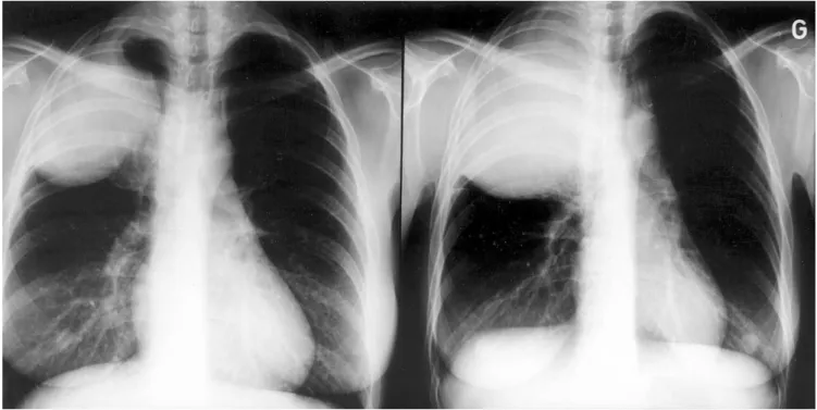
Fig. 1. Chest radiographies of fifth patient in Table 1 demonstrating extremely rapid tumor growth in only 11 days.
Table 2
Cases of pulmonary blastoma compared to all primary lung cancers operated No. of
cases
Age (mean) Sex (M/F) (%) Patients with symptoms (%)
Patients with N2 disease (%)
Median survival (years) Operated primary lung cancers 1918 27–83 (61) 1559/359 (81/19) 863 (45) 403 (21) 3.7 Pulmonary blastomas 5 32–46 (37) 1/4 4 4 , 1.0
Mediastinal N2 lymph node involvement – stage IIIA (observed in four of our five cases) would be expected to be frequent in view of the grim outcome of these tumors. Curiously, it is quite rare when noted [16]: three instances of lymph node metastasis in the 24-case American series (12%) – which may also have been N1 nodes – and three out of 11 in the Danish one [4]. Also, for some obscure reason, metastases – be it lymphatic or in another organ – do not necessarily reproduce the biphasic primary tumor architecture [4]: patients whose metastases were examined showed evenly distributed monotype and mixed histologies [6]; our 33-year old patient had her N2 lymph node composed solely of glandular cells. All in all, incidence of N2 disease in PB [9] compares closely with that of N2 disease in operated NSCLC (17% in Geneva, for instance [3]) and probably explains why N2 involvement per se was not found sufficiently predictive.
Finally, prognosis of PB is poor, since only one out of six patients survives $5 years [6], which is worse than for operated NSCLC considered as a whole. In the Danish survey of 83 cases – and quite expectedly – patients having undergone surgery (n ¼ 66) had a mean survival of 33 months, versus only two when they were deemed inoperable (n ¼ 17) [6]. Surgery should obviously aim to be curative, even though ‘subtotal’ resections backed by radio- and chemotherapy have occasionally afforded significant survi-vals (up to 7 years [17]) [16]. Interestingly, the situation is not quite so grim for children with pleuropulmonary blas-toma (quite a different entity, as mentioned earlier), despite higher numbers of inoperable tumors [9], since survival averages 30% at 5 years. Recurrences and metastases, observed in two-thirds of adult patients in the Danish series, and precocious in another one [1] are predominantly thor-acic, occurring twice as often in N2 nodes than in remaining lung tissue (after surgery).
In conclusion, when compared to NSCLC, PB are rare, larger, and symptomatic tumors; they affect younger patients and carry a worse prognosis. PB are all the more treacherous since imaging may mimic other benign conditions. Without resorting to undue work-up which is still inconclusive in two-thirds of cases, surgical excision should thus be rapidly contemplated for such large solitary pulmonary masses.
Acknowledgements
The authors thank Drs Joan Yap, Marian Stanisic, and Marcel Robert for their comments.
References
[1] Koss MN, Hochholzer L, O’Leary T. Pulmonary blastomas. Cancer 1991;67:2368–2381.
[2] Barrett NR, Barnard WG. Some unusual thoracic tumours. Br J Surg 1945;32:447–457.
[3] de Perrot M, Licker M, Bouchardy C, Usel M, Robert J, Spiliopoulos A. Sex differences in presentation, management, and prognosis of patients with non-small cell lung carcinoma. J Thorac Cardiovasc Surg 2000;119:21–26.
[4] Jacobsen M, Francis D. Pulmonary blastoma. A clinico-pathological study of 11 cases. Acta Pathol Microbiol Scand 1980;88:151–160. [5] Nakatani Y, Dickersin GR, Mark EJ. Pulmonary endodermal tumor
resembling fetal lung: a clinicopathologic study of five cases with immunohistochemical and ultrastructural characterization. Hum Pathol 1990;21:1097–1107.
[6] Francis D, Jacobsen M. Pulmonary blastoma. Curr Top Pathol 1983;73:265–294.
[7] Kodama T, Koide T, Shimosato Y, Naruke T, Watanabe S, Shimase J. Six cases of well-differentiated adenocarcinoma simulating fetal lung tubules in pseudoglandular stage. Comparison with pulmonary blas-toma. Am J Surg Pathol 1984;8:735–744.
[8] Manivel JC, Priest JR, Watterson J, Steiner M, Woods WG, Wick MR, Dehner LP. Pleuropulmonary blastoma: the so-called pulmonary blastoma of childhood. Cancer 1988;62:1516–1526.
[9] Priest JR, McDermott MB, Bhatia S, Watterson J, Manivel JC, Dehner LP. Pleuropulmonary blastoma. A clinicopathologic study of 50 cases. Cancer 1997;80:147–161.
[10] Koss MN, Moran CA, Stocker JT. Mixed epithelial-mesenchymal tumors. In: Saldana MJ, editor. Pathology of pulmonary disease, Philadelphia: JB Lippincott, 1994. pp. 617–629.
[11] Luce JA. Lymphoma, lymphoproliferative diseases, and other primary malignant tumors. In: Murray JF, Nadel JA, editors. Text-book of respiratory medicine, Philadelphia: WB Saunders, 1994. pp. 1597–1613.
[12] Barson AJ, Jones AW, Lodge KV. Pulmonary blastoma. J Clin Pathol 1968;21:480–485.
[13] Canver CC, Memoli VA, Vanderveer PL, Dingivan CA, Mentzer RM. Sex hormone receptors in non-small cell cancer in human beings. J Thorac Cardiovasc Surg 1994;108:153–157.
[14] Kohno H, Baba M, Fujizawa T, Shiba M, Nomoto Y, Shibuya K, Saitoh H, Toyazaki T, Hiroshima K, Ohwada H. Pulmonary blastoma: report of a case. Jpn J Surg 1999;29:803–806.
[15] Medbery CA, Bibro MC, Phares JC, Veach SR, Martin JE, Pasquale DN. Pulmonary blastoma. Case report and literature review of chemotherapy experience. Cancer 1984;53:2413–2416.
[16] Parker JC, Payne WS, Woolner LB. Pulmonary blastoma (embry-oma). J Thorac Cardiovasc Surg 1966;51:694–699.
[17] Cutler CS, Michel RP, Yassa M, Langleben A. Pulmonary blastoma. Case report of a patient with a 7-year remission and review of chemotherapy experience in the world literature. Cancer 1998;82:462–467.
[18] de Perrot M, Kurt AM, Robert J, Borisch B, Spiliopoulos A. Clinical behavior of solitary fibrous tumors of the pleura. Ann Thorac Surg 1999;67:1456–1459.