Sperm nuclear DNA damage and altered
chromatin structure: effect on fertilization and
embryo development
D.Sakkas
13, F.Urner
1, D.Bizzaro
2, G.Manicardi
2,
P.G.Bianchi
1, Y.Shoukir
1and A.Campana
1 'Clinic of Infertility and Gynaecological Endocrinology-WHO Collaborating Centre in Human Reproduction, Department of Obstetricsand Gynaecology, University Hospital of Geneva, Switzerland and department of Animal Biology, University of Modena, Via Berengario 14,
41100 Modena, Italy 3
To whom correspondence should be addressed at: Assisted Conception Unit, Birmingham Women's Hospital, Edgbaston, Birmingham B15 2TG, UK
In the first part of this report we investigate whether chromatin anomalies
in human spermatozoa can influence fertilization after intracytoplasmic
sperm injection (ICSI). We have examined the sperm chromatin packaging
quality using the chromomycin A
3(CMA
3) fluorochrome and the presence
of DNA damage in spermatozoa using in-situ nick translation. When
comparing the spermatozoa of patients undergoing in-vitro fertilization
(IVF) and ICSI distinct differences are evident in that ICSI males have a
higher CMA
3fluorescence, indicating spermatozoa with loosely packed
chromatin, and more spermatozoa containing endogenous DNA nicks. When
examining the unfertilized oocytes of ICSI patients we found that men who
had a high percentage of anomalies in their chromatin, i.e. >30% CMA
3fluorescence and >10% nicks, had more than double the number of
unfertilized oocytes containing spermatozoa that had remained condensed.
The observation that failed fertilized oocytes, injected with spermatozoa
from patients with a higher percentage of sperm nuclear anomalies, contain
more condensed spermatozoa indicates that a selection process against these
spermatozoa may be in place at the time of fertilization. In the second part
of the study we show that spare ICSI embryos have significantly lower rates
of development to the blastocyst stage compared with those developed after
routine IVF. These results show that a greater understanding of the molecular
basis of male infertility is therefore needed to broaden our knowledge on
the effect that abnormal spermatozoa have on fertilization and embryo
development.
Key words: fertilization/intracytoplasmic sperm injection/male infertility/sperm
chromatin/sperm nuclear decondensation
D.Sakkas et al.
Introduction
The treatment of severe male factor infertility has seen a dramatic advancement since the report of the initial pregnancies using intracytoplasmic sperm injection (ICSI) (Palermo et al, 1992). Consequently, ICSI is now widely applied to couples that have failed to achieve fertilization in conventional in-vitro fertilization (IVF) cycles, in which the husband has sperm parameters limiting or ruling out the use of IVF and in which the husband has undergone surgery to recover epididymal or testicular spermatozoa. The surprisingly high success rate in both fertilization and pregnancy rates after ICSI regardless of the severity of sperm defect (Van Steirteghem et al, 1993; Payne et al, 1994; Tournaye et al, 1994; Nagy et al, 1995; Silber et al, 1995) has subsequently caused debate on the possibility that using ICSI to force fertilization by abnormal spermatozoa may have hidden consequences for the normality of embryos and the. resulting fetus (Cummins et al, 1994; Menezo and Dale, 1995; Seamark and Robinson, 1995; Tarin and Cano, 1995). Initial data, thankfully, suggest that the normality of the babies delivered has not been jeopardized (Van Steirteghem et al, 1993; Bonduelle
et al, 1994) even though some studies report an increase in sex chromosome
anomalies (Bonduelle et al, 1996; Meschede and Horst, 1997).
ICSI in the majority of cases is applied to couples in which the male has a severe sperm defect. These defects are distinguishable as low sperm numbers, poor motility, abnormal morphology or combinations of these parameters. In addition to these normally accepted parameters, spermatozoa from certain sub-fertile men also display hidden defects in their surface proteins and/or in their chromatin organization. We and others have shown that male factor infertility patients possess anomalies in the composition of their sperm nuclei, displaying higher levels of loosely packaged chromatin and damaged DNA (Evenson et al, 1980, 1986; Foresta et al, 1992; Sailer et al, 1995; Golan et al, 1997). In our own studies we have used two methods to assess sperm chromatin quality: (i) the guanine-cytosine specific fluorochrome, chromomycin A3 (CMA3), which
evidences poor packaging quality of chromatin in human spermatozoa, as it allows an indirect visualization of protamine-deficient, nicked and partially denatured DNA, and (ii) in-situ nick translation, not preceded by endonuclease treatment, to evidence the presence of endogenous nicks in the DNA of ejaculated spermatozoa (Bianchi et al, 1993; 1996; Manicardi et al, 1995; Bizzaro
et al, 1998).
The fertilization potential of spermatozoa with abnormal chromatin organization in conventional IVF has been difficult to ascertain as results may be influenced by the initiation of the acrosome reaction and sperm membrane interactions with the oocyte. In light of this we have shown that semen with high CMA3 positivity
leads to significantly lower fertilization rates when using sub-zonal sperm injection (SUZI) (Bianchi et al, 1996). When using the ICSI technique all sperm membrane-oocyte interactions are superseded placing more importance on the quality of the sperm nucleus and the ability of the oocyte to initiate decondensation and pronuclear formation. The presence of spermatozoa containing damaged
Sperm nuclear DNA damage % 45 40 35 30 25 20 15 10 5 0 I 1
*
•=
M IVF • ICSI•
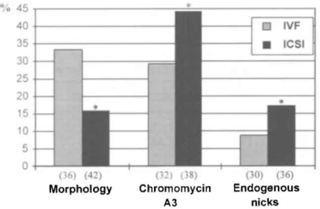
(36) (42) Morphology (32) (38) Chromomycin A3 (30) (36) Endogenous nicksFigure 1. The mean percentage of spermatozoa presenting normal morphology, chromomycin A3 (CMA3) fluorescence and endogenous nicks for the patients undergoing in-vitro fertilization (IVF) or intracytoplasmic sperm injection (ICSI). The number of patients assessed for each group is shown in parentheses.
*Significantly different (P < 0.01) from the IVF value. The means were transformed using an arcsin square root transformation and analysed using one-way analysis of variance and Scheffe's F-test.
DNA (Bianchi et al, 1993; Gorczyca et al, 1993; Manicardi et al, 1995; Sailer
et al, 1995) and the use of ICSI to force fertilization in these patients may cause
further uneasiness as to the fate of fertilized ICSI eggs. Here we therefore discuss whether the quality of a patient's spermatozoa, in terms of chromatin anomalies, can influence the outcome of ICSI and present results comparing embryo development to the blastocyst stage of patients undergoing routine IVF and ICSI.
Comparing the morphology, chromomycin A3 fluorescence and endogenous nicks of ejaculated human spermatozoa from patients
undergoing routine IVF and ICSI
When assessing the spermatozoa of patients undergoing IVF and ICSI distinct differences are evident in the mean percentage of spermatozoa presenting normal morphology, CMA3 fluorescence (an indicator of the packaging quality of sperm
chromatin) and endogenous nicks (an indicator of DNA damage in the sperm nucleus) (Figure 1).
When examining the above three parameters it could therefore be presumed that males with acceptable sperm parameters would present a normal morphology of >20%, CMA3 fluorescence of <30% and exhibit endogenous nicks in <10%
of their spermatozoa (Figure 1). The question therefore arises as to whether the above anomalies may influence fertilization after ICSI.
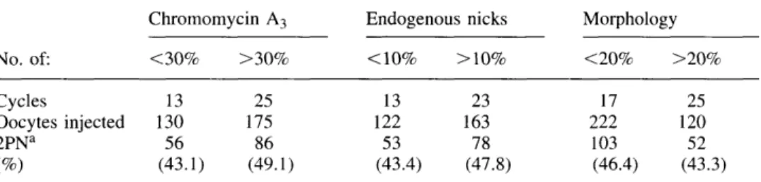
To ascertain whether a relationship existed between the sperm chromatin parameters of the patients and the ability of spermatozoa to fertilize after ICSI we separated the patients according to their sperm morphology, CMA3
fluorescence, and the presence of endogenous nicks. When ICSI patients were separated according to these criteria no overall difference was observed in their ability to achieve fertilization (Table I).
D.Sakkas et al.
Table I. Fertilization rates after intracytoplasmic sperm injection (ICSI) in male factor patients separated according to their percentage of normal morphology, chromomycin A3 positivity and
the presence of endogenous nicks
No. of: Cycles Oocytes injected 2PNa Chromomycin A3 <30% 13 130 56 (43.1) >30% 25 175 86 (49.1) Endogenous <1O% : 13 122 53 (43.4) nicks >10% 23 163 78 (47.8) Morphology <20% 17 222 103 (46.4) >20% 25 120 52 (43.3) fertilization rates are reduced as the study only involved the analysis of cycles in which more than two unfertilized oocytes were retrieved to investigate whether sperm had initiated decondensation after intracytoplasmic sperm injection (ICSI).
2PN = two pronuclear.
high CMA3 fluorescence, and/or a higher level of endogenous DNA nicks are
not limited in their ability to achieve fertilization using ICSI when compared with those from patients exhibiting low levels of these parameters. This may however mean that spermatozoa selected for ICSI were among those that did not possess anomalies in the sperm nucleus. What however is the fate of the spermatozoa selected for ICSI in oocytes that failed to fertilize?
When failed fertilized oocytes are examined after ICSI, using Hoechst 33342, and the status of the injected spermatozoa investigated, the sperm chromatin in the unfertilized oocyte can be scored as either (i) condensed; (ii) decondensing or decondensed in the cytoplasm of the oocyte and the maternal chromatin was between metaphase II (one polar body) and telophase II stage (two polar bodies); or (iii) both the maternal and paternal chromatin formed pronuclei. The relationship between the sperm chromatin patterns in the unfertilized oocytes after ICSI and the patients morphology, CMA3 fluorescence and presence of
endogenous nicks is shown in Table II. In patients who had a lower percentage of anomalies in their chromatin (<30% CMA3 fluorescence and <10% nicks) a
significantly lower number of spermatozoa remained condensed. In contrast, patients who had a high percentage of anomalies in their chromatin (>30% CMA3 fluorescence and >10% nicks) had more than double the number of
unfertilized oocytes containing spermatozoa that had remained condensed. The percentage of unfertilized oocytes containing spermatozoa that had remained condensed did not differ in relation to the patients morphology. Once sperm decondensation had been initiated, no significant difference was observed between the patient groups.
Embryo development
Although the above evidence suggests that spermatozoa with abnormal chromatin may influence the process of fertilization, the question of whether the development of an ensuing embryo is adversely affected is not known. A paternal effect has
-uooap jnjsssoons j o uoipjduio o ai p IO J snu o sq j aous q 'papuaosirex i pin? uoi;Bzifi;j3j u o VM G JO §ui§B>ped uijBuio.iip u ua ds ui saqiuiiou e jo oq j dA I su pn oj soA*jqxug spsau j9A3A\oq pajap uusds ye\\\ u^q j ssa ^ (^661 '7 sq; o j pq d j o aq rejuauidopAgp aq i u o poo§ ^ s q J3A3A\o q io u jqSiu i snouaSopua jo u 0 1 sarep i siq j MO H (il l si iS 3i J9JJ B sisXoois^^ q j o ip Suisn oj JA I P UB ISDI o\u\\o UA\ O Jti o ux 'juauidopAa p oXiqui s j o ssSej s jaqj^ s aq j U T m suiajqoi d Xu y IS3 I J^IJ ^ oA\iqui 9 ir e jo jo pajja aqi jo jusuiqsqqB d\# 9{iqA \ 'OA\ I i n %0 £> ui an 1 !^ 'sspxu % 0l < P TO aqj jo sajqj 'IS3 I JSJJ E sapu^uSaj d ^oiuq o xi s u j jo Suiuip sqi X q paouanyu i a q XBU I Xjiren b sajnpaoojd uopeuiuiasui JA I aupno j Suis n u o J9A9A\oq 3A^q p aj ja jEiiiared ^ pa^oipu i 9A^ q i^q j saipnj s "(e66T " F ^ pnButred t£66I '"/ » ^ u t ^3 -1661 '7 D ^ 13"«OH) XBUI iu9uidopA9p 0A*jqui9 pu ^ Ajiren b uaius s i^q j pajsaSSn s osr e jo jaquinu e 'Apnj s siq i o i J OI JJ •s}SA"oojsE|q UIJOJ oj repuajod pun J9A\oj Xpu^oyiuSi s B qjiA\ soAjqiu a o j as u 9AK S eozoiBima d ^ozoreuuods uazoj j jeq j paA\oq s saoqjn ^ assq x '(^66 1 'ozauajA j aqj o i so^jqiu s u^uin q j o jusuidopAs p sq j qii M pa^poss B usa q %0£< 300 = •ppnuojd Suisn pasuapuooap 'Suisuapuoosp BOZO;BUU3(JS s3pnpui D 100 = c / qP U B (6'8I) O T (8'3S) 83 (£"83) SI
es
S3 %03< (6'9I) 01 (Z'PS) 3£ (Q' Q7^ Vo o C/ L\ 6S L\ %03> 3qdiO]/\[ (S'8) i7 (9'3f) 03 (6'817) q£3 LV £Z %0l< (9"I3) 8 (8'9S) 13 (9-13) q 8 L£ ei %0 l> S 5{ o iu snouaSopug (8'Z.) P (O'lS) 93 (Z'\P) B I3 T S S3%oe<
(S'Z.1)z .
(0S9) 93 (S'Z.1) BZ. O t 7 £ T%oe>
uotuojq3 (%) sjsqio (%) D p3SU3pUO03p UU3ds (%) pssuspuoo uuads pauiuiBxa sajXoo o s 3 pX 3 (9661 '7° ^ a ^^Pl^S tuoJJ pajdBpe ) EozojBuusd s sjuax^ d aq ; U T s>pi u snousSopu s jo souassjd sqj pu e AjiApiso d £ v upAuioxuojq o 'X§opqdjou i o j uoxj^p j u i (iSDl ) uoxpsfu iD.Sakkas et al. 60 22 (36.7) 168 45 (26.8) 90 70 (77.7) 423 200 (47.3) Table III. The development of spare human embryos to the blastocyst stage after routine in-vitro fertilization (IVF) and intracytoplasmic sperm injection (ICSI)
ICSI IVF No. of cycles
No. of cycles with blastocysts (%)
No. of spare embryos No. of blastocysts
Significantly different from ICSI (P < 0.001, Fisher's exact test).
densation of the spermatozoa falls largely on a relationship between sperm chromatin organization and the ooplasm. How anomalies in the sperm nuclear structure can influence decondensation during fertilization is not established. Griveau et al. (1992) found that asthenozoospermic men displayed a high percentage of spermatozoa with nuclear abnormality, and using cytoplasmic extracts from unfertilized Xenopus laevis oocytes to induce decondensation, demonstrated that their sperm chromatin decondensed slowly and partially compared to normozoospermic men. In contrast, Perreault et al. (1987) reported that hamster sperm nuclei treated with the S-S bond reducing agent, dithiothreitol (DTT), decondensed more rapidly when microinjected into hamster oocytes. In addition, Burruel et al. (1996) showed that mouse spermatozoa with grossly misshapen heads could be injected into oocytes and contribute to the development of normal fertile adults.
The question of whether DNA damaged spermatozoa can impair the process of fertilization is not clear, however the alarming studies by the group of Robaire (see below) have indicated that damage to sperm DNA may be linked to an increase in early embryo death. They have shown that treatment of male rats with cyclophosphamide had little effect on the male reproductive system but caused single strand DNA breaks in the cauda-epididymal spermatozoa (Qiu
et al, 1995a) and altered the decondensation potential of spermatozoa (Qiu et al,
1995b). More disturbingly, similar treatment protocols using cyclophosphamide produce an increase in postimplantation loss and malformations (Trasler et al, 1985; 1986; 1987) and are transmissible to the next generation (Hales et al, 1992). A number of studies have indicated that the oocyte has the capability to repair the damaged DNA of spermatozoa. For example, Matsuda and Tobari (1988) showed that newly fertilized eggs were capable of repairing some of the deliberately damaged DNA of mouse spermatozoa irradiated with UV or treated with alkylating agents. Although there may be inbuilt mechanisms to guard against the incorporation of damaged DNA the results from the group of Robaire are highly indicative that these systems may not be foolproof.
The observation that failed fertilized oocytes injected with spermatozoa from patients with higher rates of sperm nuclear anomalies contain more condensed
spermatozoa indicates that a selection process against these spermatozoa may transpire at the time of fertilization. A high level of abnormalities in the chromatin of a spermatozoa selected for ICSI may impede the completion or initiation of decondensation therefore leading to a failure of fertilization. This may occur even though the oocyte possesses the necessary mechanism to initiate decondensation. Although we do not postulate that the failure of fertilization is entirely due to a sperm defect it seems likely that poor chromatin packaging and/or damaged DNA may contribute to a failure in the decondensation process. Furthermore, lower rates of development to the blastocyst stage by ICSI embryos compared to those developed after routine IVF indicate that a further selection may occur during the preimplantation stage. A greater understanding of the molecular basis of male infertility, is therefore needed to broaden our knowledge on the effect that abnormal spermatozoa have on fertilization and embryo development and to avoid the inappropriate use of ICSI.
References
Bianchi, P.G., Manicardi, G.C., Bizzaro, D. et al. (1993) Effect of DNA protamination on fluorochrome staining and in situ nick-translation of murine and human mature spermatozoa.
Biol. Reprod., 49, 1038-1043.
Bianchi, P.G., Manicardi, G.C., Urner, F. et al. (1996) Chromatin packaging and morphology in ejaculated human spermatozoa: evidence of hidden anomalies in normal spermatozoa. Mol.
Hum. Reprod., 2, 139-144.
Bizzaro, D., Manicardi, G.C., Bianchi, P.G. et al. (1998) In-situ competition between protamine and fluorochromes for sperm DNA. Mol. Hum. Reprod., 4, 127-132.
Bonduelle, M., Legein, J., Buysse, A. et al. (1994) Comparative follow-up study of 130 children born after ICSI and 130 children born after IVF. Hum. Reprod., 9 (Suppl. 1), 38.
Bonduelle, M., Legein, J., Buysse, A. et al. (1996) Prospective follow-up study of 432 children born after ICSI. Hum. Reprod., 11, 1558-1564.
Burruel, V.R., Yanagimachi, R. and Whitten, W.K. (1996) Normal mice develop from oocytes injected with spermatozoa with grossly misshapen heads. Biol. Reprod., 55, 709-714.
Chan, S.Y.W., Tucker, M.J., Leung, C.K.M. and Leong, M.K.H. (1993) Association between human
in vitro fertilization rate and pregnancy outcome: a possible involvement of spermatozoal quality
in subsequent embryonic viability. Asia-Oceania J. Obstet. Gynaecol, 19, 357-373.
Cummins, J., Jequier, A. and Kan, R. (1994) Molecular biology of human male infertility: links with ageing, mitochondrial genetics and oxidative stress. Mol. Reprod. Dev., 37, 345-362. Evenson, D.P., Darzynkiewicz, Z. and Melamed, M.R. (1980) Relation of mammalian sperm
chromatin heterogeneity to fertility. Science, 240, 1131-1133.
Evenson, D., Darzynkiewicz, Z., Jost, L. et al. (1986) Changes in accessibility of DNA to various fluorochromes during spermiogenesis. Cytometry, 7, 45N53.
Foresta, C , Zorzi, M., Rossato, M. and Varotto, A. (1992) Sperm nuclear instability and staining with aniline blue: abnormal persistence of histones in spermatozoa in infertile men. Int. J.
Androl, 15, 330-337.
Golan, R., Schochat, L., Weissenberg, R. et al. (1997) Evaluation of chromatin condensation in human spermatozoa: a flow cytometric assay using acridine orange staining. Mol. Reprod. Dev., 3, 47-54.
Gorczyca, W., Traganos, R, Jesionowska, H. and Darzynkiewicz, Z. (1993) Presence of DNA strand breaks and increased sensitivity of DNA in situ to denaturation in abnormal human sperm cells: Analogy to apoptosis of somatic cells. Exp. Cell Res., 207, 202-205.
D.Sakkas et al.
Griveau, J.F., Charbonneau, M., Blanchard, Y. et al. (1992) Decondensation of human sperm nuclei and HP1 protamine degradation from normospermia and asthenospermia in Xenopus egg extracts.
Arch. Androl, 29, 127-136.
Hales, B.F., Crosman, K. and Robaire, B. (1992) Increased postimplantation loss and malformations among the F2 progeny of male rats chronically treated with cyclophosphamide. Teratology, 45,
671-678.
Janny, L. and Menezo, Y.J.R. (1994) Evidence for a strong paternal effect on human preimplantation embryo development and blastocyst formation. Mol. Reprod. Dev., 38, 3 6 ^ 2 .
Manicardi, G.C., Bianchi, P.G., Pantano, S. et al. (1995) Underprotamination and nicking of DNA in ejaculated human spermatozoa are highly related phenomena. Biol. Reprod., 52, 864-867. Matsuda, Y. and Tobari, 1.(1988) Chromosomal analysis in mouse eggs fertilized in vitro with
sperm exposed to ultraviolet (UV) light and methyl and ethyl methansulfonate (MMS and EMS).
Mutat. Res., 198, 131-144.
Menezo, Y. and Dale, B. (1995) Paternal contribution to successful embryogenesis. Hum. Reprod., 10, 1326-1328.
Meschede, D. and Horst, J. (1997) Sex chromosomal anomalies in pregnancies conceived through intracytoplasmic sperm injection: a case for genetic counselling. Hum. Reprod., 12, 1125-1127. Nagy, Z., Liu, J., Joris, H. et al. (1995) The result of intracytoplasmic sperm injection is not
related to any of the three basic sperm parameters. Hum. Reprod., 10, 1123-1129.
Palermo, G., Joris, H., Devroey, P. and Van Steirteghem, A.C. (1992) Pregnancies after intracytoplasmic injection of single spermatozoon into an oocyte. Lancet, 340, 17-18. Parinaud, J., Mieusset, R., Vieitez, G. et al. (1993) Influence of sperm parameters on embryo
quality. Fertil. Steril, 60, 888-892.
Payne, D., Flaherty, S.P., Jeffrey, R. et al. (1994) Successful treatment of severe male factor infertility in 100 consecutive cycles using intracytoplasmic sperm injection. Hum. Reprod., 9, 2051-2057.
Perreault, S.D., Naish, S.J. and Zirkin, B.R. (1987) The timing of hamster sperm nuclear decondensation and male pronucleus formation is related to sperm nuclear disulfide bond content.
Biol. Reprod., 36, 239-244.
Qiu, J., Hales, B.F. and Robaire, B. (1995a) Damage to rat spermatozoal DNA after chronic cyclophosphamide exposure. Biol. Reprod., 53, 1465-1473.
Qiu, J., Hales, B.F. and Robaire, B. (1995b) Effects of chronic low dose cyclophosphamide on the nuclei of rat spermatozoa. Biol. Reprod., 53, 33^-0.
Ron-El, R., Nachum, H., Herman, A. et al. (1991) Delayed fertilization and poor embryonic development associated with impaired semen quality. Fertil. Steril., 55, 338-344.
Sailer, B.L., Jost, L.K., and Evenson, D.P. (1995) Mammalian sperm DNA susceptibility to In Situ denaturation associated with the presence of DNA strand breaks as measured by the terminal deoxynucleotidyl transferase assay. J. Androl., 16, 80-87.
Sakkas, D., Jacquenoud, N., Campana, A. and Leppens, G. (1994). Comparison of results after
in vitro fertilized human embryos are cultured in routine medium and in coculture on Vero cells:
a randomized study. Fertil. Steril., 61, 521-525.
Sakkas, D., Urner, F., Bianchi, P.G. et al. (1996) Sperm chromatin anomalies can influence decondensation after intracytoplasmic sperm injection (ICSI). Hum. Reprod., 11, 837-843. Seamark, R.F. and Robinson, J.S. (1995) Potential health problems stemming from assisted
reproduction programmes. Hum. Reprod., 10, 1321-1322.
Silber, S.J., Van Steirteghem, A.C. Liu, J. et al. (1995) High fertilization and pregnancy rate after intracytoplasmic sperm injection with spermatozoa obtained from testicle biopsy. Hum. Reprod., 10, 148-152.
Tarin, J.J. and Cano, A. (1995) Effects of manipulation in vitro of human oocytes and embryos on the birthweight of the resultant babies. Hum. Reprod., 10, 1322-1324.
Tournaye, H., Devroey, P., Liu, J. et al. (1994) Microsurgical epididymal sperm aspiration and intracytoplasmic sperm injection: a new effective approach to infertility as a result of congenital bilateral absence of the vas deferens. Fertil. Steril, 61, 1045-1051.
Trasler, J.M., Hales, B.F. and Robaire, B. (1985) Paternal cyclophosphamide treatment causes fetal loss and malformations without affecting male fertility. Nature, 316, 144-146.
Trasler, J.M., Hales, B.F. and Robaire, B. (1986) Chronic low dose cyclophosphamide treatment of adult male rats: Effect on fertility, pregnancy outcome and progeny. Biol. Reprod., 34,276-283. Trasler, J.M., Hales, B.F. and Robaire, B. (1987) A time course study of chronic paternal
cyclophosphamide treatment of rats: effects on pregnancy outcome and the male reproductive and haematologic systems. Biol. Reprod., 37, 317-326.
Van Steirteghem, A.C., Liu, J., Joris, H. et al. (1993) Higher success rates by intracytoplasmic sperm injection than by subzonal insemination. Report of a second series of 300 consecutive treatment cycles. Hum. Reprod., 8, 1055-1060.